Computer-Simulated Phacoemulsification
Carl-Gustaf Laurell, MD, PhD,
1Per So¨derberg, MD, PhD,
1Leif Nordh, MSc,
2Eva Skarman, MSc, PhD,
2Per Nordqvist, MSc
2Objective:
To develop a simulator for training in phacoemulsification to be used as a learning device for both
beginners and experienced surgeons to shorten the learning curve.
Design:
Experimental study.
Methods:
The system consists of a personal computer, a 3-dimensional visual interface, a
phacoemulsifi-cation handpiece, and a nucleus manipulator and foot pedals for control of the phacoemulsifiphacoemulsifi-cation procedure
and microscope adjustments. The simulation is based on generalized simulation software that can be also used
for the development of other medical simulations.
Main Outcome Measures:
Qualitative statements given in a questionnaire. Medical students and
ophthal-mic surgeons with varying experience of phacoemulsification were tested.
Results:
A simulator for training in phacoemulsification has been developed. The surgical procedures can be
practiced any number of times, and there is no risk to patients. The efforts of the surgeon can be evaluated
objectively.
Conclusions:
Studies have shown that the number of complications for an ophthalmic surgeon learning
phacoemulsification decreases exponentially, reaching close to the asymptote only after several hundred
procedures. Simulator training might shorten the learning period, reduce expensive supervision by an
experi-enced surgeon, and maintain and improve the skills of experiexperi-enced surgeons.
Ophthalmology 2004;111:
693– 698 © 2004 by the American Academy of Ophthalmology.
Recently, personal computers have become powerful
enough to permit real-time operator feedback simulation of
surgical procedures. This allows a widespread use of virtual
reality simulators in ophthalmic surgery for teaching new
surgeons and for training experienced surgeons on how to
manage peroperative complications. The potential benefits
of such simulators could relate to both acquisition and
assessment of surgical skills.
We believe phacoemulsification is a good application for
simulator training because the operation is a fairly complex
procedure requiring extensive training. The procedure is
essentially dependent on visual feedback. Tactile feedback
is almost absent. Both these factors make
phacoemulsifica-tion less difficult to simulate. Furthermore, cataract surgery
is the most common surgical procedure in the Western
world.
Phacoemulsification is usually taught in 2 phases. During
phase 1, the student watches surgery performed by a
teacher, an experienced colleague. The student may also
exercise the procedure on enucleated animal eyes. The
duration of the first phase may vary between approximately
6 and 12 months, depending on the type of surgical center,
the student’s learning speed, and previous experience in
ocular surgery. During phase 2, the student operates and the
teacher stands by. Because the operation is almost
exclu-sively performed with local anesthesia with the patient
awake, the possibilities for the teacher to comment to the
student during surgery are limited. Furthermore, because of
space constrictions, the teacher has no immediate access to
the operating field and cannot easily prevent erroneous
manipulations by the student. The duration of the second
phase again might vary from approximately 6 to 12 months.
Altogether, the teacher and the student might spend more
than a year together in the operating room before the student
can begin to operate independently.
The phacoemulsification procedure requires relatively
complex coordination of hands and feet, and the margins for
inaccurate manipulations are small. The success of the
op-eration is highly related to the maintenance of an intact
capsular bag. Despite careful training, residents are reported
to have an incidence of 5% to 20% of capsular ruptures
during their first 200 cases.
1– 4Similar figures have been
reported for experienced surgeons learning
phacoemulsifi-cation.
5,6A study of 1000 consecutive cases operated by 1
surgeon demonstrated that the number of complications
decreases exponentially and that the asymptote is reached
after approximately 400 cases.
7A similar study by another
experienced surgeon showed that the asymptote is reached
after approximately 1000 cases.
8Originally received: February 11, 2003.
Accepted: June 25, 2003. Manuscript no. 230088. 1St. Erik’s Eye Hospital, Stockholm, Sweden.
2Melerit AB, Berzelius Science Park, Linko¨ping, Sweden.
Presented, in part, at: American Society of Cataract and Refractive Surgery meeting, May, 2000, Boston; International Biomedical Optics Symposium, January, 2001, San Jose, California; XXth Congress of the European Society of Cataract and Refractive Surgeons, September, 2002, Nice, France.
Supported in part by Karolinska Institutet, Stockholm, Sweden, and Melerit AB, Linko¨ping, Sweden.
L. Nordh, E. Skarman, and P. Nordqvist are employed by Melerit AB. Reprint requests to Carl-Gustaf Laurell, MD, PhD, St. Erik’s Eye Hospital, SE-112 82 Stockholm, Sweden.
Efforts should be made to shorten the long learning
period for phacoemulsification and to minimize the number
of intraoperative complications. There might also be
eco-nomic gain if the time during which high-volume cataract
surgeons are occupied with teaching could be reduced.
Pedagogic feedback is facilitated in simulators, because
the metrics calculated intrinsically in the software to run the
simulation can be used to give performance feedback to
both trainees and teachers. In basic medical education and
during the training of residents, medical simulators could be
used to make objective assessments of psychomotor skills.
Hand-eye coordination may be tested and trained with
vir-tual reality applications.
9Recently, it was demonstrated that
virtual reality training leads to faster adaptation to the
psychomotor restrictions encountered by surgeons who
op-erate through small incisions.
10Materials and Methods
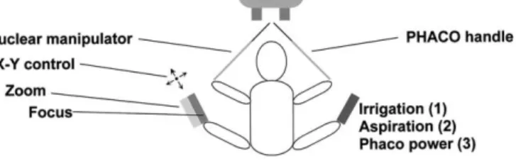
The complexity of phacoemulsification surgery is indicated in Figure 1. The surgeon gets 3-dimensional visual input from the
microscope and must provide feedback reactions with the right hand on the phacoemulsification handpiece (3 space dimensions⫹ rotation), with the left hand on the nucleus manipulator (3 space dimensions⫹rotation), with the right foot on the pedal controlling irrigation, aspiration, and phacoemulsification (movement in 1 dimension with the function related to the position of the pedal), and with the left foot on the x–y control (2 dimensions), the focus control (1 dimension), and the zoom control (1 dimension).
In the first step, the phacoemulsification procedure was simu-lated with a nucleus manipulator and a phacoemulsification hand-piece for input and a computer screen as a visual feedback inter-face to the trainee (Laurell CG, et al. Computer-simulated phacoemulsification. SPIE Proc 2001;4245:174 – 6). In a second step, a 3-dimensional visual interface, software for zooming and x–y positioning with foot pedals, and a computer algorithm for foot pedal real time focusing– defocusing have been developed.
Hardware
A simulator should contain the same transducer complexity for the surgeon as in the real situation. The system developed consists of a personal computer, a 3-dimensional visual interface, a phaco-emulsification handpiece, a nucleus manipulator, and foot pedals for control of the phacoemulsification procedure and microscope adjustments (Fig 2). The phacoemulsification handpiece and the nucleus manipulator are mounted with 4 degrees of freedom (3 space dimensions ⫹ rotation), with an electronic transducer for each degree of freedom. Similarly, the positions of the foot pedal are sensed with analog electronic transducers. All signals are converted to digital in a personal computer card. The digital signals are used as input in the simulation software.
Three-dimensional Visual Interface
To achieve a 3-dimensional visual interface, the monitor units from a virtual reality helmet (AddVisor 100, Saab Avionics, Swe-den) are mounted above the phacoemulsification handpiece and the
Figure 1. The complex interface between the surgeon and the environ-ment during cataract surgery. PHACO⫽phacoemulsification.
Figure 2. Schematic of simulator with the same interface complexity between surgeon and the environment as in real surgery. LCDs⫽liquid crystal displays; PHACO⫽phacoemulsification.
nucleus manipulator (Fig 3). Each monitor consists of a high-resolution liquid crystal display (LCD). In the simulator system, these units are mounted on a commercial Carl Zeiss (Stockholm, Sweden) microscope stand. There is a forehead phantom for hand support during surgery.
Foot Pedal Control of Phacoemulsification
The position of the transducer for foot pedal control of phacoemul-sification power is fed into the algorithm that determines the efficacy of the virtual phacoemulsification process, implying linear response in phacoemulsification power.
Zoom and x–y Positioning
The position of the zoom and x–y is fed from the foot pedal transducer into the software. For zooming, the magnification of the virtual image presented on the LCD is altered. When moving the
x–y stick on the foot pedal, the virtual cameras monitoring the eye model are moved in relation to the eye model, thus moving the center of the image on the LCD.
Focusing
The data input is a 3-dimensional model of the surgical field with a defined focal plane. A blurry image and a sharp image of the focal plane are generated.
Averaging over several pixels generates the blurry image. For each pixel the distance between the focal plane and the image is measured. The final image is generated as a mixture of the sharp and the blurred image, using the distance to the focal plane as the weight for luminous intensity of the images.
Software
The simulation is based on generalized simulation software (M-base, Melerit AB, Linko¨ping, Sweden), which is working on top of
Cosmo3D/Optimizer (Silicon Graphics Inc., Mountain View, CA). On top of M-base, a module has been created for the phacoemul-sification procedure.
Mode of Action for Phacoemulsification
A 3-dimensional model of the field of surgery is generated (Fig 4) and presented to the surgeon on the LCDs. The surgeon can sculpt the nucleus with the phacoemulsification handpiece, rotate or push on the nucleus with either of the instruments, divide the nucleus, and aspirate the pieces. The action of the 2 instruments is imme-diately fed back to the visual interface as image information with an update frequency of 25 Hz, thus directing the next move of the surgeon.
Evaluation of the Simulator
Cataract surgeons of various experience levels tested the realism of the simulator. In a pilot study, 7 medical students, who had passed the course in ophthalmology, were interviewed before and after performing phacoemulsification in the simulator. The study was performed in cooperation with the pedagogic institution of the University of Linko¨ping, Sweden.
Results
A simulator for training in phacoemulsification has been devel-oped. In the simulator, the most important steps in learning the
phacoemulsification procedure might be reproduced. Sculpting and dividing of the lens nucleus can be performed in a realistic manner, followed by aspiration of the nuclear fragments. The movements of the pieces of nucleus are similar to real surgery because of simulated flow and aspiration, attracting the lens pieces to the phacoemulsification tip. The hardness of the nucleus (nuclear sclerosis) can be altered freely and correlates to different nuclear colors. Rotation of the nucleus can be made more or less difficult, simulating the situation after ineffective hydrodissection or a loose lens capsule. Breaks in the posterior capsule may be simulated. Total procedure time and total phacoemulsification energy can be measured, and the movements of the instruments inside the eye may be registered by the computer. During the procedure, air bubbles are generated exiting from the irrigation holes in the sleeve of the phacoemulsification handpiece.
According to the structured interviews with the medical stu-dents, they were positive regarding the use of medical simulators, because simulators make possible surgical training without risk to patients, and the procedures can be exercised any number of times. The movements on the computer screen were considered to agree with the real movements and felt natural. The students thought that simulators might be used to test disposition and interest for oph-thalmic surgery. A rapid understanding of a procedure and its complexity may be achieved. According to the students, realism of function is more important than photorealism of the visual inter-face. The students stressed the importance of receiving continuous feedback in the form of changes in the picture and text messages, as well as a comprehensive judgment at the end of the procedure. The possible risk of a false feeling of security and the importance of training on real patients were mentioned.
The cataract surgeons found the realism of the simulator ade-quate for further studies in which the validity and educational value of the device are to be evaluated.
Discussion
The first surgical simulators appeared in the early 1990s.
11Today, the fast development of information technology and
computer graphics presents opportunities to create new
tools for surgical training. Virtual reality simulators are
known to provide a safe training environment for high-risk
work environments. The simulators are available at all times
and provide a structured curriculum that can be
standard-ized, repeated, and optimized toward the learner’s needs.
Skills may be assessed objectively and repeatedly. In
addi-tion to the improved training opportunities, simulators
might shorten residency training programs and perhaps
lower educational expenses.
One problem is that the development of virtual reality
applications is expensive and time consuming. Close
col-laboration among physicians, computer scientists, and
en-gineers is essential. However, the success of virtual reality
in pilot and military training suggests strong potential in
medical education. Experienced airline pilots maintain their
skills continuously through the use of flight simulators.
Until now, surgical education has mainly been based on
the apprentice model. The residents gain progressive
expe-rience through supervised training on patients. However,
this model might fail to provide skill acquisition in an
organized fashion. Concerns about cost and risks to patients
have been raised, and the importance and potential danger
of learning curves have been alluded to.
12Although
alter-native training methods such as surgery on artificial eye
models and cadaver eyes are available to a certain extent,
there are significant drawbacks to these methods. The
ac-cessibility to training on pig eyes is limited, and the
char-acteristics of the lens nucleus and capsule in these eyes are
different from what we find in human eyes with senile
cataract. Sculpting and dividing the nucleus is much more
realistic in the simulator, because the lens nucleus of the pig
eye is too soft for practicing this procedure. Furthermore,
the lens capsule of the pig lens is difficult to break, in sharp
contrast to the capsule in senile cataracts. Recently, the risk
for transmission of infectious diseases has been said to
impose practical problems and increase costs on setting up
wet laboratories.
As long as there is a learning curve involved in the
training of phacoemulsification, something should be done
to improve the situation. However, medical simulators will
have to be evaluated carefully to confirm whether virtual
reality training improves surgical education. Recently, such
training was demonstrated to improve operating room
per-formance during laparoscopic cholecystectomy in human
patients. Surgeons trained in a simulator made 6 times fewer
errors and were 5 times less likely to injure nontarget tissue
according to a randomized, double-masked study
(Gal-lagher T, presented at the Annual Meeting of the American
Surgical Association, Hot Springs, Virginia, 2002). Even
experienced surgeons with more than 7 years of experience
demonstrated a significantly improved performance with the
added experience of simulator training. Other recent studies
have shown improved skills by residents after training in a
bronchoscopy simulator
13and a laparoscopic simulator.
14It is necessary to determine whether a given simulation
actually measures the performance parameters it is
sup-posed to measure. The simulator should reveal the
differ-ence in skills between experidiffer-enced surgeons and beginners.
This issue was addressed in recent studies of simulators for
laparoscopy
15and arthroscopy,
16in which the experienced
surgeons performed significantly better than the novices.
Another study demonstrated that the simulator was able to
assess the psychomotor skills in residents.
17Experience with ophthalmic virtual reality simulators is
limited. Previously, an eye surgery simulator was
present-ed,
18and more recently, a vitreous surgery simulator
19and
an intraocular surgery workstation using a mechanical eye
model.
20The educational value of virtual reality
applica-tions for eye surgery has still to be demonstrated and must
be compared with currently used training methods. These
studies will put the spotlight on what constitutes good
surgical skills and how these should be measured
objec-tively. Next, the validity of the phacoemulsification
simu-lator will be tested in a study comparing the results of
experienced cataract surgeons, residents in ophthalmology,
and medical students. The computer will perform objective
measurements of variables such as phacoemulsification
time, distance covered by the phacoemulsification tip inside
the eye, and number of inappropriate or dangerous
move-ments performed with the phacoemulsification tip. The
ed-ucational value should be verified by comparing the
learn-ing curves in 2 groups of novices, namely, those who have
and those have not undergone simulator training.
In conclusion, we believe that in the near future virtual
reality surgery will be an important supplement to what is
currently considered the best training approach to learn
phacoemulsification. Our pilot study on medical students
indicates the importance of the teacher being present with
the novice during the exercises. Ultimately, the value of
simulators will be measured by their ability to improve
patient outcomes.
References
1. Cruz OA, Wallace GW, Gay CA, et al. Visual results and complications of phacoemulsification with intraocular lens implantation performed by ophthalmology residents. Ophthal-mology 1991;99:448 –52.
2. Tarbet KJ, Mamalis N, Theurer J, et al. Complications and results of phacoemulsification. J Cataract Refract Surg 1995; 21:661–5.
3. Yang YC, Kirwan JF, Foster PJ, Pereira AM. Cataract surgery by junior ophthalmologists. Eye 1995;9(suppl):22–5. 4. Robin AL, Smith SD, Natchiar G, et al. The initial
complica-tion rate of phacoemulsificacomplica-tion in India. Invest Ophthalmol Vis Sci 1997;38:2331–7.
5. Seward HC, Dalton R, Davis A. Phacoemulsification during the learning curve: risk/benefit analysis. Eye 1993;7:164 – 8. 6. Thomas R, Braganza A, Raju R, et al. Phacoemulsification—a
senior surgeon’s learning curve. Ophthalmic Surg 1994;25: 504 –9.
7. Ng DT, Rowe NA, Francis IC, et al. Intraoperative complica-tions of 1000 phacoemulsification procedures: a prospective study. J Cataract Refract Surg 1998;24:1390 –5.
8. Martin KR, Burton RL. The phacoemulsification learning curve: per-operative complications in the first 3000 cases of an experienced surgeon. Eye 2000;14:190 –5.
9. McDonald P. Training for surgeons after the year 2000. J R Soc Med 1998;91:401.
10. Jordan JA, Gallagher AG, McGuigan J, McClure N. Virtual reality training leads to faster adaptation to the novel psy-chomotor restrictions encountered by laparoscopic surgeons. Surg Endosc 2001;15:1080 – 4.
11. Meier AH, Rawn CL, Krummel TM. Virtual reality: surgical application— challenge for the new millennium. J Am Coll Surg 2001;192:372– 84.
12. Moore MJ, Bennett CL, The Southern Surgeons Club. The learning curve for laparoscopic cholecystectomy. Am J Surg 1995;170:55–9.
13. Rowe R, Cohen RA. An evaluation of a virtual reality airway simulator. Anesth Analg 2002;95:62– 6.
14. Taffinder N, Sutton C, Fishwick RJ, et al. Validation of virtual
reality to teach and assess psychomotor skills in laparoscopic surgery: results from randomised controlled studies using the MIST VR laparoscopic simulator. Stud Health Technol In-form 1998;50:124 –30.
15. McNatt SS, Smith CD. A computer-based laparoscopic skills assessment device differentiates experienced from novice laparoscopic surgeons. Surg Endosc 2001;15:1085–9. 16. Pedowitz RA, Esch J, Snyder S. Evaluation of a virtual reality
simulator for arthroscopy skills development. Arthroscopy 2002;18(6):E29.
17. Grantcharov TP, Rosenberg J, Pahle E, Funch-Jensen P. Vir-tual reality computer simulation: an objective method for the evaluation of laparoscopic surgical skills. Surg Endosc 2001; 15:242– 4.
18. Sinclair MJ, Peifer JW, Haleblian R, et al. Computer-simu-lated eye surgery: a novel teaching method for residents and practitioners. Ophthalmology 1995;102:517–21.
19. Hikichi T, Yoshida A, Igarashi S, et al. Vitreous surgery simulator. Arch Ophthalmol 2000;118:1679 – 81.
20. Wagner C, Schill M, Hennen M, et al. Virtual reality in ophthalmological education [in German]. Ophthalmologe 2001;98:409 –13.