Morphometric Parameters of the Small and Large Intestine of
the Ostrich
(Struthio Camelus Var. Domesticus)
from Day 38 of Embrionic
Development to the Age of 60 Days
Strausu
(Struthio camelus var. domesticus)
tievās un resnās zarnas
morfometriskie rādītāji no 38. embrionālās attīstības dienas
līdz 60 dienu vecumam
Ilmars Duritis, Arnis Mugurevics
Preclinical institute, LLU LLU Preklīniskais institūts e-mail: ilmars.duritis@llu.lv
Abstract. The anatomic intestine structure of the ostrich (Struthio camelus var. domesticus) is characterized by a number of essential intrinsic properties related to the climatic conditions of their natural distribution area (desert) and the relatively high fibre content in feed. The objective of the present research was to establish the mass-metric and morphometric parameters of the large and small intestine of the ostrich in the period from the 38th day of embryonic development till the 60th day of life. A total of 42 ostrich specimens of both genders were used, including six embryos obtained on the 38th incubation day and 36 chicks at the age of one day, 3, 7, 14, 30, and 60 days, distributed in groups of 6 birds in each group. The total absolute and relative weight and length of separate segments of the large and small intestine were established. The small and large intestine relative weight of the ostrich chicks increased with the birds advancing in age and reached its maximum on day 30; however, by the age of 60 days the relative weight diminished. A substantial increase in the absolute length of the small and large intestine was observed starting with day 3 of life (p<0.05; 0.001), and with chicks advancing in age it continued to elongate. Assessing the length proportions of separate intestine segments it was found that they had not significantly changed over the whole period of ontogenesis.
Key words: the ostrich, small intestine, large intestine, morphometric parameters.
Introduction
The intestine structure of domestic poultry has been studied in great detail already in the middle of the last century; however, in case of the ostrich, especially its gastro-intestinal tract and development of its parts in ontogenesis period, the research information is quite scarce. The anatomic intestine structure of the ostrich (Struthio camelus var. domesticus) is characterized by a number of essential intrinsic properties related to the climatic conditions of their natural distribution area (desert) and the relatively high fibre content in feed (Sales, 2006; Cooper, Mahroze, 2004). In comparison with other bird species, the Struthioniformes are characterized by a strongly developed large intestine which constitutes the largest part of the gastro-intestinal tract for an adult ostrich (Bezuidenhout 1993; Fowler, 1991; Порческу, 2007) and, as is
common knowledge, the large intestine plays an especially important role in fibre fermentation processes.
The development stage of the digestion tract at the time of hatching, especially that of the stomach and intestines, determines the ability of chicks to consume feed, thus enabling to deduce the abilities of the organism to digest the feed consumed or the functionality of the digestive organs. Development of intestines plays a leading role in the growing and development processes of the organism also in later ontogenesis periods.
Each species of birds has a characteristic (natural) type and composition of feed determining anatomical differences of the digestion tract between ostriches and other poultry species (Bezuidenhout, 1993; Fowler, 1991; Порческу, 2007; Wang, Peng et al., 2007). Therefore, in order to facilitate the development of
optimum, age-adjusted feeding programmes, it is essential to understand the gastro-intestinal tract anatomy and physiology of the ostrich at different periods of ontogenesis.
The objective of the present research study was to establish the mass-metric and morphometric parameters of the large and small intestine of the ostrich in the period from day 38 of embryonic development to the 60th day of life.
Materials and Methods
In the research study, 42 ostrich specimens of both genders raised in the farm “Ozoliņi AB” of Jēkabpils district, Latvia, were used: six embryos obtained on the 38th incubation day, and 36 chicks at the age of one day, 3, 7, 14, 30, and 60 days. Ostriches were distributed in groups of 6 birds each. Chicks were obtained over the period of May–October of 2009. The first 3 days following the hatching, the chicks were kept in the hatchery. Starting with day 4, the chicks were placed in a heated box with sand bedding and they started to receive the commercial ostrich chick feed “Strus Premium – Strus 1”. Feed and water were supplied ad libidum.
Before euthanasia, the birds of age group in question (7, 14, 30, and 60 days old) were taken off feed for 12 hours. After that, they were anaesthetised by intra-muscular injection of 0.5 ml of 10% ketamine and 0.5 ml of 2% xylasine solution and afterwards euthanized by intracardial injection of 0.5 ml of 20% pentobarbital solution. After euthanasia, each carcass was weighted on electronic scales (±1 g) and subjected to necropsy for further examination.
The total absolute weight of the intestines (together with their contents), as well as the absolute weight of the large and small intestine separately was determined using scales (±0.01 g).
Also the length of separate segments of the large and small intestine was established: for the duodenum – from ostium pyloricoduodenale to flexura duodenojejunalis; for the jejunum – from flexura duodenojejunalis to the apex level of caecum; for the ileum – from the apex level of caecum to ostium ileocecale; for the caecum – from ostium ileocecale to the apex; for the colon – from the connection point of caecum and colon to the cloacal extension (Baumel, 1993). A tape measure (±1 mm) was applied for measuring the length of intestines.
The relative weight of each segment of the intestines separately (in relation to body weight) as well as their relative length (in relation to the total
intestinal length) was calculated. The data obtained in the study were statistically processed by the SPSS 11.5 software program. The mean arithmetic value and standard error (SEM) were calculated for each parameter. For comparison of the mean parameters among age groups, the multifactor dispersion analysis ANOVA was applied.
Results
Over the ontogenesis period studied, the total intestine weight for the ostrich chicks (small and large intestine) increased 67 times (from 8.8±1.00 g on the 38th embryonic development day to 593.6±82.66 g on the 60th day of life). A critical weight increase was observed between days 3 and 14 (p<0.05), days 7 and 30 (p<0.01), and days 30 and60 (p<0.001) (Table 1).
Having determined the absolute and relative weight of the small intestine it was established that on day 38 of embryonic development the small intestine weighed 3.0±0.39 g on average, constituting 0.3±0.04% of the chick body weight. On the day of hatching, these parameters increased to 5.8±0.45 g and 0.7±0.08% respectively (Table 1).
Over the first days after hatching, increase in the absolute weight of the small intestine was quite slow; however, after the 3rd day of hatching a rapid increase was observed. The absolute weight of the small intestine continued to increase over the whole ontogenesis period. On the earlier stages of postnatal ontogenesis, the weight increase of the small intestine was more rapid. For instance, between day 30 and day 60, the weight of the small intestine increased 2.5 times (p<0.001), but between days 3 and 14 – 5 times (p<0.001) (Table 1).
With chicks advancing in age, the relative weight of the small intestine increased reaching its maximum at the age of 30 days when it constituted already 4.9±0.37% of the body weight; the most rapid increase could be observed between days 7 and 14 (p<0.01). Afterwards, from the 30th to 60th day of life, the relative weight of the small intestine of the ostrich chicks decreased to 3.8±0.14% of their body weight (Table 1).
The absolute and relative weight of the large intestine, in its turn, on the 38th day of embryonic development was 5.8±0.62 g or 0.7±0.07% of the body weight. On the day of hatching, the absolute weight of the large intestine had already increased almost threefold (14.3±1.55 g;
Table 1
The weight of the small and large intestine of the ostrich chicks from 38th day of embryonic development to 60th day of life
Age, days
Small intestine Large intestine
Total, g±SEM g±SEM* %±SEM,of body
weight g±SEM %±SEM, of body weight 38 (embr.)** 3.0±0.39 0.3±0.04 5.8±0.62 0.7±0.07 8.8±1.00 1 5.8±0.45 0.7±0.08 14.3±1.55 1.7±0.25 20.2±1.84 3 8.0±0.85 0.9±0.07 14.1±1.92 1.6±0.17 22.1±2.70 7 16.2±1.77 2.1±0.21 51.2±5.00 6.6±0,.7 67.5±6.04 14 42.2±7.22 4.0±0.62 129.6±16.94 12.2±1.29 171.6±23.17 30 72.7±5.49 4.9±0.37 204.2±17.95 13.6±0.71 276.9±22.40 60 182.1±28.62 3.8±0.14 411.6±58.20 8.9±1.10 593.6±82.66
* – standard error of mean
** – 38th day of embryonic development
Table 2
Morphometric parameters of the small and large intestine of the ostrich chicks from 38th day of embryonic development to 60th day of life
Age,
days intestine, mm±SEMLength of the small intestine, mm±SEMLength of the large length, mm±SEMTotal intestine
38 (embr.) 509±24.1 680±72.9 1189±99.9 1 576±24.9 978±61.2 1555±66.6 3 762±42.7 1036 ±84.4 1798±81.1 7 1002±65.5 1605±81.1 2607±133.7 14 1424±138.5 2733±81.9 4157±206.5 30 2043±81.9 3654±178.7 5697±210.9 60 2960±314.6 4987±475.8 7947±773.7
1.7±0.25%). After the 3rd day of life it increased much more rapidly and reached a ninefold size on the 14th day thus constituting 129.6±16.94 g or 12.3±1.29% on average (p<0.05) (Table 1).
At the age of 30 days, the relative weight of the large intestine reached its maximum, constituting 13.6±0.71% of the body weight. A significant decrease in the weight of large intestine was observed at the age of 60 days (p<0.01), when it constituted only 8.9±1.10% of the chick body weight (Table 1).
The total intestine length for the ostrich chicks over the studied ontogenesis period increased 6.7 times on average (from 1189±99.9 mm on the 38th embryonic development day to 7947.00±773.7 mm on the 60th day of life). An important intestine length increase could be observed between days 7 and 14 (p<0.05), days
14 and 30 (p<0.05), as well as between days 30 and 60 (p<0.01) (Table 2).
Analysing the total length of the small
intestine it was found that on the 38th embryonic development day it had reached 509±24.1 mm on average or 43.4±2.04% of the total intestine length (Table 2). A rapid growth in the length of the small intestine started after hatching, when on the 7th day of life its length had already doubled compared to the 38th day of embryonic development making up 1002±65.5 mm or 38.4±1.09% of the total intestine length. A critical growth in the length of the small intestine could be observed between days 3 and 14 (p<0.05), as well as between days 30 and 60 (p<0.001).
The relative length of the small intestine for the ostrich chicks of all age groups ranged from
33.9±1.76% at the age of 14 days to 43.4±2.04% on the 38th day of embryonic development with a decreasing trend as chicks advanced in age (Table 2).
The large intestine formed the longest portion of the intestine length in the ostrich chicks of all age groups. On the 38th embryonic development day, the large intestine length (679.50±72.87 mm) only slightly exceeded length of the small intestine, but on the 14th day of chick life the large intestine was two times longer than the small intestine. At this age, length of the small intestine was 1423.67±138.49 mm, whereas that of the large intestine – 2733.17±81.85 mm respectively; furthermore, this correlation persisted also for 30- and 60-day-old birds (Table 2). A substantial increase in the absolute length of the large intestine was observed between days 3 and 14 (p<0.001), days 7 and 14 (p<0.01), and days 30 and 60 (p<0.01).
The relative length of the large intestine for the ostrich chicks included in the study ranged from 56.58±2.07% (38th embryonic development day) to 66.15±1.76% (14th day). The relative length of the large intestine increased as chicks advanced in age (Table 2).
For birds of all age groups included in the research, all three parts (segments) of the small intestine, namely, duodenum, jejunum, and ileum, indicated an adequate and well-marked development.
Assessing the morphometric parameters of each part of the small intestine we established that
the jejunum formed the greatest weight and length proportion.
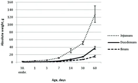
Figure 1 demonstrates that the jejunum had a pronounced increase in weight starting with the 3rd day of life of the ostrich chicks. In comparison with other segments of the small intestine, the weight increase of jejunum was faster.
On the 38th day of embryonic development, the absolute weight of the jejunum was 2.3±0.30 g on average or 0.25±0.03% of the body weight, on the day of hatching it almost doubled (4.0±0.35 g), but at the age of 30 days it was 51.1±4.00 g. A significant increase in the weight of jejunum was observed between days 1 and 30 (p<0.01), and days 30 and 60 (p<0.001) (Fig. 1).
The weight of the duodenum and ileum was increasing slower and more evenly. A rapid increase in the weight of these two parts could be observed between days 7 and 30 (p<0.01), and days 30 and 60 (p<0.001).
Of the three segments of the small intestine, the jejunum made up also the relatively greater weight proportion.
Relative weight of the duodenum on the 38th embryonic development day constituted only 0.06±0.007% of the body weight, and reached its maximum (0.96±0.023%) on the 30th day of life, after which a downwards trend set in (in a similar way as for the total relative weight of the small intestine) (Fig. 2). Relative
Fig. 1. The dynamics of absolute weight (g±SEM) of the small intestine segments (duodenum, jejunum, and ileum) of the ostrich chicks in the period between 38th day of embryonic development and 60th day of life.
Fig. 3. The dynamics of absolute weight (g±SEM) of the large intestine segments (colon and caecum) of the ostrich chicks between 38th day of embryonic development and 60th day of life.
Fig. 2. The dynamics of relative weight (%±SEM) of the small intestine segments (duodenum, jejunum, and ileum) of the ostrich chicks in the period between 38th day of embryonic development and 60th day of life.
weight of the jejunum reached 3.47±0.34% on the 30th day of ostrich chick life, but at the age of 60 days decreased to 2.65±0.12% of the body weight.
On the 38th day of embryonic development, the relative weight of the ileum was only 0.03±0.005% of the body weight on average, between the age of 1 day to 14 days (p<0.001) it increased significantly, but in 30-day-old ostrich chicks reached its maximum – 0.49±0.04% of the body weight (Fig. 2).
Analysis of the absolute weight of the large intestine segments showed that the weight of the colon had grown faster than that of the caecum (Fig. 3).
The weight of the caecum on the 38th embryonic development day was 0.9±0.10 g on average, relatively constituting
0.10±0.01% of the chick body weight, but on the day of hatching it had more than doubled (2.2±0.21 g and 0.26±0.03% respectively). Over further development, a substantial absolute weight increase of the caecum was established between days 3 and 14 (p<0.01), as well as between days 30 and 60 (p<0.001) (Fig. 3). A substantial relative weight increase of the caecum was established between days 7 and 14 of chick life (p<0.01). At the age of 30 days, the relative weight of the caecum was 1.96±0.28% on average, but on the 60th day it decreased significantly – to 1.14±0.09% (p<0.01) (Fig. 4).
For the ostrich chicks of all age groups, the colon both in length and weight was developed better than other intestine parts and formed the largest portion of the total intestine length and weight.
On the 38th embryonic development day, the absolute weight of the colon was 4.9±0.53 g on average, composing 0.55±0.06% of the body weight (Figs 3 and 4). On the day of hatching, the weight of the colon reached 12.2±1.52 g or 1.47±0.23% on average. Over the first three days of life, the absolute and relative weight of the colon showed a downward trend, but afterwards a rapid increase was observed. On the 30th day, the weight of the colon was 174.9±16.38 g on average, or 11.7±0.70% of the body weight. It should be noted that a substantial growth in the relative weight of the colon could be observed between days 3 and 7, as well as between days 7 and 14 (p<0.01). On the 60th day of ostrich chick life it was
established that the absolute weight of the colon had increased significantly (p<0.001) (358.7±55.13 g), but its relative weight had decreased (p<0.05) to 7.7±1.04% of the body weight (Fig. 4).
Determining the length of the duodenum it was found that on the embryonic development day 38 it was 94±6.2 mm on average, but on the day of hatching it already reached 125±5.3 mm (Table 3). A rapid increase in the length of the duodenum was observed also in further ontogenesis period. Thus, for instance, between days 3 and 14 of life, the intestine length almost doubled and at the end of this period constituted 278±30.8 mm on average (p<0.05), but at the age of 60 days the intestine length reached 526±40.5 mm on average.
Fig. 4. The dynamics of relative weight of the large intestine segments (%±SE) of the ostrich chicks from 38th day of embryonic development to the age of 60 days.
Table 3
The length of the small and large intestine segments of the ostrich chicks from 38th day of embryonic development to 60th day of life
Age, days Small intestine length, mm±SEM Large intestine length, mm±SEM
duodenum jejunum ileum caecum colon
38 (embr.) 93±5.2 363±15.9 53±8.1 67±4.6 613±69.8 1 125±5.3 391±22.6 60±2.3 85±5.2 893±59.9 3 156±5.9 535±37.7 72±14.5 100±6.3 936±78.8 7 207±14.8 677±47.7 118±15.0 150±10.2 1 456±71.5 14 278±30.8 1 004±99.0 142±12.7 226±9.5 2 507±79.1 30 372±19.4 1 463±61.0 209±22.5 311±17.9 3 343±165.9 60 526±40.5 2 142±254.6 301±24.6 409±23.0 4 568±455.5
The jejunum was the longest of all small intestine parts, and its length on the 38th embryonic development day was 363±15.9 mm. At the age of 14 days it extended significantly (p<0.01) to 1004±99.1 mm, but on the 60th day of life reached 2142±254.7 mm on average (p<0.001) (Table 3).
Length of the ileum at the age of 30 days had increased 4 times (208.5±22.49 mm) on average (p<0.001) compared to that in the ostriches on the 38th embryonic development day (52.5±8.12 mm). It should be noted that the most significant growth in the length of ileum had occurred already on the 14th day of the chick life (p<0.05) (Table 3).
The length of the caecum on the 38th day of embryonic development was 67.0±4.60 mm on average, but a substantial increase in the caecum length was observed from the moment of hatching till day 7 (p<0.05) when it reached already 149.7±10.21 mm (Table 3). At the end of the second month of life (60 days of age) the caecum was on average 409.17±23.03 mm long.
As mentioned above, the colon was the longest of all intestine parts in the ostriches of all age groups. On the 38th embryonic development day its average length was 612.5±69.80 mm or 50.9±2.10% of the total intestine length (Table 3). A substantial increase in the length of the colon was observed between days 7 and 14 (p<0.05), as well as between days 30 and 60 (p<0.01), but on the 60th day it reached 4568.0±455.47 mm or 57.5±1.25% of the total intestine length.
The length of the duodenum in all age groups of ostrich chicks ranged within 6–9% of the total intestine length; furthermore, while chicks advanced in age, its growing slowed down. Thus, at the age of 3 days, the length of duodenum composed 8.7±0.20% of the total intestine length, whereas at the age of 30 days it was only 6.5±0.25% (p<0.01) (Fig. 5).
The relative length of the jejunum showed a downwards tendency with chicks advancing in age (Fig. 5). Ostrich chicks of different age groups displayed no statistically significant difference as to this parameter.
The relative length of the ileum was 4.0±0.17% of the total intestine length on average, and it did not substantially change over the ontogenesis period studied (Fig. 5).
The relative length of caecum and colon did not change substantially in all age groups staying on average 5.5±0.91% and 56.1±0.81% of the total intestine length respectively (Fig. 5).
Discussion
The relative weight of the small intestine of the birds included in the trial reached its maximum at the age of 30 days constituting 4.9% of the body weight on average, but at the age of 60 days it reduced to 3.8%. Whereas researchers Iji, Van der Walt et al. (2003) have observed that on the 27th day of life the relative weight of the small intestine in ostrich chicks was 4.6%, then it increased slightly to 4.7% till day 41, but afterwards decreased, and on the 72nd day composed only 4.2% of the chick Fig. 5. The relative length of the small and large intestine segments of the ostrich chicks from 38th day of
small intestine reached its maximum already in the first week of life and afterwards gradually decreased (Soriano, Rovira et al., 1993; King, Asem, Adeola, 2000).
Wang and Peng (2008) have indicated that the length of the small intestine for ostrich chicks in the period between days 1 and 45 increased much more rapidly than between days 45 and 90. This is confirmed also by data obtained in our research. During the first month of ostrich life, the length of the small intestine increased by 1467 mm (3.5 times), but in the second month – only by 917 mm (1.4 times). On the whole, our data showed that during the first two months of life the length of the small intestine in ostrich chicks had increased 5 times. Whereas other authors in their researches on the development of intestinal canal in birds during first three months of life, have found that the length of small intestine increased 6 times in ostriches (Wang, Peng, 2008), and only 3.4 times in chicken of Gallus domesticus (Soriano, Rovira et al., 1993). The relatively rapid growth of the small intestine over the first months of ostrich life point to an important anatomically physiological trait of this species, which has to be taken into account when evaluating the characteristic pathologies and selecting the optimum feeding conditions for the ostriches of this age.
Wang and Peng (2008) have established that the length of duodenum for 24-hour-old chicks was 17.3 cm, of jejunum – 43.2 cm, but of ileum – 7.9 cm; whereas in our study the same parameters were 12.5±0.53 cm, 39.1±2.25 cm, and 6.0±0.23 cm respectively.
The relative weight dynamics of the large intestine of ostrich chicks of different age groups have been pointed out by several authors. According to Iji, Van der Walt et al. (2003), relative weight of the large intestine on the 3rd day of ostrich life constituted 4%, but on the 72nd day – 19.8%; whereas according to Porchesku (Порческу, 2007) it reached 3.17% of the body weight in adult ostriches. Several authors have established that in one-month-old ostriches the length of the colon was 1.6 m or 57% of the total intestine length (Cho, Brown, Anderson, 1984; Fowler, 1991), but in adult ostriches it reached 11–13 m or 57% of the total intestine length (Порческу, 2007; Skadhauge, Warü, 1984; Fowler, 1991).
The results of our research demonstrated that for 3-day-old chicks the relative weight of the body weight. Decrease in the relative weight of the
small intestine over the second month of life was associated with an overall rapid increase in the body weight during that time.
The results of our study indicated that for 3-day-old ostrich chicks the relative weight of the small intestine constituted only 0.9% of the body weight on average, while Iji, Van der Walt et al. (2003), on the contrary, have established that at the age of 3 days this parameter reached 2.1%; whereas for more advanced age periods the data of these researchers are similar to our results. Differences in the results might be explained by the different ages of chicks when their feeding started (day 3 of chick life – in the research of Iji, Van der Walt et al., day 4 – in our research).
In ostrich, similarly to other bird species, the jejunum constitutes the longest portion of the small intestine. Wang and Peng (2008) have established that in 24-hour-old chicks the relative weight of the jejunum was 0.65% of the body weight, of duodenum – 0.1 %, and of ileum – 0.08%. Examining one-day, 45, 90, and 334 days old birds, the researchers have found that the relative weight of the jejunum and ileum reached its maximum at the age of 45 days (4.42% and 0.54% of the body weight respectively), but relative weight of the duodenum increased till 90th day of life constituting 1.07% of the ostrich body weight. Also Iji, Van der Walt et al. (2003) studying 3, 27, 41, 55, and 72 days old chicks have established that the largest relative weight of the small intestine was in 41-day-old chicks (4.7%).
Regarding relative weight of separate segments of the small intestine for 24-hour-old chicks, our research findings are rather similar to the data obtained by Wang and Peng (2008). In our research, on the 30th day of ostrich chick life the highest relative weight was observed for all segments of the small intestine (duodenum – 0.81%, jejunum – 3.47%, ileum – 0.49%), but on the 60th day of life these parameters had decreased. Whereas data of Iji, Van der Walt et al. (2003) and Wang and Peng (2008) demonstrate a further increase in the relative weight of the small intestine up to 41st–45th day of life.
The distinct results of the relative intestine weight point to necessity to carry out research also over the further period of ontogenesis, as well as to continue to study the relation between the weight gain of chicks and the feeding and raising conditions.
Research on chicks of Gallus domesticus and ducklings has shown that the relative weight of the
large intestine constituted 1.6% of the body weight. During the growth period, this parameter increased and reached its maximum at the age of 1 month (13.6%), but at the end of the second month it had already diminished. Decrease in the relative weight of the large intestine during the second month of life could be related to the rapid body weight gain due to fast development of the muscles.
The longest part of the large intestine is the colon. In our research its length in one-month-old ostrich chicks reached 3.3 metres or 58% of the total intestine length. Rather disparate parameters of the absolute length of the colon can probably be explained by the critical impact of individual factors (growing development) and feeding on the absolute parameters (Shanawany, 1996; Jamroz, Wertelecki et al., 2006).
Conclusions
The relative weight of the small intestine of the ostrich chicks increased with the birds advancing in age, reaching its maximum on day 30 when it constituted 4.9% of the total body weight, but by the age of 60 days it reduced to 3.8%.
The relative weight of the large intestine also reached its maximum at the age of 30 days when it constituted 13.6% of the total body weight, but by the age of 60 days it reduced to 8.9%.
The reduction in the relative weight of the small and large intestine over the second month of life can be associated with the rapid overall increase in ostrich chick body weight during that period.
A substantial increase in the absolute length of the small and large intestine was observed starting with day 3 of ostrich life (p<0.05; 0.001), and it continued to increase with chicks advancing in age.
Over all the age periods observed, the large intestine of the ostrich chicks constituted the largest part of the total intestine length, and its relative length increased faster than that of the small intestine.
Analysis of the length proportions of different intestine segments showed that they did not substantially change over the whole period of ontogenesis.
References
1. Baumel, J.J.(1993) Apparatus digestorius. Handbook of avian anatomy: Nomina anatomica avium. 2nd ed. Cambridge, Massachusetts, Joint Nature Conservation Committee, 301–327.
2. Bezuidenhout, A. (1993) The spiral fold of the cecum in the ostrich (Struthio Camelus).Journal of Anatomy, Vol. 183, No. 3, 587–592.
3. Cho, P., Brown, R., Anderson, M. (1984) Comparative gross anatomy of ratites. Zoo Biology, 3, 133–144.
4. Cooper, R.G., Mahroze, K.M. (2004) Anatomy and physiology of the gastro-intestinal tract and growth curves of the ostrich (Struthio camelus). Animal Science Journal, 75, 491–498.
5. Fowler, M.E. (1991) Comparative clinical anatomy of ratites. Journal of Zoo and Wildlife Medicine, Vol. 22, No. 2, 204–227.
6. Iji, P.A., Van der Walt, J.G., Brand, T.S., Boomker, E.A., Booyse, D. (2003) Development of the digestive tract in the ostrich (Struthio camelus). Archives of Animal Nutrition, 57(3), 217–228.
7. Jamroz, D., Wertelecki, T., Houszka, M., Kamel, C. (2006) Influence of diet type on the inclusion of plant origin active substances on morphological and histochemical characteristics of the stomach and jejunum walls in chicken. Journal of Animal Physiology and Animal Nutrition, 90, (5/6), 255–268.
8. King, D.E., Asem, E.K., Adeola, O. (2000) Ontogenic development of intestinal digestive functions in white Pekin ducks. The Journal of Nutrition, Vol. 130, 57–62.
9. Sales, J. (2006) Digestive physiology and nutrition of ratites. Avian and poultry biology reviews, 17(3), 41–55.
10. Shanawany, M.M. (1996) Principles and practice of ostrich feeding. Feed Mix, 4, 44–46.
11. Skadhauge, E., Warü, C.N., Kamau, J.M.Z., Maloiy, G.M.O. (1984) Function of the lower intestine and osmoregulation in the ostrich: preliminary anatomical and physiological observations. Quarter Jornal of Experimental Physiology, Vol. 69, 809–818.
12. Soriano, M.E., Rovira, N., Pedros, N., Planas, J.M. (1993) Morphometric changes in chicken small intestine during development. Gastroenterology, Vol. 31, p. 578.
13. Wang, J.X., Peng, K.M. (2008) Developmental morphology of the small intestine of African ostrich chicks. Poultry Science, Vol. 87, 2629–2635.
14. Wang, J.X., Peng, K.M., Du, A.N., Tang, L., Wei, L. (2007) Histological study on the digestive ducts of African ostrich chicks. Chinese Journal of Zoology, 42(3), 131–135.
Anotācija
Āfrikas strausu (Struthio camelus var. domesticus) zarnu trakta anatomiskajā uzbūvē ir vērojama virkne būtisku īpatnību, kas saistītas ar šīs sugas dabīgās izplatības areāla klimatiskajiem apstākļiem (tuksnesis) un samērā augsto kokšķiedras īpatsvaru barībā. Pētījuma mērķis bija noskaidrot strausa cāļu tievās un resnās zarnas un to daļu masometriskos un morfometriskos rādītājus no 38. embrionālās attīstības dienas līdz 60 dienu vecumam. Pētījumā izmantoti 42 abu dzimumu Āfrikas strausu īpatņi, no kuriem seši embriji bija 38. inkubācijas dienā un 36 cāļi – 1, 3, 7, 14, 30 un 60 dienu veci, katrā grupā attiecīgi seši putni. Tika noteikta atsevišķu tievās un resnās zarnas segmentu absolūtā un relatīvā masa un garums. Strausu cāļu tievās un resnās zarnas relatīvā masa palielinājās, putnam pieaugot, un maksimumu sasniedza 30 dienu vecumā, taču 60 dienu vecumā visu zarnu segmentu relatīvā masa samazinājās. Būtisks tievās un resnās zarnas absolūtā garuma pieaugums bija vērojams, sākot ar trešo dzīves dienu (p<0.05; 0.001), un tas turpināja pieaugt, palielinoties strausu cāļu vecumam. Vērtējot atsevišķu zarnu segmentu garuma attiecības, tās būtiski nemainījās visā pētītajā ontoģenēzes periodā.
15. Порческу, Г.С. (2007) Сравнительная морфология пищеварительного тракта Африканского черного страуса, курицы и индейки. Автореферат диссертации на соискание ученой степени доктора ветеринарных наук. Кишинев, 40 с.