Expiratory CT: Correlation with
Pulmonary Function Tests and Value
for Discriminating Lung Diseases
ABSTRACT
Aim: To evaluate the factors affecting air trapping on expiratory CT, its correlation with pulmonary function tests and its value for discriminating pulmonary diseases.
Method: A total of seventy-five patients 28 chronic obstructive pulmonary disease, 21 asthma, 17 interstitial lung disease, and 9 bronchiectasis patients were included in this study. All patients underwent inspiratory HRCT, expiratory HRCT, and pulmonary function tests. Expiratory scans were evaluated for the presence of air trapping. The cross-sectional area and the ratio of air-trapping was calculated. Smoking history, duration of illness were noted. Statistically a correlation between the level and extent of air trapping, its correlation with pulmonary function tests and factors affecting air trapping were evaluated.
Result:Air trapping was detected in 59 patients. In ten of these patients there was no mosaic pattern on inspiratory images. Air trapping on expiratory images was mostly seen in asthma patients (7 out of 10). The level of air-trapping showed a good correlation with the extent of air trapping in general, but in asthma patients the level of air-trapping did not correlate with the extent of trapping. There was a good correlation between pulmonary function tests and the level and extent of air trapping. The duration of illness affected the extent of air-trapping.
Conclusion: Expiratory images are effective for discriminating asthma from other obstructive lung disease. The level and extent of air trapping detected on expiratory images which are mainly affected by illness duration, are good predictors of pulmonary function tests.
Key words: CT, Chronic Obstructive Airways Disease, Lung, Thorax, Adults
Erciyes university, Medical Faculty, De -partments of Radiology1 and Pulmonol-ogy2, Kayseri, Turkey.
Eur J Gen Med 2010;7(1):56-62
Correspondence: Ertuğrul Mavili
Erciyes University Medical Faculty
Department of Radiolgy 38039 Kayseri/Turkey Phone: +90524374901 (23783) E-mail: [email protected]
INTRODUCTION
The advent of CT has been a revolution for the evaluation of pulmonary parenchymal disease. High resolution CT (HRCT) images obtained at full inspirium are invaluable for morphological evaluation of interstitial and parenchymal abnormalities (1). HRCT obtained at full expiration has been used as a complement in patients suffering from emphysema, bronchiectasis, asthma, and small airways disease. Expiration leads to a marked and homogeneous increase in lung attenuation. No or minor increase of lung attenuation on scans obtained at full expiration is referred to as air trapping which is supposed to be an ancillary finding of obstructive lung disease. (1-4). The pulmonary function tests (PFT) which are routinely used provide a global overview of both lungs without local information. A significant deterioration of both lungs is generally needed for a change in PFT parameters (5). Therefore expiratory HRCT might be valuable in patients with normal or slightly abnormal PFT by showing the presence and distribution of air trapping (5). Although there are many studies performed for the correlation of PFT and expiratory HRCT scans their results are controversial (3-16). Moreover the significance of air trapping is not known since many factors affect the presence of air trapping (6,8).
We conducted this study for showing the factors affecting the level and extent of air trapping and evaluated whether the level and extent of air trapping has a correlation with pulmonary function tests and whether it is valuable for discriminating pulmonary diseases.
MATERIAL AND METHODS
Seventy five patients 28 chronic obstructive pulmonary disease (COPD), 21 asthma, 9 bronchiectasis, 17 interstitial lung disease (ILD) were included in this study. Ethical committee approval and informed consent was obtained from all patients. Age of the patient, duration of the disease and smoking history were noted. Inspiratory HRCT scans, expiratory HRCT scans and pulmonary function tests (PFT) were obtained from the patients.
All images were obtained with SCHIMADZU. SCT 7000-TX BT machine. Inspiratory HRCT scans were obtained at full inspiration with 1mm thick sections at 10 mm intervals from the lung apices to below the costophrenic angles. 512*512 reconstruction matrix was used. Examinations were performed with
240-300 mAs, 130 kVp. Same parameters were used for expiratory images; only the interval was taken 20 mm. All scans were obtained in supine position. Contrast agent was not used. All images were reconstructed with a bone algorithm. Window settings appropriate for the assessment of the bronchi and the lung parenchyma (level –500 to –700; width 1000-1500) were used.
Interpretation of CT scans
All inspiratory HRCT scans were evaluated for the presence of mosaic perfusion on inspiratory images and for air trapping on expiratory images. When the lung attenuation did not show any decrease on corresponding expiratory images it was accepted as air trapping. Involvement in only few pulmonary lobules, air trapping in the vicinity of the minor fissures and relatively low attenuating apical segments of the lower lobes were accepted as normal. A visual and a semiquantitative classification were used for air trapping. Visually the level of air trapping was classified as subsegmental, segmental or lobar. The cross-sectional area of air trapping was assessed at three levels aortic arch, tracheal carina, and above the diaphragm on the expiratory scans by superimposing a grid of 3x3 mm squares semiquantitavely as the method used by Lucidarme et al. (7). The number of squares overlying the parenchyma and squares containing air trapping were calculated. The ratio of air-trapping was calculated by determining the ratio of squares containing an area of decreased attenuation to the total number of squares overlying the lung parenchyma. The ratio of air trapping was classified as 0-24%, 25-49%, 50-74%, and 75-100% at each level. Afterwards points ranging from1 to 4 respectively were given according to the ratio of air trapping. The points at each level were summed and the extent of air trapping was found. Extent of air trapping was classified as mild 0-6 point, moderate 7-12 point or severe 13-24 point. A correlation between the level and extent of the disease was evaluated.
All patients underwent respiratory function tests with Sensor Medics V-max 20 or Sensor Medics V-max 22 machine FEV1, FVC, FEV1/FVC were evaluated. In patients with FEV1/FVC lower than %70; FEV1 between 70-85% represents mild, 50-69% represents moderate, 35-50% represents severe and lower than %35 represents very severe airway obstruction. The correlation between the PFT and air trapping was evaluated.
Statistical analysis was performed with SPSS 9. 0 software program nonparametric spearman correlation
(NPS) and chi-square tests were used. All P values less than 0. 05 were considered to be significant.
RESULTS
Our study group consisted of 38 men, 37 women with a mean age of 53.3±13.7 (20-78) years. 21 (28%) were followed as asthma, 28 (37.3%) as COPD, 9 (% 12) as bronchiectasis and 17 (22.7%) as ILD (4 sarcoidosis, 1 hypersensitivity pneumonitis and 12 interstitial fibrosis). The asthma group consisted of 14 (66.66%) women and seven (33.34%) men whose mean age was 39.10±10.5 (20-62) years. Duration of illness was 9.2±5.0 (3-20) years. Six patients had smoking history. On inspiratory images decreased attenuation was detected in 10 patients whereas on expiratory images air trapping was detected in 17 patients.
The COPD group consisted of 21 (75%) men and seven (25%) women with a mean age of 61.6±7.6 (47-78) years, Duration of illness was 10,9±4.5 (3-19) years. 19 (67.85%) patients had smoking history. On inspiratory images decreased attenuation was detected in 24 patients whereas on expiratory images air trapping was detected in 26 patients.
The bronchiectasis group consisted of 5 (55.5%) men and 4 (44.5%) women. with a mean age of 54.6±14. 5 (35-70) years. Duration of illness was 10±5.15 (4-17) years. Five (55.5%) patients had smoking history. All 9 patients had air trapping on inspiratory and expiratory images. The ILD group consisted of 5 (29.4%) men and 12 (70.6%) women. with a mean age of 56.65±11.48 (34-74) years. Duration of illness was 2.17±2.21 (1-8) years. 4 (22.18%) patients had smoking history. On inspiratory images decreased attenuation was detected in 6 patients
whereas on expiratory images air trapping was detected in 7 patients.
Air trapping
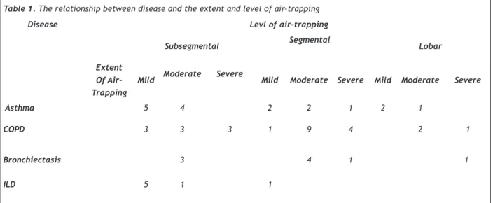
Mosaic pattern was detected in 49 patients on inspiratory images whereas on expiratory images air trapping was seen in 59 patients. The extent and level of air trapping is shown in table 1. In patients with subsegmental airtrapping the extent was mild in 13 (48.1%) and moderate in 11 (40.7%) patients. In patients with segmental air trapping the extent of air trapping was moderate in 15 (60%) patients. NPS showed correlation between the level and extent of air trapping (p<0.0001). In COPD patients the extent of air trapping correlated well with the level of air trapping. But in asthma patients the extent of air trapping did not correlate well with the level of air trapping. It was mild or moderate in patients with lobar involvement (Figure 1). And air trapping on expiratory images without mosaic pattern on inspiratory images was mostly detected in asthma (7 patients) (Figure 2). Two COPD and one ILD (Figure 3) patient had also expiratory air trapping.
Table 1. The relationship between disease and the extent and level of air-trapping
Disease Levl of air-trapping
Subsegmental Segmental Lobar
Extent Of Air-Trapping
Mild Moderate Severe Mild Moderate Severe Mild Moderate Severe
Asthma 5 4 2 2 1 2 1
COPD 3 3 3 1 9 4 2 1
Bronchiectasis 3 4 1 1
ILD 5 1 1
Table 2: Relationship between illness duration and
extent of air-trapping
Illness duration
Extent of Air-Trapping normal mild moderate severe
<5 years 12 7 7
5-10 years 4 8 9 1 >10 year 4 13 10
Fig1-A
Fig1-B
Figure 1: 63 y, F, Asthma: On inspiratory images (A), mosaic attenuation together with bronchiectasis is seen and on expiratory images (B) lobar air trapping on the right upper lobe
is seen. The extent of air trapping was mild in this patient.
Fig2-A
Fig2-B
Figure 2: 45 y, F, Asthma, on inspiratory images
(A), homogeneous attenuation is seen whereas on
expiratory images (B) subsegmental air trapping is seen.
Pulmonary function tests were normal in six patients, obstructive in 28 patients restrictive in14 patients, and mixed type in 27 patients. In five (6,7%) patients with normal PFT air-trapping with mild extension was detected. In six (8%) patients four obstructive and two mixed pattern no air trapping was detected. The PFT of all patients with mosaic pattern was abnormal. The extent of air-trapping was not severe in any of the patients with normal FEV1. All the patients with severely decreased FEV1 had air-trapping. In seven out of 11 patients with severely decreased FEV1 the extent of air trapping was severe. NPS showed significant correlation between FEV1 and extent of air-trapping. (p<0. 001)
There was no significant difference in the level of air trapping between smokers and non-smokers. Segmental and subsegmental involvement was dominant in both groups. Table 2 shows duration of illness and the extent of air-trapping on CT. Twelve (%46.1) of the patients with a history of less than five years illness duration did not show any trapping. And the extent of air-trapping was not severe in any of these patients.All the patients with a history of more than 10 years illness had air-trapping. In 10 (%37) of these patients the extent of air trapping was severe. There was correlation between duration of illness and the extent of air-trapping (p=0. 002). There was also correlation between duration of illness and the level of air-trapping (p=0.038).
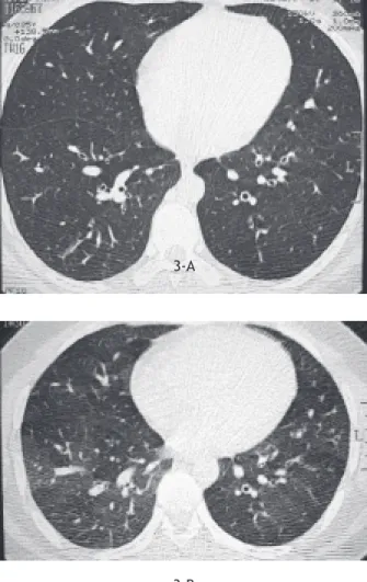
3-A
3-B
Figure 3: 35 y, F, ILD, on inspiratory images (A), homogeneous
attenuation together with interstitial thickening is seen
whereas on expiratory images (B) subsegmental air trapping
is seen.
DISCUSSION
Mosaic pattern presents as patchy decreased and increased attenuation areas. This finding is non-specific and may be seen in airway, vascular and infiltrative diseases. In airway and vascular diseases the diameter of the vascular structures in high attenuation areas is greater than the ones in low attenuation areas (7,8). In infiltrative diseases ground glass appearance (patchy decreased and increased attenuation areas) due to filling of alveolus is seen. The diameter of the vascular structures is equal in normal and abnormal appearing areas. Sometimes it is impossible to differentiate between ground-glass opacity and mosaic perfusion. In this case, expiratory
images may be helpful, no attenuation increase on expiratory images means air-trapping which is the cause of mosaic attenuation (7, 9). also in our study expiratory images were helpful for detecting air-trapping.
The inspiratory images are mostly abnormal in patients with air-trapping (10, 11). Yang et al proposed that the presence of air trapping on inspiratory images was a predictor of severe airway disease (12). In concordance to this none of our patients with mosaic perfusion had normal FEV1 levels. Arakawa et al (11) reported that %20 of patients with normal inspiratory images had air trapping on expiratory images. They also concluded that air trapping on expiratory images is a finding of mild airway disease. Also in our study the patients with expiratory air trapping had mild airway disease.Lucidarme et al. (7) reported that 41%of the patients with obstructive pattern on PFT had normal expiratory images. In the present study six (8%) patients with obstructive pattern on PFT had normal expiratory images. The presence of air trapping in patients with normal PFT and abnormal PFT without air trapping suggests that this two modalities are complementary.
Chung et al. (13) and Hansell et al. (14) found good correlation between FEV1 and air trapping. Whereas Lucidarme et al (7) reported a weak correlation. Tanaka et al. (15) found significant correlation between extent of air trapping and FEV1/FVC. This study also shows that there is a good correlation between FEV1 and the extent of air trapping.
Expiratory air trapping may also be seen in interstitial lung diseases such as hypersensitivity pneumonitis, sarcoidosis, histiocytosis-x and smokers (10,11,16,17). The bronchiolar obstruction is the result of fibrosis, bronchiolar, or peribronchiolar granulomas, interstitial infiltration, or inflammatory luminal exudates. Magkanas et al. (18) reported that %83 of sarcoiodosis patients had air trapping on expiratory images whereas only minority of them had mosaic pattern on inspiratory images. In the present study one sarcoidosis patient with normal inspiratory HRCT findings had expiratory air trapping.
The patterns of obstructive airway disease may discriminate diseases. Particularly air-trapping on expiratory images suggests asthma and obliterative bronchiolitis (10, 19). Our findings also supports that expiratory air trapping suggests asthma. We also think that lobar involvement without severe extent of air trapping suggests central involvement and is a pattern of
asthma. Kraft et al. (20) who performed transbronchial biopsies found that the peripheral airways were mostly affected in asthma patients. The studies performed with excised lungs concluded that central involvement was predominant. (21). But now a days it is widely accepted that the airway inflammation in asthma affects the central and peripheral airways. Although COPD is a disease of central airways it mostly affects the small airways (22). In our study the predominant involvement was subsegmental or segmental in 14 (%67) of asthma patients and 23 (83%) of COPD patients which favours small airway involvement. Lobar involvement was seen in three asthma and COPD patients. But the patients with lobar involvement without extensive air-trapping had the diagnosis of asthma which favours central involvement. Therefore our study suggests central and peripheral involvement in asthma patients and peripheral involvement in COPD patients.
The frequency of air trapping increases with age, and its severity increases with age and duration of illness and smoking (6, 22). Lee et al (6) found significant correlation between age, illness duration and the extent of air trapping . In our study there was no correlation between patient age and extent of air trapping This discrepancy is due to the heterogeneous patient population. Asthma is seen in younger people whereas COPD is seen in older people. This study showed that the duration of illness was a good predictor for air trapping and patients with more then 10 year illness had more extensive and mainly segmental or lobar involvement. Mastora et al (23) reported that segmental and lobar air trapping was more frequently observed among smokers whereas Tanaka et al. (15) found no correlation between smoking history and level of air trapping. Also in our study there was no correlation between the level of air trapping and smoking history, but this is probably the result of patient selection. In conclusion: Expiratory images are helpful for showing air trapping in patients with obstructive lung diseases. Expiratory air trapping without mosaic perfusion mainly suggests asthma. It seems that the illness duration is the most important predictor for the extent of air-trapping. The extent of air trapping correlates well with pulmonary function tests
REFERENCES
1. Kazerooni EA. High-resolution CT of the lungs. Am J
Roentgenol 2001;177:501-19
2. Worthy SA, Müller NL, Hartman TE, Swensen SJ, Padley SPG, Hansel DM. Mosaic attenuation pattern on thin section CT scan of the lung: differentiation among infiltrative, airway and vascular diseases as a cause. Radiology 1997; 205:465-70.
3. Arakawa H, Webb RW, McCowin M, Katsou G, Lee K, Seitz RF. Inhomogeneous lung attenuation at thin section CT: diagnostic value of expiratory scans. Radiology 1998; 206:89-94.
4. Takahashi M, Murata K, Takazakura R, et al. Bronchiolar
disease: spectrum and radiological findings. Eur J Radiol 2000; 35:15-29.
5. Kauczor HU, Hast J, Heussel CP, Schlegel J, Mildenberger P, Thelen M. CT attenuation of paired HRCT scans obtained at full inspiratory/expiratory position: comparison with pulmonary function tests. Eur Radiol 2002;12: 2757-63. 6. Lee KW, chung SY, Yang I, Lee Y, Ko YE, Park MJ. Correlation
of aging and smoking with air trapping at thin section CT
of the lung in asymptomatic subjects. Radiology 2000; 214:831-6.
7. Lucidarme O, Coche E, Cluzel P, Mourey-Gerosa I, Howarth
N, Grenier P. Expiratory CT scans for chronic airway
disease: correlation with pulmonary function test results. Am J Roentgenol 1998; 170:301-7.
8. Webb WR, Stern EJ, Nalini K. Dynamic pulmonary CT: findings in healthy adult men. Radiology 1993;186:117-24. 9. Stern EJ, Frank MS. Small airway diseases of the lungs: findings at expiratory CT. Am J Roentgenol1994;163:37-41. 10. Arakawa H, Webb WR. Air trapping on expiratory high
resolution CT scans in the absence of inspiratory scan abnormalities: correlation with pulmonary function tests and differential diagnosis. Am J Roentgenol 1998; 170:1349-53.
11. Arakawa H, Niimi H, Kurihara Y, Nakajima Y, Webb RW. Expiratory high resolution CT: diagnostic value in diffuse lung diseases. Am J Roentgenol 2000;175:1537-43. 12. Yang CF, Wu MT, Chiang AA, La, et al. Correlation of high
resolution CT and pulmonary function in bronchiolitis obliterans. A study based on 24 patients associated with
consumption of sauropus androgynus. Am J Roentgenol
1997;168:1045-50.
13. Chung MH, Edinburgh KJ, Webb EM, McCowin M, Webb WR. Mixed infiltrative and obstructive disease on high
resolution CT differential diagnosis and functional
correlates in consecutive series. J Thorac Imaging 2001; 16:69-75.
14. Hansell DM, Rubens MB, Padley SPG, Wells AU. Obliterative bronchiolitis: individual CT signs of small airway disease and functional correlations. Radiology 1997;203:721-6. 15. Tanaka N, Matsumoto T, Miura G, et al. Air trapping at CT:
high prevalence in asymptomatic subjects with normal pulmonary function. Radiology 2003;227:776-85. 16. Davies CWH, Tasker AD, Padley SPG, Davies RJO, Gleeson
FV. Air trapping in sarcoidosis on computed tomography: correlation with lung function. Clin Radiol 2000;55:217-21. 17. Terasaki H, Fujimoto K, Muller NL, et al. Pulmonary
sarcoidosis: comparison of findings of inspiratory and
expiratory high-resolution CT and pulmonary function
tests between smokers and nonsmokers. Am J Roentgenol 2005;185:333-8.
18. Magkanas E, Voloudaki A, Bouros D, et al. Pulmonary Sarcoıdosıs: Correlation of expiratory high-resolution CT findings with inspiratory patterns and pulmonary function tests. Acta Radiol 2001;42:494-501.
19. Copley SJ, Wells AU, Muller NL, et al. Thin section CT in obstructive pulmonary disease: discriminatory value. Radiology 2002;223:812-9
20. Kraft M, R. Djukanovic, Wilson S, Holgate S, Martin R. Alveolar tissue inflammation in asthma. Am J Respir Crit Care Med 1996;154:1505-11.
21. Carroll N, Cooke C, James A. The distribution of
eosinophils and lymphocytes in the large and small
airways of asthmatics. Eur Respir J 1997;10:292-300. 22. O’Brien C, Guest PJ, Hill SL, Stockley RA. Physiological
and radiological characterisation of patients diagnosed
with chronic obstructive pulmonary disease in primary care. Thorax 2000;55:635-42.
23. Mastora I, Remy-Jardin M, Sobaszek A, Boulenguez C, Remy J, Edme JL. Thin-section CT finding in 250 volunteers: assessment of the relationship of CT findings
with smoking history and pulmonary function test results.