R E S E A R C H A R T I C L E
Open Access
Analysis of complement biomarkers in
systemic sclerosis indicates a distinct
pattern in scleroderma renal crisis
Marcin Okrój
1,2, Martin Johansson
3, Tore Saxne
4, Anna M. Blom
1*†and Roger Hesselstrand
4†Abstract
Background:The complement system has been implicated in pathogenesis of systemic sclerosis (SSc). The goal of
the present study was to evaluate improved complement biomarkers in SSc.
Methods:The presence of C4d, reflecting activation of the classical/lectin pathways, C3bBbP corresponding to activation of the alternative pathway, and soluble terminal complement complexes (all complement pathways), was measured in plasma samples by enzyme-linked immunosorbent assay and correlated to clinical parameters. The study included 81 patients with limited cutaneous SSc and 41 with diffuse cutaneous SSc, as well as 47 matched healthy controls and 81 patients with rheumatoid arthritis, 22 with psoriatic arthritis and 20 with ankylosing spondylitis. Skin and kidney biopsies of selected patients were stained to detect deposited C3b as a marker of local complement activation.
Results:Biomarkers of activation of all complement pathways were increased in SSc compared with healthy controls and were similar to those in other rheumatic diseases. When patients with SSc were divided into subgroups, a distinct pattern of complement markers was observed in individuals with scleroderma renal crisis (SRC). By functional assay, we confirmed a significant decrease in complement haemolytic activity in SRC vs. non-SRC patients, indicating complement consumption. Further, we detected glomerular deposits of C3b in some patients with SRC.
Conclusions: The data indicate that complement activation is an important feature of SRC.
Keywords: Scleroderma, Complement, Biomarkers, Renal crisis, Systemic sclerosis
Background
Scleroderma, also termed systemic sclerosis (SSc), is an autoimmune disease of connective tissue. Its pathology involves excessive collagen production, resulting in fibro-sis of skin and internal organs [1, 2]. This condition is accompanied by microangiopathy of varying severity and locations, most obviously seen as Raynaud’s phenomenon. The most widely accepted classification distinguishes two main subtypes: limited cutaneous SSc (lcSSc) and diffuse cutaneous SSc (dcSSc) [3]. In the latter case, internal organs, most typically the kidneys, gastrointestinal tract, heart and lungs, are more severely affected. There is an
ongoing discussion about the primary cause of SSc be-cause many molecular patterns and various pathways have been found to be involved in the pathogenesis. Import-antly, 90% of patients with SSc present with autoanti-bodies to intracellular components such as topoisomerase, centromeres, histones, RNA polymerases or ribonucleases, and these patients also show an increase in surface density of CD19 on their B cells [2]. It has been shown that the presence of these autoantibodies represents specific phenotypes of the disease, but less is known about their pathogenic role.
Data from in vivo models show that low expression of CD19 affects B-cell proliferation, whereas overexpression potentiates antibody production and increases the degree of autoantibodies [4]. Indeed, whole-genome microarray analysis has demonstrated that gene expression patterns characteristic of plasma cells decreases more than 90% * Correspondence:[email protected]
†Equal contributors
1Department of Translational Medicine, Section of Medical Protein Chemistry,
Lund University, Inga Marie Nilssons Street 53, Malmö S-20502, Sweden Full list of author information is available at the end of the article
upon anti-CD19 treatment and correlates with inhibition of collagen expression [5]. Apart from intracellular com-ponents, protein complexes present on the surface of fibroblasts, lymphocytes and endothelial cells are also targets of autoantibodies in SSc [6]. These autoantibodies may activate fibroblasts to produce collagen, either directly or indirectly, by fuelling local inflammation and release of pro-inflammatory cytokines. However, it is un-clear to what extent the complement system, for which antibodies are a main trigger, contributes to SSc pathogen-esis. CD21 (CR2), a receptor on the surface of B cells, binds activation products of the main complement factor C3b. Following complement activation, C3b covalently binds target surfaces and forms transient enzymatic com-plexes: complement convertases such as C3bBbP, which fuel downstream events of the cascade such as release of the potent pro-inflammatory anaphylatoxin C5a, and for-mation of terminal complement complexes (TCCs), which can cause cell lysis. CD21 and CD19 associate and form a signal transduction complex capable of enhancing B-cell responses to antigen once CD21 binds complement degradation fragments [7].
In fact, over the last 30 years, researchers have tried to correlate the levels of complement proteins, markers of complement activation and circulating immune com-plexes in patients’ bloodstream with severity of SSc and different subtypes of the disease. Elevated immune com-plexes were found only in some patients and were not associated with clinical or serological features [8, 9]. In another study, low-molecular-weight markers of comple-ment activation—Ba, C3d and C4d—were measured by nephelometry in plasma of patients with SSc [10]. The results showed that C3d, C4d and Ba fragments, as well as C3d:C3 and C4d:C4 ratios, were clearly higher in patients with SSc than in healthy control subjects, indicating in-creased complement activation. Also, patients with dcSSc showed significantly higher values than those with lcSSc [10]. On the basis of observations of higher C4d values in patients with SSc and subendothelial deposition of im-mune complexes [11], the classical complement pathway may indeed play a role in the pathogenesis of SSc. How-ever, this should be confirmed in a larger number of patients and with validated methods capable of specifically measuring products of complement activation.
We recently established a novel enzyme-linked im-munosorbent assay (ELISA) that can reliably detect soluble C4d in plasma [12]. In the present study, we re-examined the usefulness of C4d as biomarker in a cohort of 122 patients with SSc. Also, we measured a soluble marker of alternative pathway activation, C3bBbP, and soluble TCC (sTCC), a marker of the final step of com-plement activation via any pathway in the same cohort. In order to compare the results obtained for patients with SSc with those for patients with other autoimmune
diseases in whom activation of the complement system was previously reported, we performed the same ana-lyses in patients with rheumatoid arthritis (RA) [13], psoriatic arthritis (PsoA) [14] and ankylosing spondylitis (AS) [15], as well as in healthy control subjects.
Methods Patients
Plasma samples were collected from 122 patients who fulfilled the American College of Rheumatology (ACR) criteria for SSc [16]. The disease was classified as dcSSc (n= 41) or lcSSc (n= 81), based on the extent of skin involvement [3]. Samples were collected within a mean (±SD) of 7.5 (8.5) years of disease onset, which was de-fined as the first non-Raynaud’s manifestation. A sum-mary of baseline characteristics of patients with SSc is presented in Table 1. Healthy control plasma samples were collected from 47 age-matched (mean 52.3 ± 8.7 years) and sex-matched (14 males, 33 females) healthy volunteers with no history of rheumatologic disease. All plasma samples were retrieved in a standardised fashion (non-fasting) and were stored at −80 °C after centrifuga-tion until the experiments. Addicentrifuga-tionally, we included plasma samples from age- and sex-matched patients with RA (n= 81), PsoA (n= 22) and AS (n= 20). All patients with RA fulfilled the 1987 ACR criteria [17]. Diagnoses of PsoA and AS were based on clinical judgment by the treating physician and included radiographic examinations when applicable. All patients with AS had axial involve-ment but no clinical signs of peripheral arthritis, whereas the patients with PsoA had peripheral arthritis but no clinical signs of axial involvement.
Clinical data
[image:2.595.306.539.584.689.2]Anti-nuclear antibodies (ANA) and anti-centromeric antibodies (ACA) were analysed by an indirect immuno-fluorescence technique using the human Hep-2 cell line as a substrate; anti-topoisomerase I antibodies (ATA)
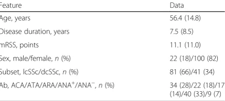
Table 1Clinical features at the time of blood sampling of 122 patients with systemic sclerosis
Feature Data
Age, years 56.4 (14.8)
Disease duration, years 7.5 (8.5)
mRSS, points 11.1 (11.0)
Sex, male/female,n(%) 22 (18)/100 (82)
Subset, lcSSc/dcSSc,n(%) 81 (66)/41 (34)
Ab, ACA/ATA/ARA/ANA+/ANA−,n(%) 34 (28)/22 (18)/17 (14)/40 (33)/9 (7)
Abbreviations:AbAutoantibodies,mRSSModified Rodnan skin score,lcSSc Limited cutaneous systemic sclerosis,dcSScDiffuse cutaneous systemic sclerosis, AbAutoantibodies,ACAAnti-centromere,ATAAnti-topoisomerase I,ARAAnti-RNA polymerase III,ANA+Anti-nuclear but not anti-centromere, anti-topoisomerase I
were analysed by ELISA; and anti-RNA polymerase III antibodies (ARA) were analysed by immunoprecipita-tion. Serum cartilage oligomeric matrix protein (COMP) was measured with a commercial sandwich ELISA using two monoclonal antibodies directed against separate antigenic determinants (AnaMar, Lund, Sweden) [18]. Subjects were classified as having dcSSc or lcSSc accord-ing to LeRoy et al.’s criteria [3]. The severity of skin in-volvement was determined by the modified Rodnan skin score (mRSS) [19]. Disease duration was determined as the time from the first non-Raynaud’s manifestation.
Measurement of complement activation markers
Plasma concentrations of C4d [12], C3bBbP [20] and sTCC [20] were measured by specific sandwich ELISAs in plasma ethylenediaminetetraacetic acid (EDTA) samples as described previously. Readout of each of these assays was given in complement activation units (CAU), a de-fined arbitrary unit set for the International Complement Standard #2 (ICS#2) sample, which is serum-pooled from about 1000 healthy individuals and incubated with activa-tors of all three complement pathways [20].
Haemolytic assays
Plasma EDTA samples collected from patients with SSc were tested for activity of classical or alternative comple-ment pathways by haemolytic assays performed as described elsewhere [21], with small modifications. Briefly, the classical complement pathway was activated on antibody-sensitised sheep erythrocytes. To overcome the inhibitory effect of EDTA, plasma samples were diluted 1:100 in DGVB2+buffer (2.5 mM veronal buffer, pH 7.35, 72 mM NaCl, 140 mM glucose, 0.1% gelatin,
1 mM MgCl2 and 0.15 mM CaCl2). For the
alterna-tive pathway, plasma samples were diluted 1:20 in Mg-ethylene glycol-bis(2-aminoethylether)-N,N,N′,N′ -tet-raacetic acid (Mg-EGTA) buffer (2.5 mM veronal buffer, pH 7.3, 70 mM NaCl, 140 mM glucose, 0.1% gelatin,
7 mM MgCl2and 10 mM EGTA) and added directly to
rabbit erythrocytes.
Immunohistochemical staining of C3
Kidney and skin biopsies collected from patients with SSc were fixed in formalin and embedded in paraffin. Sections of 4-μm thickness were cut and automatically pre-treated using the PT Link system (Dako/Agilent Technologies, Santa Clara, CA, USA) and then stained in an Autostainer Plus (Dako) with rabbit anti-human C3c polyclonal antibody (P0062; Dako) at a final dilution of 1:5000 for 30 minutes. Subsequently, the EnVision Flex HRP kit (Dako) was applied to the sections for 20 minutes to detect primary antibodies, followed by 3,3′-diaminobenzidine (DAB) reagent for visualisation.
Statistical analysis
Obtained data on the appearance of complement activa-tion markers and haemolytic activity were not distributed normally; in each complement measurement there were outliers, and some patient subgroups were rather small. Therefore, we used non-parametric statistical methods throughout the whole analysis. The Kruskal-Wallis and
Mann-Whitney U tests were used to compare multiple
groups or two groups, respectively. Spearman’s correlation was used to analyse relationships. p values <0.05 were considered significant. Calculations were performed with Prism 5.0 (GraphPad Software, La Jolla, CA, USA) and IBM SPSS Statistics version 20 (IBM, Armonk, NY, USA) software.
Results
The complement system is activated in patients with autoimmune diseases
We tested three different complement activation markers: (1) C4d, corresponding to activation of the classical pathway; (2) the fluid-phase alternative C3 convertase (C3bBbP), reflecting activation of the alternative pathway as well as the amplification loop enhancing the cascade at the level of convertases; and (3) sTCC, which evaluates the lytic (terminal) pathway (Fig. 1). All autoimmune pa-tients, regardless of diagnosis, had 5- to 15-fold increased levels of all three markers, thus confirming ongoing com-plement activation. Regarding SSc, C4d was increased to a similar degree as for AS and PsoA, but to a lesser extent than in RA (p= 0.05 by Kruskal-Wallis test). Interestingly, the alternative pathway was activated significantly more strongly in patients with SSc than in patients with RA (p< 0.001). All groups of patients showed similar levels of sTCC. As expected, healthy control subjects showed low levels of all three markers.
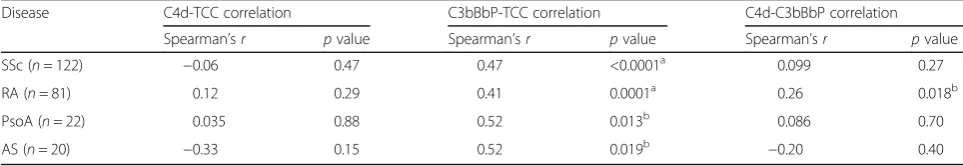
Correlations of complement activation markers in patients with autoimmune diseases
Because sTCC is a marker of the terminal complement pathway, which is fuelled by all complement pathways, we analysed which of the early complement markers correlate to appearance of sTCC in patients with SSc and other autoimmune diseases. Only C3bBbP, and not C4d, was significantly correlated to sTCC in all groups of patients. However, in patients with RA, C4d was significantly corre-lated to C3bBbP, which may indicate the role of the ampli-fication of the classical pathway via alternative convertases in overall complement activation in this patient group. Correlation coefficients andpvalues are given in Table 2.
Correlations of complement activation markers to mRSS and COMP
Spearman’s correlation analysis and found weak, bor-derline significant correlation between mRSS and C4d (Table 3). Furthermore, COMP, which is expressed by fibroblasts in SSc, is associated with mRSS [22] (in the present cohort, Spearman’s r= 0.47, p= 0.0005) and is known to activate the alternative pathway [23]. How-ever, we did not observe a statistically significant correl-ation between complement activcorrel-ation markers and COMP levels in plasma, implying that it is likely not a major complement trigger in these patients, at least not when released into blood.
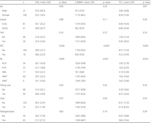
Analyses of complement activation markers in the subgroups of patients with SSc
We classified the patients into subgroups depending on the form of disease (lcSSc vs. dcSSc); typical complica-tions, such as pulmonary arterial hypertension (PAH), scleroderma renal crisis (SRC), pitting scars, ulcers, telan-giectasias; or the presence of certain types of anti-nuclear autoantibodies, including ACA, ATA, ARA, ANA without ACA, ATA or ARA, and finally ANA-negative. We did not observe significant differences in C4d, C3bBbP and
sTCC levels between men and women or between patients with lcSSc and those with dcSSc (Table 4). Also, concentra-tions of these markers were equally distributed within patients with and without PAH, pitting scars, ulcers, and telangiectasias. Importantly, a different distribution of complement activation markers was detected in pa-tients with SRC, who had significantly higher amounts
of C4d (p= 0.036) and significantly lower levels of
[image:4.595.60.538.88.260.2]C3bBbP (p< 0.001) and sTCC (p= 0.003) than patients without kidney involvement. When overall distribution of autoantibodies was analysed by the Kruskal-Wallis test, we found a significant difference for sTCC (p= 0.016) and a difference on the border of significance for C3bBbP (p= 0.055). Although not statistically signifi-cant (p= 0.095), there was a trend for C4d, according to which patients without any antinuclear autoantibodies presented with low levels of this marker. Furthermore, pa-tients with ARA positivity showed a pattern of complement activation markers similar to that of the patients with SRC (high C4d, low C3bBbP and TCC). Additional Kruskal-Wallis analysis performed for a single type of autoanti-body revealed that the ARA-positive group differed
Table 2Correlations between complement biomarkers C4d, C3bBbP and terminal complement complex in plasma samples from patients with autoimmune diseases
Disease C4d-TCC correlation C3bBbP-TCC correlation C4d-C3bBbP correlation
Spearman’sr pvalue Spearman’sr pvalue Spearman’sr pvalue
SSc (n= 122) −0.06 0.47 0.47 <0.0001a 0.099 0.27
RA (n= 81) 0.12 0.29 0.41 0.0001a 0.26 0.018b
PsoA (n= 22) 0.035 0.88 0.52 0.013b 0.086 0.70
AS (n= 20) −0.33 0.15 0.52 0.019b −0.20 0.40
Abbreviations: ASAnkylosing spondylitis,PsoAPsoriatic arthritis,RARheumatoid arthritis,SScSystemic sclerosis,TCCTerminal complement complex
a
p<0.001
b
p< 0.05
[image:4.595.56.538.624.707.2]significantly from patients without any antinuclear anti-bodies (ANA-negative) at a p level <0.05 for all
comple-ment markers. Also, the ARA-positive group had
significantly lower amounts of C3bBbP and TCC than ARA-negative patients (p< 0.01). Interestingly, 8 (47%) of 17 patients with ARA presented also with SRC, a group
that accounted only for 15% of all patients with SSc (18 of 122). Taken together, patients with ARA were, as ex-pected, overrepresented among those with SRC and, importantly, manifested the same pattern of comple-ment activation markers.
Haemolytic activity of plasma samples collected from patients with SSc
[image:5.595.57.289.110.166.2]The levels of C3bBbP and sTCC in the SRC group (means 656 CAU and 4.22 CAU, respectively) were lower than in other patients with SSc; however, these levels were still considerably higher than analogous means for healthy control subjects (79.7 CAU and 1.64 CAU, respectively), indicating that complement activa-tion takes place during the renal crisis. At the same time, C4d, which is a marker of classical pathway activation
Table 3Correlations of complement activation markers with age, modified Rodnan skin score and cartilage oligomeric matrix protein
n C4d pvalue C3bBbP pvalue TCC pvalue
Age 122 −0.14 0.13 −0.083 0.36 0.017 0.86
mRSS 118 0.17 0.06 −0.16 0.087 −0.082 0.38
COMP 53 0.16 0.26 −0.052 0.71 0.092 0.51
Abbreviations: COMPCartilage oligomeric matrix protein,mRSSModified Rodnan skin score,TCCTerminal complement complex
Table 4Analyses of complement activation markers in the subgroups of patients with systemic sclerosis
n C4d, mean (SD) pvalue C3bBbP, mean (SD) pvalue TCC, mean (SD) pvalue
Sex 0.96 0.20 0.87
Male 22 35.6 (40.3) 913 (574) 5.80 (3.69)
Female 100 29.2 (18.3) 1119 (661) 8.39 (15.8)
Subset 0.88 0.11 0.36
lcSSc 81 30.1 (25.2) 1143 (655) 8.56 (16.4)
dcSSc 41 30.8 (20.7) 962 (629) 6.66 (9.34)
PAH 0.19 0.75 0.76
No 96 31.8 (25.3) 1069 (645) 7.50 (12.4)
Yes 26 25.0 (16.0) 1131 (676) 9.49 (20.5)
SRC 0.036 <0.001 0.003
No 104 28.9 (23.7) 1156 (655) 8.57 (15.4)
Yes 18 38.6 (22.9) 656 (423) 4.22 (5.54)
Ab 0.095 0.055 0.016
ACA 34 30.7 (16.9) 1029 (594) 5.83 (5.70)
ATA 22 32.7 (18.8) 1140 (744) 10.8 (22.0)
ARA 17 35.0 (22.5) 761 (560) 3.19 (2.59)
ANA+ 40 29.5 (32.2) 1195 (642) 10.6 (18.0)
ANA− 9 18.1 (10.0) 1244 (705) 5.62 (3.36)
Pitting scar 0.50 0.35 0.76
No 68 31.0 (26.1) 1071 (658) 9.30 (18.6)
Yes 50 28.6 (19.0) 1155 (633) 6.53 (5.62)
Ulcer 0.35 0.36 0.54
No 105 30.5 (23.9) 1090 (656) 8.31 (15.3)
Yes 14 25.5 (17.8) 1183 (574) 6.19 (6.53)
Telangiectasias 0.83 0.19 0.39
No 56 28.5 (17.8) 1003 (586) 6.07 (3.80)
Yes 63 31.2 (27.2) 1189 (687) 9.84 (19.6)
Abbreviations:PAHPulmonary arterial hypertension,SRCScleroderma renal crisis,AbAutoantibodies,ACAAnti-centromere,ATAAnti-topoisomerase I,ARAAnti-RNA polymerase III,ANA+Anti-nuclear but not anti-centromere, anti-topoisomerase I or anti-RNA polymerase III,ANA−No anti-nuclear autoantibodies,dcSScDiffuse cutaneous
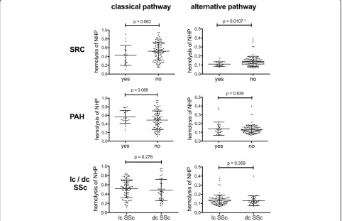
[image:5.595.58.541.306.699.2]and the end-degradation product of C4b component, in-creased to significantly higher levels than in the non-SRC group. One possible explanation for these observa-tions is that during SRC the classical pathway is strongly initiated by autoantibodies, and then an amplification loop driven by the alternative pathway takes over the cascade, leading to systemic depletion of its components, including C3 and factor B proteins. Such depletion is also reflected in a reduction of soluble sTCC compared with patients with no SRC episodes. To confirm the sce-nario of ongoing complement consumption in SRC, we analysed haemolytic activity of plasma samples from pa-tients with SSc. Residual complement activity was exam-ined in assays testing the classical and alternative pathways (Fig. 2). Of all classifications made for the whole SSc cohort, only the SRC-positive group showed signifi-cantly lower haemolytic activity in the alternative pathway (p= 0.0107) and a borderline significant drop of haemo-lytic activity in the classical pathway (p= 0.063), which confirmed systemic complement depletion, most probably at the level of alternative convertases.
C3 staining in kidney biopsies from patients with SRC
Because the SRC subgroup showed a distinct pattern of complement activation markers, we searched for the pri-mary source of a putative strong complement activation deduced from lower levels of C3bBbP and lower haemo-lytic activity of plasma samples. Kidney biopsies from 5 of 18 patients with SRC included in the study were available, and we examined these for C3b deposition by immunohis-tochemistry. C3b deposits were detected in glomeruli of two patients and in tubules of one more patient (Fig. 3), whereas C3b staining was negative for kidneys of two remaining patients. These findings indicate that ongoing complement activation in kidneys may be one of the hall-marks of SRC. However, it does not explain all cases, and other sources of activation are also plausible.
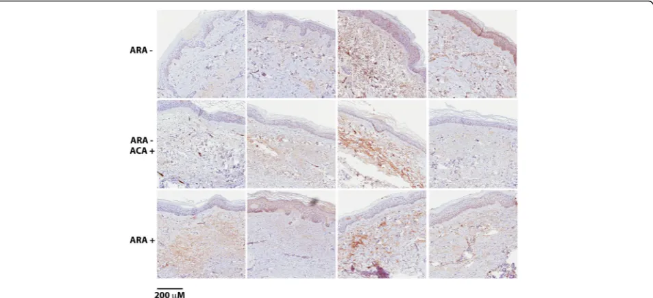
C3b staining in skin biopsies from ARA+, ARA−and ARA−/ACA+patients
Searching for further explanations for complement acti-vation in patients with SRC, we followed the overrepre-sentation of ARA-positive patients in the SRC group
[image:6.595.57.540.354.665.2]and analysed C3b deposition in skin biopsies obtained from ARA+, ARA− and ARA−/ACA+ patients. Material from four patients in each group was available. We
observed C3b deposition in skin tissue from all ARA+
patients, but other groups also had at least two of four slides that were positive (Fig. 4), so there was no dra-matic difference between the groups. The staining was localised to the dermal layer and most prominent in the endothelium of small dermal arteries. Less staining was associated with the collagenous fibres of the dermis. In the basal layer of the epidermis, cells were discovered containing what seemed to be stain. However, these cells were melanocytes, and the pigment was melanin, not DAB chromogen (Fig. 4).
Discussion
Reliable measurement of complement activation is not a trivial task. Of two possible strategies, measurement of na-tive complement protein consumption and measurement of markers appearing only after complement activation, the latter seems to be more reliable. This is due partly to large interpersonal differences in some complement pro-teins, such as that normal concentration of the main com-plement protein C3 ranges from 0.8 to 1.8 mg/ml. On one hand, these levels are affected by both consumption and a changing rate of synthesis because many comple-ment factors are acute-phase response proteins. On the other hand, challenges in the strategy of measuring products of activation relate to the specificity of
Fig. 3C3b deposition in kidney biopsies from patients with scleroderma renal crisis (SRC). Shown are representative micrographs of kidney biopsy cores immunostained for C3b.aAn example of a biopsy obtained from a patient with SRC, which was negative for C3b.bExample of a biopsy positive for C3b in glomeruli.cExample of a biopsy positive for C3b in tubules.dPositive control biopsy from patient diagnosed for immunoglobulin A nephropathy with distinct glomerular C3 positivity
Fig. 4C3b staining of skin biopsied from anti-RNA polymerase III (ARA+), ARA−and anti-centromere-positive (ACA+)/ARA−patients. Shown are
[image:7.595.62.538.89.214.2] [image:7.595.61.538.463.681.2]antibodies, which must target only neoepitopes local-ised on processed complement proteins and not epi-topes on ubiquitous, native molecules. Previous studies performed with 34 patients with SSc revealed higher levels of C4d in blood of patients with dcSSc than in patients with lcSSc, suggesting that C4d content may be associated with clinical severity of SSc [10]. How-ever, the authors of that study did not use specific anti-C4d antibodies; instead, they performed precipitation of high-molecular-weight plasma proteins and detected C4d in supernatant with whole antiserum by nephelom-etry. We re-examined this hypothesis using plasma samples of 122 patients with our recently designed C4d ELISA. The advantage of this method over nephelome-try and other, previously used C4d immunoassays is that it uses a novel antibody specific to a 5-aa linear cleavage site neoepitope. Previously, we showed that the readout of this assay is unaffected by repeated freezing-thawing or heating of samples, which is not the case for some other techniques based on conform-ational neoepitopes [12]. We noticed a clear increase of C4d as well as the complement activation markers C3bBbP and sTCC in patients with SSc as well as in patients with other rheumatic diseases compared with healthy control subjects (Fig. 1). However, we could confirm neither a link between clinical severity of SSc and C4d content nor associations of complement acti-vation markers with particular syndromes characteristic for SSc, except for SRC.
Renal crisis is reported typically in 5–10% patents with SSc [24, 25]. Historically, this group of patients had a high risk of death (up to 85% mortality after 1 year [26]), and even after introducing treatment with angiotensin-converting enzyme inhibitors, mortality remains rela-tively high at 18% after the first year and 41–58% after 5 years [27, 28]. To prevent loss of kidney function and other associated complications, such as systemic hyper-tension, retinopathy or pulmonary oedema, patients re-quire early identification and aggressive treatment [25]. Therefore, increasing knowledge about underlying pathomechanisms of SRC is necessary. Our data sug-gest that activation of complement, which is a feature common to rheumatic and/or autoimmune diseases [13, 29], presents differently in patients with SSc with SRC. Another important observation was that the same pattern of complement activation markers seen in SRC was observed in the group of ARA-positive patients, which accounted for 44% of patients with SRC in our study and 33% [30] to 59% [27] in other studies. Trends and a signifi-cant increase of C4d marker in these two groups of patients linked to each other speak for enhanced activation of the classical pathway. C3bBbP is a marker that may indicate ac-tivation of the alternative pathway alone or as an amplifica-tion loop of the classical or lectin pathway. At first glance,
elevated C4d and decreased C3bBbP levels in the same pa-tients may appear contradictory, but the nature of these markers may provide a logical explanation. Whereas C4d is the end degradation product of an early component of the classical pathway C4b and may accumulate over time, C3bBbP is a fluid-phase convertase, an enzymatic complex that converts available C3 into C3b. Activation of the alter-native pathway leads to depletion of C3 and factor B and, by doing so, limits de novo formation of C3bBbP. However, C3bBbP decays both spontaneously and with the aid of sev-eral complement inhibitors [20]. As a result of limited replacement of alternative pathway components and pathway exhaustion, patients with acute episodes of complement activation may present with lower C3bBbP content than those with chronic disease. The same is true for sTCC.
On the basis of our results, we hypothesised that acute complement activation takes place during the onset of SRC and that depletion of available complement is an-other consequence of such scenario. Our hypothesis was confirmed by haemolytic assays in which residual com-plement activity of plasma from patients with SRC was significantly lower than that in non-SRC subjects. In conjunction with a previous report showing increased C4d deposition in peritubular capillaries of patients with SSc with SRC compared with normotensive controls and hypertensive non-SRC control subjects [24], we should consider local complement activation as an important element of the SRC pathomechanism. This hypothesis is strengthened by presence of C3b deposits, which reflect complement activation via all pathways, in three of five analysed kidney biopsies from patients with SRC. We also tested whether ARA-positive patients overrepre-sented in the SRC group had increased C3b deposits in skin. Although all ARA-positive individuals showed C3b staining in skin biopsies, we also found it in some ARA-negative patients. Therefore, we hypothesise that local complement activation in kidneys and skin takes place during SRC, but that this phenomenon is probably not limited only to this subgroup and that other, distinct features may be characteristic for renal involvement. Nonetheless, complement activation may be a strongly contributing factor in some patients with SRC, and these patients could be considered for treatment with emer-ging complement inhibitors such as eculizumab, which is now used in therapy for another kidney disease with complement involvement: haemolytic uremic syndrome.
Conclusions
indicating ongoing complement consumption. This, finding, together with glomerular deposits of C3b found in some of these patients, indicates that complement activation is an important feature of SRC.
Abbreviations
ACA:centromere; ACR: American College of Rheumatology; ANA: nuclear; ARA: RNA polymerase III; AS: Ankylosing spondylitis; ATA: Anti-topoisomerase I; CAU: Complement activation units; COMP: Cartilage oligomeric matrix protein; DAB: 3,3′-Diaminobenzidine; dcSSc: Diffuse cutaneous systemic sclerosis; EDTA: Ethylenediaminetetraacetic acid; EGTA: Ethylene glycol-bis(2-aminoethylether)-N,N,N′,N′-tetraacetic acid; ELISA: Enzyme-linked immunosorbent assay; lcSSc: Limited cutaneous systemic sclerosis; mRSS: Modified Rodnan skin score; NHP: Normal human plasma; PAH: Pulmonary arterial hypertension; PsoA: Psoriatic arthritis; RA: Rheumatoid arthritis; SRC: Scleroderma renal crisis; SSc: Systemic sclerosis; sTCC: Soluble terminal complement complex; TCC: Terminal complement complex
Acknowledgements
We thank Marie Wildt for excellent technical assistance and Dr. Ben King for proofreading the manuscript.
Funding
This study was supported by the Swedish Research Council (K2012-66X-14928-09-5), the Polish National Science Centre (2014/14/E/NZ6/00182), the Alfred Österlund Foundation, the Kock Foundation, King Gustav V’s 80th Birthday Fund, the Knut and Alice Wallenberg Foundation, and the IngaBritt and Arne Lundberg Foundation, as well as by grants for clinical research (Medical Training and Research Agreement [ALF] and Skåne University Hospital).
Availability of data and materials
Not applicable: no supplementary files, no databases.
Authors’contributions
AMB, RH and MO were responsible for study conception and design. MO was responsible for the acquisition of experimental data. RH and TS recruited patients and provided samples and clinical data. MO, RH and MJ performed the data analysis and interpretation. All authors were involved in drafting the manuscript or revising it critically for important intellectual content, and all authors read and approved the final manuscript.
Competing interests
AMB and MO are named as inventors in a patent application (Antibodies specific for complement component C4d and uses thereof; application number PCT/EP2015/070526). The other authors state that they have no competing interests.
Consent for publication Not applicable.
Ethical approval and consent to participate
Informed consent was obtained from all participants involved in the study, and permissions were obtained from the regional ethical review board in Lund.
Author details
1Department of Translational Medicine, Section of Medical Protein Chemistry,
Lund University, Inga Marie Nilssons Street 53, Malmö S-20502, Sweden.
2Department of Medical Biotechnology, Intercollegiate Faculty of
Biotechnology UG-MUG, Medical University of Gdańsk, Gdańsk 80210, Poland.
3Department of Translational Medicine, Section of Clinical Pathology, Lund
University, Jan Waldenströms street 59, Malmö S-20502, Sweden.
4Department of Clinical Sciences, Lund, Section of Rheumatology, Lund
University, Skåne University Hospital, Lund S-22185, Sweden.
Received: 20 June 2016 Accepted: 27 October 2016
References
1. Balbir-Gurman A, Braun-Moscovici Y. Scleroderma–new aspects in pathogenesis and treatment. Best Pract Res Clin Rheumatol. 2012;26:13–24. 2. Yoshizaki A, Sato S. Abnormal B lymphocyte activation and function in
systemic sclerosis. Ann Dermatol. 2015;27:1–9.
3. LeRoy EC, Black C, Fleischmajer R, Jablonska S, Krieg T, Medsger Jr TA, et al. Scleroderma (systemic sclerosis): classification, subsets and pathogenesis. J Rheumatol. 1988;15:202–5.
4. Engel P, Zhou LJ, Ord DC, Sato S, Koller B, Tedder TF. Abnormal B lymphocyte development, activation, and differentiation in mice that lack or overexpress the CD19 signal transduction molecule. Immunity. 1995;3:39–50.
5. Streicher K, Morehouse CA, Groves CJ, Rajan B, Pilataxi F, Lehmann KP, et al. The plasma cell signature in autoimmune disease. Arthritis Rheumatol. 2014;66:173–84.
6. Chizzolini C, Raschi E, Rezzonico R, Testoni C, Mallone R, Gabrielli A, et al. Autoantibodies to fibroblasts induce a proadhesive and proinflammatory fibroblast phenotype in patients with systemic sclerosis. Arthritis Rheum. 2002;46:1602–13.
7. Tedder TF, Poe JC, Fujimoto M, Haas KM, Sato S. The CD19-CD21 signal transduction complex of B lymphocytes regulates the balance between health and autoimmune disease: systemic sclerosis as a model system. Curr Dir Autoimmun. 2005;8:55–90.
8. Swierczynska Z, Rdultowska H, Blaszczyk M, Jablonska S, Luft S. Circulating immune complexes in systemic scleroderma. Immunol Commun. 1984; 13:433–8.
9. Siminovitch K, Klein M, Pruzanski W, Wilkinson S, Lee P, Yoon SJ, et al. Circulating immune complexes in patients with progressive systemic sclerosis. Arthritis Rheum. 1982;25:1174–9.
10. Senaldi G, Lupoli S, Vergani D, Black CM. Activation of the complement system in systemic sclerosis: relationship to clinical severity. Arthritis Rheum. 1989;32:1262–7.
11. Chen M, Daha MR, Kallenberg CG. The complement system in systemic autoimmune disease. J Autoimmun. 2010;34:J276–86.
12. Blom AM, Osterborg A, Mollnes TE, Okroj M. Antibodies reactive to cleaved sites in complement proteins enable highly specific measurement of soluble markers of complement activation. Mol Immunol. 2015;66:164–70.
13. Okroj M, Heinegard D, Holmdahl R, Blom AM. Rheumatoid arthritis and the complement system. Ann Med. 2007;39:517–30.
14. Chimenti MS, Perricone C, Graceffa D, Di Muzio G, Ballanti E, Guarino MD, et al. Complement system in psoriatic arthritis: a useful marker in response prediction and monitoring of anti-TNF treatment. Clin Exp Rheumatol. 2012;30:23–30.
15. McGuigan LE, Geczy AF, Edmonds JP. The immunopathology of ankylosing spondylitis—a review. Semin Arthritis Rheum. 1985;15:81–105.
16. Subcommittee for scleroderma criteria of the American Rheumatism Association Diagnostic and Therapeutic Criteria Committee. Preliminary criteria for the classification of systemic sclerosis (scleroderma). Arthritis Rheum. 1980;23:581–90.
17. Arnett FC, Edworthy SM, Bloch DA, McShane DJ, Fries JF, Cooper NS, et al. The American Rheumatism Association 1987 revised criteria for the classification of rheumatoid arthritis. Arthritis Rheum. 1988;31:315–24. 18. Otteby KE, Holmquist E, Saxne T, Heinegard D, Hesselstrand R, Blom AM.
Cartilage oligomeric matrix protein-induced complement activation in systemic sclerosis. Arthritis Res Ther. 2013;15:R215.
19. Clements P, Lachenbruch P, Siebold J, White B, Weiner S, Martin R, et al. Inter and intraobserver variability of total skin thickness score (modified Rodnan TSS) in systemic sclerosis. J Rheumatol. 1995;22:1281–5. 20. Bergseth G, Ludviksen JK, Kirschfink M, Giclas PC, Nilsson B, Mollnes TE. An
international serum standard for application in assays to detect human complement activation products. Mol Immunol. 2013;56:232–9. 21. Okroj M, Holmquist E, Sjolander J, Corrales L, Saxne T, Wisniewski HG, et al.
Heavy chains of inter alpha inhibitor (IαI) inhibit the human complement system at early stages of the cascade. J Biol Chem. 2012;287:20100–10. 22. Hesselstrand R, Kassner A, Heinegard D, Saxne T. COMP: a candidate
molecule in the pathogenesis of systemic sclerosis with a potential as a disease marker. Ann Rheum Dis. 2008;67:1242–8.
24. Batal I, Domsic RT, Shafer A, Medsger Jr TA, Kiss LP, Randhawa P, et al. Renal biopsy findings predicting outcome in scleroderma renal crisis. Hum Pathol. 2009;40:332–40.
25. Denton CP, Lapadula G, Mouthon L, Müller-Ladner U. Renal complications and scleroderma renal crisis. Rheumatology (Oxford). 2009;48 Suppl 3:iii32–5. 26. Steen VD, Costantino JP, Shapiro AP, Medsger Jr TA. Outcome of renal crisis in
systemic sclerosis: relation to availability of angiotensin converting enzyme (ACE) inhibitors. Ann Intern Med. 1990;113:352–7.
27. Penn H, Howie AJ, Kingdon EJ, Bunn CC, Stratton RJ, Black CM, et al. Scleroderma renal crisis: patient characteristics and long-term outcomes. QJM. 2007;100:485–94.
28. Hesselstrand R, Scheja A, Wuttge DM. Scleroderma renal crisis in a Swedish systemic sclerosis cohort: survival, renal outcome, and RNA polymerase III antibodies as a risk factor. Scand J Rheumatol. 2012;41:39–43.
29. Sturfelt G, Truedsson L. Complement in the immunopathogenesis of rheumatic disease. Nat Rev Rheumatol. 2012;8:458–68.
30. Bunn CC, Denton CP, Shi-Wen X, Knight C, Black CM. Anti-RNA polymerases and other autoantibody specificities in systemic sclerosis. Br J Rheumatol. 1998;37:15–20.
• We accept pre-submission inquiries
• Our selector tool helps you to find the most relevant journal
• We provide round the clock customer support
• Convenient online submission
• Thorough peer review
• Inclusion in PubMed and all major indexing services
• Maximum visibility for your research
Submit your manuscript at www.biomedcentral.com/submit