Study of fingertip pattern in Carcinoma Cervix patients
Dr. Asutosh Pramanik
*, Dr. Arnab Bhattacharya**
*Demonstrator, Department of Anatomy, Murshidabad Medical College & Hospital, Berhampore, West Bengal. **
Assistant Professor, Department of Anatomy, Institute of Post Graduate Medical Education & Research, Kolkata, West Bengal.
DOI: 10.29322/IJSRP.8.5.2018.p7723 http://dx.doi.org/10.29322/IJSRP.8.5.2018.p7723
Abstract- Dermatoglyphic study to correlate a particular dermatoglyphic pattern with occurrence of cervical carcinoma in the Northern Bengal population was done for a period of one year (July2015 to June 2016). Fingertip patterns of 72 cases of cervical carcinoma were tested against 72 controls. The results showed a statistically significant decrease in the frequency of ulnar loop pattern in cervical cancer patients(52.78%) compared to control group(60.83%) in both the hands. There is decrease in the percentage of Radial loops in cervical cancer patients (3.19%) compared to control group (7.36%) in both hands and the difference is statistically significant. The percentage of whorls decreased in control group (27.50%) compared to cervical cancer patients (38.89%) and the difference is statistically significant in both hands.
Index Terms- dermatoglyphics, cervical cancer, finger print, fingertip pattern.
I. INTRODUCTION
ermatoglyphics is the scientific study of epidermal ridges and their configurations on the palmar region of hand and fingers and plantar region of foot and toes. The term dermatoglyphics was coined by Cummins H and Midlo C in 1926 and was derived from Greek words „derma‟ means skin and „glyphics‟ means carvings (Penrose LS, 1963)[1,2]. Development of dermatoglyphic pattern is under genetic control. This is evident from the clear resemblance of dermatoglyphics among related person (Schaumann B and Alter M, 1976)[3]. There are many diseases known to be caused by abnormal genes. Whenever there is any abnormality in the genetic make up of parents it is inherited to the children and is reflected in dermatoglyphic pattern. (Walkar JFA, 1941)[4]. Dermatoglyphics as a diagnostic aid is now well established in a number of diseases, which have a strong hereditary basis, and is employed as a method of screening abnormal anomalies (Holt SB, 1961)[5] ; (Holt SB and Lindsten J, 1964)[6].
Dermatoglyphic analysis as a diagnostic tool has many advantages:
Epidermal ridge pattern on the palm are fully developed at birth and each individual‟s ridge configurations are unique & remains unchanged from womb to tomb except in the dimension in proportion to the growth of an individual. Question of chance of similarity is a theoretical possibility being 1:64 billion[7].
Patterns are readily accessible.
Recordings are quick, simple & inexpensive.
There is no trauma to individual during recording.
Ridge patterns can quickly be analyzed.
Ridge patterns can be inspected for abnormalities immediately after birth.
The cervical cancer is one of the most extensively studied cancers & its genetic basis was well established[8]. High mortality & morbidity rate poses a heavy economic burden on families[9] as well the country[10]. Additionally high medical costs that are incurred by families due to cervical cancer further impoverish individuals and communities[11]. The screening procedures (i.e. Pap smear test etc.) have been effectively reducing the incidence rate by 80% and mortality by 70%[12]. But they are invasive procedures & expensive also and an equipped set up is required.
The etiology of cervical carcinoma is multi-factorial with genetics playing an important role. Taking into consideration of genetic predisposition of dermatoglyphics and cervical carcinoma, the study was undertaken to find out correlation between them. Being inexpensive and noninvasive procedure (in a country like India) like dermatoglyphics may be useful investigatory or screening procedures for the population at risk in cervical carcinoma.
II. AIMS & OBJECTIVES
To determine various fingertip patterns in women suffering from cervical carcinoma and compare them with normal subjects in North Bengal.
To find out fingertip patterns in women having cervical carcinoma in North Bengal.
To compare the specific fingertip patterns in normal women and women having cervical cancer.
III. MATERIALS & METHODS
3.1 STUDY TYPE AND DESIGN: This was an Analytical and
Cross-sectional study.
3.2 STUDY AREA: In collaboration with the departments of
Radiotherapy (OPD), Gynecology (OPD & IPD) of North Bengal Medical College & Hospital.
3.3 STUDY PERIOD: The tenure of the study was one year
from July 2014-June 2015.
3.4 STUDY POPULATION: Women having Cervical
ISSN 2250-3153
Gynecology OPD & IPD in North Bengal Medical College and Hospital.
3.5 INCLUSION CRITERIA:
Histopathologically confirmed cases of cervical carcinoma of above 15 years of age[13].
The first degree relatives were selected for normal group who are above 15 years of age.
3.6 EXCLUSION CRITERIA: Very seriously ill patients.
Women having any obvious genetic disorder and having any other carcinoma in body and diseases causing dermatoglyphic changes[7].
History of any type of skin hypersensitivity.
Women not willing to give consent.
Cervical cancer patient with no first degree relatives.
3.7 SAMPLE SIZE:
72 patients with cervical cancer.
72 women with no cervical cancer.
Total 144 subjects.
3.8 SAMPLING DESIGN: Sampling design by complete enumeration method. After taking proper history of 108 histopathologically confirmed cervical cancer patients, 36 patients were excluded. So remaining 72 patients‟ dermatoglyphics were taken. The 72 control women were also found from the first degree relatives of cervical cancer patients. 3.9 STUDY TOOLS: Black duplicating gel, „T‟ shaped rubber roller, Inking slab, Reducer, One side glazed mop lithography paper(A-3), Doctors‟ spirit, Soap, Drawing board and board clips, Cotton, Protractor, magnifying lens, scale, calculator, pencil and pen, Needle with a sharp point, for ridge counting, Format for Consent and History taking.
3.10 STUDY TECHNIQUES: Among the various number of methods used for recording dermatoglyphics, the most routinely used one (that having no harm onto human body) is the INK METHOD (described by Cummins H in 1936[14] and Cummins H and Midlo C, 1961[15]) was used for this study.
3.11 STEPS IN THE PRINTING METHOD:
1. After taking proper consent and brief history, the subjects were asked to clean their hands with soaps and water to remove any oil or dirt and dry their hands but to leave some moisture.
2. The requisite amount of Black Duplicating Ink was placed on the glass slab. It was uniformly spread by the „T‟ shaped rubber roller to get a thin even ink film on the glass slab. 3. The thin film of ink was applied on the palm by passing the
inked rubber roller uniformly over the palm and digits. 4. Left hand of the subject was then placed on the sheet of
paper (kept over the pressure pad) from proximal to distal end. The palm was then lifted from the paper in reverse order, from the distal to proximal end. The fingers were also printed below the palmar print by rolled finger print method.
(Figure 1, 2).
5. The same procedure was repeated for right hand.
6. The prints were then subjected for detail dermatoglyphic statistical analysis (Figure 3).
Figure1: Print of Finger taken by rolled finger print method
Figure 3: The Completed set of both sides palm and all ten fingertip prints taken on mop-lithography paper.
3.12 PARAMETERS/VARIABLES STUDIED:
Fingertip patterns i.e. loops (radial and ulnar), Whorls, arches. 3.13 FINGERTIP PATTERN CONFIGURATION:
Galton F (1892)[16], divided fingertip patterns into 3 groups - Loops, Arches and Whorls. Henry ER (1900)[17], added 4th group „Composites‟ to demarcate more complex patterns.
3.14 Fingertip Pattern:
1. Arch (A): An arch is the simplest pattern. It consists of more or less parallel ridges. This pattern is present in 6-7% cases. 2. Loop (L): It is the most frequent pattern on fingertip. This pattern is present in 65-67% cases. It is subdivided into two types: Ulnar Loop (Lu), Radial Loop (Lr)
3. Whorl (W): According to Galton‟s classification, whorl
is any ridge configuration with two
or more triradii. According to Henry‟s classification whorl is a ridge configuration in which ridges actually encircle core and more complex patterns are called as „Composites‟[7].
3.15 STATISTICAL ANALYSIS:
Chi Squared test was used for analysis the qualitative values. Statistical analyses were done using a computer-based on-line Programmer[18] and Microsoft Office Excel 2007. Differences were considered significant if P values were less than 0.05.
IV. RESULTS & ANALYSIS
ISSN 2250-3153
Table 1: Distribution of study population and statistical analysis according to fingertip pattern of both hands(72x10=720) among patients(n1=72) & controls(n2=72).
Groups Fingertip patterns
Ulnar loops
Radial loops
Arches Whorls
Patients Frequency 380 23 37 280
Percentage 52.78 3.19 5.14 38.89
Controls Frequency 438 53 31 198
Percentage 60.83 7.36 4.31 27.50 Chi square value 4.112 11.842 0.530 14.067 P value <0.05 <0.001 >0.05 <0.001
Remarks S ES NS ES
Legend Table 1:The above table 1 shows that the frequencies of ulnar loop and radial loop are more in control groups than cervical cancer patients. The differences are statistically significant for ulnar loop and radial loop. The
[image:4.612.182.431.314.432.2]frequency of whorl pattern is more in patients than controls, and this difference is statistically significant. The frequencies of arch pattern are statistically non-significant.
Table 2: Distribution of study population and statistical analysis according to fingertip pattern of right hand (72 x 5=360) among patients(n1=72) & controls(n2=72).
Groups Fingertip patterns
Ulnar loops
Radial loops
Arches Whorls
Patients Frequency 175 15 22 148
Percentage 48.61 4.17 6.11 41.11
Controls Frequency 196 34 20 110
Percentage 54.44 9.44 5.56 30.56 Chi square value 1.188 7.368 0.096 5.596 P value >0.05 <0.01 >0.05 <0.05
Remarks NS HS NS S
Legend Table 2: The above table 2 shows the differences of frequencies of whorls and radial loops between patients and control groups statistically significant for right hands. But the frequencies of other two patterns are not statistically non-significant between two groups.
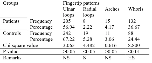
Table 3: Distribution of study population and statistical analysis according to fingertip pattern of left hand (72 x 5=360) among patients(n1=72) & controls(n2=72).
Legend Table 3:The above table 3 shows the differences of frequencies of radial loops and whorls are statistically significant between cervical cancer patients and controls in left hands. The differences of frequencies of ulnar loops and arches are statistically non-significant between two groups.
V. DISCUSSION
The study consisted of 72 histopathologically confirmed cervical carcinoma patients (above 15 years of age) and equal numbers of normal healthy females were selected from the first degree relatives who were above 15 years of age, were included
Groups Fingertip patterns
Ulnar loops
Radial loops
Arches Whorls
Patients Frequency 205 8 15 132
Percentage 56.94 2.22 4.17 36.67
Controls Frequency 242 19 11 88
Percentage 67.22 5.28 3.06 24.44
Chi square value 3.063 4.482 0.616 8.800
P value >0.05 <0.05 >0.05 <0.01
[image:4.612.162.455.523.645.2]as controls for comparison. The prints were obtained by “Ink method” on the one side glazed mop lithography paper (A-3) and analyzed to find out variations in dermatoglyphic features among cervical cancer females and control group. The dermatoglyphic patterns were analyzed under following heading:
5.1 Ulnar loops:
In the present study there is decrease in the frequency of ulnar loop pattern in cervical cancer patients(52.78%) compared to control group(60.83%) in both hands. The difference is statistically significant(x2 value=4.112, P<0.05).
The percentage of ulnar loops in right hand is decreased in cervical carcinoma patients(56.94%) than control group(67.22%), but the difference is not statistically significant. The difference of percentage of ulnar loop pattern in left hand of cervical cancer patients(48.61%) and control group(54.44%) is statistically not significant.
Pal GP et al. (1985)[19] observed significant decrease frequency of ulnar loop on fingertips of carcinoma of cervix patients.
Inamdar VV et al. (2006)[20]noticed decrease frequency of ulnar loops in both hands of cancer cervix patients.
Kashinathappa BS. Et al.(2013)[21] reported significant decrease in frequency of ulnar loops in both hands of carcinoma cervix patients as compared to controls.
However Umana U. et al(2012)[22] found that the cervical cancer group presented with 74.9% of loop pattern on the right compared to the 64.0% in the normal, while the left hands had 68.5% in the case compared to the 61.2% (P<0.001).
5.2 Radial loops:
There is decrease in the percentage of Radial loops in cervical cancer patients (3.19%) compared to control group(7.36%) in both hands and the difference is extremely statistically significant(x2 value=11.842, P<0.001) .
There is decrease in the percentage of radial loops of right hand in cervical cancer patients(2.22%) compared to control group(5.28%) and the difference is statistically significant(x2 value= 4.482, P<0.05).
There is decrease in the percentage of radial loops in left hands in cervical cancer patients(4.17%) compared to control group(9.44%) and the difference is highly statistically significant(x2value=7.368, P<0.01).
The increased and decreased percentages of cancer cervix patients and control groups radial loop could not be compared as no workers found any statistical significance for radial loop pattern.
5.3 Arches:
In the present study the percentage of arches decreased in control groups compared to cervical cancer patients in both hands, right hand, left hand, But the differences are not statistically significant.
Pal GP et al. (1985)[19] observed significant increase in arches on fingertips of carcinoma of cervix patients.
Inamdar VV et al. (2006)[20]studied dermatoglyphic features in carcinoma cervix and observed significant increase in the frequency of arches in left hand.
5.4 Whorls:
The percentage of whorls decreased in control group(27.50%) compared to cervical cancer patients(38.89%)
and the difference is extremely statistically significant(x2 value=14.067, P<0.001) in both hands.
There is decreased percentage of whorls in control group(24.44%) compared to cervical cancer patients(36.67%), this difference is highly statistical significant(x2 value=8.800, P<0.01) in right hand.
There is decreased percentage of whorls in control group(30.56%) compared to cervical cancer patients(41.11%) and the difference is statistically significant(x2 value=5.596, P<0.05) in left hand.
Floris G. et al.(1990)[23] explains the increase in whorls in cervical cancer patients compared to normal.
Inamdar VV et al. (2006)[20] studied dermatoglyphic features in carcinoma cervix and observed significant increase in the frequency of whorls in both hands of cervical cancer patients.
Kashinathappa BS. et al.(2013)[21] showed significant increase in frequency of whorls in both hands of carcinoma cervix patients as compared to controls.
VI. CONCLUSION
The present work on dermatoglyphics in cervical cancer patients has determined few significant parameters applicable to the cervical cancer patients.
Significant findings in qualitative analysis of cervical cancer patients include:
1. Decrease in frequency of ulnar loop in both hands.
2. Decrease in frequency of radial loop in both hands(extremely significant).
3. Decrease in frequency of radial loop in right hand.
4. Decrease in frequency of radial loop in left hand(highly significant).
5. Increase in frequency of whorl in both hands(extremely significant).
6. Increase in frequency of whorl in right hand(highly significant).
7. Increase in frequency of whorl in left hand.
Thus from the present study, it appears that there do exists a variation in the fingertip patterns in cervical cancer patients and it is possible to a certain extent to predict the individual‟s tendency for acquiring cervical cancer with an advantage of
being very simple and economical
„Ink method‟. Moreover the materials required for the dermatoglyphic procedure are easily available and portable. As the specific features of fingertip patterns are present in the cervical cancer patients, it can be use for mass screening program for prevention of cervical cancer.
REFERENCES
[1] Penros LS. Finger prints, palms and chromosomes. J Nature, 1963; 197: 933-938.
[2] Mavalwala J. Harold Cummins--and the birth, growth and development of dermatoglyphics. Am J Phys Anthropol. 1975; 42(2):177-81.
ISSN 2250-3153
[4] Walker JFA. Sex linked recessive finger print pattern. J. Hered., 1941; 32: 279-280.
[5] Holt SB. Dermatoglyphic patterns (Ed) Genetical variation in human population. 1961; Oxford, Pregamon, pp: 791.
[6] Holt SB and Lindsten J. Dermatoglyphic anomalies in Turner‟s syndrome. Ann Hum Genet London 1964; 28: 87-100.
[7] Basu R. Fundamentals of Forensic Medicine and Toxicology, 2004, 1st ed,
Revised reprint, Books & allied publisher(P) Ltd., Kolkata; pp: 40-44. [8] https://www.cancer.gov/news-events/press-releases/2013/ReportNation last
assessed on May 21, 2018
[9] Arrossi S, Matos E, Zengarini N, Roth B, Sankaranayananan R, and Parkin M, “The socio-economic impact of cervical cancer on patients and their families in Argentina, and its influence on radiotherapy compliance. Results from a cross-sectional study”, Gynecol Oncol, (2007); 105(2): 335-340. [10] National Commission on Macroeconomics and Health. “Report of the
National Commission on Macroeconomics and Health”, NCMH, Ministry of Health and Family Welfare, Government of India, August 2005. [11] Bishop A, Sherris J, Tsu VD, and Kilbourne-Brook M, “Cervical dysplasia
treatment: key issues for developing countries”, Bulletin of the Pan American Health Organization, (December 1996); 30(4): 378-86. [12] Konar H. DC Dutta‟s Textbook of Gynecology including contraception,
November 2013, 6th ed, Enlarged and Revised Reprint, Jaypee brothers medical publishers(P) Ltd., New Delhi, pp: 110-116.
[13] IARC, Globocon 2002 I WHO GBD 2004(for WHO region estimates only)
[14] Cummins H. Dermataglyphics stigmata in Mongolism. Anat. Record, 1936; 64 (suppl.2):11.
[15] Cummins H and Midlo C. Finger prints of palms and soles: An introduction to dermatoglyphics. 1961; Dovar pub. INC, New York.
[16] Galton F. (1892). Fingerprints. MacMillon, London.
[17] Henry ER. Classification and uses of finger prints. 1900; Routlege and Sons, London.
[18] http://www.socscistatistics.com/ last assessed on May 21, 2018.
[19] Pal GP, Roufal RV, Bhagwat SS. Dermatoglyphics in carcinoma cervix. J. Anat Soc. India, 1985, 34(3): 157-161.
[20] Inamdar VV, Vaidya SA, Kulkarni PR, Devarshi DB, Kulkarni S, Tungikar SL. Dermatoglyphics in Carcinoma Cervix. J. Anat Soc. India, 2006, 55(1): 57-59.
[21] Kashinathappa BS, Khanzode LS. Study of palmer dermatoglyphics in carcinoma of cervix, Int J Cur Res Rev, Feb 2013; 5(4):136-140.
[22] Umana U, Ahunna CO, Timbuak JA, Ibegbu AO, Musa SA, Hamman WO. Dermatoglyphics and Cheiloscopic patterns in cancer patients, CRJBS, Sep2013; 5(5): 220-225.
[23] Floris G, Sanciu MG, Sanna E. Dermatoglyphics in pathology with emphasis on breast cancer and cervix carcinoma; Int journal of Anthropology, 1990; vol5,n 2:125-128.
AUTHORS
First Author- Dr. Asutosh Pramanik (MBBS, MD),
Demonstrator, Department of Anatomy, Murshidabad Medical College & Hospital, Berhampore, West Bengal, India
Second Author- Dr. Arnab Bhattacharya (MBBS, MD),
Assistant Professor, Department of Anatomy, Institute of Post Graduate Medical Education & Research, Kolkata, West Bengal, India.