reviews
reports
deposited research
interactions
information
refereed research
Joachim Frank
Address: Howard Hughes Medical Institute, Health Research, Inc., at the Wadsworth Center and Department of Biomedical Sciences, State University of New York at Albany, Empire State Plaza, Albany, NY 12201-0509, USA. E-mail: [email protected]
Abstract
The recently solved X-ray crystal structures of the ribosome have provided opportunities for studying the molecular basis of translation with a variety of methods including cryo-electron microscopy - where maps give the first glimpses of ribosomal evolution - and fluorescence spectroscopy techniques.
Published: 19 November 2003
GenomeBiology2003, 4:237
The electronic version of this article is the complete one and can be found online at http://genomebiology.com/2003/4/12/237
© 2003 BioMed Central Ltd
The bacterial ribosome, a 2.5 MDa assembly formed by three large RNA molecules and 51 proteins, is the most complex biological structure that is currently known at atomic resolu-tion. It is surpassed by some large viruses in sheer number of atoms (250,000 in the ribosome, if we include the hydro-gen atoms), but such viruses are typically formed by sym-metrically arranged repeats of a unique structure whose size is easily dwarfed by the ribosome, which has no repeated units. The ribosome’s function - to translate the genetic code into protein - is one of the most fundamental processes of life, and intense efforts are going into elucidating the under-lying mechanisms. These efforts are reminiscent of the heroic struggle to solve another of life’s riddles, the question of how genetic information is stored and replicated, which resulted in the discovery of the structure of DNA 50 years ago. A review of research on the ribosome from its discovery in 1957 until the turn of the century is found in Spirin’s com-prehensive book [1].
In the ribosome, functional complexity - known from several decades of biochemical studies by thousands of researchers -is matched by structural complexity. Initial v-isualizations of negatively stained ribosomes by electron microscopy [2] showed little more than a particle that is subdivided into subunits of unequal size. But a first appreciation of the structural complexity came from cryo-electron microscopy (cryo-EM), which in 1995 started to produce density maps revealing an intricate topology [3,4] and allowed mapping of the binding positions of critical ligands such as tRNA [5,6]
and elongation factors Tu (EF-Tu [7]) and G (EF-G [8]). Subsequently, many years of hard work by several X-ray crystallography groups bore fruit in the year 2000, when three groups published X-ray structures of the small [9,10] and large [11] ribosomal subunits of the eubacterium
and biophysical experiments is available: Escherichia coli.
Secondly, evolutionary comparisons of structures at atomic resolution are currently possible only for a small number of species that are not widely separated in evolution. As yet there is no X-ray structure available for any eukaryotic some, although an atomic model has been built for the ribo-some from yeast on the basis of a cryo-EM map and homology modeling [14]. To date, therefore, the bulk of what we can say about the structures of ribosomes from different species and different kingdoms has come from cryo-EM.
The basic structure and function of the
ribosome and its functional centers
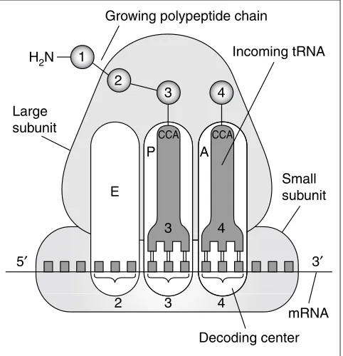
Protein synthesis is accomplished by an interaction between the ribosome and amino-acid-bearing tRNAs selected and lined up according to the genetic instructions of the mRNA, in the course of which the amino acids are strung together to form a polypeptide. The basic model of translation, postulated by James Watson [15], remains valid today in its essential features (see, for instance, Wilson and Noller [16] and Ramakrishnan [17]). In this model, there are two sites on the ribosome where tRNAs are bound: the aminoacyl (A) site and the peptidyl (P) site (see Figure 1). Each site is made up of regions on both subunits. Interaction of the tRNA with the mRNA at the A site of the small subunit the decoding center -determines whether the tRNA is rejected or accepted. The other functional center, the peptidyltransferase center, consists of pocket-like structure on the large subunit. Upon acceptance and accommodation of a cognate tRNA in the A site, we have the following situation: the tRNA residing in the P site is bound through its anticodon to the downstream mRNA codon, while the other end of this tRNA (the CCA end) is bound to the nascent polypeptide; the tRNA residing in the A site is bound to the current codon, while its CCA end carries the new-coming amino acid (Figure 1). This state is short-lived and is immediately followed by the transfer of the peptide to the aminoacyl end of the A-site tRNA, leaving the tRNA that is bound to the P site lacking an amino acid (deacylated). The subsequent step of tRNA translocation, catalyzed by EF-G, moves the entire complex formed by the mRNA and the two tRNAs by one codon relative to the ribosome so that the former P-site tRNA leaves the ribosome; the former A-site tRNA occupies the P site; and the A site is vacated and ready to accept a tRNA cognate to the codon that has just moved in. The most significant departure from this original model came from Knud Nierhaus’ discovery of a separate site into which the deacylated P-site tRNA moves - the exit or E site [18] (Figure 1). In addition, the existence of hybrid states that combine different binding sites on the two subunits, such as ‘A/P’ and ‘P/E’ sites, has been inferred from some experiments [19]. Finally, the decoding process is much more complicated than is portrayed in this simple model, as it also involves the participation of EF-Tu and a proofread-ing step in which near-cognate tRNAs are rejected (see [16]).
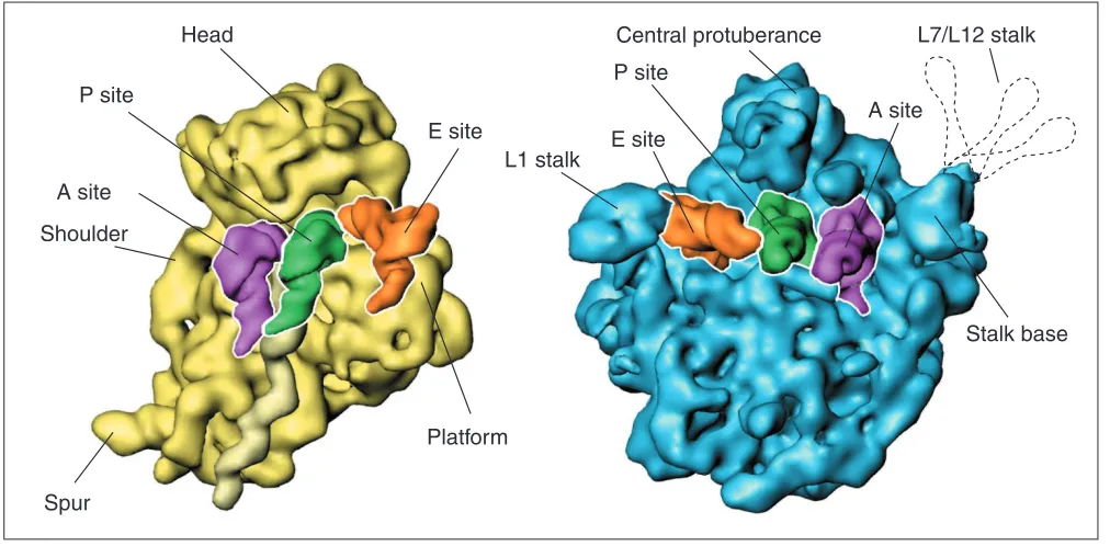
The architecture of the ribosome is determined by the shapes of its two subunits (Figure 2) and their spatial arrangement, which result in a space of complex topology, the inter-subunit cavity. This cavity is tailored for the passage of tRNA, an L-shaped molecule that comes in basi-cally 20 varieties, one for each amino acid. The small subunit has an elongate shape and a pronounced domain structure, with its anthropo-morphically named domains ‘head,’ ‘plat-form,’ ‘shoulder,’ and ‘body’ being distinctly recognizable. The large subunit, in contrast, is rather round and mono-lithic, except for three protrusions that give the subunit the characteristic ‘crown’ appearance in one of its views. The association of the subunits is mediated by multiple bridges [3,12,20], mostly formed by RNA-RNA contacts, although a few peripheral bridges involve proteins as well. The large and small subunits are made up of the ribosomal proteins L1-L36 and S1-S21, respectively.
[image:2.609.315.556.88.338.2]The inter-subunit cavity, which is open on both sides, is suit-ably shaped to act as a conduit for the tRNA’s migration through the ribosome from the A to the E site. The ‘tRNA entrance’ is roughly funnel-shaped and constitutes the binding site for several protein factors: EF-Tu, which deliv-ers the tRNA as part of a ternary complex with GTP to the ribosome; EF-G, which catalyzes the translocation of tRNAs Figure 1
The basic function of the ribosome during the elongation phase of protein synthesis. Amino acids and their corresponding tRNAs and codons are numbered from the amino terminus of the polypeptide. The amino acid is attached to the CCA end of the tRNA and is then transferred to the previous amino acid in the chain. See text for more details. Adapted from [43].
1
2
3
Growing polypeptide chain
Incoming tRNA
Large subunit
Small subunit
mRNA
Decoding center 4
CCA CCA
2 3 4
3 E
A P
4 H2N
and mRNA by one codon; release factors (RF1, RF2, and RF3), which release the polypeptide in response to the appearance of a stop codon at the A site and prepare the ribosome for recycling; and finally, recycling factor (RRF), which in concert with EF-G separates the subunits from each other and liber-ates the mRNA and the remaining tRNAs. A prominent feature of the ribosome on the tRNA entrance side is a long, flexible stalk formed by proteins L7 and L12, whose function is still unclear; the base of that stalk is formed by ribosomal protein L10 and a 58-nucleotide region of RNA that is tightly associated with the carboxy-terminal domain of protein L11. This component of the ribosome is instrumental in its GTPase activity, which is required both for EF-G-dependent translocation and accommodation of tRNAs.
The ‘tRNA exit,’ at the opposite end of the inter-subunit space, is gated by a large mushroom-shaped stalk, the L1 stalk, which is formed from an RNA helix and a globular protein, L1. This stalk is mobile and can pivot along an arc (by at least 30 degrees [13,21,22]) around a flex point on the RNA helix. It can apparently alternate between an ‘open’ position, in which it allows tRNA to exit, and a ‘closed’ posi-tion, in which it blocks the exit (see below).
It has been possible, first by cryo-EM [5,6,23] and then by X-ray crystallography [12,15], to visualize tRNA bound at these sites and to follow the progress of tRNA through the inter-subunit space. As it travels, the tRNA interacts tran-siently with the two regions of the ribosome called
‘func-tional centers’: first the anticodon end interacts with the decoding center, located on the ‘neck’ region of the small subunit, and second the CCA end interacts with the peptidyl-transferase center, a pocket-like structure on the large subunit made entirely of RNA that also forms the entrance of the polypeptide exit tunnel. The X-ray and cryo-EM maps show many additional contacts of the tRNA with the ribo-some in the different stations of its trajectory, suggesting that its movement is tightly controlled for stereochemical precision at the functional binding sites.
After the X-ray structures appeared [9-13], initial attention focused on the molecular processes at the two functional centers. It was suggested that the peptidyl-transferase activ-ity of the ribosome involved a predominantly chemical catal-ysis of peptide-bond formation [11], but this hypothesis has given way to the view that the proper positioning of the sub-strates may be all that is required; this issue is still not resolved [24]. Valuable insights into the decoding process have been gained by the group of Ramakrishnan, who have studied the interaction of a tRNA anticodon loop with the decoding center of the isolated 30S subunit in the presence of mRNA at atomic detail [25]. Decoding and accommoda-tion involve a complex sequence of steps that consist of a dynamic interplay between the ribosome, tRNA, and EF-Tu. Many ‘snapshots’ will need to be analyzed to get the full picture. Toward this end, cryo-EM is starting to provide the first low-resolution (around 10 Å) density maps of the trans-lating ribosome [26,27].
comment
reviews
reports
deposited research
interactions
information
[image:3.609.54.560.87.336.2]refereed research
Figure 2
The two ribosomal subunits, as visualized by cryo-EM, with tRNAs attached in the A, P and E sites. The L7/L12 stalk, normally invisible because of its flexibility, is indicated by the dashed contour lines.
Head
Platform
Spur
A site
P site
E site
L1 stalk
E site
P site
A site
Stalk base
Shoulder
Interaction with elongation factors, and
functional dynamics
In vitrotranslation systems mimic the conditions in the cell by supplying ribosomal subunits and the basic ligands, as well as GTP, in a carefully balanced buffer solution (see [28]). Although not as efficient as in the cell, in which protein synthesis proceeds at 20 amino acids per second, protein synthesis in an in vitrosystem still works at a rate of about 5 amino acids per second, which means that a protein with a length of 200 amino-acid residues can be made in about 40 seconds. Such in vitrosystems, in which ribosome complexes are free in solution and unimpeded by crystal contacts, can be used to study their dynamic behavior. The notion that the ribosome changes its conformation during the elongation cycle goes back to early studies [29-31]. Functional dynamics can be studied by various biophysi-cal techniques, such as cryo-EM, hydroxyl radibiophysi-cal probing, fluorescence stopped-flow and quench-flow analysis, and single-molecule fluorescent resonance energy transfer (FRET). These techniques complement one another in giving different aspects of the system’s time course and in looking at bulk or individual molecules. Significantly, studies using the different approaches all come to the conclusion that the ribosome undergoes periodic changes in confirmation during the elongation cycle ([32,33] and J. Puglisi, personal communication).
So far, the most detailed observations have come from cryo-EM. Here, the dynamic behavior is inferred from a series of three-dimensional ‘snapshots’ that show the ribosome at dif-ferent stages of the process. Given that cryo-EM visualization is based on the formation of an average over particles with supposedly identical structures, the taking of a meaningful three-dimensional snapshot requires that a large fraction of the ribosomes is trapped in the same state. This can be accomplished by using either antibiotics or non-hydrolyzable GTP analogs. Antibiotics have been likened to wrenches or ‘spanners’ that are ‘thrown into the works’ [34] of the ribo-some. They are small molecules that bind at strategic sites and cause the arrest of one or several steps of the dynamic process, mostly by interfering with the required conforma-tional changes. For instance, tetracycline binds to the small subunit at the decoding center and prevents a cognate tRNA from moving into the A site; kirromycin arrests EF-Tu in a conformation that does not allow tRNA accommodation; and fusidic acid prevents a conformational change in EF-G that is required for the factor to leave the ribosome. Nonhydrolyz-able GTP analogs, another means of trapping a particular state, bind with high affinity to the GTP-binding site without allowing GTPase activity to take place, resulting in perma-nent binding of the ligand.
Following such studies, it has been possible, among other results, to observe a large conformational reorganization of the ribosome that occurs in response to the binding of EF-G,
termed the ratchet motion [27,32]. The two subunits rotate relative to each other, and this motion apparently represents the first step in the two-step process of translocation. Early on, Spirin [30] suggested that the very architecture of ribo-somes, as a complex formed by two loosely linked massive subunits, implied the existence of a relative motion between these building blocks. Currently, atomic modeling is being used to study the molecular basis of the conformational reorganization [35]. There is the intriguing possibility, brought out by normal-mode analysis of the ribosome struc-ture (an analysis based on classical mechanics, predicting the most important modes of motion), that the observed motions are closely related to the dynamic properties of the ribosome’s gross architecture ([36] and Y. Wang, A.J. Rader, I. Bahar and R. Jernigan, personal communication).
Ribosomes from different species and kingdoms
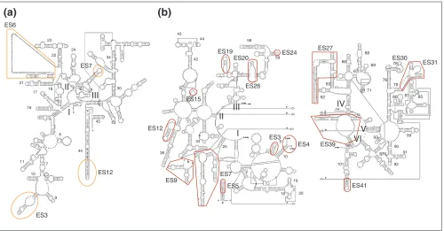
Secondary-structure comparisons indicate that ribosomes from eukaryotes have acquired insertions of additional RNA, the so-called expansion segments (for example, see Figure 3 for the yeast expansion segments). When the locations of the insertion points are traced back to the X-ray structures of the bacterial ribosomes, they prove to be situated exclusively at the periphery of the ribosome, away from the more centrally located functional centers. Other evolutionary developments in the eukaryotes are the addition of new proteins and the expansion and modification of existing ones, again mostly in peripheral locations. Organellar ribosomes, on the other hand, appear to be specialized through the loss of some pro-teins and substitution of some RNA by new propro-teins [37]. Although relatively little is known about the structures of the ribosomes of eukaryotes, a cryo-EM look at the ribosome of yeast (Figure 4c) [14] allows us to make some generaliza-tions. The new peripheral acquisitions can be put into three categories: those conveying topological properties that tailor the ribosome for membrane interactions; those involved with protein export; and those required for translational reg-ulation and control. The discovery, mentioned above, that functionally important motions can be reproduced in a normal-mode analysis of the bacterial ribosome structure invites the speculation that some of the added mass might also be required as strategically placed ‘counterweights’ to other functionally necessary additions, so as to preserve the motions that are required for ribosome function.[14,39,40]; rabbit reticulocyte [38,40]; and human (C.M.T. Spahn, E. Jan, A. Mulder, R.A. Grassucci, P. Sarnow and J.F., unpublished data)), and it appears to be a universal feature. Regarding additions affecting protein export, we know through Günther Blobel’s seminal work (see [41]) of the existence of a complex apparatus (the ‘translocon’) for protein insertion into, or export through, the endoplasmic
reticulum membrane. The structural implications of this concept are that some of the new ribosomal proteins and RNA additions near the exit site of the polypeptide tunnel must have a role in interacting with the protein exit channel and the signal recognition particle [39,40,42]. The addi-tional mechanisms for translaaddi-tional regulation and control that have evolved in eukaryotes are least understood and will
comment
reviews
reports
deposited research
interactions
information
[image:5.609.57.557.86.347.2]refereed research
Figure 3
Expansion segments in the secondary structure of yeast ribosomal RNA.(a) 18S rRNA; (b)5.8/25S rRNA [44]. Expansion segments (ES) are labeled using Gerbi’s nomenclature [45]. Small numbers refer to the helix numbering convention; Roman numerals refer to the RNA domains. Reproduced with permission from [14].
ES6
ES3
ES12 ES7
ES24
ES26 ES19
ES20
ES15
ES12
ES9
ES7 ES5
ES3 ES4
ES41 ES39
ES31 ES30
ES27
I II
III
I II
III
VI IV
V
23
24
21
27 34
30
45 43
6
11
10 7
43 44
42
58
59
38
34 25
20 19
18 10 9
89
90
92 101
SRL 98
93 62
63 66
67 68
69
71 76
78
79
80 81
9
85
91 22
44 16
17 18
(a)
(b)
Figure 4
Structures of ribosomes from different species. The small subunit is on the left. (a)X-ray structure of the T. thermophilus70S ribosome [12]. (b) Cryo-EM map of the E. coli70S ribosome [46]. (c)Cryo-EM map of the yeast 80S ribosome [14]. Expansion regions are darker. The dashed line indicates a flat surface that suggests eukaryotic specialization of 60S subunit for association with a planar membrane. (d)Cryo-EM map of the mammalian mitochondrial ribosome [37]. Reproduced with permission from (a) [12], (b) [46], (c) [14] and (d) [37].
[image:5.609.54.562.532.682.2]be a rich area for structural probing by cryo-EM once an X-ray structure of the eukaryotic ribosome is available. Much more work is clearly needed before a full picture of the eukaryotic ribosome can emerge.
In conclusion, thanks to recent advances in X-ray crystallog-raphy and cryo-EM of the ribosome, we now have extensive information on its structure, on the makeup of functional centers, and the beginning of an understanding of its func-tional dynamics. This picture, however, comes mainly from a few bacterial species. Much less is known about the structural basis of translation in eukaryotes, but first insights have come from cryo-EM of the ribosome of yeast and mammals.
Acknowledgements
This work was supported by HHMI and grants NIH R37 GM29169 and R01 GM55440. I thank Michael Watters for assistance with the prepara-tion of figures.
References
1. Spirin AS: Ribosomes.New York: Kluwer Academic/Plenum Publish-ers; 1999.
2. Huxley HE, Zubay G: Electron microscope observations on the structure of microsomal particles from Escherichia coli. J Mol Biol1960, 2:10-18.
3. Frank J, Zhu J, Penczek P, Li Y, Srivastava S, Verschoor A, Raderma-cher M, Grassucci R, Lata RK, Agrawak RK: A model of protein synthesis based on cryo-electron microscopy of the E. coli ribosome. Nature 1995, 376:441-444.
4. Stark H, Mueller F, Orlova EV, Schatz M, Dube P, Erdemir T, Zemlin F, Brimacombe R, van Heel M: The 70S Escherichia coli ribo-some at 23 Å resolution: fitting the ribosomal RNA. Structure 1995, 3:815-821.
5. Agrawal RK, Penczek P, Grassucci RA, Li Y, Leith A, Nierhaus KH, Frank J: Direct visualization of A-, P-, and E-site transfer RNAs in the Escherichia coli ribosome. Science 1996, 271:1000-1002.
6. Stark H, Orlova EV, Rinke-Appel J, Junke N, Müller F, Rodnina M, Wintermeyer W, Brimacombe R, van Heel M: Arrangement of tRNAs in pre- and posttranslocational ribosomes revealed by electron cryomicroscopy.Cell1997, 88:19-28.
7. Stark H, Rodnina MV, Rinke-Appel J, Brimacombe R, Wintermeyer W, van Heel M: Visualization of elongation factor Tu on the Escherichia coli ribosome. Nature 1997, 389:403-406.
8. Agrawal RK, Penczek P, Grassucci RA, Frank J: Visualization of elongation factor G on the Escherichia coli 70S ribosome: the mechanism of translocation. Proc Natl Acad Sci USA1998, 95:6134-6138.
9. Wimberly BT, Brodersen DE, Clemons WM Jr, Morgan-Warren RJ, Carter AP, von Rhein C, Hartsch T, Ramakrishnan V: Structure of the 30S ribosomal subunit.Nature 2000, 407:327-339.
10. Schlünzen F, Tocilj A, Zarivach R, Harms J, Glühmann M, Janell D, Bashan A, Bartels H, Agmon I, Franceschi F, Yonath A: Structure of functionally activated small ribosomal subunit at 3.3 Å reso-lution.Cell 2000, 102:615-623.
11. Nissen P, Hansen J, Ban N, Moore PB, Steitz TA: The structural basis of ribosome activity in peptide bond synthesis.Science 2000, 289:920-930.
12. Yusupov MM, Yusopova GZ, Baucom A, Lieberman K, Earnest TN, Cate JN, Noller HF: Crystal structure of the ribosome at 5.5 Å resolution.Science2001, 292:883-896.
13. Harms J, Schluenzen F, Zarivach R, Bashan A, Gat S, Agmon I, Bartels H, Franceschi F, Yonath A: High resolution structure of the large ribosomal subunit from a mesophilic eubacterium.Cell 2001, 107:679-688.
14. Spahn CMT, Beckmann R, Eswar N, Penczek PA, Sali A, Blobel G, Frank J: Structure of the 80S ribosome from Saccharomyces cerevisiae-tRNA-ribosome and subunit-subunit interactions. Cell2001, 107:373-386.
15. Watson JD: The synthesis of proteins upon ribosomes.Bull Soc Chim Biol1964, 46:1399-1425.
16. Wilson KS, Noller HF: Mapping the position of translational elongation factor EF-G in the ribosome by directed hydroxyl radical probing.Cell1998, 92:131-139.
17. Ramakrishnan V: Ribosome structure and the mechanism of translation.Cell2002, 108:557-572.
18. Rheinberger H-J, Sternbach H, Nierhaus KH: Three tRNA binding sites on E. coliribosome.Proc Natl Acad Sci USA1981, 78:5310-5314.
19. Moazed D, Noller HF: Interaction of tRNA with 23S RNA in ribosomal A, P and E sites.Cell1989, 57:585-597.
20. Cate JH, Yusupov MM, Yusopova GZh, Earnest TN, Noller HF: X-ray crystal structures of 70S ribosome functional com-plexes.Science1999, 285:2095-2104.
21. Valle M, Zavialov A, Li W, Stagg SM, Sengupta J, Nielsen RC, Nissen P, Harvey SC, Ehrenberg M, Frank J: Incorporation of aminoacyl-tRNA into the ribosome as seen by cryo-EM.Nat Struct Biol 2003, 10:899-906.
22. Vila-Sanjurjo A, Ridgeway WK, Seymaner V, Zhang W, Santoso S, Yu K, Cate JH: X-ray crystal structures of the WT and a hyper-accurate ribosome from Escherichia coli. Proc Natl Acad Sci USA 2003, 100:8682-8687.
23. Agrawal RK, Spahn CMT, Penczek P, Grassucci RA, Nierhaus KH, Frank J: Visualization of tRNA movements on the Escherichia coli 70S ribosome during the elongation cycle. J Cell Biol2000, 150:447-459.
24. Moore PB, Steitz TA: The structural basis of large ribosomal subunit function.Annu Rev Biochem2003, 72:813-850.
25. Ogle JM, Brodersen DE, Clemons WM Jr, Tarry MJ, Carter AP, Ramakrishnan V: Recognition of cognate transfer RNA by the 30S ribosomal subunit.Science2001, 292:897-902.
26. Valle M, Sengupta J, Swami NK, Grassucci RA, Burkhardt N, Nier-haus KH, Agrawal RK, Frank J: Cryo-EM reveals an active role for the aminoacyl-tRNA in the accommodation process. EMBO J 2002, 21:3557-3567.
27. Valle M, Zavialov A, Sengupta J, Rawat U, Ehrenberg M, Frank J: Locking and unlocking of ribosomal motions. Cell 2003, 114:123-134.
28. Blaha G, Stelzl U, Spahn CMT, Agrawal RK, Frank J, Nierhaus KH: Preparation of functional ribosomal complexes and effect of buffer conditions on tRNA positions observed by cryoelec-tron microcsopy. In Methods of Enzymology. Edited by Celander DW, Abelsen JN. San Diego: Academic Press; 2000:292-309. 29. Bretscher MS: Translocation in protein synthesis: a hybrid
structure model.Nature 1968, 218:675-677.
30. Spirin AS: On the mechanism of ribosome function. The hypothesis of locking-unlocking of subparticles. Dokl Akad Nauk USSR1968, 179:1467-1470.
31. Schreier MH, Noll H: Conformational changes in ribosomes during protein synthesis. Proc Natl Acad Sci USA1971, 68:805-809.
32. Frank J, Agrawal RK: A ratchet-like inter-subunit reorganiza-tion of the ribosome during translocareorganiza-tion. Nature 2000, 406:318-322.
33. Polacek N, Patzke S, Nierhaus KH, Barta A: Periodic conforma-tional changes in rRNA: monitoring the dynamics of trans-lating ribosomes.Mol Cell2000, 6:159-171.
34. Spahn CMT, Prescott CD: Throwing a spanner in the works: antibiotics and the translation apparatus. J Mol Med 1996, 74:423-439.
35. Gao H, Sengupta J, Valle M, Korostelev A, Eswar N, Stagg SM, Van Roey P, Agrawal RK, Harvey SC, Sali A, et al.: Study of the struc-tural dynamics of the E. coli 70S ribosome using real space refinement.Cell2003, 113:789-801.
36. Tama F, Valle M, Frank J, Brooks CL III: Dynamic reorganization of the functionally active ribosome explored by normal mode analysis and cryo-electron microscopy. Proc Natl Acad Sci USA2003, 100:9319-9323.
37. Sharma MR, Koc EC, Datta PP, Booth TM, Spremulli LL, Agrawal RK: Structure of the mammalian mitochondrial ribosome reveals an expanded functional role for its component pro-teins. Cell2003, 115:97-108.
38. Dube P, Bacher G, Stark H, Müller F, Zemlin F, van Heel M, Brima-combe R: Correlation of the expansion segments in mam-malian rRNA with the fine structure of the 80S ribosome; a cryoelectron microscopic reconstruction of the rabbit retic-ulocyte ribosome at 21 Å resolution. J Mol Biol 1998, 279:403-421.
40. Morgan DG, Ménétret JF, Radermacher M, Neuhof A, Akey IV, Rapoport TA, Akey CW: A comparison of the yeast and rabbit 80S ribosome reveals the topology of the nascent chain exit tunnel, inter-subunit bridges and mammalian rRNA expan-sion segments.J Mol Biol2000, 301:301-321.
41. Simon SM, Blobel G: A protein-conducting channel in the endoplasmic reticulum.Cell1991, 65:371-380.
42. Beckmann R, Spahn CMT, Eswar N, Helmers J, Penczek PA, Sali A, Frank J, Blobel G: Architecture of the protein-conducting channel associated with the translating 80S ribosome. Cell 2001, 107:361-372.
43. Alberts B, Johnson A, Lewis J, Raff M, Roberts K, Walter P: Molecular Biology of the Cell.4th edition. New York: Garland; 2002.
44. Gutell Lab Comparative RNA website [http://www.rna.icmb.utexas.edu]
45. Gerbi SA: Expansion segment: regions of variable size that interrupt the universal core secondary structure of riboso-mal RNA.In Ribosomal RNA: Structure, Evolution, Processing, and Func-tion in Protein Synthesis. Edited by Zimmermann RA, Dahlberg AE. New York: CRC Press; 1996:71-87.
46. Gabashvili IS, Agrawal RK, Spahn CMT, Grassucci RA, Svergun DI, Frank J, Penczek P: Solution structure of the E. coli70S ribo-some at 11.5 Å resolution.Cell2000, 100:537-549.
comment
reviews
reports
deposited research
interactions
information