C A S E R E P O R T
Open Access
Delayed surgical treatment for a traumatic
bilateral cervical facet joint dislocation using a
posterior-anterior approach: a case report
Takashi Shimada
1, Seiji Ohtori
2*, Gen Inoue
2, Junichi Nakamura
2, Izumi Nakada
1, Hiroshi Saiki
1, Song Ho Chang
1,
Koui Kawamura
1, Kazuhisa Takahashi
2and Hiroshi Sugiyama
1Abstract
Introduction:There have been few reports of patients with bilateral cervical facet dislocations that remain
untreated for eight weeks or more. We report the case of a 76-year-old man with an old bilateral cervical facet joint dislocation fracture that was treated by posterior-anterior reduction and fixation.
Case presentation:A 76-year-old Asian man was involved in a road traffic accident. He presented with neck pain and arm pain on his right side, but motor weakness and paralysis were not observed. He was treated
conservatively; however, instability and spondylolisthesis at the C5 to C6 joint increased eight weeks after the injury. We performed a posterior-anterior reduction and fixation. After surgery, bony union was achieved, and his neck pain and arm pain disappeared.
Conclusion:We recommend reduction and fixation surgery if a patient has an old bilateral facet joint dislocation fracture in the cervical spine.
Introduction
Several authors have reported failures to correctly diag-nose cervical spine injuries. The rate of misdiagnosis ranges from 5% to 20% [1-3]. Of missed spinal injuries, misdiagnosis of an injury to the cervical spine has been most frequently reported [3]. The use of computed tom-ography (CT) and magnetic resonance imaging (MRI) can improve the accuracy of cervical spine injury diag-nosis. However, an analysis of the clinical records of 367 patients with cervical spine injuries revealed a diagnostic failure rate of 4.9% [4].
Treatment for acute cervical dislocation fracture in-cludes conservative treatment and surgery. Whether conservative treatment is sufficient is controversial [5-7]. Recently, the incidence of surgical treatment to reduce dislocation and for fixation has increased compared with conservative treatment.
An injury is considered as ‘old’ when the interval be-tween the accident and correct diagnosis is longer than
three weeks [8]. There are several reports on the man-agement of old dislocations of the subaxial cervical spine in the English literature [9-13]. Among these, only three articles were found in which the delayed treatment of bilateral cervical facet dislocations is described after a diagnostic delay of more than one month [9-11].
Here, we report a case of a 76-year-old man with an old bilateral cervical facet joint dislocation fracture that was treated by posterior-anterior reduction and fixation.
Case presentation
A 76-year-old Asian man was involved in a road traffic accident. He presented with neck and arm pain on his right side, but motor weakness and paralysis were not observed. His arm pain corresponded to the right C6 and C7 dermatomes.
We carried out an X-ray image examination of our patient’s cervical spine and diagnosed a slight cervical spondylolisthesis (Figure 1a). CT and MRI were not per-formed. Because our patient did not show motor weak-ness or paralysis, his neck and arm pain were treated conservatively. However, the pain did not change over the six weeks following the injury. We conducted further * Correspondence:sohtori@faculty.chiba-u.jp
2
Department of Orthopedic Surgery, Graduate School of Medicine, Chiba University, 1-8-1 Inohana, Chuo-ku, Chiba 260-8670, Japan
Full list of author information is available at the end of the article
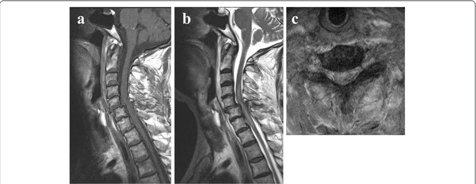
X-ray imaging, MRI and CT eight weeks after the injury (Figures 1a,b,c, 2 and 3). Plain X-ray film images obtained at this time showed increased instability at the C5 to C6 joint when compared with those taken imme-diately after the accident (Figure 1a,b,c). MRI revealed central spinal canal stenosis at the C5 to C6 joint and high signal intensity in the spinal cord on T2-weighted imaging (Figure 2). A sagittal CT showed bilateral dis-location of facet joints (Figure 3).
We planned posterior-anterior surgery eight weeks after the injury. We performed a partial resection of both C5 to C6 facet joints for reduction, using a posterior ap-proach. Half of the C5 to C6 facet joint was resected on the right side, and one quarter of the C5 to C6 facet joint was resected on the left side.
Lateral mass screws on the left side were used for fi-xation, and bilateral local bone was grafted between the posterior surface of the C5 and C6 laminae. Lateral mass
screws on the right side were not used for fixation be-cause most of the lateral bone mass was resected. Be-cause fixation was insufficient, we added an anterior approach by removing the C5 to C6 intervertebral disc, and grafted iliac bone into the C5 to C6 space. We used a titanium plate for fixation.
Our patient gradually became symptom-free after sur-gery. He presented with a transient C5 palsy on his right side; however, he had recovered three months after the surgery. Plain X-ray film images obtained six months after surgery showed good stability (Figure 4). MRI revealed recovery of the central spinal canal stenosis at the C5 to C6 joint and showed normal intensity in the spinal cord on T2-weighted imaging (Figures 2 and 5).
Discussion
[image:2.595.57.539.89.231.2]We present the case of an old bilateral cervical facet joint dislocation fracture found in a 76-year-old man, Figure 1Plain X-ray film images showing slight spondylolisthesis of C5 immediately after a traffic accident. (a)The extent of the spondylolisthesis increased over the following eight weeks.(b)Flexion position;(c)extension position.
[image:2.595.57.539.517.704.2]treated by posterior-anterior reduction and fixation. After surgery, bony union was achieved, and our patient’s neck pain and arm pain disappeared.
In one study, cervical spine injuries were identified in 740 patients and the diagnosis was delayed or missed in 34 patients (4.6%). Ten of the 34 patients (29%) deve-loped permanent sequelae as a result of these delays [1]. In another study, 1331 patients were evaluated following blunt trauma injury; recognition of their cervical spine injury was delayed in 8% of patients [2]. In a review of
253 patients with 274 spinal injuries, delays in diagnosis were documented in 22.9% of cervical injuries and 4.9% of thoracolumbar injuries [3]. We therefore carefully consider the potential misdiagnosis of cervical injury be-fore therapy. In our patient, even with slight cervical spondylolisthesis, plain X-rays were not sufficient for a proper diagnosis. In general, such a finding requires fur-ther diagnostic efforts including a CT scan.
[image:3.595.58.290.88.283.2]Currently, there is evidence in several articles that closed reduction in an awake and alert patient is relatively safe [14,15]. There is, however, no agreement regarding whether such a maneuver is safe in an obtunded or intu-bated patient. Most surgeons would agree that an open reduction with or without a decompression should be
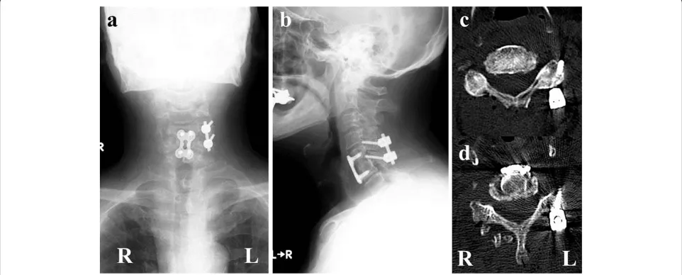
[image:3.595.304.540.89.238.2]Figure 4Plain X-ray imaging and computed tomography scans obtained six months after surgery.We performed surgery to remove a part of both sides of the facet joints at the C5 to C6 joint, and posterior fusion with lateral mass screws on the left side. After posterior fusion, anterior fusion using a plate and screws was performed.(a)X-ray anterior-posterior view;(b)X-ray lateral view;(c)computed tomography scan of C5;(d)computed tomography scan of C6. R: right side. L: left side.
[image:3.595.57.538.491.685.2]Figure 3Sagittal computed tomography performed eight weeks after the traffic accident showing dislocation of both facet joints at C5 to C6. (a)R: right side (arrow).(b)L: left side (arrow).
formed in 12 patients with an old dislocation of the lower cervical spine, including bilateral dislocation of the facet joints, and all patients developed bone fusion and showed neurological improvement [9]. There has been a report of three patients who had older (>8 weeks) untreated bila-teral cervical facet dislocation. All three patients were sur-gically treated [10]. In a neurolosur-gically intact 51-year-old patient, surgical treatment was performed 10 weeks after the trauma upon diagnosis of bilateral cervical facet dislocation at the C5 to C6 joint. That patient remained neurologically intact and radiographic fusion was observed [11]. In our case, our patient with an old bilateral cervical facet joint dislocation fracture underwent surgery; complete reduction was performed, and bony fusion was observed. We therefore recommend reduction and fixation surgery if the patient has an old bilateral cer-vical facet joint dislocation fracture.
Several authors have reported surgical methods for patients with old bilateral cervical facet joint dislocation fractures using anterior and posterior approaches, and a combination of the two with instrumentation [9-11]. The use of pedicle screws for patients with old bilateral cervical facet joint dislocation fractures has not been fre-quently reported. Yamazaki et al. reported a case of an old fracture-dislocation unilaterally at a C4 to C5 facet joint. Reduction of the deformity and spinal fusion were successfully performed using a pedicle screw-rod system [12]. Leeet al. reported that, when patients were neuro-logically intact, an anterior approach was more com-monly chosen than a posterior approach, and combined approaches were more commonly chosen for bilateral facet injuries [16]. In our case, we selected a bilateral posterior facetectomy, unilateral lateral mass screws and anterior plating for our patient. To reduce the facet joint, half of the C5 to C6 facet joint was resected on the right side, and one quarter of the C5 to C6 facet joint was resected on the left. We considered an anterior plate to be insufficient for stabilization. Furthermore, half of the C5 to C6 facet joint was resected, so we could not use lateral mass screws on the right side.
Several surgical methods are recommended for old bila-teral cervical facet dislocation fractures. We recommend
Consent
Written informed consent was obtained from the patient for publication of this case report and accompanying images. A copy of the written consent is available for re-view by the Editor-in-Chief of this journal. The protocols for human procedures used in this study were approved by the ethics committee of our institution.
Competing interests
The authors declare that they have no competing interests.
Authors’contributions
SO, TS and GI performed the surgery. HSs and KT evaluated the imaging. The other authors were key contributors in writing the manuscript. All authors read and approved the final manuscript.
Author details 1
Department of Orthopedic Surgery, Asahi General Hospital, i-1326, Asahi-shi, Chiba 289-2511, Japan.2Department of Orthopedic Surgery, Graduate School
of Medicine, Chiba University, 1-8-1 Inohana, Chuo-ku, Chiba 260-8670, Japan.
Received: 17 July 2012 Accepted: 20 November 2012 Published: 9 January 2013
References
1. Davis JW, Phreaner DL, Hoyt DB, Mackersie RC:The etiology of missed cervical spine injuries.J Trauma1993,34:342–346.
2. Gerrelts BD, Petersen EU, Mabry J, Petersen SR:Delayed diagnosis of cervical spine injuries.J Trauma1991,31:1622–1626.
3. Reid DC, Henderson R, Saboe L, Miller JD:Etiology and clinical course of missed spine fractures.J Trauma1987,27:980–986.
4. Platzer P, Hauswirth N, Jaindl M, Chatwani S, Vecsei V, Gaebler C:Delayed or missed diagnosis of cervical spine injuries.J Trauma2006,61:150–155. 5. Beyer CA, Cabanela ME:Unilateral facet dislocations and fracture
dislocations of the cervical spine: a review.Orthopedics1992,15:311–315. 6. Fehlings MG, Sekhon LHS, Tator C:The role and timing of decompression
in acute spinal cord injury.Spine2001,26:S101–S110.
7. Lee AS, MacLean JCB, Newton DA:Rapid traction for reduction of cervical spine dislocations.J Bone Joint Surg Br1994,76B:352–356.
8. Roy-Camille R, Edward B, Zeller R, Lapresle P:Les Lesions Traumatiques Anciennes du Rachis Cervical Inferieure. InRachis cervical inferieure, 6mes journees d’Orthopedie de la Pitie. Paris: Masson; 1988:139–146. 9. Hassan MG:Treatment of old dislocations of the lower cervical spine.
Int Orthop2002,26:263–267.
10. Bartels RH, Donk R:Delayed management of traumatic bilateral cervical facet dislocation: surgical strategy. Report of three cases.J Neurosurg 2002,97:362–365.
12. Yamazaki M, Okawa A, Akazawa T, Koda M:Usefulness of 3-dimensional full-scale modeling for preoperative simulation of surgery in a patient with old unilateral cervical fracture-dislocation.Spine2007,32:E532–E536. 13. Korres DS, Nikiforidis P, Babis GC, Vlachou C, Lykomitros V, Andreakos A:
Old injuries of the lower cervical spine treated surgically.J Spinal Disorders1995,8:509–515.
14. Vaccaro AR, Falatyn SP, Flanders AE, Balderston RA, Northrup BE, Cotler JM:
Magnetic resonance evaluation of the intervertebral disc, spinal ligaments, and spinal cord before and after closed traction reduction of cervical spine dislocations.Spine1999,24:1210–1217.
15. Grant GA, Morza SK, Chapman JR, Winn HR, Newell DW, Jones DT, Grady MS:Risk of early closed reduction in cervical spine subluxation injuries.
J Neurosurg1999,90:13–18.
16. Lee JY, Nassr A, Eck JC, Vaccaro AR:Controversies in the treatment of cervical spine dislocations.Spine J2009,9:418–423.
doi:10.1186/1752-1947-7-9
Cite this article as:Shimadaet al.:Delayed surgical treatment for a traumatic bilateral cervical facet joint dislocation using a posterior-anterior approach: a case report.Journal of Medical Case Reports20137:9.
Submit your next manuscript to BioMed Central and take full advantage of:
• Convenient online submission
• Thorough peer review
• No space constraints or color figure charges
• Immediate publication on acceptance
• Inclusion in PubMed, CAS, Scopus and Google Scholar
• Research which is freely available for redistribution