C A S E R E P O R T
Open Access
Anaplastic carcinoma of the pancreas
diagnosed by endoscopic
ultrasound-guided fine-needle aspiration: a case report
and review of the literature
Kohei Oka
1, Ken Inoue
1*, Satoshi Sugino
1, Taishi Harada
1, Toshifumi Tsuji
1, Shingo Nakashima
2, Takayuki Katayama
1,
Takashi Okuda
1, Syuichi Kin
2, Akihiro Nagata
3, Toshiyuki Komaki
1and Keizo Kagawa
1Abstract
Background:Anaplastic carcinoma of the pancreas is a rare pancreatic neoplasm with a poor prognosis. It is classified as a variant of ductal adenocarcinoma, but the clinical features and treatment of it remain unknown because of its rarity and aggressiveness. Endoscopic ultrasonography and endoscopic ultrasound-guided fine-needle aspiration are useful techniques for the diagnosis of pancreatic tumors with high sensitivity and specificity. Case presentation:A 72-year-old Japanese woman presented with a diagnosis of acute pancreatitis, and a cystic lesion with slightly high density area was observed by computed tomography in her pancreatic head. In addition, endoscopic ultrasound revealed a heterogeneous lesion. Endoscopic ultrasound-guided fine-needle aspiration showed pleomorphic atypical cells. We diagnosed anaplastic carcinoma of the pancreas. We resected the lesion, and she has shown no sign of recurrence for > 6 months. There are few reports of anaplastic carcinoma of the pancreas diagnosed by endoscopic ultrasound-guided fine-needle aspiration and treated by surgery. Our analysis indicates that anaplastic carcinoma of the pancreas is more likely than typical ductal carcinomas to have cystic lesions with the tumor.
Conclusions:We report a case of anaplastic carcinoma of the pancreas diagnosed by endoscopic ultrasound-guided fine-needle aspiration and subsequently resected with a clear margin. We speculate that anaplastic carcinoma of the pancreas is more likely to have cystic changes than pancreatic ductal adenocarcinoma. When we diagnose pancreas tumor as having cystic changes, anaplastic carcinoma of the pancreas should be considered one of the differential diagnoses.
Keywords:Anaplastic carcinoma of the pancreas, EUS-FNA, Cystic change
* Correspondence:keninoue71@koto.kpu-m.ac.jp
1Department of Gastroenterology and Hepatology, Fukuchiyama City
Hospital, 231 Atsunaka-cho, Fukuchiyama-city, Kyoto 620-8505, Japan Full list of author information is available at the end of the article
Background
Anaplastic carcinoma of the pancreas (ACP) is a rare pan-creatic neoplasm with a poor prognosis [1,2]. It is classi-fied as a variant of ductal adenocarcinoma, but the clinical features and treatment of ACP remain unknown because of its rarity and aggressiveness. Endoscopic ultrasonog-raphy (EUS) and endoscopic ultrasound-guided fine-needle aspiration (EUS-FNA) are useful techniques for the diagnosis of pancreatic tumors with high sensitivity and specificity [3, 4]. There are case reports describing ACP, but only a few cases of ACP diagnosed by EUS-FNA have been reported. Here, we report the case of a patient with ACP diagnosed by EUS-FNA who subsequently underwent resection. We also discuss the characteristics of ACP, espe-cially in EUS imaging, in a comparison with pancreatic ductal adenocarcinoma (PDAC).
Case presentation
A 72-year-old Japanese woman presented with complaints of epigastric pain and nausea. The pain had started 3 days before admission and gradually worsened. Laboratory test-ing showed a high level of serum amylase (AMY) 838 IU/l and pancreatic amylase (P-AMY) 778 IU/l. CA 19-9 was elevated to 86.4 U/ml, but other tumor markers were nor-mal. Computed tomography (CT) scanning showed in-flammation localized in the pancreatic head and dilatation of the main pancreatic duct (MPD) (Fig.1). In addition, a cystic lesion with a slightly high density area was observed by CT in the pancreatic head.
Transabdominal ultrasonography and magnetic reson-ance cholangiopancreatography (MRCP) were performed. Both ultrasonography and MRCP demonstrated a 14 mm
cystic lesion in the pancreatic head. They showed the dila-tation of the MPD from the body to the tail of her pan-creas. We could not identify a connection between the cystic lesion and the MPD. EUS showed the cystic lesion more clearly than other modalities. EUS revealed that the cystic lesion consisted of both solid and cystic lesions. The solid area was shown as a hypoechoic and heterogeneous tumor, and the cystic area was shown as an anechoic le-sion. The EUS also showed that the MPD was dilated to 5 mm, and it was cut off around the mass in the pancreatic head. Endoscopic retrograde cholangiopancreatography (ERCP) showed > 12-mm-long stenosis of the MPD in the pancreatic head. The stenosis prevented a brush for cytology passing the stricture, and it was not possible to obtain a cytology specimen.
We performed EUS-FNA (Fig. 2). An echoendoscope (GF-UCT260, Olympus; Tokyo, Japan) with a 22-gauge needle was used to obtain cytological material: EchoTip ProCore® HD Ultrasound Needle (ECHO-HD-22-C; Cook Medical, USA). We carried out two fine-needle as-piration (FNA) punctures, moving ten times in each puncture. We aspirated the solid area of the tumor, and then the cells were histologically revealed as pleo-morphic atypical cells. Immunohistochemical stains were positive for cytokeratin (CK) AE1/AE3 and CK CAM5.2, which confirmed they were epithelial cells. Based on these findings, we diagnosed the tumor as an ACP.
Our patient’s abdominal pain and the elevation of pan-creatic enzyme improved (Fig. 3). CT images suggested tumor invasion to the front of her pancreas. Positron emission tomography (PET)-CT and gadolinium-ethoxybenzyl-diethylenetriamine pentaacetic acid
(Gd-c
f
a
b
d
e
[image:2.595.56.542.481.665.2]EOB-DTPA)- enhanced MRI showed no obvious metas-tasis. We performed a subtotal stomach-preserving pancreaticoduodenectomy, and no major complications occurred. On macroscopic examination, the tumor con-sisted of a yellow nodular mass with a cystic lesion, as had
been shown by EUS. The cystic lesion was pathologically confirmed as a pancreatic duct with some blood pooling. Hematoxylin-eosin staining showed spindle cells, pleo-morphic cells, and multinuclear osteoclast-like giant cells (OCGCs), which are characteristic of ACP (Fig.4).
The resection was completed with a clear margin. The final diagnosis was ACP according to Japan Pancreas Society (JPS) classification, staged pathologically T3(pS +)N0 M0, pStageIIA, according to the Union for International Cancer Control (UICC) TNM staging system. After discharge from our hospital, she began oral tegafur/gimeracil/oteracil (S-1) intake as adjuvant chemotherapy. Due to anorexia and diarrhea as adverse effects, however, the S-1 administration was discontin-ued after 2 weeks. Although she declined gemcitabine therapy as an alternative, she has shown no evidence of recurrence 6 months after the resection.
Discussion
ACP is a variant of ductal adenocarcinoma with poor differentiation. It accounts for only 2–7% of all pancre-atic adenocarcinomas [1]. It is so aggressive that its me-dian survival time is only 5.7 months (n= 18) [2]. Our patient’s ACP showed rapid progression. Although the EUS indicated that the tumor size was 15 mm, it turned out to be 25 mm at the time of surgery at approximately 1 month after the EUS-FNA. This is characteristic of sarcoma-like progression, but ACP is classified as a sub-type of ductal tumors because of the presence of ductal carcinoma cells. ACPs are pathologically classified into three variants: pleomorphic type, spindle cell type, and ACP with OCGCs. ACP with OCGCs has an exception-ally better prognosis than the other subtypes.
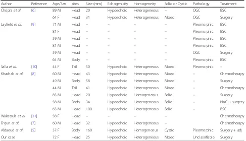
There are many case reports describing ACP, but only a few cases of ACP diagnosed by EUS-FNA have been re-ported. We performed a literature search with the terms “Anaplastic carcinoma pancreas” and “EUS-FNA”, using PubMed database for the period from 2006 to September 2016. We found seven reports of 19 cases of ACP during that period diagnosed by EUS-FNA (including our patient; Table 1) [5–11]. In most of the cases, the tumors were shown as hypoechoic and heterogeneous by EUS. The fea-tures of their components varied; of the components, 30% (3 of 10) were a fully solid mass and 60% (6 of 10) were mixed with solid and cystic lesions.
It is reported that ACPs are more likely than PDACs to have a cystic appearance [2, 12]. It is assumed that the aggressiveness of ACP may induce cystic change be-cause of central necrosis or degeneration [2]. Although these cystic changes may occur not only in ACP but also PDAC, such a change occurs in < 1% of PDACs [13]. ACPs should thus be considered a differential diagnosis in patients with a pancreatic tumor with a cystic lesion. We can also find cystic degeneration in other pancreatic
b
a
c
Fig. 2Findings obtained by endoscopic ultrasound-guided fine-needle aspiration.aEndoscopic ultrasound-guided fine-needle aspiration was performed to obtain cytology for the solid mass in the pancreatic head.
[image:3.595.57.290.87.585.2]tumors such as adenosquamous carcinoma, neuroendo-crine tumors, solid and pseudopapillary tumors, acinar cell carcinoma, and so on [14–16]. In our case, ACP might have induced cystic change due to central necrosis or degeneration into a pancreatic duct, because the cys-tic lesion was pathologically a pancreacys-tic duct.
In contrast, CT images of an ACP may show features comparable to those of PDACs. Compared to PDACs, ACPs are more hypervascular tumors [8]. It is reported that ACPs often present as a mass with peripheral contrast enhancement in the portal venous phase [2, 12]. In our patient’s case, a cystic lesion with slightly high density area was observed by CT. EUS showed the cystic lesion more clearly than other modalities. EUS also might be useful for
detecting ACP because of the high resolution of images, as has been reported for PDAC. EUS-FNA is useful for making a pathological diagnosis of pancreatic tumors, but there are some risks for dissemination [3,17].
There is no consensus regarding the optimal treatment for ACPs, due to the lack of data and evidence. It is re-ported that ACP is rare and aggressive with shorter overall survival times than PDAC [2]. This result sug-gests that an early diagnosis of ACP should be made for better outcomes. Chemotherapy for ACP is also uncer-tain. There is a report of a patient with ACP who passed a chemosensitivity test and was treated with paclitaxel [11]. Khashabet al. suggested that in light of the hyper-vascularity of ACPs, investigational therapies with agents
Fig. 3The patient’s clinical course. This graph shows serum level of amylase and 10-point numerical rating scale on abdominal pain. Both of them improved immediately, except for temporary elevation of serum level of amylase after the endoscopic retrograde cholangiopancreatogra-phy procedure.AMYserum amylase,ERCPendoscopic retrograde cholangiopancreatography,EUS-FNAendoscopic ultrasound-guided fine-needle aspiration,NRSnumerical rating scale
f
e
c
d
b
a
[image:4.595.57.539.88.281.2] [image:4.595.57.540.524.685.2]such as bevacizumab may improve the outcome of pa-tients with ACP [8]. There is no evidence regarding ad-juvant chemotherapy for ACP. We administered S-1 to our patient based on a phase 3 trial for PDAC.
Conclusions
We report a case of ACP diagnosed by EUS-FNA and subsequently resected with a clear margin. We speculate that ACPs are more likely to have cystic changes than PDAC, and we found that EUS-FNA was a useful tech-nique to detect this characteristic change and make a def-inite diagnosis. When we diagnose the pancreas tumor as having cystic changes, ACP should be considered one of the differential diagnoses. A further accumulation of cases of ACP must be examined to clarify this disease.
Abbreviations
ACP:Anaplastic carcinoma of the pancreas; AMY: Serum amylase; CK: Cytokeratin; CT: Computed tomography; ERCP: Endoscopic retrograde cholangiopancreatography; EUS: Endoscopic ultrasonography; EUS-FNA: Endoscopic ultrasound-guided fine-needle aspiration; EUS-FNA: Fine-needle aspiration; Gd-EOB-DTPA: Gadolinium-ethoxybenzyl-diethylenetriamine pentaacetic acid; JPS: Japan Pancreas Society; MPD: Main pancreatic duct; MRCP: Magnetic resonance cholangiopancreatography; OCGCs: Osteoclast-like giant cells; P-AMY: Pancreatic amylase; PDAC: Pancreatic ductal adenocarcinoma; PET: Positron emission tomography; S-1: Tegafur/gimeracil/ oteracil; UICC: Union for International Cancer Control
Acknowledgements
We are grateful to our patient, who cooperated with the publication of this report.
Funding
Not applicable.
Availability of data and materials
Not applicable.
Authors’contributions
All authors were involved in the care of the patient. KO, KI, TO, and KK contributed equally to drafting, revision, and preparation of the manuscript. KO, KI, and TO performed the EUS-FNA. SN and SK performed the operation. SN has followed up this patient as an out-patient. AN performed the pathological diagnosis. All authors read and approved the final manuscript.
Ethics approval and consent to participate
Not applicable.
Consent for publication
Written informed consent was obtained from the patient for publication of this case report and any accompanying images. A copy of the written consent is available for review by the Editor-in-Chief of this journal.
Competing interests
The authors declare that they have no competing interests.
Publisher’s Note
Springer Nature remains neutral with regard to jurisdictional claims in published maps and institutional affiliations.
Author details
1Department of Gastroenterology and Hepatology, Fukuchiyama City
Hospital, 231 Atsunaka-cho, Fukuchiyama-city, Kyoto 620-8505, Japan. 2
[image:5.595.55.542.98.378.2]Department of Surgery, Fukuchiyama City Hospital, 231 Atsunaka-cho, Fukuchiyama-city, Kyoto 620-0056, Japan.3Department of Pathology, Fukuchiyama City Hospital, 231 Atsunaka-cho, Fukuchiyama-city, Kyoto 620-0056, Japan.
Table 1Reported cases of anaplastic carcinoma of the pancreas diagnosed by endoscopic ultrasound-guided fine-needle aspiration
Author Reference Age/Sex sites Size (mm) Echogenicity Homogeneity Solid or Cystic Pathology Treatment
Chopraet al. [6] 89 M Head 20 Hypoechoic Heterogeneous – OGC BSC
64 F Head 31 Hypoechoic Heterogeneous Mixed OGC Surgery
Layfieldet al. [9] 71 M Head – – – – Pleomorphic BSC
81 F Head – – – – Pleomorphic BSC
59 M Head – – – – Pleomorphic BSC
81 M Head – – – – Pleomorphic BSC
59 M Head – – – – OGC Surgery
64 M Body – – – – Pleomorphic BSC
Sallaet al. [10] 44 F Tail 50 Hypoechoic Heterogeneous Mixed Pleomorphic –
Khashabet al. [8] 60 M Head 43 Hypoechoic Heterogeneous Mixed – Chemotherapy
49 M Body 58 Hypoechoic Heterogeneous Mixed – Surgery
44 M Tail 41 Hypoechoic Heterogeneous Mixed – Chemotherapy
85 M Head 20 Hypoechoic Homogeneous Solid – Surgery
58 M Body 34 Hypoechoic Heterogeneous Solid – NAC + surgery
65 M Head 100 Hypoechoic Heterogeneous Solid – BSC
Wakatsukiet al. [11] 58 F Head – – – – Chemotherapy
Ergunet al. [7] 60 M Head 32 Hypoechoic Heterogeneous – Chemotherapy
Aldaoudet al. [5] 37 F Body 160 Hypoechoic Homogeneous Cystic Pleomorphic Surgery + adj
Our case 72 F Head 25 Hypoechoic Heterogeneous Mixed Unclassifiable Surgery
Received: 5 June 2017 Accepted: 12 February 2018
References
1. Paal E, Thompson LD, Frommelt RA, Przygodzki RM, Heffess CS. A clinicopathologic and immunohistochemical study of 35 anaplastic carcinomas of the pancreas with a review of the literature. Ann Diagn Pathol. 2001;5:129–40.
2. Strobel O, Hartwig W, Bergmann F, Hinz U, Hackert T, Grenacher L, et al. Anaplastic pancreatic cancer: Presentation, surgical management, and outcome. Surgery. 2011;149:200–8.
3. Chen G, Liu S, Zhao Y, Dai M, Zhang T. Diagnostic accuracy of endoscopic ultrasound-guided fine-needle aspiration for pancreatic cancer: a meta-analysis. Pancreatology. 2013;13:298–304.
4. Hewitt MJ, McPhail MJ, Possamai L, Dhar A, Vlavianos P, Monahan KJ. EUS-guided FNA for diagnosis of solid pancreatic neoplasms: a meta-analysis. Gastrointest Endosc. 2012;75:319–31.
5. Aldaoud N, Joudeh A, Al-Momen S, Alnahawi M, Al-Abbadi MA. Anaplastic Carcinoma Arising in a Mucinous Cystic Neoplasm Masquerading as Pancreatic Pseudocyst. Diagn Cytopathol. 2016;44:538–42.
6. Chopra S, Wu ML, Imagawa DK, Lee J, Gu M. Endoscopic ultrasound-guided fine-needle aspiration of undifferentiated carcinoma with osteoclast-like giant cells of the pancreas: a report of 2 cases with literature review. Diagn Cytopathol. 2007;35:601–6.
7. Ergun M, Aydog G, KayaÇEtin E, Sasmaz N. A case of anaplastic carcinoma of pancreas diagnosed with endoscopic ultrasound-guided fine needle aspiration cytology. Turk J Gastroenterol. 2012;23:828–9.
8. Khashab MA, Emerson RE, DeWitt JM. Endoscopic ultrasound-guided fine-needle aspiration for the diagnosis of anaplastic pancreatic carcinoma: a single-center experience. Pancreas. 2010;39:88–91.
9. Layfield LJ, Bentz J. Giant-cell containing neoplasms of the pancreas: an aspiration cytology study. Diagn Cytopathol. 2008;36:238–44. 10. Salla C, Sakellariou S, Doumani I, Kakiopoulos G, Chatzipantelis P,
Karoumpalis I. Neuroendocrine differentiation of anaplastic carcinoma of the pancreas in a case report diagnosed by endoscopic ultrasound-guided fine-needle aspiration cytology. Pancreas. 2009;38:103–5.
11. Wakatsuki T, Irisawa A, Imamura H, Terashima M, Shibukawa G, Takagi T, et al. Complete response of anaplastic pancreatic carcinoma to paclitaxel treatment selected by chemosensitivity testing. Int J Clin Oncol. 2010;15:310–3. 12. Hoshimoto S, Matsui J, Miyata R, Takigawa Y, Miyauchi J. Anaplastic
carcinoma of the pancreas: Case report and literature review of reported cases in Japan. World J Gastroenterol. 2016;22:8631–7.
13. Adsay NV. Cystic neoplasia of the pancreas: pathology and biology. J Gastrointest Surg. 2008;12:401–4.
14. Hu S, Hu S, Wang M, Wu Z, Miao F. Clinical and CT imaging features of pancreatic acinar cell carcinoma. Radiol Med. 2013;118:723–31. 15. Jamali M, Serra S, Chetty R. Adenosquamous carcinoma of the pancreas
with clear cell and rhabdoid components. A case report. JOP. 2007;8:330–4. 16. Thosani N, Thosani S, Qiao W, Fleming JB, Bhutani MS, Guha S. Role of EUS-FNA-based cytology in the diagnosis of mucinous pancreatic cystic lesions: a systematic review and meta-analysis. Dig Dis Sci. 2010;55:2756–66. 17. Hartwig W, Schneider L, Diener MK, Bergmann F, Buchler MW, Werner J.
Preoperative tissue diagnosis for tumours of the pancreas. Br J Surg. 2009;96:5–20.
• We accept pre-submission inquiries
• Our selector tool helps you to find the most relevant journal
• We provide round the clock customer support
• Convenient online submission
• Thorough peer review
• Inclusion in PubMed and all major indexing services
• Maximum visibility for your research
Submit your manuscript at www.biomedcentral.com/submit

![Table 1) [5–11]. In most of the cases, the tumors were](https://thumb-us.123doks.com/thumbv2/123dok_us/9234799.991295/3.595.57.290.87.585/table-cases-tumors.webp)