Swine by Use of Minimum Core Genome Sequence Typing
Han Zheng,aShaobo Ji,aRuiting Lan,bZhijie Liu,aXuemei Bai,aWen Zhang,aMarcelo Gottschalk,cJianguo Xua
Collaborative Innovation Center for Diagnosis and Treatment of Infectious Diseases, State Key Laboratory for Infectious Disease Prevention and Control, National Institute for Communicable Disease Control and Prevention, Chinese Center for Disease Control and Prevention, Changping, Beijing, Chinaa; School of Biotechnology and Biomolecular Sciences, University of New South Wales, Sydney, New South Wales, Australiab; Groupe de Recherche sur les Maladies Infectieuses du Porc, Faculté de Médecine Vétérinaire, Université de Montréal, Montréal, Québec, Canadac
Streptococcus suis, an important zoonotic pathogen, is a highly diverse species with only a subset of strains that cause disease in humans. Our previous study proposed a minimum core genome (MCG) sequence typing method and defined seven MCG groups, with MCG group 1 as the prevalent group causing human infections. In this study, we identified a set of 10 single nucleo-tide polymorphisms (SNPs) distributed in six genes that were used to identify the seven MCG groups. The 10 SNPs were typed for 179S. suisisolates collected from slaughtered pigs. The most prevalent groups among the tested isolates were MCG groups 6 and 7. Most of the isolates (147/179) were genotyped asmrpnegative,epfnegative,slynegative, and CDS2157 positive. The 179 isolates were also typed by multilocus sequence typing (MLST) and divided into 115 sequence types (STs), 111 of which were new. The 6 serotypes (29, 11, 5, 12, 30, and 2) represented 72.3% of the serotyped isolates. Our data show that the typing assay facilitates the application of genome data to the surveillance ofS. suis.
S
treptococcus suisis an important pathogen of pigs (1) and may cause serious human disease (2–4). However, little is known of theS. suispopulation structure. High genetic diversity within the species adds complexity to the understanding of the clonal rela-tionships between strains, especially low-virulence strains (5–7). In a previous study, we developed a whole-genome sequence-based typing scheme to identify and classifyS. suispopulations as one of seven minimum core genome (MCG) groups (8). In that study, 13 epidemic strain-specific single nucleotide polymor-phisms (SNPs), 553 group-specific SNPs, and 129 lineage-specific SNPs were found. None of them had reverse or parallel mutations. We also found that the isolates causing severe human infections, death, and outbreaks fell into MCG group 1.In order to provide an interim means of MCG typing in labo-ratories where whole-genome sequencing is not yet available and an economical alternative for identification of the MCG groups, we developed a simplified MCG typing method based on core genome SNPs with a minimum of 10 SNPs distributed in six genes. Healthy pigs often are carriers ofS. suis, which is considered a potential health hazard for workers in the pig and pork industry (9–15). In this study, we characterized 179S. suisisolates collected from slaughtered pigs by MCG SNP typing, multilocus sequence typing (MLST), virulence marker profiles, and serotyping.
MATERIALS AND METHODS
Specimen collection.Throat swabs were collected from 3,000 different
healthy pigs from January to December 2011 and from 1,500 different healthy pigs from May to August 2012. All of the samples were collected in China from different slaughter houses in Beijing and Jiangsu, Sichuan, and Guizhou provinces. This study was reviewed and approved by the ethics committee of the National Institute for Communicable Disease Control and Prevention, Chinese Center for Disease Control and Preven-tion. The rights and the welfare of the pigs used in the study were ade-quately protected.
Isolation ofS. suis.Selective Todd-Hewitt broth (THB) (Oxoid, Ltd.,
London, United Kingdom) with 3.75% fetal bovine serum (FBS) contain-ing polymyxin B (10g/ml), nitrofurantoin (15g/ml), and aztreonam
(50g/ml) was used to enrich the throat swab specimens. Twenty micro-liters of culture broth with growth was spread on Todd-Hewitt agar with polymyxin B (10g/ml), nitrofurantoin (15g/ml), aztreonam (50g/ ml), and neutral red (25g/ml). We obtained 80S. suisisolates in 2011 and 99S. suisisolates in 2012. Each isolate came from an individual pig.
Bacterial isolates and chromosomal DNA preparation.All of the
iso-lates were confirmed to belong to theS. suisspecies usinggdhgene PCR, 16S rRNA sequencing, and the biochemical identification system API 20 Strep (bioMérieux, Hazelwood, MO). Chromosomal DNA was prepared from all of the isolates as described previously (16). Thirty-five isolates were whole-genome sequenced by Solexa sequencing and were used in a previous study (8).
PCR amplification and nucleotide sequence determination.The
ge-nome sequence of strain GZ1 (GenBank accession numberCP000837) was used as the reference in this study for primer design. The SNPs and primers used in this study are shown inTable 1.
PCR was performed using standard conditions: 30 cycles at 94°C for 30 s,x°C for 30 s, and 72°C for 1 min per kilobase of the predicted product size (wherex°C represents an annealing temperature appropriate for the particular primer set used) (Table 1). Details of all oligonucleotides used in this study are shown inTable 1. PCR products were directly sequenced by Sangon Biotech (Shanghai, People’s Republic of China). The sequences were analyzed using DNASTAR software.
MLST analysis.MLST was performed using PCR amplification and
DNA sequencing of the genesaroA,cpn60,dpr,gki,mutS,recA, andthrA, as described previously (17). For each isolate, the alleles at each of the
Received26 February 2014 Returned for modification29 March 2014
Accepted12 July 2014
Published ahead of print23 July 2014
Editor:E. Munson
Address correspondence to Jianguo Xu, [email protected].
Supplemental material for this article may be found athttp://dx.doi.org/10.1128 /JCM.00536-14.
Copyright © 2014, American Society for Microbiology. All Rights Reserved.
doi:10.1128/JCM.00536-14
on May 16, 2020 by guest
http://jcm.asm.org/
seven loci defined the sequence type (ST). Field isolates were grouped into clonal complexes in a minimum spanning tree of the 435 STs in theS. suis MLST database, defined as groups of profiles differing by no more than one gene from at least one other profile in the group.
Sequencing ofmrpgene and PCR assay forsly,epf, and CDS2157.
The full-lengthmrpgene was amplified and sequenced using a previously described method (18) or extracted from the draft sequence (GenBank accession numbersPRJNA171448toPRJNA171478andPRJNA171480to PRJNA171483), as done in our previous study (8). Amplification of thesly andepfgenes was performed using a previously described method (19, 20). The CDS2157 gene (GenBank accession numberJX978834) was am-plified using the primers 5=-CACCATTCCCTTTATCGC-3= (forward) and 5=-GAACAGTCCAGCCTCACG-3= (reverse). Amplification was performed at 94°C for 5 min, followed by 30 cycles at 94°C for 30 s, 53°C for 30 s, and 72°C for 30 s, and then 72°C for 5 min. The length of the PCR amplicon was 277 bp, as determined using agarose gel electrophoresis.
Serotyping.The serotypes of the isolates were determined using a
multiplex PCR (mPCR) assay that identified 33 serotypes ofS. suisbased on genes coding for capsular polysaccharides (cps) developed by our lab-oratory (16). The serotypes were confirmed by the seroagglutination test. Sera were purchased from Statens Serum Institut (Copenhagen, Den-mark).
RESULTS
Development of MCG SNP typing.In our previous study using genome sequencing, we identified 7 MCG groups. Based on the genome data of 85 isolates, we first identified the minimum num-ber of SNPs needed to assign the 7 MCG groups. The SNPs chosen
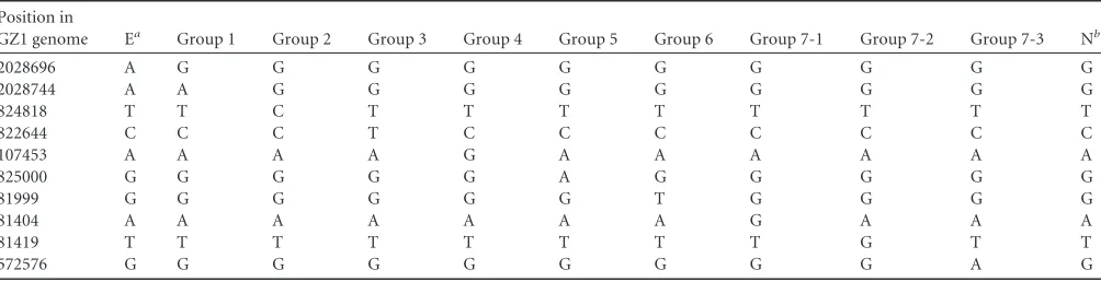
were based on the following criteria: (i) the target gene contained as many specific SNPs for different groups as possible and there-fore required the fewest number of genes, (ii) the length of the amplified product was less than the reading length for the se-quence reaction (900 bp), and (iii) the genes selected contained conserved regions without sequence variation among the 85 ge-nome sequences available forS. suis(8) so that primers suitable for amplifying a diverse range of strains could be designed. MCG group 7 is defined by 3 subgroups, 7-1, 7-2, and 7-3, as there are no unique SNPs that define MCG group 7. To further differentiate the epidemic ST7 clone, we added an extra SNP to type this clone within MCG group 1. Therefore, a total of 10 SNPs distributed in six genes were selected (Table 2), includingSSGZ1_0088 (encod-ing a putative preprotein translocase SecY protein, SNP positions 81404, 81419, and 81999),SSGZ1_0114(encoding the glycosyl transferase family, SNP position 107453),SSGZ1_0530(encoding a predicted membrane protein, SNP position 572576),SSGZ1_ 0776 (encoding carbamoyl-phosphate synthase, large subunit, SNP position 822644),SSGZ1_0777(encoding a SAM-dependent methyltransferase, SNP positions 824818 and 825000), and SSGZ1_1981 (encoding a hypothetical protein, SNP positions 2028696 and 2028744).
[image:2.585.40.550.79.235.2]Six primer pairs were designed to identify the region covering the 10 SNPs. The primer pairs could be amplified in all the isolates tested. We used sequencing of the PCR product to identify the TABLE 1Primers used in the study
Primer no. MCG group(s) Sequence (5=to 3=)
Targeted gene
Annealing temp (°C)
SNP position(s) (GZ1 genome)
PCR product size (bp)
1 1 Forward: TCCATTCATAAGCGACTC SSGZ1_1981 48 2028696, 2028744 557
Reverse: AAGGAAGACAAGCCCAAG
2 2/5 Forward: GGCTGTCTTGGTTCTTTA SSGZ1_0777 48 824818, 825000 626
Reverse: TTCATACTTCCTCCTCCA
3 3 Forward: ACCACAATGAAATGCCAGAA SSGZ1_0776 51.7 822644 468
Reverse: GACCAGAACCCAGCACCA
4 4 Forward: AAACCAGCGAAAGACTTGACT SSGZ1_0114 53.7 107453 866
Reverse: TACGCTTTGGCATCTACACCT
5 6/7-1/7-2 Forward: GAGTAAACGCAAAGAGTC SSGZ1_0088 48 81404, 81419, 81999 795
Reverse: GAACCGTCAATAACCAC
6 7-3 Forward: GCAAAGAGGAAGCGATGA SSGZ1_0530 48 572576 457
Reverse: AGGCTGGAAATACTGAACC
TABLE 2SNPs used in this study
Position in
GZ1 genome Ea Group 1 Group 2 Group 3 Group 4 Group 5 Group 6 Group 7-1 Group 7-2 Group 7-3 Nb
2028696 A G G G G G G G G G G
2028744 A A G G G G G G G G G
824818 T T C T T T T T T T T
822644 C C C T C C C C C C C
107453 A A A A G A A A A A A
825000 G G G G G A G G G G G
81999 G G G G G G T G G G G
81404 A A A A A A A G A A A
81419 T T T T T T T T G T T
572576 G G G G G G G G G A G
a
E, epidemic strains (ST7 strains). bN, ungroupable.
on May 16, 2020 by guest
http://jcm.asm.org/
[image:2.585.42.544.578.707.2]SNPs of interest. The 169 isolates were categorized as belonging to 1 of the 7 MCG groups, and 10 were MCG ungroupable. The most prevalent group was MCG group 6, representing⬎65% of the isolates (118/179). The next most prevalent groups were MCG group 7 (7-1, two isolates; 7-2, five isolates; and 7-3, 17 isolates), MCG groups 4 and 5 (nine isolates each), MCG group 1 (six lates), MCG group 2 (two isolates), and MCG group 3 (one iso-late) (see Table S1 in the supplemental material).
Relationship between MCGs and STs.We also typed 179 iso-lates by MLST; 115 sequence types (STs) were found, among which 111 were newly assigned. Only four of the STs (ST1, ST17, ST28, and ST32) were reported previously, a considerably differ-ent observation from that of a previous study (21). Thirteen iso-lates were untypeable due to the deletion of one of the seven housekeeping genes (see Table S1 in the supplemental material).
A minimum spanning tree of the 435 STs in theS. suisMLST database identified 27 clonal complexes (defined as groups of pro-files differing by no more than one gene from at least one other ST in the group) (see Fig. S1 in the supplemental material). Within the 27 clonal complexes, 15 included 27 STs and 48 isolates in this study.
MCG groups 1, 2, 3, 4, 5, 6, 7-1, 7-2, and 7-3 and MCG un-groupable contained 2, 1, 1, 2, 4, 80, 1, 4, 13, and 7 STs, respec-tively. The 15 clonal complexes were distributed in MCG groups 1 (2 complexes), 2 (1 complex), 4 (2 complexes), 5 (1 complex), 6 (7 complexes), and 7-3 (2 complexes) (see Fig. S1 in the supplemen-tal material). Each ST and clonal complex were represented in only one MCG group or lineage. Isolates of the same ST and ST complex were clustered into the same MCG group.
Genotypes ofmrp,epf, andslyand their distribution in dif-ferent MCGs.Most of the isolates (150/179, 83.8%) were mrp negative. Twenty-nine isolates contained putative full-lengthmrp gene copies; five were MCG group 1, two were MCG group 2, seven were MCG group 4, nine were MCG group 5, two were MCG group 7-1, and four were MCG ungroupable. Based on the mrpsubtypes reported in North America (18), the sequences of mrpwere grouped into one of three subtypes, EU (four isolates), NA1 (22 isolates), or NA2 (three isolates). The three genotypes are very similar at the 5=and 3=ends, while variation has been ob-served in the central portion of the gene. Only nine isolates con-tained theslygene. Six were from MCG group 1, two were from MCG group 4, and one was MCG ungroupable. Isolates positive forepf(n⫽6) were all from MCG group 1.
There were eight genotypes ofmrp,epf, andsly, primarily based onmrpvariation:mrpEUepf⫹sly⫹;mrpnegativeepf⫹sly⫹;mrpNA2
epf⫹sly⫹;mrpNA2epfnegativeslynegative;mrpnegativeepf neg-ativesly⫹;mrpNA1epfnegativeslynegative;mrpNA1epfnegative
sly⫹; andmrpnegativeepfnegativeslynegative. Of the isolates, 82.1% (147/179) were genotyped mrpnegative epfnegative sly negative. Isolates from MCG group 1 were genotypedmrpEUepf⫹ sly⫹,mrpnegativeepf⫹sly⫹, ormrpNA2epf⫹sly⫹. All the isolates
from MCG group 2 harboredmrpNA2(see Table S2 in the supple-mental material).
All but ST1 isolates harbored the CDS2157 gene.In our pre-vious study, the CDS2157 gene annotated as RNA binding S1 was present in all intermediate and weakly virulent (I/WV) and viru-lent (V) groups (except for the ST13 strain) but not in the epi-demic and highly virulent (E/HV) group (22). We also tested for the presence of this gene in the 179 isolates. Our results showed
that all except five ST1 isolates from MCG group 1 harbored the CDS2157 gene.
Relationship between MCG groups and serotypes.The sero-types of 83 isolates were identified using the mPCR assay (16). The result was confirmed using the seroagglutination test. Within the known serotypes, serotype 29 (n⫽19) was the most prevalent, followed by serotypes 11 (n⫽9), 2 (n⫽8), 5 (n⫽8), 12 (n⫽8), 30 (n⫽8), and 24 (n⫽4). All of them but serotype 24 were found to be associated with multiple MCG groups. In addition, three isolates (each) of serotypes 1/2, 7, and 10, two isolates (each) of serotypes 8 and 31, and a single isolate each of serotypes 9, 14, 15, 16, 21, and 22 were also identified (see Table S3 in the supplemen-tal material).
A number of MCG groups which contain isolates of multiple serotypes were also identified. MCG groups 6 and 7 each con-tained 7 serotypes, MCG ungroupable included 4 serotypes, MCG groups 1 and 4 each contained 4 serotypes, and MCG ungroupable included 2 serotypes.
mPCR showed that 96 isolates were not assigned to a known type. Sequencing of thecpsgene cluster showed that these isolates possess undescribedwzy genes compared to those of currently known serotypes (16,23) (data not shown) and may represent new serotypes. Of the 96 unserotypeable isolates, 10 newwzy se-quences were found. Theircpsgene clusters were also greatly dif-ferent from those of the known 33 serotypes (16,23) (data not shown). Almost all of the untypeable isolates were from MCG group 6 (84.4%, 81/96) or 7 (14.6%, 14/96 isolates).
DISCUSSION
Studying the population structure ofS. suismay reveal important information about the epidemiology ofS. suisinfection and facil-itate the rapid identification of potentially virulent strains. The ability to cluster related isolates and differentiate unrelated iso-lates is crucial for understanding the population biology ofS. suis. Whole-genome comparative analysis and collection of high-con-fidence global SNPs provide ideal typing targets for investigating the population structure of bacterial species (24–26). The reliabil-ity of phylogenetic inference based on MLST is adversely affected by the frequency of recombinations that obscure phylogenetic sig-nals, asS. suisis a weakly clonal species. We showed previously that MCG groups are the ideal units for investigating the popula-tion structure ofS. suisbased on genome sequence analysis of 85 isolates (8). In this study, we translated genome-based MCG group assignment by identifying a set of 10 SNPs distributed in six genes which can accurately assign the isolates to a unique clade or lineage and distinguish epidemically associated strains. This ap-proach offers a practical, rapid, and cost-effective assay that is suitable for laboratories where whole-genome sequencing is not yet available.
In the present study, we were unable to group 10 isolates. These isolates possibly had a significant recombinant history that cannot be reliably allocated. The most prevalent MCG groups among the tested isolates were groups 6 and 7. Only six isolates were from MCG group 1, to which the epidemic clone belongs, and no iso-lates associated with the epidemic clone were found. Using Strep-tococcus pneumoniaestrain R6 as an out group, groups 6 and 7 diverged the earliest, while group 1 diverged most recently. This indicates that most isolates from carrier animals were from the groups that diverged earlier.
The presence of some genes, such asmrp,epf, andsly, has been
on May 16, 2020 by guest
http://jcm.asm.org/
associated with virulence (27,28). We previously reported that MCG group 1 includes highly virulent isolates and contains the greatest number of virulence genes. Conversely, MCG groups 6 and 7 carry the lowest number of virulence genes (8). Here, we found that 98.6% of the isolates from MCG groups 6 and 7 were genotypedmrpnegativeepfnegativeslynegative, which is consis-tent with our previous findings.
Based on the presence/absence of genes using comparative genomic hybridization (CGH), we previously clustered 40 isolates into the E/HV, V, and I/WV groups (22). The virulence levels of the three groups decreased incrementally from the E/HV group to the I/WV group. There was a clear tendency in the association of isolates belonging to the E/HV group with MCG group 1. The genotype ofmrpharbored by all isolates from the E/HV group was mrpEU(our unpublished observations) and CDS2157 negative. Of the 12 MCG group 1 isolates, 11 were genotyped mrpEU and CDS2157 negative. Only one isolate (NCTC 10446, serotype 15, ST81) was genotyped asmrpNA2and CDS2157 positive. The iso-late was also clustered into the V group by CGH (22). We also found a similar isolate (YS12) in this study. These two isolates, although assigned to MCG group 1, may have diverged earliest (8) and shared some characteristics with MCG group 2, since MCG group 2 was predominantly of the genotypemrpNA2. Studies on more isolates from diseased pigs and patients will be required to evaluate the public health significance of MCG group 2.
CDS2157 belongs to the Tex family of proteins that contain the S1 RNA-binding domain at the C terminus. We found in our previous study that this gene was specific to the I/WV group (22). We proposed that the presence of this gene may be negatively associated with virulence (22). In this study, we found that the gene was absent in all highly virulent ST1 isolates, further support-ing our hypothesis.
The mechanisms used byS. suisto colonize the host are poorly understood. It is proposed thatS. suisgains entry to the systemic circulation primarily through the palatine tonsils after adhesion and invasion of the epithelial cells (29). OnceS. suisreaches deep tissues and/or the bloodstream, it is able to resist phagocytosis and persist in the blood at high concentrations with inflammatory consequences. CPS and suilysin may play important roles in these stages (30–32). Most invasiveS. suisdiseases are caused by sero-types 1/2, 2, 3, 4, 8, 9, and 14 (18,33–35). In our previous study, the blood bacterial loads of mice infected with strains belonging to these serotypes were higher than those of other serotype strains (22). We found in this study that⬍10% of the isolates from healthy pigs were suilysin positive or belonged to serotypes 1/2, 2, 3, 4, 7, 8, 9, or 14. Therefore, most of the isolates from carrier pigs found in this study may be less capable of causing serious infec-tions in humans.
Considerable genetic diversity between strains of the same se-rotype has been observed, and there is also far more sese-rotype di-versity with 10 novelwzysequences from the 96 isolates that can-not be serotyped using current PCR molecular serotyping (16).
In conclusion, we developed an SNP-based typing method that can categorize isolates into the correct MCG groups, defined by whole-genome sequencing, for a robust, economical, and simple means of differentiating the population structure ofS. suis. Our data show that MCG SNP typing assay facilitates the application of genome data to the surveillance ofS. suis. Our analysis of 179 isolates isolated from pigs using MCG SNP typing and other
mo-lecular typing also expanded our knowledge on the population structure and genetic diversity ofS. suis.
ACKNOWLEDGMENTS
This work was supported by the Ministry of Science and Technology, People’s Republic of China (grants 2012ZX10004-215, 2013ZX10004221, 2013ZX10004216-001-002, and 81261120559).
We thank Yan Wang for technical assistance.
REFERENCES
1.Gottschalk M, Xu J, Calzas C, Segura M.2010.Streptococcus suis: a new emerging or an old neglected zoonotic pathogen? Future Microbiol. 5:371–391.http://dx.doi.org/10.2217/fmb.10.2.
2.Gottschalk M, Segura M, Xu J.2007.Streptococcus suis infections in humans: the Chinese experience and the situation in North America. Anim. Health Res. Rev. 8:29 – 45. http://dx.doi.org/10.1017/S146625 2307001247.
3.Ye C, Zhu X, Jing H, Du H, Segura M, Zheng H, Kan B, Wang L, Bai X, Zhou Y, Cui Z, Zhang S, Jin D, Sun N, Luo X, Zhang J, Gong Z, Wang X, Wang L, Sun H, Li Z, Sun Q, Liu H, Dong B, Ke C, Yuan H, Wang H, Tian K, Wang Y, Gottschalk M, Xu J.2006.Streptococcus suis sequence type 7 outbreak, Sichuan, China. Emerg. Infect. Dis.12:1203– 1208.http://dx.doi.org/10.3201/eid1708.060232.
4.Yu H, Jing H, Chen Z, Zheng H, Zhu X, Wang H, Wang S, Liu L, Zu R, Luo L, Xiang N, Liu H, Liu X, Shu Y, Lee SS, Chuang SK, Wang Y, Xu J, Yang W.2006. HumanStreptococcus suisoutbreak, Sichuan, China. Emerg. Infect. Dis.12:914 –920.http://dx.doi.org/10.3201/eid1206 .051194.
5.Baums CG, Verkuhlen GJ, Rehm T, Silva LM, Beyerbach M, Pohlmeyer K, Valentin-Weigand P.2007. Prevalence ofStreptococcus suisgenotypes in wild boars of northwestern Germany. Appl. Environ. Microbiol.73: 711–717.http://dx.doi.org/10.1128/AEM.01800-06.
6.de Greeff A, Wisselink HJ, de Bree FM, Schultsz C, Baums CG, Thi HN, Stockhofe-Zurwieden N, Smith HE.2011. Genetic diversity of Strepto-coccus suisisolates as determined by comparative genome hybridization. BMC Microbiol.11:161.http://dx.doi.org/10.1186/1471-2180-11-161. 7.Wu Z, Li M, Wang C, Li J, Lu N, Zhang R, Jiang Y, Yang R, Liu C, Liao
H, Gao GF, Tang J, Zhu B.2011. Probing genomic diversity and evolu-tion ofStreptococcus suisserotype 2 by NimbleGen tiling arrays. BMC Genomics12:219.http://dx.doi.org/10.1186/1471-2164-12-219. 8.Chen C, Zhang W, Zheng H, Lan R, Wang H, Du P, Bai X, Ji S, Meng
Q, Jin D, Liu K, Jing H, Ye C, Gao GF, Wang L, Gottschalk M, Xu J. 2013. Minimum core genome sequence typing of bacterial pathogens: a unified approach for clinical and public health microbiology. J. Clin. Mi-crobiol.51:2582–2591.http://dx.doi.org/10.1128/JCM.00535-13. 9.Brisebois LM, Charlebois R, Higgins R, Nadeau M.1990. Prevalence of
Streptococcus suisin four to eight week old clinically healthy piglets. Can. J. Vet. Res.54:174 –177.
10. Callens BF, Haesebrouck F, Maes D, Butaye P, Dewulf J, Boyen F.2013. Clinical resistance and decreased susceptibility inStreptococcus suis iso-lates from clinically healthy fattening pigs. Microb. Drug Resist.19:146 – 151.http://dx.doi.org/10.1089/mdr.2012.0131.
11. Dee SA, Carlson AR, Winkelman NL, Corey MM.1993. Effect of man-agement practices on theStreptococcus suiscarrier rate in nursery swine. J. Am. Vet. Med. Assoc.203:295–299.
12. Martinez G, Harel J, Lacouture S, Gottschalk M.2002. Genetic diversity ofStreptococcus suisserotypes 2 and 1/2 isolates recovered from carrier pigs in closed herds. Can. J. Vet. Res.66:240 –248.
13. Ngo TH, Tran TB, Tran TT, Nguyen VD, Campbell J, Pham HA, Huynh HT, Nguyen VV, Bryant JE, Tran TH, Farrar J, Schultsz C. 2011. Slaughterhouse pigs are a major reservoir ofStreptococcus suis sero-type 2 capable of causing human infection in southern Vietnam. PLoS One 6:e17943.http://dx.doi.org/10.1371/journal.pone.0017943.
14. Silva LM, Baums CG, Rehm T, Wisselink HJ, Goethe R, Valentin-Weigand P.2006. Virulence-associated gene profiling ofStreptococcus suis isolates by PCR. Vet. Microbiol.115:117–127.http://dx.doi.org/10.1016/j .vetmic.2005.12.013.
15. Wang K, Zhang W, Li X, Lu C, Chen J, Fan W, Huang B. 2013. Characterization ofStreptococcus suisisolates from slaughter swine. Curr. Microbiol.66:344 –349.http://dx.doi.org/10.1007/s00284-012-0275-4. 16. Liu Z, Zheng H, Gottschalk M, Bai X, Lan R, Ji S, Liu H, Xu J.2013.
on May 16, 2020 by guest
http://jcm.asm.org/
Development of multiplex PCR assays for the identification of the 33 serotypes ofStreptococcus suis. PLoS One8:e72070.http://dx.doi.org/10 .1371/journal.pone.0072070.
17. King SJ, Leigh JA, Heath PJ, Luque I, Tarradas C, Dowson CG, Whatmore AM.2002. Development of a multilocus sequence typing scheme for the pig pathogenStreptococcus suis: identification of virulent clones and potential capsular serotype exchange. J. Clin. Microbiol.40: 3671–3680.http://dx.doi.org/10.1128/JCM.40.10.3671-3680.2002. 18. Fittipaldi N, Fuller TE, Teel JF, Wilson TL, Wolfram TJ, Lowery DE,
Gottschalk M.2009. Serotype distribution and production of murami-dase-released protein, extracellular factor and suilysin by field strains of Streptococcus suisisolated in the United States. Vet. Microbiol.139:310 – 317.http://dx.doi.org/10.1016/j.vetmic.2009.06.024.
19. Staats JJ, Plattner BL, Stewart GC, Changappa MM.1999. Presence of theStreptococcus suissuilysin gene and expression of MRP and EF corre-lates with high virulence inStreptococcus suistype 2 isolates. Vet. Micro-biol.70:201–211.http://dx.doi.org/10.1016/S0378-1135(99)00147-9. 20. Wisselink HJ, Reek FH, Vecht U, Stockhofe-Zurwieden N, Smits MA,
Smith HE.1999. Detection of virulent strains ofStreptococcus suistype 2 and highly virulent strains ofStreptococcus suistype 1 in tonsillar speci-mens of pigs by PCR. Vet. Microbiol.67:143–157.http://dx.doi.org/10 .1016/S0378-1135(99)00036-X.
21. Wang S, Liu P, Li C, Tan Y, Cai X, Zhou D, Jiang Y.2012. Isolation and characterization of 89K pathogenicity island-positive ST-7 strains of Streptococcus suisserotype 2 from healthy pigs, northeast China. Scientific World Journal.2012:302386.http://dx.doi.org/10.1100/2012/302386. 22. Zheng H, Lan R, Zheng X, Cui Z, Liu Z, Bai X, Ji S, Gottschalk M, Xu
J.2014. Comparative genomic hybridization identifies virulence differ-ences inStreptococcus suis. PLoS One9:e87866.http://dx.doi.org/10.1371 /journal.pone.0087866.
23. Okura M, Takamatsu D, Maruyama F, Nozawa T, Nakagawa I, Osaki M, Sekizaki T, Gottschalk M, Kumagai Y, Hamada S.2013. Genetic analysis of capsular polysaccharide synthesis gene clusters from all sero-types ofStreptococcus suis: potential mechanisms for generation of capsu-lar variation. Appl. Environ. Microbiol.79:2796 –2806.http://dx.doi.org /10.1128/AEM.03742-12.
24. Kuroda M, Serizawa M, Okutani A, Sekizuka T, Banno S, Inoue S.2010. Genome-wide single nucleotide polymorphism typing method for identi-fication ofBacillus anthracisspecies and strains amongB. cereusgroup species. J. Clin. Microbiol.48:2821–2829.http://dx.doi.org/10.1128/JCM .00137-10.
25. Leekitcharoenphon P, Lukjancenko O, Friis C, Aarestrup FM, Ussery DW.2012. Genomic variation inSalmonella entericacore genes for epi-demiological typing. BMC Genomics 13:88. http://dx.doi.org/10.1186 /1471-2164-13-88.
26. Pandya GA, Holmes MH, Petersen JM, Pradhan S, Karamycheva SA, Wolcott MJ, Molins C, Jones M, Schriefer ME, Fleischmann RD, Peterson SN.2009. Whole genome single nucleotide polymorphism based phylogeny ofFrancisella tularensisand its application to the devel-opment of a strain typing assay. BMC Microbiol.9:213.http://dx.doi.org /10.1186/1471-2180-9-213.
27. Takeuchi D, Akeda Y, Nakayama T, Kerdsin A, Sano Y, Kanda T, Hamada S, Dejsirilert S, Oishi K.2014. The contribution of suilysin to the pathogenesis ofStreptococcus suismeningitis. J. Infect. Dis.10:1509 – 1519.http://dx.doi.org/10.1093/infdis/jit661.
28. Vecht U, Wisselink HJ, Jellema ML, Smith HE.1991. Identification of two proteins associated with virulence ofStreptococcus suistype 2. Infect. Immun.59:3156 –3162.
29. Madsen LW, Bak H, Nielsen B, Jensen HE, Aalbaek B, Riising HJ.2002. Bacterial colonization and invasion in pigs experimentally exposed to Streptococcus suisserotype 2 in aerosol. J. Vet. Med. B Infect. Dis. Vet. Public Health 49:211–215. http://dx.doi.org/10.1046/j.1439-0450.2002 .00491.x.
30. Chabot-Roy G, Willson P, Segura M, Lacouture S, Gottschalk M.2006. Phagocytosis and killing ofStreptococcus suisby porcine neutrophils. Mi-crob. Pathog.41:21–32.http://dx.doi.org/10.1016/j.micpath.2006.04.001. 31. Gottschalk M, Segura M. 2000. The pathogenesis of the meningitis caused byStreptococcus suis: the unresolved questions. Vet. Microbiol. 76:259 –272.http://dx.doi.org/10.1016/S0378-1135(00)00250-9. 32. Smith HE, Damman M, van der Velde J, Wagenaar VF, Wisselink HJ,
Stockhofe-Zurwieden N, Smits MA.1999. Identification and character-ization of the cps locus ofStreptococcus suisserotype 2: the capsule protects against phagocytosis and is an important virulence factor. Infect. Immun. 67:1750 –1756.
33. Gottschalk M, Lacouture S, Bonifait L, Roy D, Fittipaldi N, Grenier D. 2013. Characterization ofStreptococcus suisisolates recovered between 2008 and 2011 from diseased pigs in Quebec, Canada. Vet. Microbiol. 162:819 – 825.http://dx.doi.org/10.1016/j.vetmic.2012.10.028.
34. Schultsz C, Jansen E, Keijzers W, Rothkamp A, Duim B, Wagenaar JA, van der Ende A.2012. Differences in the population structure of invasiveStreptococcus suisstrains isolated from pigs and from humans in the Netherlands. PLoS One7:e33854.http://dx.doi.org/10.1371/journal .pone.0033854.
35. Wisselink HJ, Smith HE, Stockhofe-Zurwieden N, Peperkamp K, Vecht U.2000. Distribution of capsular types and production of muramidase-released protein (MRP) and extracellular factor (EF) ofStreptococcus suis strains isolated from diseased pigs in seven European countries. Vet. Mi-crobiol.74:237–248.http://dx.doi.org/10.1016/S0378-1135(00)00188-7.