0095-1137/08/$08.00
⫹
0
doi:10.1128/JCM.01202-08
Copyright © 2008, American Society for Microbiology. All Rights Reserved.
Identities of
Microbacterium
spp. Encountered in Human Clinical Specimens
䌤
Kathrina Gneiding, Reinhard Frodl, and Guido Funke*
Department of Medical Microbiology and Hygiene, Ga
¨rtner & Colleagues Laboratories, Ravensburg, Germany
Received 25 June 2008/Returned for modification 19 August 2008/Accepted 7 September 2008
In the present study, 50 strains of yellow-pigmented gram-positive rods that had been isolated from human
clinical specimens and collected over a 5-year period were further characterized by phenotypic and molecular
genetic methods. All 50 strains belonged to the genus
Microbacterium
, and together they represented 18
different species.
Microbacterium oxydans
(
n
ⴝ
11),
M. paraoxydans
(
n
ⴝ
9), and
M. foliorum
(
n
ⴝ
7) represented
more than half of the strains included in the present study. The isolation of strains belonging to
M.
hydro-carbonoxydans
(
n
ⴝ
2),
M. esteraromaticum
(
n
ⴝ
1),
M. oleivorans
(
n
ⴝ
1),
M. phyllosphaerae
(
n
ⴝ
1), and
M.
thalassium
(
n
ⴝ
1) from humans is reported for the first time.
Microbacterium
sp. strain VKM Ac-1389 (
n
ⴝ
1)
and the previously uncultured
Microbacterium
sp. clone YJQ-29 (
n
ⴝ
1) probably represent new species.
Comprehensive antimicrobial susceptibility data are given for the 50
Microbacterium
isolates. This study is, so
far, the largest on
Microbacterium
spp. encountered in human clinical specimens and outlines the heterogeneity
of clinical
Microbacterium
strains.
Among the coryneform bacteria, the phenotypically and
phy-logenetically closely related genera
Microbacterium
and
Aureo-bacterium
have been united in the redefined genus
Microbacte-rium
(20). At present, the genus
Microbacterium
comprises
55 species (www.bacterio.cict.fr/m/microbacterium.html), all of
which exhibit more or less yellow-pigmented gram-positive
rods. Despite this large number of species, only in the
mid-1990s was the presence of microbacteria in human clinical
specimens recognized (7, 8, 11). Since then, only eight other
reports on microbacteria have appeared in the relevant clinical
microbiology literature (1, 2, 9, 12–16). The aim of the present
study was to reveal the distribution of individual
Microbacte-rium
species in human clinical specimens by applying
pheno-typic and molecular genetic methods. Because no
comprehen-sive data on the antimicrobial susceptibility patterns of
Microbacterium
spp. were available, we also determined the
MICs of 10 antimicrobial agents against all 50 strains included
in the present study. We observed that three species, namely,
Microbacterium oxydans
,
M. paraoxydans
, and
M. foliorum
,
ac-counted for more than 50% of all strains included in the
present study, but overall, 18 different taxa were encountered,
indicating the heterogeneity of microbacteria isolated from
clinical specimens.
(This paper is part of the medical doctoral thesis of K.
Gneiding at the medical faculty of the University of Ulm, Ulm,
Germany.)
MATERIALS AND METHODS
Strains.During a 5-year period, the 50 strains investigated in the present study were isolated in the routine clinical microbiology laboratories of Ga¨rtner & Colleagues Laboratories, Ravensburg, Germany, or referred to the reference laboratory for coryneform bacteria at this institution by collaborating laborato-ries. None of the isolates had been included in any of our previous studies (7–9, 11). None of the patients were epidemiologically linked. The strains had been
stored at⫺20°C in skim milk. For the investigations, strains were grown on Columbia sheep blood agar plates (BD, Heidelberg, Germany) and passaged twice on Columbia sheep blood agar at 35°C in ambient air before use.
Biochemical identification.The techniques used have been described in detail previously (10). The commercial API Coryne and API ZYM kits (both from bioMe´rieux, Marcy l’Etoile, France) were used according to the manufacturer’s instructions, and reading was done after 48 h of incubation at 35°C for the API Coryne and after 4 h for the API ZYM system.
Molecular genetic investigations.The 16S rRNA gene sequences were ana-lyzed according to a published protocol (3). Almost complete (⬎1,400-bp) 16S rRNA gene sequences were determined for each clinical strain by aligning multiple overlapping sequences by use of the Lasergene 5 package (DNAStar Inc., Madison, WI). The 16S rRNA genes of the differentMicrobacteriumspecies were aligned and compared by using the Web-based BLAST 2 Sequences soft-ware tool (www.ncbi.nlm.nih.gov/blast/bl2seq/wblast2.cgi).
Identification.A strain was identified to the species level if its 16S rRNA gene sequence shared⬎98.70% base pair homology with the type strain or with other representative strains of a valid species (19) and if phenotypic testing did not indicate any aberrant reactions relative to the published data for this particular species.
Antimicrobial susceptibility testing.The CLSI standard for the determination and interpretation of antimicrobial MICs forCorynebacteriumspp. (5) was ap-plied. Briefly, by use of a broth microdilution method, bacterial cells with an inoculum equivalent to a 0.5 McFarland standard were grown in cation-adjusted Mueller-Hinton broth with lysed horse blood and were incubated for as long as 48 h. MICs were read by two independent researchers.
Nucleotide sequence accession numbers.The GenBank accession numbers of the almost complete 16S rRNA gene sequences of all 50 clinical isolates included in the present study are given in Table 1.
RESULTS
Table 1 outlines the patients’ data as well as the identities of
the 50
Microbacterium
strains included in the present study.
Twenty-nine patients were male and 21 female. The ages of the
patients ranged from 1 to 79 years, with an average of 43.1
years. Sixteen strains came from blood cultures; 13 strains were
isolated from wounds; 11 strains came from normally sterile
anatomical sites or sterile materials; 6 strains came from
urines; and 4 strains were isolated from miscellaneous
materi-als.
The 50 strains were found to belong to 18 different taxa:
M.
oxydans
(
n
⫽
11),
M. paraoxydans
(
n
⫽
9),
M. foliorum
(
n
⫽
7),
M. aurum
(
n
⫽
3),
M. lacticum
(
n
⫽
3), “
M. binotii
” (
n
⫽
2),
M.
* Corresponding author. Mailing address: Department of Medical
Microbiology and Hygiene, Ga
¨rtner & Colleagues Laboratories,
Elisa-bethenstrasse 11, D-88212 Ravensburg, Germany. Phone:
49-751-502-230. Fax: 49-751-502-385. E-mail: ldg.funke@t-online.de.
䌤
Published ahead of print on 17 September 2008.
3646
on May 16, 2020 by guest
http://jcm.asm.org/
hydrocarbonoxydans
(
n
⫽
2),
M. testaceum
(
n
⫽
2),
M.
tricho-thecenolyticum
(
n
⫽
2),
M. esteraromaticum
(
n
⫽
1),
M.
lae-vaniformans
(
n
⫽
1),
M. oleivorans
(
n
⫽
1),
M. phyllosphaerae
(
n
⫽
1),
M. resistens
(
n
⫽
1),
M. schleiferi
(
n
⫽
1),
M. thalassium
(
n
⫽
1),
Microbacterium
sp. strain VKM Ac-1389 (
n
⫽
1), and
the uncultured
Microbacterium
sp. clone YJQ-29 (
n
⫽
1). For
all 50 strains, the 16S rRNA gene homology of the individual
clinical strain with the type strain or another representative
strain of the corresponding species ranged from 98.84% to
100%, with a mean homology of 99.60%.
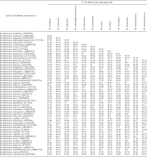
The 16S rRNA gene homologies between all 55
Microbac-terium
species defined to date are given in Table 2. A total of
1,485 16S rRNA gene homologies were calculated. Two
differ-ent clinically relevant
Microbacterium
species always shared
less than 98.70% homology except for the species
M.
arbore-scens
and
M. imperiale
(99.73% homology),
M. oxydans
and M.
paraoxydans
(99.25%),
M. foliorum
and
M. phyllosphaerae
(99.19%),
M. lacticum
and
M. schleiferi
(98.91%),
M. foliorum
and
M. hydrocarbonoxydans
(98.85%),
M. hydrocarbonoxydans
and
M. oxydans
(98.77%),
M. oleivorans
and
M. phyllosphaerae
(98.73%),
M. hydrocarbonoxydans
and
M. phyllosphaerae
(98.72%), and
M. foliorum
and
M. oxydans
(98.70%).
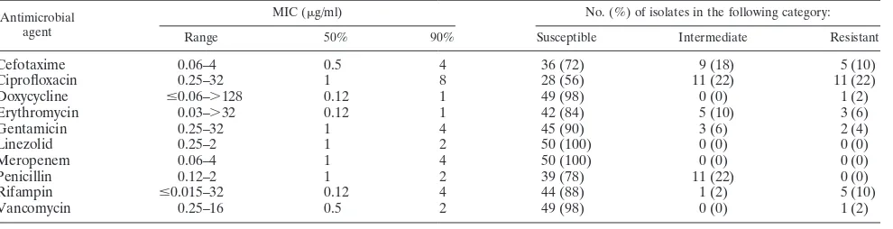
Table 3 shows the antimicrobial susceptibility patterns of
Microbacterium
spp. All 50 isolates were susceptible to
lin-TABLE 1. Strains included in the present study
Running no.
Strain collection no.
Patient’s age
(yr), sexa Clinical source Identification
GenBank accession no.
1
1
31, f
Knee puncture fluid
M. paraoxydans
EU714331
2
9
34, m
Wound swab
M. foliorum
EU714380
3
58
45, f
Urine
M. paraoxydans
EU714372
4
76
56, m
Wound swab
M. paraoxydans
EU714377
5
118
74, f
Gall bladder
M. schleiferi
EU714332
6
150
70, m
Wound swab
M. foliorum
EU714333
7
297
8, m
Throat swab
M. oxydans
EU714348
8
314
1, m
Blood culture
M. aurum
EU714355
9
327
36, m
Wound swab
M. foliorum
EU714358
10
331
65, f
Prosthetic hip infection
M. phyllosphaerae
EU714359
11
332
55, f
Blood culture
M. trichothecenolyticum
EU714360
12
343
5, f
Urine
M. testaceum
EU714365
13
346
66, m
Blood culture
M. laevaniformans
EU714366
14
407
25, f
Superficial wound
M. oxydans
EU714369
15
428
79, m
Pleural fluid
M. paraoxydans
EU714370
16
556
49, m
Wound swab
M. foliorum
EU714371
17
591
39, m
Blood culture
M. paraoxydans
EU714373
18
698
47, m
Endophthalmitis
M. oxydans
EU714374
19
699
25, f
Urine
M. oxydans
EU714375
20
720
74, f
Pleural fluid
M. foliorum
EU714376
21
768
68, m
Wound swab
Microbacterium
sp. strain VKM Ac-1389
EU714378
22
798
24, f
Blood culture
M. foliorum
EU714379
23
985
18, m
Blood culture
M. oleivorans
EU714381
24
2083
40, f
Sinus aspirate
M. oxydans
EU714335
25
2121
5, m
Superficial wound
“
M. binotii
”
EU714336
26
2122
59, m
Blood culture
M. esteraromaticum
EU714337
27
2229
23, m
Bone infection
“
M. binotii
”
EU714338
28
2345
54, m
Dialysis fluid
M. oxydans
EU714339
29
2350
28, f
Blood culture
M. oxydans
EU714340
30
2400
67, m
Wound swab
M. foliorum
EU714341
31
2470
54, f
Urine
M. paraoxydans
EU714342
32
2588
65, m
Blood culture
M. aurum
EU714343
33
2704
41, m
Lymph node
M. oxydans
EU714344
34
2761
45, f
Wound swab
Microbacterium
sp. strain YJQ-29
EU714345
35
2833
54, f
Blood culture
M. lacticum
EU714346
36
2841
8, m
Wound swab
M. oxydans
EU714347
37
3043
29, f
Blood culture
M. oxydans
EU714349
38
3047
NK, f
Urine
M. lacticum
EU714350
39
3075
66, m
Wound swab
M. lacticum
EU714351
40
3084
7, m
Blood culture
M. hydrocarbonoxydans
EU714352
41
3109
35, m
Tracheal secretion
M. paraoxydans
EU714353
42
3131
75, f
Urine
M. paraoxydans
EU714354
43
3200
11, m
Blood culture
M. paraoxydans
EU714356
44
3227
74, m
Blood culture
M. oxydans
EU714357
45
3352
31, m
Wound swab
M. resistens
EU714361
46
3370
60, m
Blood culture
M. trichothecenolyticum
EU714362
47
3373
49, m
Urethral swab
M. thalassium
EU714363
48
3388
51, f
Conjunctival swab
M. aurum
EU714364
49
3502
42, f
Blood culture
M. testaceum
EU714367
50
3517
45, f
Catheter tip
M. hydrocarbonoxydans
EU714368
am, male; f, female; NK, not known.
on May 16, 2020 by guest
http://jcm.asm.org/
[image:2.585.47.540.80.561.2]ezolid and meropenem. Only strain 3352 was resistant to
van-comycin, and only strain 985 was resistant to doxycycline.
Cip-rofloxacin had the weakest activity against microbacteria; 22%
of the isolates were intermediately susceptible, and 22% were
resistant.
DISCUSSION
From the work of Stackebrandt and Ebers, it has been clear
that a cutoff of 98.7% 16S rRNA gene homology is appropriate
for species differentiation within a genus (19). As is evident
from Table 2, the genus
Microbacterium
is a very tight genus
regarding the 16S rRNA gene homology between two valid
species. However, applying the recommendations of
Stack-ebrandt and Ebers, we were able to easily identify every
Mi-crobacterium
strain included in the present study.
Of note is the molecular genetic differentiation between
M.
oxydans
and
M. paraoxydans
, the two most frequently
[image:3.585.47.550.79.608.2]encoun-tered species in the present study. Compared to the
M. oxydans
TABLE 2. Percentages of 16S rRNA gene homologies of
Microbacterium
spp.
Species (GenBank accession no.)
% 16S rRNA gene homology with:
M.
aerolatum
M.
aoyamense
M.
aquimaris
M.
arabinogalactanolyticum
M.
arborescens
M.
aurantiacum
M.
aurum
M.
barkeri
M.
chocolatum
M.
deminutum
M.
dextranolyticum
M.
esteraromaticum
Microbacterium aerolatum(AJ309929)
Microbacterium aoyamense(AB234028) 96.43
Microbacterium aquimaris(AM778449) 96.58 96.22
Microbacterium arabinogalactanolyticum(Y17228) 97.43 96.53 96.99
Microbacterium arborescens(X77443) 95.94 97.16 95.88 95.83
Microbacterium aurantiacum(AB004726) 96.37 96.54 96.72 96.65 95.44
Microbacterium aurum(Y17229) 96.09 98.89 95.81 96.17 96.67 96.39
Microbacterium barkeri(X77446) 95.51 94.19 95.05 96.16 96.19 95.03 93.81
Microbacterium chocolatum(AB004725) 95.93 95.75 96.06 96.21 95.12 98.42 95.92 94.5
Microbacterium deminutum(AB234026) 96.08 99.09 95.99 96.23 97 96.93 98.39 94.23 96.3
Microbacterium dextranolyticum(Y17230) 96.72 96.94 96.28 97.33 96.13 97.09 96.54 95.24 96.77 97.49
Microbacterium esteraromaticum(Y17231) 97.65 96.54 96.86 99.18 95.83 96.52 96.18 96.37 96.08 96.24 97.19
Microbacterium flavescens(Y17232) 96.58 96.68 96.5 97.13 96.39 97.34 96.33 95.44 96.84 97 97.82 97.13
Microbacterium flavum(AB286029) 97.43 98.75 96.35 96.56 97.04 96.88 98.34 94.58 96.21 98.4 97.38 96.56
Microbacterium foliorum(AJ249780) 98.53 97.37 97.14 97.4 96.67 97 97.15 95.38 96.24 97.08 96.68 97.27
Microbacterium ginsengisoli(AB271048) 94.73 97.23 94.77 94.67 98.55 95.01 96.49 95.46 94.36 97.21 95.48 94.67
Microbacterium gubbeenense(AF263563) 94.42 94.53 94.77 93.6 93.9 93.82 94.9 95.42 92.95 94.3 92.62 94.61
Microbacterium halophilum(AB004714) 95.22 94.95 97.14 95.97 95.38 95.58 94.64 94.77 95.08 95.33 95.66 95.84
Microbacterium halotolerans(AY376165) 95.02 94.27 95.12 95.4 93.87 95.3 93.93 94.55 94.14 94.44 94.97 95.34
Microbacterium hatanonis(AB274908) 95.59 97.23 95.96 96.01 96.51 95.66 96.93 94.95 94.61 97.01 96.39 96.02
Microbacterium hominis(AB004727) 96.93 96.81 96.65 96.84 97.06 97.15 96.38 95.57 96.61 97.07 97.63 96.7
Microbacterium hydrocarbonoxydans(AJ698726) 97.95 97.58 97 97.47 96.6 97.37 97.42 95.45 96.65 97.22 97.23 97.2
Microbacterium imperiale(X77442) 96.08 97.09 95.95 95.83 99.73 95.71 96.6 96.05 95.2 97 96.27 95.83
Microbacterium indicum(AM158907) 94.6 94.29 95.05 95.33 95.08 94.63 94.46 96.4 93.55 93.76 94.54 95.13
Microbacterium keratanolyticum(Y17233) 97.43 97.02 96.79 96.24 96.3 96.58 96.99 94.25 95.53 96.59 96.16 96.38
Microbacterium ketosireducens(AB004724) 97.08 96.39 96.74 98.44 96.1 97.73 96.1 96.04 97.02 96.95 97.73 97.59
Microbacterium kitamiense(AB013907) 96.51 96.68 96.86 96.92 95.92 99.39 96.88 95.31 98.17 97.07 97.43 96.79
Microbacterium koreense(AY962574) 96.39 98.32 96.86 96.1 96.66 96.31 98.05 93.96 95.52 97.83 96.16 95.9
Microbacterium lacticum(X77441) 96.58 98.62 96.44 96.51 96.94 96.53 98.44 94.15 96.09 98.26 96.95 96.52
Microbacterium lacus(AB286030) 96.22 99.45 96.09 96.4 97.04 96.79 98.99 94.09 95.62 98.95 96.71 96.41
Microbacterium laevaniformans(Y17234) 96.71 97.49 96.01 96.79 96.44 97.64 97.3 95.02 97.29 97.99 98.72 96.65
Microbacterium liquefaciens(X77444) 97.58 97.23 97 97.2 96.53 96.66 97.08 95.17 95.88 96.86 96.61 97.34
Microbacterium luteolum(Y17235) 96.73 96.75 96.79 97.06 96.24 96.51 96.72 95.01 95.74 96.38 96.64 97.2
Microbacterium luticocti(AM747814) 94.38 94.32 95.26 94.59 94.62 94.8 94.07 95.32 94.68 94.22 93.96 94.32
Microbacterium marinilacus(AB286020) 95.52 95.16 95.05 95.65 97.11 95.84 94.71 96.43 95.21 95.06 95.55 95.58
Microbacterium maritypicum(AB004728) 96.81 97.16 95.88 96.84 96.24 97.55 96.72 95.23 97.13 97.63 98.31 96.84
Microbacterium natoriense(AY566291) 97.87 97.19 97.18 97.54 96.83 97.19 96.84 95.5 96.68 96.9 97.11 97.26
Microbacterium oleivorans(AJ698725) 97.56 97.65 97.97 97.4 97.14 97.17 97.15 95.3 96.79 97.42 97.22 97.27
Microbacterium oxydans(Y17227) 97.8 97.44 97.2 97.4 96.72 96.86 96.72 95.35 96.09 97.07 96.92 97.54
Microbacterium paludicola(AJ853909) 94.94 95.25 94.84 95.25 97.53 95.03 97.53 96.37 94.51 94.94 94.8 95.06
Microbacterium paraoxydans(AJ491806) 98.4 97.23 97.56 97.74 96.66 96.96 96.66 95.92 96.35 97.01 97.08 98.02
Microbacterium phyllosphaerae(AJ277840) 98.24 97.3 97.14 97.81 97.01 97.28 97.01 95.78 96.65 96.93 97.35 97.68
Microbacterium pumilum(AB234027) 96.22 99.29 96.02 96.37 97.15 97.01 97.15 94.25 96.5 99.79 97.58 96.3
Microbacterium resistens(Y14699) 97.22 96.75 97.48 97.67 96.3 96.92 96.3 95.82 96.49 96.45 97.33 97.68
Microbacterium saperdae(Y17236) 97.36 96.89 96.79 96.86 96.33 96.46 96.33 94.77 95.67 96.59 96.34 97
Microbacterium schleiferi(Y17237) 96.86 98.89 96.92 96.65 97.74 96.92 97.74 94.73 96.42 98.53 97.46 96.79
Microbacterium sediminicola(AB286021) 96.01 96.4 95.53 95.71 98.28 96.86 98.28 95.88 95.76 96.17 96.1 95.72
Microbacterium terrae(AB004720) 96.23 95.71 96.46 97.06 95.42 96.72 95.42 95.22 96.35 96.37 97.45 96.79
Microbacterium terregens(AB004721) 96.3 98.95 96.03 96.45 96.87 96.11 96.87 94.24 95.51 98.6 96.39 96.24
Microbacterium terricola(AB234025) 96.58 97.23 96.69 96.87 96.93 96.73 96.93 94.85 96.03 98.33 97.08 96.87
Microbacterium testaceum(X77445) 97.65 97.02 97.76 97.74 96.67 97.75 96.67 95.98 97.18 96.72 97.48 97.74
Microbacterium thalassium(AB004713) 96.66 96.55 97.35 96.84 96.25 97.15 96.25 95.38 96.28 97.08 97.76 97.12
Microbacterium trichothecenolyticum(AB004722) 96.29 96.12 96.16 97.47 96.3 97.47 96.3 95.75 96.9 96.3 97.19 97.33
Microbacterium ulmi(AY062021) 95.31 95.93 95.81 95.83 97.76 95.22 97.76 95.78 94.86 95.76 95.61 95.57
Microbacterium xylanilyticum(AJ853908) 96.73 96.13 96.16 97.04 95.96 96.74 95.96 95.63 96.05 96.45 96.66 97.04
on May 16, 2020 by guest
http://jcm.asm.org/
type strain sequence (GenBank accession no. Y17227 [18]), all
nine
M
.
paraoxydans
strains from the present study showed the
following nucleotide differences: at position 168, T instead of
C; at position 177, T instead of A; at position 181, T instead of
a deletion; at position 374, T instead of C; at position 555, C
instead of G; at position 569, G instead of C; at position 588,
G instead of N; and at position 1211, T instead of C. In general,
we can confirm the data of Laffineur et al. (15) for the
bio-chemical differentiation of
M. oxydans
and
M. paraoxydans
: in
the present study, 9 of 11
M. oxydans
strains expressed

-glu-cosidase activity (10 of 10 in reference 15), whereas all
M.
paraoxydans
strains were negative in both studies. Another
distinguishing reaction might be the strong pyrrolidonyl
[image:4.585.48.544.80.187.2]aryl-amidase activity detected in the present study for 8 of 11
M.
TABLE 2—
Continued
% 16S rRNA gene homology with:
M.
flavescens
M.
flavum
M.
foliorum
M.
ginsengisoli
M.
gubbeenense
M.
halophilum
M.
halotolerans
M.
hatanonis
M.
hominis
M.
hydrocarbonoxydans
M.
imperiale
M.
indicum
M.
keratanolyticum
M.
ketosireducens
M.
kitamiense
M.
koreense
M.
lacticum
M.
lacus
96.83 96.74 97.24 95.66 97.08 95.53 93.07 94.09 94.37 93.39 96.4 95.05 95.55 94.17 92.76 95.29 94.73 95.02 92.97 94.29 93.93 96.31 97.23 96.59 96.65 93.18 95.16 94.03 98.02 97.22 96.82 95.83 93.42 96.36 94.93 96.35 96.67 98 98.85 95.97 93.37 95.56 95.06 96.75 96.85 96.32 96.97 96.68 98.41 93.75 95.26 93.8 96.44 96.95 96.54 94.53 94.68 94.59 94.21 96.27 94.47 95.24 94.15 94.56 95.52 94.94 96.51 97.18 98.02 95.22 94.41 95.08 94.51 96.84 96.15 97.67 96.17 94.56 98.09 96.67 97.03 94.97 93.38 96.32 95.76 95.83 97.45 97.17 96.11 94.77 96.18 97.62 96.83 97.29 95.21 94.06 95.62 95.41 96.02 97.21 97.59 96 94.97 97.2 97.73 96.24 98.18 97.08 96.1 94.69 94.96 94.3 95.97 96.31 97.43 96.59 94.71 96.45 96.25 96.38 96.67 98.55 97.36 96.84 93.97 95.19 94.07 96.86 96.81 97.76 96.95 94.25 96.99 96.39 96.88 98.05 96.49 98.61 97.53 97.12 94.69 94.86 94.41 97.54 96.72 97.75 96.97 94.62 97.58 96.24 97.27 98.05 98.35 97.29 98.22 97.22 95.98 93.59 95.37 95.27 96.51 97.58 98.15 96.37 94.71 96.65 97.14 97.72 96.81 97.44 97.35 96.67 97.38 98.51 96.84 94.33 95.26 94.95 97.27 96.47 98.58 96.4 94.66 97.74 96.89 96.95 96.8 97.56 97.39 96.51 96.97 98.29 95.09 94.09 95.09 94.79 96.84 96.36 98.36 96.1 94.42 98.02 96.6 96.79 96.38 96.99 96.89 94.07 94.5 94.69 94.78 94.91 94.16 94.04 94.13 94.26 95.21 94.55 95.45 93.83 94.22 95.01 95 94 94.25 95.94 95.77 95.6 96.44 94.36 94.45 94.89 94.49 95.57 96.32 97.04 96.14 94.89 95.97 96.05 94.92 95.26 95.09 97.13 97.85 96.69 95.76 93.57 95.35 95.08 96.49 98.12 97.59 96.2 94.62 96.15 97.24 97.6 96.38 97.15 97.06 97.11 97.47 98.66 95.57 93.01 93.01 95.02 96.49 97.11 98.45 96.83 94.74 97.61 97.59 97.33 96.91 97.33 97.04 97.62 97.94 98.11 96.17 93.58 94.49 94.99 97.01 97.65 98.06 97.22 94.66 97.67 97.52 97.38 97.15 97.9 97.54 96.93 97.59 98.7 95.78 94.49 93.58 95.13 97.46 96.66 98.77 96.59 94.77 97.95 97.1 97.14 97 97.61 97.58 95.27 95.59 95.63 96.35 94.78 94.78 94.22 94.86 95.05 98.36 97.25 95.94 95.11 95.37 95.41 95.05 95.07 95.03 97.13 97.59 98.5 95.76 95.28 94.53 95.08 97.31 96.96 98.65 96.6 95.18 97.6 97.38 97.18 96.66 97.41 97.14 97.41 97.45 99.19 95.66 93.3 93.3 95.57 96.86 97.63 98.72 96.88 95.36 98.15 97.73 97.69 97.01 97.49 97.46 97.08 98.58 97.16 97.22 94.35 94.35 94.6 96.87 97.22 97.37 97.08 94.58 96.65 97.08 97.15 98.01 98.29 99.14 97.26 97.18 97.74 95.15 94.21 94.21 95.06 96.77 96.91 97.74 96.24 95.46 97.47 97.17 97.33 96.38 97.06 97.06 96.46 97.17 98.03 95.19 94.25 94.25 94.54 97 96.32 98.1 96.19 94.26 98.15 96.46 96.68 96.59 97.08 97.08 97.33 98.62 97.61 97.23 93.85 93.85 94.24 97.32 97.25 98.02 97.74 94.42 97.33 96.88 97.19 98.4 98.91 96.61 96.56 96.53 96.36 97.74 95.32 95.32 94.43 95.87 96.53 96.86 98.21 95.66 96.27 95.96 97.22 95.83 96.5 96.98 97.81 95.94 96.31 94.26 93.17 93.17 94.79 95.13 96.7 96.38 95.42 94.01 95.83 98.58 96.99 95.55 95.76 95.76 96.39 98.32 97.16 97.01 94.45 94.45 93.84 96.95 96.6 97.36 96.8 93.96 96.59 98.46 96.11 97.71 98.13 98.13 97.08 97.98 97.43 96.72 94.61 94.61 93.98 97.64 96.59 97.71 96.93 94.67 97.08 96.45 96.87 97.83 97.98 97.98 97.62 97.32 97.76 95.46 94.29 94.29 95.16 97.13 97.47 97.89 96.8 95.22 97.4 97.45 98.03 96.73 97.21 97.21 97.47 96.75 96.9 95.23 93.83 93.83 94.8 96.56 96.95 97.06 96.39 94.63 96.78 97.67 97.09 96.25 96.66 96.66 98.43 96.14 96.58 95.43 93.89 93.89 95.13 95.87 97.66 96.79 96.17 94.42 96.03 97.8 97.74 95.82 96.31 96.31 95.72 95.95 96.22 96.78 94.59 94.59 94.07 95.62 96.36 95.92 97.76 95.17 95.22 95.54 95.52 95.83 95.6 95.6 97.46 96.18 97.6 94.95 93.45 94.78 95.36 95.59 97.83 96.8 95.9 94.56 96.07 97.03 96.88 95.62 96.03 96.03
Continued on next page
on May 16, 2020 by guest
http://jcm.asm.org/
oxydans
strains, whereas weak activity was detected for 1 of 9
M. paraoxydans
strains. The reason why
M. oxydans
and
M.
paraoxydans
were the most frequently encountered species in
our series is unclear but might be related to the distribution of
these species in the environment.
In their important study of microbacteria, Laffineur et al.
(15) observed that
M. oxydans
(9 of 30 strains) and
M.
paraoxy-dans
(5 of 30 strains) were the microbacteria most frequently
found in clinical specimens. These authors also detected
M.
aurum
(4 of 30 strains),
M. lacticum
(4 of 30 strains),
M.
schleiferi
(1 of 30 strains), and
M. testaceum
(1 of 30 strains),
but only 1 of 30 strains was identified as
M. foliorum
, whereas
in our study, 7 of 50 strains belonged to this species.
In the present study, we describe the second and third
M.
trichothecenolyticum
strains from humans, whereas to date,
only one strain had been isolated from clinical specimens and
TABLE 2—
Continued
% 16S rRNA gene homology with:
M.
laevaniformans
M.
liquefaciens
M.
luteolum
M.
luticocti
M.
marinilacus
M.
maritypicum
M.
natoriense
M.
oleivorans
M.
oxydans
M.
paludicola
M.
paraoxydans
M.
phyllosphaerae
M.
pumilum
M.
resistens
M.
saperdae
M.
schleiferi
M.
sediminicola
M.
terrae
M.
terregens
M.
terricola
M.
testaceum
M.
thalassium
M.
trichothecenolyticum
M.
ulmi
97 97 98.97 94.66 94.42 94.24 95.98 95.12 94.96 94.38 99 96.61 96.5 94.33 95.91 97.93 97.75 97.61 94.39 95.99 97.11 97.58 97.83 97.68 94.46 95.63 97.45 98.58 97.22 99.66 99.18 94.59 95.3 96.8 97.96 98.02 95.65 95.21 95.04 94.48 98.6 95.47 96.07 95.73 95.39 97.57 99.05 98.84 95.08 95.77 97.24 98.24 98.38 99.25 95.82 97.43 98.37 98.22 94.35 96.22 97.36 98.73 98.73 98.56 95.84 98.3 98.17 97.01 96.51 94.19 95.09 97.79 97.01 97.01 97.22 95.14 97.01 97.08 97.58 97.74 97.61 95.35 95.71 97.25 97.82 97.82 97.95 95.81 98.36 97.88 96.51 97.15 98.78 99.11 94.15 94.79 96.46 97.47 97.47 99.04 94.86 98.5 97.96 96.73 97.61 97.79 97.67 97.33 94.24 95.65 97.46 97.82 97.82 97.88 95.82 97.67 97.88 98.57 97.26 97.47 96.48 95.88 95.72 94.93 97.82 96.47 96.41 96.41 96.06 97.14 96.27 96.91 96.23 96.26 95.75 96.75 96.94 96.1 96.31 94.04 95.17 96.5 96.77 96.77 96.31 97.47 96.58 96.85 96.44 96.37 95.97 96.3 95.23 97.04 96.95 96.45 94.05 95.21 96.74 96.91 96.91 97.14 95.27 96.95 97.08 98.86 96.45 96.6 98.26 96.04 95.34 97.34 97.63 97.22 94.63 95.76 97.21 97.26 97.26 97.84 95.79 97.77 97.42 98.36 97.71 97.43 98.4 96.66 95.89 98.32 97.51 98.03 97.88 94.97 96.43 97.54 97.96 97.96 98.22 95.84 98.64 98.3 96.79 98.5 97.83 97.54 96.91 96.72 96.6 97.84 97.51 97.21 96.92 94.4 95.38 97.26 97.26 97.26 97.28 95.55 97.84 97.1 97.16 98.01 96.87 97.19 96 96.78 96.33 97.5 97.82 96.81 96.58 96.45 93.79 95.85 96.91 96.9 96.9 96.79 95.46 97.12 97.13 96.37 97.47 96.51 96.65 96.54 97.26 96.03 96.8 98.01 97.19 96.3 95.47 95.29 95.28 96.66 96.07 96.28 96.28 95.65 96.52 96.12 96.08 95.81 95.76 95.26 95.9 97.15 94.95 95.91 95.76 96.27 95.38 95.7 96.92 96.65 96.49 94.46 95.5 96.54 96.91 96.91 96.83 95.52 97.15 97.26 96.51 97.38 96.31 96.28 95.98 96.28 96.05 96.52 96.78 96.95 97.31 95.86
on May 16, 2020 by guest
http://jcm.asm.org/
another from soil (16, 22). We report on the first two
M.
hydrocarbonoxydans
strains and the first
M. oleivorans
strain
from humans, whereas only one strain of
M.
hydrocarbonoxy-dans
had been isolated from oil-contaminated soil and only
one strain of
M. oleivorans
had been isolated from an oil
storage cavern (17).
M. esteraromaticum
also has not been
reported for humans but had been used as an aroma-producing
bacterium (22), and
M. thalassium
had been isolated from soil
(21). One
M. laevaniformans
strain (previously CDC group A-5
coryneform bacteria) isolated from blood had been described
previously (7).
M. foliorum
and
M. phyllosphaerae
cannot be distinguished
phenotypically but were reported to share 12 differences in
1,480 bp (10 substitutions and 2 additional bases) of their 16S
rRNA genes (4). All seven
M. foliorum
strains from the present
study shared the following mismatches with the type strain of
M. phyllosphaerae
(AJ277840): at positions 45 to 47, CAG
instead of GCC; at positions 49 and 50, GG instead of C and
a deletion; at position 60, T instead of G; and at position 65, G
instead of a deletion. The latter two
Microbacterium
species
were isolated from phyllospheres and grasses and from
decay-ing grasses of a litter layer (4). It is not unlikely that our
patients acquired their
M. foliorum
and
M. phyllosphaerae
strains from grasses.
Strains 2121 and 2229 were identified as “
Microbacterium
binotii
,” a taxon that has been proposed as a new species by D.
Clermont, S. Diard, L. Motreff, C. Vivier, F. Bimet, C.
Bouchier, M. Welker, W. Kallow, and C. Bizet (unpublished
data) (GenBank accession no. EF567306) but has not been
validated so far. Strain 768 is a member of a presently
unde-scribed
Microbacterium
species of which strain VKM Ac-1389,
isolated from an interacting plant and nematode (GenBank
accession no. AB0402070), is a representative. Finally, strain
2761 is a representative of the uncultured
Microbacterium
sp.
clone YJQ-29, which had been isolated from a hot spring
(GenBank accession no. AY569297).
It should be noted that, except for
M. resistens
,
M. hominis
,
M. paraoxydans
, and “
M. binotii
,” all microbacteria were
ini-tially defined by strains that originated from the environment.
It is not known at present whether microbacteria have a habitat
in humans or are solely acquired from the environment.
The 50
Microbacterium
isolates of our current series
exhib-ited a level of susceptibility to penicillin (78%) similar to that
of isolates reported in previous publications, about 80 to 90%
of which showed susceptibility (7, 11). Interestingly, higher
MICs for gentamicin (range, 1 to 64
g/ml) were reported in
previous studies of microbacteria (7, 11) than in the present
study. The reason for this is unclear, but the different results
might result from different MIC determination methods
(mi-crodilution in the present study versus agar dilution in the
previous studies). The results of the present study correlated
well with the antimicrobial MIC data obtained for six
M.
paraoxydans
strains by use of Etest strips (15). In contrast to
other coryneform bacteria and, in particular, other
yellow-pigmented strains (6), for which rifampin usually has very low
MICs, the MICs were slightly higher than usual (i.e.,
ⱖ
0.12
g/ml), although 88% of the
Microbacterium
strains were still
fully susceptible. The present study reports only the third
iso-late of
M. resistens
, which shows the vancomycin resistance
inherent in this species (9).
It is acknowledged that microbacteria are not frequently
found as pathogens in human clinical specimens, as evidenced
by the fact that just 50 isolates were collected in a reference
center over a 5-year period.
Because of the heterogeneity of clinical isolates belonging to
the genus
Microbacterium
, we strongly recommend to clinical
microbiology laboratories that for yellow-pigmented
gram-pos-itive rods, of which
Microbacterium
is the most frequently
en-countered genus (6), almost-complete (i.e.,
⬎
1,400-bp) 16S
rRNA gene sequences should be determined in order to
iden-tify the strains, if indicated, to the species level, although the
present study, together with the study of Laffineur et al. (15),
indicates that
M. oxydans
and
M. paraoxydans
are the most
frequently isolated microbacteria in human clinical specimens.
REFERENCES
1.Adderson, E. E., J. W. Boudreaux, and R. T. Hayden.2008. Infections caused by coryneform bacteria in pediatric oncology patients. Pediatr. Infect. Dis. J.
27:136–141.
2.Alonso-Echanove, J., S. S. Shah, A. J. Valenti, S. N. Dirrigl, L. A. Carson, M. J. Arduino, and W. R. Jarvis.2001. Nosocomial outbreak of Microbac-teriumspecies bacteremia among cancer patients. J. Infect. Dis.184:754–760. 3.Beck, M., R. Frodl, and G. Funke.2008. Comprehensive study of strains previously designatedStreptococcus bovisconsecutively isolated from human blood cultures and emended description ofStreptococcus gallolyticusand
Streptococcus infantariussubsp.coli. J. Clin. Microbiol.46:2966–2972. 4.Behrendt, U., A. Ulrich, and P. Schumann.2001. Description of
Microbac-terium foliorumsp. nov. andMicrobacterium phyllosphaeraesp. nov., isolated from the phyllosphere of grasses and the surface litter after mulching the sward, and reclassification ofAureobacterium resistens(Funke et al. 1998) as
Microbacterium resistenscomb. nov. Int. J. Syst. Evol. Microbiol.51:1267– 1276.
[image:6.585.45.540.80.207.2]5.Clinical and Laboratory Standards Institute.2006. Methods for antimicro-bial dilution and disk susceptibility testing of infrequently isolated or fastid-ious bacteria. Document M45-A. CLSI, Wayne, PA.
TABLE 3. Antimicrobial susceptibility patterns of
Microbacterium
strains (
n
⫽
50)
Antimicrobial agent
MIC (g/ml) No. (%) of isolates in the following category: Range 50% 90% Susceptible Intermediate Resistant
Cefotaxime
0.06–4
0.5
4
36 (72)
9 (18)
5 (10)
Ciprofloxacin
0.25–32
1
8
28 (56)
11 (22)
11 (22)
Doxycycline
ⱕ
0.06–
⬎
128
0.12
1
49 (98)
0 (0)
1 (2)
Erythromycin
0.03–
⬎
32
0.12
1
42 (84)
5 (10)
3 (6)
Gentamicin
0.25–32
1
4
45 (90)
3 (6)
2 (4)
Linezolid
0.25–2
1
2
50 (100)
0 (0)
0 (0)
Meropenem
0.06–4
1
4
50 (100)
0 (0)
0 (0)
Penicillin
0.12–2
1
2
39 (78)
11 (22)
0 (0)
Rifampin
ⱕ
0.015–32
0.12
4
44 (88)
1 (2)
5 (10)
Vancomycin
0.25–16
0.5
2
49 (98)
0 (0)
1 (2)
on May 16, 2020 by guest
http://jcm.asm.org/
6.Funke, G., and K. A. Bernard.2007. Coryneform gram-positive rods, p. 485–514.InP. R. Murray, E. J. Baron, J. H. Jorgensen, M. L. Landry, and M.A. Pfaller (ed.), Manual of clinical microbiology, 9th ed. ASM Press, Washington, DC.
7.Funke, G., E. Falsen, and C. Barreau. 1995. Primary identification of
Microbacteriumspp. encountered in clinical specimens as CDC coryneform group A-4 and A-5 bacteria. J. Clin. Microbiol.33:188–192.
8.Funke, G., G. Haase, N. Schnitzler, N. Schrage, and R. R. Reinert.1997. Endophthalmitis due toMicrobacteriumspecies: case report and review of mycobacterium infections. Clin. Infect. Dis.24:713–716.
9.Funke, G., P. A. Lawson, F. S. Nolte, N. Weiss, and M. D. Collins.1998.
Aureobacterium resistenssp. nov., exhibiting vancomycin resistance and teico-planin susceptibility. FEMS Microbiol. Lett.158:89–93.
10.Funke, G., G. Martinetti Lucchini, G. E. Pfyffer, M. Marchiani, and A. von Graevenitz.1993. Characteristics of CDC group 1 and group 1-like coryne-form bacteria isolated from clinical specimens. J. Clin. Microbiol.31:2907– 2912.
11.Funke, G., A. von Graevenitz, and N. Weiss.1994. Primary identification of
Aureobacteriumspp. isolated from clinical specimens as “Corynebacterium aquaticum.” J. Clin. Microbiol.32:2686–2691.
12.Giammanco, G. M., S. Pignato, P. A. D. Grimont, F. Grimont, C. Santangelo, G. Leopardi, A. Giuffrida, V. Legname, and G. Giammanco.2006. Interstitial pulmonary inflammation due toMicrobacteriumsp. after heart transplanta-tion. J. Med. Microbiol.55:335–339.
13.Hirji, Z., R. Saragosa, H. Dedier, M. Crump, N. Franke, L. Burrows, F. Jamieson, S. Brown, and M. A. Gardam.2003. Contamination of bone marrow products with an actinomycete resemblingMicrobacteriumspecies and reinfusion into autologous stem cell and bone marrow transplant recip-ients. Clin. Infect. Dis.36:e115–e121.
14.Ko, K. S., W. S. Oh, M. Y. Lee, K. R. Peck, N. Y. Lee, and J. H. Song.2007.
A newMicrobacteriumspecies isolated from the blood of a patient with fever:
Microbacterium pyrexiaesp. nov. Diagn. Microbiol. Infect. Dis.57:393–397. 15.Laffineur, K., V. Avesani, G. Cornu, J. Charlier, M. Janssens, G. Wauters,
and M. Delme´e.2003. Bacteremia due to a novelMicrobacteriumspecies in a patient with leukemia and description ofMicrobacterium paraoxydanssp. nov. J. Clin. Microbiol.41:2242–2246.
16.Lau, S. K. P., P. C. Y. Woo, G. K. S. Woo, and K.-Y. Yuen.2002. Catheter-relatedMicrobacteriumbacteremia identified by 16S rRNA gene sequencing. J. Clin. Microbiol.40:2681–2685.
17.Schippers, A., K. Bosecker, C. Spro¨er, and P. Schumann.2005. Microbacte-rium oleivoranssp. nov. andMicrobacterium hydrocarbonoxydanssp. nov., novel crude-oil-degrading gram-positive bacteria. Int. J. Syst. Evol. Micro-biol.55:655–660.
18.Schumann, P., F. A. Rainey, J. Burghart, E. Stackebrandt, and N. Weiss.
1999. Reclassification of Brevibacterium oxydans (Chatelain and Second 1966) asMicrobacterium oxydanscomb. nov. Int. J. Syst. Bacteriol.49:175– 177.
19.Stackebrandt, E., and J. Ebers.2006. Taxonomic parameters revisited: tar-nished gold standards. Microbiol. Today33:152–155.
20.Takeuchi, M., and K. Hatano.1998. Union of the generaMicrobacterium
Orla-Jensen andAureobacteriumCollins et al. in a redefined genus Microbac-terium. Int. J. Syst. Bacteriol.48:739–747.
21.Takeuchi, M., and K. Hatano.1998. Proposal of six new species in the genus
Microbacteriumand transfer ofFlavobacterium marinotypicumZoBell and Upham to the genusMicrobacteriumasMicrobacterium maritypicumcomb. nov. Int. J. Syst. Bacteriol.48:973–982.
22.Yokota, A., M. Takeuchi, T. Sakane, and N. Weiss.1993. Proposal of six new species in the genusAureobacteriumand transfer ofFlavobacterium esteraro-maticumOmelianski to the genusAureobacteriumasAureobacterium ester-aromaticumcomb. nov. Int. J. Syst. Bacteriol.43:555–564.