Quantification of
Cryptosporidium
spp. and Identification of
Cryptosporidium parvum
and
Cryptosporidium hominis
C. Mary,aE. Chapey,bE. Dutoit,cK. Guyot,dL. Hasseine,eF. Jeddi,aJ. Menotti,gC. Paraud,fC. Pomares,eM. Rabodonirina,bA. Rieux,f F. Derouin,gfor the ANOFEL
CryptosporidiumNational Network
Aix-Marseille Université, Faculté de Médecine, UMR MD3, and APHM, Hôpital de la Timone, Laboratoire de Parasitologie-Mycologie, Marseille, Francea; Laboratoire de
Parasitologie-Mycologie, Hospices Civils de Lyon, Université Lyon 1, Lyon, Franceb; Laboratoire de Parasitologie-Mycologie, Centre Hospitalo-Universitaire de Lille, Lille,
Francec; Biologie et Diversité des Pathogènes Eucaryotes Émergents, Centre d’Infection et d’Immunité de Lille, Institut Pasteur de Lille, INSERM U1019, CNRS UMR 8204,
Université Lille-Nord-de-France, Lille, Franced; Laboratoire de Parasitologie-Mycologie, Centre Hospitalo-Universitaire de Nice, Nice, Francee; Agence Nationale de Sécurité
Sanitaire de l’Alimentation, de l’Environnement, et du Travail, Niort, Francef; Laboratoire de Parasitologie-Mycologie, Hôpital Saint-Louis, Assistance Publique-Hôpitaux de
Paris and Université Denis Diderot, Paris, Franceg
Cryptosporidiumis a protozoan parasite responsible for gastroenteritis, especially in immunocompromised patients. Laboratory diagnosis of cryptosporidiosis relies on microscopy, antigen detection, and nucleic acid detection and analysis. Among the nu-merous molecular targets available, the 18S rRNA gene displays the best sensitivity and sequence variations between species and can be used for molecular typing assays. This paper presents a new real-time PCR assay for the detection and quantification of all
Cryptosporidiumspecies associated with the identification ofCryptosporidium hominisandCryptosporidium parvum. The sen-sitivity and specificity of this new PCR assay were assessed on a multicentric basis, using well-characterizedCryptosporidium -positive and -negative human stool samples, and the efficiencies of nine extraction methods were comparatively assessed using
Cryptosporidium-seeded stool samples and phosphate-buffered saline samples. A comparison of extraction yields showed that the most efficient extraction method was the Boom technique in association with mechanical grinding, and column extraction showed higher binding capacity than extraction methods based on magnetic silica. Our PCR assay was able to quantify at least 300 oocysts per gram of stool. Satisfactory reproducibility between laboratories was observed. The two main species causing hu-man disease,Cryptosporidium hominisandCryptosporidium parvum, were identified using a duplex real-time PCR assay with specific TaqMan minor-groove-binding ligand (MGB) probes for the same amplicon. To conclude, this one-step quantitative PCR is well suited to the routine diagnosis of cryptosporidiosis since practical conditions, including DNA extraction, quantifica-tion using well-defined standards, and identificaquantifica-tion of the two main species infecting humans, have been positively assessed.
C
ryptosporidiumis a protozoan parasite involved in waterborne gastrointestinal infections. The disease usually is mild in im-munocompetent patients but can have serious consequences in immunocompromised patients (1). To date, no treatment has been able to eradicateCryptosporidium, especially in immuno-compromised patients (2).Several species are causative agents of diarrhea in humans and animals. In humans, Cryptosporidium parvum and Cryptospo-ridium hominisare the most frequently detected species, account-ing for almost 90% of cases of diagnosed cryptosporidiosis but displaying different prevalence rates in different areas. The two species are almost equally represented in Europe and the United States (3–8), whereasC. hominisis predominant in tropical re-gions, reaching a prevalence of up to 88% of identified cases in some countries (9–12). Several other species also have emerged as causes of cryptosporidiosis in immunocompromised and immu-nocompetent patients, but at much lower prevalence rates than C. parvumorC.hominis(5,10,11).
Laboratory methods for detectingCryptosporidiumwere first based on microscopy in combination with various staining meth-ods, the most widely used being the modified Ziehl-Neelsen stain-ing method for oocysts, the sensitivity of which has been estimated at 75% (13,14). A direct fluorescent antibody assay (DFA) im-proved the sensitivity of conventional microscopy, as its sensitiv-ity is about 1,000 oocysts per g of stool (15).
Antigen detection methods have been developed and marketed as immunochromatographic assays (16, 17), possibly coupled withGiardiaantigen detection. These methods are easier to use as rapid detection tests but do not allow for quantification or typing of the parasites. Higher sensitivity of detection in environmental and clinical samples can be achieved by performing molecular diagnosis of cryptosporidiosis using PCR. The most commonly used targets are the 18S rRNA gene (18), the Cryptosporidium oocyst wall protein (19), the 60-kDa glycoprotein (20), heat shock protein 70, the Laxer locus, and microsatellite loci (reviewed in reference21). Several studies agree on the higher sensitivity of PCR targeting the 18S rRNA gene, in relation to its copy number (22). Nested PCR has been put forward as a means of improving the sensitivity of detection (23) and reverse transcription-PCR as a method for studying viability in environmental samples (24). Real-time PCR presently is the most extensively used method,
Received2 January 2013 Returned for modification8 January 2013 Accepted12 May 2013
Published ahead of print29 May 2013
Address correspondence to C. Mary, cmary@ap-hm.fr.
Copyright © 2013, American Society for Microbiology. All Rights Reserved.
doi:10.1128/JCM.03458-12
on May 16, 2020 by guest
http://jcm.asm.org/
allowing for both accurate quantification of the molecular target and typing under technical conditions that avoid cross contami-nation and DNA carryover (25,26). However, DNA extraction remains a limiting condition for PCR technologies, since DNA polymerase inhibitors (which often are found in environmental and clinical samples) are copurified with nucleic acids and the yield of extraction depends on the technical conditions (27,28).
In this context, four university hospital laboratories of medical parasitology and one veterinary laboratory of the French Agency for Food, Environmental, and Occupational Health and Safety (ANSES) (Niort, France), all belonging to the French ANOFEL CryptosporidiumNational Network (5), set up a multicentric eval-uation of a new sensitive real-time quantitative PCR (qPCR) assay capable of easily discriminating and quantifying the main Crypto-sporidiumspecies involved in human pathology. This organiza-tion made it possible (i) to share well-defined clinical samples and positive controls, (ii) to compare the extraction rates of nine methods using a single independent quantification process, and (iii) to assess comparatively the performance of a new qPCR assay among laboratories using the same DNA samples (extracted in the coordinating laboratory), primers, fluorescent probes, and stan-dards.
MATERIALS AND METHODS
Study design.The Marseilles parasitology laboratory coordinated the study. The Lyons, Paris, and Nice parasitology laboratories and the ANSES veterinary laboratory were participating laboratories. Well-char-acterized positive stool samples were provided by the French ANOFEL
CryptosporidiumNational Network. The ANSES laboratory providedC. parvumoocysts. The coordinating laboratory prepared the natural stool samples and the spiked samples, performed the DNA extractions for test-ing of the different PCR methods, and prepared the natural and plasmid DNA standards. All of the participating laboratories received identical sets of materials, including 20 samples to test the DNA extraction methods and 100 DNA solutions from positive and negative biological samples to test the diagnosis/quantification and typing PCR assays.
Stool samples.Forty human stool specimens (N1 to N40) were se-lected on the basis of negative microscopic results forCryptosporidium
with the Ziehl-Nielsen modified staining method and for other protozoa or helminths with the formalin-ether concentration method. All of these specimens served as negative controls, and one of them was used to pre-pare samples seeded withCryptosporidiumoocysts. DNA from human stool specimens containingCystoisospora belli(n⫽3),Cyclospora cayet-anensis(n⫽1),Enterocytozoon bieneusi(n⫽3),Entamoeba histolytica
(n⫽2),Giardia intestinalis(n⫽6), andCandidaspp. (n⫽10) was used for the specificity assessment.
Sixty Cryptosporidium-positive human or animal stool specimens (P41 to P100) were provided by the ANOFELCryptosporidiumNational Network. The diagnosis was established by microscopy, and stool samples were preserved in 2.5% potassium dichromate (final concentration).
Cryptosporidiumspecies were determined by PCR sequencing at the 18S rRNA gene locus (29) and includedC. hominis(n⫽23),C. parvum(n⫽
20),Cryptosporidium felis(n⫽8),Cryptosporidium bovis(n⫽4), Cryp-tosporidium cuniculus(n⫽2),Cryptosporidium canis(n⫽2), and Cryp-tosporidiumchipmunk genotype (n⫽1). Ten additional fresh stool sam-ples that were positive by microscopy forCryptosporidium(7 withC. parvum[E1, E3, E4, E5, E6, E9, and E10] and 3 withC. hominis[E2, E7, and E8]) were provided by the participating laboratories in order to test the DNA extraction protocols.
Artificial positive samples were prepared withC. parvumoocysts that had been purified from heavily infected calf stools, after concentration using ethyl acetate and purification on a discontinuous Percoll gradient (30). Ten samples were prepared by adding 100, 200, 500, 1,000, or 10,000 purifiedC. parvumoocysts to either 200 mg of a negative human stool sample (E11 to E15) or 200l of phosphate-buffered saline (PBS) (E16 to E20), which resulted in final parasite loads of 500, 1,000, 2,500, 5,000, and 50,000 oocysts per g, respectively. The 20 samples numbered E1 to E20 were sent to the participating laboratories, which were asked to perform the DNA extraction upon receipt.
DNA extraction.DNA from the 40 negative samples (N1 to N40) and the 60 positive samples (P41 to P100) was extracted in the coordinating laboratory using a NucliSENS easyMAG device (bioMérieux, Marcy l’Etoile, France), based on the method described by Boom et al. (31). The extraction protocol was adapted for stool processing as follows: 400 mg of stool samples was added to 1 ml of NucliSENS lysis buffer in a tube con-taining ceramic beads (lysing matrix D; MP Biomedicals, Illkirch, France) and then disrupted in a FastPrep-24 grinder (MP Biomedicals) at maxi-mum power for 1 min. After 10 min of incubation at room temperature to allow complete lysis, tubes were centrifuged at 10,000⫻gfor 10 min and three extractions were performed, each with 250l of supernatant. Each extraction included five washing steps with NucliSENS extraction buffer 2 (reference no. 280131; bioMérieux). Elution was performed at 70°C with 100l of elution buffer; 1l of the resulting eluate corresponded to 1 mg of starting material. The DNA eluates from each sample then were pooled, aliquoted into five microtubes, frozen, and sent to the participating labo-ratories. Using a similar extraction procedure, aCryptosporidiumDNA standard solution was prepared from purifiedC. parvumoocysts resus-pended in PBS (106oocysts/extraction); the resulting eluate contained 10,000 equivalent oocysts of DNA per microliter.
One hundred DNA samples to be tested (60 positive and 40 negative) and DNA standard solutions were sent to each participating laboratory and stored at⫺20°C. These samples were tested within 4 months after receipt.
[image:2.585.37.548.77.176.2]CryptosporidiumqPCR.Two new real-time PCR assays were devel-oped in the coordinating laboratory. The first assay was designed to detect the presence ofCryptosporidiumDNA and amplified a DNA fragment located in the 18S rRNA gene (GenBank accession no.EU675853.1, posi-tions 33 to 211). The direct and reverse primer sequences were CATGGA TAACCGTGGTAAT and TACCCTACCGTCTAAAGCTG, respectively. Amplification resulted in a 178-bp amplicon that included a polymorphic region (nucleotides 157 to 162) (Table 1). A TaqMan probe homologous to a conserved region of the sequence (Pan-crypto, FAM-CTAGAGCTA TABLE 1Cryptosporidiumsequence alignment of the polymorphic region of the amplicon (ClustalW2 multiple sequence alignment file)
Species Sequence GenBank accession no.
C. hominis TTTACGGATCACAATT---AATGTGACA EU675853.1
C. parvum TTTACGGATCACATAA---ATTGTGACA AF112570.1
C. felis TTTACGGATCACAATAATTTATTTTGTGACA AF112575.1
C. bovis TTTACGGATCACATTA---TGTGACA EF514234.1
C. cuniculus TTTACGGATCACAATT---AATGTGACA HQ397716.1
C. canis TTTACGGATCACATTT---TATGTGACA AF112576.1
Cryptosporidiumchipmunk genotype
TTTACGGATCACATTTTG---ATGTGACA EF641026.1
on May 16, 2020 by guest
http://jcm.asm.org/
ATACATGCGAAAAAA-MGB-BHQ [FAM, 6-carboxyfluorescein; MGB, minor-groove-binding ligand; BHQ, black hole quencher]) was designed to detect allCryptosporidiumspecies. The second assay (duplex PCR) was designed to differentiateC. hominisandC. parvumby means of two hy-bridization probes, i.e., FAM-ATCACAATTAATGT-MGB-BHQ (C. hominis) and 6-carboxyrhodamine (VIC)-ATCACATTAAATGT-MGB-BHQ (C. parvum).
Primers and probes were used at 0.2M and 0.1M (final concen-trations), respectively. One microliter of sample DNA was added to the reaction tubes (final volume, 25l). The amplification consisted of acti-vation of theTaqDNA polymerase for 10 min at 94°C, followed by 45 cycles of 94°C for 10 s, 54°C for 30 s, and 72°C for 10 s, with thermal transitions between denaturation and primer annealing of⬍1.2°C/s, in order to enable probe hybridization. Multiplex assays including the typing probes were performed under the same technical conditions, in order to confirm that there was no interference between probes.
Control DNA and plasmids.Control DNA was prepared from puri-fiedC. parvumoocysts as described above and by cloning of theC. hominis
sequence in a plasmid using a TOPO TA cloning kit (Invitrogen reference no. K4520-01; Life Technologies SAS, Saint Aubin, France). This plasmid was amplified, purified from bacteria, and quantified by spectrophotom-etry. In the qPCR assays, these controls were used to establish a standard curve using a dilution range of 0.1 to 10,000 equivalent oocysts and/or 1 to 107copies of plasmid DNA. A set of plasmid dilutions was included in each quantification experiment for calculation of the number of target copies for one oocyst and assessment of the sensitivity of the assay.C. parvumandC. hominiscontrol DNAs were included in each typing assay as positive controls.
An M13 plasmid containing nonrelevant DNA (reference no. 360364; Applied Biosystems, Villebon-sur-Yvette, France) was used to detect in-hibitors in extracted DNA. Twenty copies of this plasmid were added to each reaction tube and then a real-time PCR targeting this plasmid was performed, using M13 universal primers and a TaqMan probe specific to the inserted DNA. A positive result was considered indicative of the ab-sence of inhibitors in the sample.
Interlaboratory comparison of nine extractions methods.Nine ex-traction protocols were applied to 10 natural (E01 to E10) and 10 seeded (E11 to E15 in stool and E16 to E20 in PBS) samples. The extraction processes were applied to the entire contents of the tubes (200 mg or 200 l), and the elution volume was set at 100l; the different protocols used are summarized inTable 2. Three extraction kits (NucliSENS easyMAG, QIAamp DNA Mini, and NucleoSpin Tissue) were used with and without preliminary mechanical grinding; the others (QIAamp DNA Stool mini-kit, UltraClean fecal DNA isolation mini-kit, and Maxwell 16 Tissue DNA pu-rification kit) were used according to the manufacturer’s instructions.
Following DNA extractions by the participating laboratories, the elu-ates were sent to the coordinating laboratory to be tested by qPCR. All eluates were tested in duplicate in a single qPCR run, for more accurate comparison of the extraction rates. Results were expressed as the number of oocysts per gram of starting material by using serial dilutions of the
CryptosporidiumDNA standard solution as a reference.
Interlaboratory qPCR assessment.Primers, probes, and DNA con-trols were sent to the participating laboratories. Each laboratory was asked to perform quantification assays in duplicate with stool sample DNA us-ing the Pan-crypto TaqMan probe and then to genotype the positive sam-ples using theC. hominisandC. parvumprobes. The participating labo-ratories performed all of the tests blind to the microscopic and qPCR results of the coordinating laboratory. The threshold cycle (CT) values and
quantities were recorded and analyzed.
Statistical analysis.A regression analysis of the quantitative results was performed with raw data to examine the relationships between the two series of quantification assays in each center. Analysis of variance (ANOVA) was carried out to examine the influence of the extraction methods on the results and to identify a possible center effect on quanti-fication. ANOVA was performed after logarithmic transformation of TABLE
2 Comparison of the main characteristics of the DNA extraction methods Abbreviated name Manufacturer Commercial name Mechanical breakage beads Automation protocol Direct lysis Proteinase K digestion
Specific adsorption of
inhibitors Silica support Elution temp (°C) easyMAG bioMérieux NucliSENS easyMag No Yes, specific B Yes No No Magnetic silica 70 easyMAG ⫹ grinding bioMérieux NucliSENS easyMag FastPrep-24 (MP Biomedicals) with lysing matrix D Yes, specific B Yes No No Magnetic silica 70 Maxwell Promega Maxwell 16 Tissue DNA purification kit No Yes, tissue Yes No No Magnetic silica 70 MO BIO MO BIO Laboratories UltraClean fecal DNA isolation kit Yes, included No Yes No Yes Silica column Room temp NucleoSpin Macherey-Nagel NucleoSpin Tissue No No After washing Yes No Silica column Room temp NucleoSpin ⫹ grinding Macherey-Nagel NucleoSpin Tissue MagNA Lyser (Roche) Green beads No After washing Yes No Silica column Room temp QIAamp DNA Qiagen QIAamp DNA Mini No No Yes Yes No Silica column Room temp QIAamp Stool Qiagen QIAamp DNA Stool Mini No No Yes Yes Yes Silica column Room temp QIAamp DNA ⫹ grinding Qiagen QIAamp DNA Mini TissueLyser (Qiagen) with steel beads No Yes Yes No Silica column Room temp
on May 16, 2020 by guest
http://jcm.asm.org/
[image:3.585.322.530.58.727.2]quantitative data. All statistical analyses were conducted using StatView 5 software (SAS Institute, Cary, NC).
RESULTS
DNA extraction methods present variable yields.All extraction products obtained with the nine extraction methods from the 15 Cryptosporidium-positive fresh fecal samples (E01 to E15) were positive by PCR, except for five obtained with QIAamp DNA Stool (E01, E11, E12, E13, and E14), two with NucliSENS easyMAG without grinding (E11 and E12), one with QIAamp DNA without grinding (E01), one with NucleoSpin with grinding (E12), one with Maxwell (E12), and three with UltraClean (E01, E12, and E13) (Table 3). The results of the ANOVA performed on the 15 sets of data were not conclusive because of a large variance due to the results for three samples with high parasite loads (E7, E9, and E10). These loads were estimated by qPCR to be around 106
oocysts/g following NucliSENS easyMAG extraction, ⬎108 oocysts/g with QIAamp DNA, and⬍5⫻105oocysts/g with the
other extraction protocols. This variation is probably related to the saturation of silica rather than to other components of the extraction process. Consequently, these samples were excluded from the ANOVA extraction rate comparison.
The ANOVA performed on the results of the 12 remaining samples showed that the extraction protocol significantly influ-enced the quantification results (P⫽0.006). The effect of the method on the extraction rate is summarized inFig. 1, where the mean values of parasite loads are presented. With a significance level of 5%, the post-ANOVA Tukey-Kramer test showed that the mean difference was greater than the critical difference for
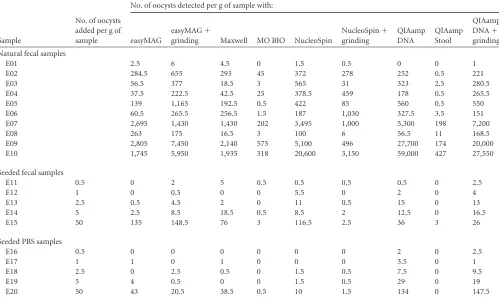
three pairs of results, i.e., QIAamp DNA Stool compared with NucliSENS easyMAG, QIAamp DNA Stool compared with easy-MAG plus grinding, and QIAamp DNA Stool compared with QIAamp DNA plus grinding. For these 12 fecal samples with low-TABLE 3Quantification ofCryptosporidiumDNA from 10 natural fecal samples (E1 to E10), five seeded fecal samples (E11 to E15), and
suspensions of oocysts in PBS (E16 to E20)
Sample
No. of oocysts added per g of sample
No. of oocysts detected per g of sample with:
easyMAG
easyMAG⫹
grinding Maxwell MO BIO NucleoSpin
NucleoSpin⫹ grinding
QIAamp DNA
QIAamp Stool
QIAamp
DNA⫹
grinding
Natural fecal samples
E01 2.5 6 4.5 0 1.5 0.5 0 0 1
E02 284.5 655 293 45 372 278 252 0.5 221
E03 56.5 377 18.5 3 565 31 323 2.5 280.5
E04 37.5 222.5 42.5 25 378.5 459 178 0.5 265.5
E05 139 1,165 192.5 0.5 422 85 560 0.5 550
E06 60.5 265.5 256.5 1.5 187 1,030 327.5 3.5 151
E07 2,695 1,430 1,430 202 3,495 1,000 5,300 198 7,200
E08 263 175 16.5 3 100 6 56.5 11 168.5
E09 2,805 7,450 2,140 575 5,100 496 27,700 174 20,000
E10 1,745 5,950 1,935 318 20,600 3,150 59,000 427 27,550
Seeded fecal samples
E11 0.5 0 2 5 0.5 0.5 0.5 0.5 0 2.5
E12 1 0 0.5 0 0 5.5 0 2 0 4
E13 2.5 0.5 4.5 2 0 11 0.5 15 0 13
E14 5 2.5 8.5 18.5 0.5 8.5 2 12.5 0 16.5
E15 50 135 148.5 76 3 116.5 2.5 36 3 26
Seeded PBS samples
E16 0.5 0 0 0 0 0 0 2 0 2.5
E17 1 1 0 1 0 0 0 3.5 0 1
E18 2.5 0 2.5 0.5 0 1.5 0.5 7.5 0 9.5
E19 5 4 0.5 0 0 1.5 0.5 29 0 19
E20 50 43 20.5 38.5 0.5 10 1.5 134 0 147.5
FIG 1Comparison of the mean log values for oocyst quantification (no. of oocysts per gram of stool) obtained from 12 samples, with medium or low parasite burdens, after nine different extractions protocols. ANOVA detected a significant influence of the technique on quantification results (P⫽0.0059). G, grinding.
on May 16, 2020 by guest
http://jcm.asm.org/
[image:4.585.43.542.89.387.2] [image:4.585.317.525.475.674.2]to-medium parasite burdens, the highest extraction rate was ob-served with NucliSENS easyMAG plus grinding, whereas the low-est extraction rates were observed with QIAamp DNA Stool and UltraClean (⬍2%, compared with NucliSENS easyMAG).
Surprisingly, NucliSENS easyMAG plus grinding was more ef-ficient with fecal specimens than with saline suspensions of oocysts, as the mean values for the samples seeded with identical quantities of oocysts were 32,800 and 4,700 oocysts/g, respec-tively. The extraction yield from PBS suspensions was 39%, lead-ing to overestimated quantification results as the standard curve was established on the basis of DNA extracted from saline suspen-sions of oocysts. Taking this correction into account, we found a mean of 12,650 oocysts/g in spiked fecal samples, and comparison with the actual quantities added (mean, 11,800 oocysts/g) showed that the extraction yield from fecal samples was about 100%.
Mechanical grinding improves DNA extraction.Three ex-traction kits (NucliSENS easyMAG, QIAamp DNA, and Nucleo-Spin) were tested with and without mechanical grinding. To esti-mate the effects of mechanical grinding, we performed DNA extraction (in duplicate) from 15 fecal samples with and without grinding, and the differences between the meanCTvalues were
calculated. Mechanical grinding using a FastPrep instrument sig-nificantly improved the NucliSENS easyMAG extraction yield by 2.17-fold (P⬍0.0001), while no significant difference was ob-served with the QIAamp DNA or NucleoSpin Tissue kits (P⫽0.6 and 0.21, respectively).
Sensitivity and specificity.Using the Pan-crypto probe and plasmid dilutions, the sensitivity of detection was estimated at 10 gene copies per reaction tube, since this quantity was always de-tected, while a single copy was amplified in two of 12 experiments. DNA corresponding to 0.3 oocysts was amplified in all cases. By taking the quantity of starting material and the elution volume into account, the practical sensitivity was estimated as 300 oocysts per g of stool. The number of target copies per oocyst was esti-mated by comparing the standard curves obtained with plasmid and oocyst DNA dilutions. Using data from six independent ex-periments, the mean ratio was estimated at 25 copies of the 18S rRNA gene per oocyst.
In silicoanalysis of this amplicon revealed no significant se-quence homology with other parasitic or mammalian DNA. The absence of cross-amplification was confirmed by the negative re-sults obtained from human, parasitic, and fungal DNA, as well as DNA from microorganisms of the normal intestinal flora.
Quantitative results for the 60 positive samples.The first step involved examining the 60 positive stool samples for the presence
ofCryptosporidiumDNA by means of a SYBR green PCR in the coordinating laboratory. All samples contained amplifiable DNA, and melting point analysis revealed a single melting peak at 77°C. These samples were then tested by the participating laboratories with qPCR, using the Pan-crypto probe. All laboratories except the ANSES laboratory performed two independent experiments. All samples except one (P59,C. parvumwith a low parasite burden by microscopy) were amplified in all experiments, with parasite loads ranging from 300 to 35⫻106oocysts per gram of stool.
The intralaboratory reproducibility was estimated through the correlation between theCTvalues and the deducted oocyst counts
from duplicate experiments.R2values ranged from 0.92 to 0.96
forCTvalues and from 0.80 to 0.99 for oocyst counts, attesting to
the variability induced by the standard curve for oocyst quantifi-cation.
The interlaboratory variability with respect to quantification of the parasite burdens in stools (mean values from the two sets of data) was examined for four laboratories by using a correlation matrix and ANOVA. The correlation coefficients ranged from 0.807 to 0.999 (Table 4), with a median of 0.931, attesting to the good reproducibility of quantification between laboratories. ANOVA showed no laboratory effect (P⫽0.313).
Molecular discrimination betweenC. hominisandC. par-vum.The 60 positive DNA samples were tested by means of a duplex PCR using theC. hominisandC. parvumprobes. DNA extracted from samples containingC. hominisorC. parvum hy-drolyzed the corresponding probes (23 and 19 samples, respec-tively). The two samples containingC. cuniculusDNA hydrolyzed theC. hominisprobe, since the two species share the same se-quence (Table 1). No signal was observed with samples containing C. bovis(4 isolates), C. felis(8 isolates),Cryptosporidium chip-munk genotype (1 isolate), orC. canis(2 isolates).
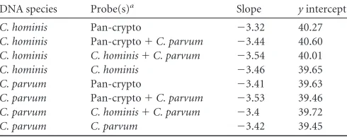
As these specific probes hybridize to the amplicon produced by the detection PCR, we tested the possibility of performing duplex PCR using the Pan-crypto probe and one species-specific probe. No negative interaction between the different probes and no loss of sensitivity were observed, as shown by the similaryintercepts and standard curve slopes obtained with the different combina-tions tested, compared with PCR using a single probe (Table 5).
DISCUSSION
[image:5.585.42.545.89.201.2]Real-time quantitative PCR (qPCR) is a unique tool for the sensi-tive detection, quantification, and specific characterization of Cryptosporidiumspecies of medical and veterinary importance. However, qPCR is a multiple-step procedure, each step of which is TABLE 4Correlation matrix for the parasite loads found in two independent series of quantifications (a and b) of 60 positive samples in four participating laboratories
Assay designationa
Correlation coefficient
1.a 1.b 2.a 2.b 3.a 3.b 4.a 4.b
1.a 1.00 0.896 0.911 0.905 0.914 0.896 0.926 0.879
1.b 0.896 1.00 0.906 0.900 0.807 0.849 0.896 0.909
2.a 0.911 0.906 1.00 0.999 0.945 0.968 0.991 0.986
2.b 0.905 0.900 0.999 1.00 0.944 0.968 0.991 0.986
3.a 0.914 0.807 0.945 0.944 1.00 0.944 0.951 0.917
3.b 0.896 0.849 0.968 0.968 0.944 1.00 0.959 0.937
4.a 0.926 0.896 0.991 0.991 0.951 0.959 1.00 0.974
4.b 0.879 0.909 0.986 0.986 0.917 0.937 0.974 1.00
aLaboratory designations were as follows: 1, Lyons; 2, Marseilles; 3, Nice; 4, Paris.
on May 16, 2020 by guest
http://jcm.asm.org/
sensitive to several technical parameters and needs to be opti-mized to improve accuracy. In this context, our multicenter study enabled a thorough assessment of several crucial steps in the qPCR procedure.
The first crucial step is DNA extraction. By comparing nine DNA extraction protocols applied to the same samples, we found marked variations, in the range of 2 log units, in extraction rates. The UltraClean and QIAamp DNA Stool kits displayed poor ex-traction rates. For the latter, our results coincide with those ob-tained for water samples by Jiang et al. (27), who concluded that the QIAamp DNA Stool Mini kit was not able to remove inhibitors efficiently despite the use of a specific adsorption system. The extraction rates for the other methods were significantly better, but each method had distinctive features depending on the prin-ciple of purification and the parasite burden. The two kits using silica columns, i.e., QIAamp DNA Mini and NucleoSpin kits, were characterized by higher saturation limits than the methods based on magnetic silica, as already observed for Mycobacterium (32), while NucliSENS easyMAG gave higher extraction yields with fecal samples containing low parasite burdens. Curiously, NucliSENS easyMAG demonstrated a better extraction rate with fecal samples than with saline suspensions of oocysts. This differ-ence was not observed with the Qiagen or Maxwell systems.
Mechanical grinding improved DNA extraction with NucliSENS easyMAG but not with the QIAamp DNA Mini or NucleoSpin Tissue kits. This means that proteinase K digestion was effective enough to alter the oocyst wall and allowed DNA release with simple cell lysis. By comparing NucliSENS easyMAG with mechanical grinding with the QIAamp DNA Stool kit, Masny et al. found that chemical lysis plus grinding was more effective than enzymatic lysis (33). Our results are in line with such obser-vations; Elwin et al. recently reported that semipurification of oocysts associated with column extraction was more effective than direct extraction with mechanical grinding in guanidinium thio-cyanate buffer (3). These studies emphasize the importance of sample pretreatment.
A second determining step in qPCR is the use of reliable stan-dards for quantification. Like other laboratories, we encountered difficulties in relation to the purification of oocysts, their preser-vation, and the variable extraction yield. In order to overcome these limitations, we estimated the number of target copies per oocyst (i.e., around 25 copies per oocyst) and proposed standard-izing positive controls using plasmid DNA. We checked that the use of oocyst or plasmid DNA standards resulted in similar sensi-tivity limits in our system. The actual sensisensi-tivity was estimated to
be about 300 oocysts per g of stool but might be lower with an extraction protocol leading to a more-concentrated DNA solu-tion. Other PCR methods based on the same target sequence dis-played comparable sensitivity values (22,26). The sensitivity of the 18S PCR determined by Stroup et al. (34) was 100 to 1,000 oocysts/200 mg of stool; that technique used the QIAamp DNA Stool kit and Scorpion probes. From our experience, this lower sensitivity would be related mainly to the DNA extraction method, rather than the molecular probe used.
Compared to microscopy, the sensitivity threshold of our PCR assay is probably lower than that of the conventional DFA, but a comparison between the two methods was not performed. Previ-ously, Xiao and Herd (15) and Valdez et al. (35) found a detection limit of 1,000 oocysts per gram using a DFA. More recently, Pe-reira et al. (36) estimated a limit of detection for a DFA of 1,000 to 6,000 oocysts per gram in bovine feces. However, those authors showed that concentrating oocysts by immunomagnetic separa-tion before the DFA resulted in a 2-log-unit increase in sensitivity (36), thus achieving the sensitivity of PCR.
Designing appropriate targets and probes for the concomitant quantification and identification of infecting species is a third step that might improveCryptosporidiumqPCR. Several authors have already developed or combined species-specific qPCRs but have found it difficult to ensure that the sensitivity of typing matches the sensitivity of detection (22). The authors were thus confronted with the risk that a sample with a low parasite load would be detected as positive but the parasites could not be typed. This difficulty has been resolved in our qPCR assay by using the same amplicon for detection and typing. Indeed, the polymorphism of the selected target made species discrimination possible with the use of different TaqMan probes with a single set of primers, which makes multiplexing easier. In order to facilitate species-specific hybridization at the polymorphic region, the minor-groove-bind-ing ligand (MGB) was used as a meltminor-groove-bind-ing point enhancer in order to shorten the nucleotide sequence.
With this method, all cases of cryptosporidiosis were reliably detected using the Pan-crypto probe andC. hominisandC. par-vumwere identified using the specific probes. The participating laboratories were consistent in this regard, since 59 of the 60 pos-itive samples were detected by PCR in all of them. Some individual variations were found in the estimation of oocyst counts, but the coefficient of variation between laboratories was⬍2%. The delay between DNA extraction in the coordinating laboratory and per-formance of PCR in the participating laboratories (1 week to 4 months) did not significantly influence the results, since ANOVA showed no laboratory effect on the quantification of parasite bur-dens in stool specimens. This observation confirms the robustness of the assay and indicates that variability depends essentially on DNA extraction rather than the PCR process itself.
However, other species that are uncommon in humans could not be characterized, because of either sequence homologies or lack of hybridization with theC. hominisorC. parvumprobes. This constitutes the main limitation of our assay, although the presence of such uncommon species might be suspected with the discrepancy between hydrolysis of the Pan-crypto probe and the negative results of the typing assay.
A set of PCRs targeting another part of the 18S rRNA gene has been proposed by Hadfield et al. (26). In that assay, detection is based on amplification of the rRNA gene and typing is performed at another locus (LIB13), using two MGB probes for species iden-TABLE 5Influence of multiplexing on the main characteristics of the
qPCR
DNA species Probe(s)a Slope yintercept
C. hominis Pan-crypto ⫺3.32 40.27
C. hominis Pan-crypto⫹C. parvum ⫺3.44 40.60
C. hominis C. hominis⫹C. parvum ⫺3.54 40.01
C. hominis C. hominis ⫺3.46 39.65
C. parvum Pan-crypto ⫺3.41 39.63
C. parvum Pan-crypto⫹C. parvum ⫺3.53 39.46
C. parvum C. hominis⫹C. parvum ⫺3.4 39.72
C. parvum C. parvum ⫺3.42 39.45
a
The combination of the Pan-crypto andC. hominisprobes was not tested because the probes used the same fluorescent ligand.
on May 16, 2020 by guest
http://jcm.asm.org/
[image:6.585.41.286.88.185.2]tification. As these two species-specific probes are not compatible, a first multiplex assay was required for detection of all species and C. parvumidentification and a second assay was necessary forC. hominisidentification. Application of this technique to routine analysis seems complicated because the specificity of the detec-tion/quantification PCR has to be confirmed by sequencing since the yeast 18S rRNA gene (GenBank accession no.JN940588.1) shares sequence homologies with the selected primers and probe, which might lead to nonspecific amplification. Moreover, neither the 18S nor LIB13 PCR displayed the same sensitivity as shown by Elwin et al. (28). Recently, specific assays have been proposed in order to identify uncommon species. Hadfield and Chalmers (37) proposed a C. cuniculusreal-time PCR that can overcome the inability to discriminate between C. hominisandC. cuniculus. New specific PCRs and probes are required to differentiate be-tween other zoonotic species.
In conclusion, through our multicentric evaluation, we have been able to assess the performance of a new sensitive one-step qPCR for the diagnosis of cryptosporidiosis, discrimination be-tween the two major species infecting humans, and quantification of parasite burdens. This assay is well suited to routine use as practical conditions have been improved, including DNA extrac-tion and the use of well-defined standards.
ACKNOWLEDGMENTS
This work was supported by the ANOFELCryptosporidiumNational Net-work and the French Institute for Public Health Surveillance (InVS).
We declare no conflicts of interest in relation to this work.
REFERENCES
1.Chalmers RM, Davies AP.2010. Minireview: clinical cryptosporidiosis. Exp. Parasitol.124:138 –146.
2.Derouin F, Lagrange-Xelot M.2008. Treatment of parasitic diarrhea in HIV-infected patients. Expert Rev. Anti Infect. Ther.6:337–349. 3.Elwin K, Hadfield SJ, Robinson G, Chalmers RM.2012. The
epidemi-ology of sporadic human infections with unusual cryptosporidia detected during routine typing in England and Wales, 2000 –2008. Epidemiol. In-fect.140:673– 683.
4.Geurden T, Levecke B, Cacció SM, Visser A, De Groote G, Casaert S, Vercruysse J, Claerebout E.2009. Multilocus genotyping of Cryptospo-ridiumandGiardiain non-outbreak related cases of diarrhoea in human patients in Belgium. Parasitology136:1161–1168.
5.ANOFEL Cryptosporidium National Network.2010. Laboratory-based surveillance forCryptosporidiumin France, 2006 –2009. Euro Surveill.15: pii⫽19642. http://www.eurosurveillance.org/ViewArticle.aspx?ArticleId
⫽19642.
6.Chalmers RM, Elwin K, Thomas AL, Guy EC, Mason B.2009. Long-termCryptosporidiumtyping reveals the aetiology and species-specific ep-idemiology of human cryptosporidiosis in England and Wales, 2000 to 2003. Euro Surveill. 14:pii⫽19086.http://www.eurosurveillance.org /ViewArticle.aspx?ArticleId⫽19086.
7.Wielinga PR, de Vries A, van der Goot TH, Mank T, Mars MH, Kortbeek LM, van der Giessen JW.2008. Molecular epidemiology of
Cryptosporidiumin humans and cattle in The Netherlands. Int. J. Parasi-tol.38:809 – 817.
8.Xiao L.2010. Molecular epidemiology of cryptosporidiosis: an update. Exp. Parasitol.124:80 – 89.
9.Ajjampur SS, Liakath FB, Kannan A, Rajendran P, Sarkar R, Moses PD, Simon A, Agarwal I, Mathew A, O’Connor R, Ward H, Kang G.2010. Multisite study of cryptosporidiosis in children with diarrhea in India. J. Clin. Microbiol.48:2075–2081.
10. Cama VA, Ross JM, Crawford S, Kawai V, Chavez-Valdez R, Vargas D, Vivar A, Ticona E, Navincopa M, Williamson J, Ortega Y, Gilman RH, Bern C, Xiao L.2007. Differences in clinical manifestations among Cryp-tosporidiumspecies and subtypes in HIV-infected persons. J. Infect. Dis. 196:684 – 691.
11. Cama VA, Bern C, Roberts J, Cabrera L, Sterling CR, Ortega Y, Gilman
RH, Xiao L. 2008.Cryptosporidiumspecies and subtypes and clinical manifestations in children, Peru. Emerg. Infect. Dis.14:1567–1574. 12. Gatei W, Wamae CN, Mbae C, Waruru A, Mulinge E, Waithera T,
Gatika SM, Kamwati SK, Revathi G, Hart CA.2006. Cryptosporidiosis: prevalence, genotype analysis, and symptoms associated with infections in children in Kenya. Am. J. Trop. Med. Hyg.75:78 – 82.
13. Cacciò SM, Pozio E.2006. Advances in the epidemiology, diagnosis and treatment of cryptosporidiosis. Expert Rev. Anti Infect. Ther.4:429 – 443. 14. Chalmers RM, Campbell BM, Crouch N, Charlett A, Davies AP.2011. Comparison of diagnostic sensitivity and specificity of seven Cryptospo-ridiumassays used in the UK. J. Med. Microbiol.60:1598 –1604. 15. Xiao L, Herd RP.1993. Quantitation ofGiardiacysts and
Cryptospo-ridiumoocysts in fecal samples by direct immunofluorescence assay. J. Clin. Microbiol.31:2944 –2946.
16. Agnamey P, Sarfati C, Pinel C, Rabodoniriina M, Kapel N, Dutoit E, Garnaud C, Diouf M, Garin JF, Totet A, Derouin F, ANOFEL Crypto-sporidiumNational Network.2011. Evaluation of four commercial rapid immunochromatographic assays for detection ofCryptosporidium anti-gens in stool samples: a blind multicenter trial. J. Clin. Microbiol.49: 1605–1607.
17. Garcia LS, Shimizu RY.1997. Evaluation of nine immunoassay kits (en-zyme immunoassay and direct fluorescence) for detection ofGiardia lam-bliaandCryptosporidium parvumin human fecal specimens. J. Clin. Microbiol.35:1526 –1529.
18. Nichols RA, Campbell BM, Smith HV.2003. Identification of Crypto-sporidiumspp. oocysts in United Kingdom noncarbonated natural mineral waters and drinking waters by using a modified nested PCR-restriction fragment length polymorphism assay. Appl. Environ. Microbiol. 69:4183– 4189.
19. Spano F, Putignani L, McLauchlin J, Casemore DP, Crisanti A.1997. PCR-RFLP analysis of theCryptosporidiumoocyst wall protein (COWP) gene discriminates betweenC. wrairiandC. parvum, and betweenC. par-vumisolates of human and animal origin. FEMS Microbiol. Lett.150:209 – 217.
20. Abe N, Matsubayashi M, Kimata I, Iseki M.2006. Subgenotype analysis ofCryptosporidium parvumisolates from humans and animals in Japan using the 60-kDa glycoprotein gene sequences. Parasitol. Res.99:303–305. 21. Skotarczak B.2010. Progress in the molecular methods for the detection and genetic characterization ofCryptosporidiumin water samples. Ann. Agric. Environ. Med.17:1– 8.
22. Jothikumar N, da Silva AJ, Moura I, Qvarnstrom Y, Hill VR.2008. Detection and differentiation ofCryptosporidium hominisand Cryptospo-ridium parvumby dual TaqMan assays. J. Med. Microbiol.57:1099 –1105. 23. Sturbaum GD, Reed C, Hoover PJ, Jost BH, Marshall MM, Sterling CR. 2001. Species-specific, nested PCR-restriction fragment length polymor-phism detection of singleCryptosporidium parvumoocysts. Appl. Environ. Microbiol.67:2665–2668.
24. Johnson AM, Giovanni GD, Rochelle PA.2012. Comparison of assays for sensitive and reproducible detection of cell culture-infectious Crypto-sporidium parvumandCryptosporidium hominisin drinking water. Appl. Environ. Microbiol.78:156 –162.
25. Di Giovanni GD, LeChevallier MW.2005. Quantitative-PCR assessment ofCryptosporidium parvumcell culture infection. Appl. Environ. Micro-biol.71:1495–1500.
26. Hadfield SJ, Robinson G, Elwin K, Chalmers RM.2011. Detection and differentiation ofCryptosporidiumspp. in human clinical samples by use of real-time PCR. J. Clin. Microbiol.49:918 –924.
27. Jiang J, Alderisio KA, Singh A, Xiao L.2005. Development of procedures for direct extraction ofCryptosporidiumDNA from water concentrates and for relief of PCR inhibitors. Appl. Environ. Microbiol.71:1135–1141. 28. Elwin K, Robinson G, Hadfield SJ, Fairclough HV, Iturriza-Gómara M, Chalmers RM.2012. A comparison of two approaches to extracting Cryp-tosporidiumDNA from human stools as measured by a real-time PCR assay. J. Microbiol. Methods89:38 – 40.
29. Xiao L, Bern C, Limor J, Sulaiman I, Roberts J, Checkley W, Cabrera L, Gilman RH, Lal AA.2001. Identification of 5 types ofCryptosporidium
parasites in children in Lima, Peru. J. Infect. Dis.183:492– 497. 30. Lorenzo-Lorenzo MJ, Ares-Mazas ME, Villacorta-Martinez de
Mat-urana I, Duran-Oreiro D.1993. Effect of ultraviolet disinfection of drink-ing water on the viability ofCryptosporidium parvumoocysts. J. Parasitol. 79:67–70.
31. Boom R, Sol CJ, Salimans MM, Jansen CL, Wertheim-van Dillen PM,
on May 16, 2020 by guest
http://jcm.asm.org/
van der Noordaa J.1990. Rapid and simple method for purification of nucleic acids. J. Clin. Microbiol.28:495–503.
32. Pontiroli A, Travis ER, Sweeney FP, Porter D, Gaze WH, Mason S, Hibberd V, Holden J, Courtenay O, Wellington EM.2011. Pathogen quantitation in complex matrices: a multi-operator comparison of DNA extraction methods with a novel assessment of PCR inhibition. PLoS One 6:e17916. doi:10.1371/journal.pone.0017916.
33. Masny A, Rozej W, Gołab E.2009. Development of efficient DNA iso-lation procedures forCryptosporidiumandTrichinellaPCR detection in fecal samples. Med. Dosw. Mikrobiol.61:259 –265.
34. Stroup SE, Roy S, Mchele J, Maro V, Ntabaguzi S, Siddique A, Kang G, Guerrant RL, Kirkpatrick BD, Fayer R, Herbein J, Ward H, Haque R,
Houpt ER.2006. Real-time PCR detection and speciation of Cryptospo-ridiuminfection using Scorpion probes. J. Med. Microbiol.55:1217–1222. 35. Valdez LM, Dang H, Okhuysen PC, Chappell CL.1997. Flow cytometric detection ofCryptosporidiumoocysts in human stool samples. J. Clin. Microbiol.35:2013–2017.
36. Pereira MD, Atwill ER, Jones T. 1999. Comparison of sensitivity of immunofluorescent microscopy to that of a combination of immunoflu-orescent microscopy and immunomagnetic separation for detection of
Cryptosporidium parvumoocysts in adult bovine feces. Appl. Environ. Microbiol.65:3236 –3239.
37. Hadfield SJ, Chalmers RM.2012. Detection and characterization of Cryp-tosporidium cuniculusby real-time PCR. Parasitol. Res.111:1385–1390.