Copyright © 2003, American Society for Microbiology. All Rights Reserved.
Genetic Diversity among
Borrelia
Strains Determined by Single-Strand
Conformation Polymorphism Analysis of the
ospC
Gene and Its
Association with Invasiveness
Vanessa Lagal,
1* Danie`le Postic,
1Eva Ruzic-Sabljic,
2and Guy Baranton
1 Unite´ de Bacte´riologie Mole´culaire et Me´dicale, Institut Pasteur, 75724 Cedex 15, France,1and Institute ofMicrobiology and Immunology, 1000 Ljubljana, Slovenia2
Received 30 October 2002/Returned for modification 5 March 2003/Accepted 8 June 2003
Lyme borreliosis (LB) is a tick-borne spirochetal infection caused by threeBorreliaspecies:Borrelia afzelii,
B.garinii, andB.burgdorferisensu stricto. LB evolves in two stages: a skin lesion called erythema migrans and
later, different disseminated forms (articular, neurological, cardiac. . .). Previous research based on analysis of
ospC sequences allowed the definition of 58 groups (divergence of <2% within a group and >8% between groups). Only 10 of these groups include all of the strains isolated from disseminated forms that are considered invasive. The aim of this study was to determine whether or not invasive strains belong to restricted ospC
groups by testing human clinical strains isolated from disseminated forms. To screen forospCgenetic diversity, we used single-strand conformation polymorphism (SSCP) analysis. Previously knownospCsequences from 44 different strains were first tested, revealing that eachospCgroup had a characteristic SSCP pattern. Therefore, we studied 80 disseminated-form isolates whoseospCsequences were unknown. Of these, 28 (35%) belonged to previously known invasive groups. Moreover, new invasive groups were identified: six ofB.afzelii, seven ofB.
garinii, and one ofB.burgdorferisensu stricto. This study confirmed that invasive strains are not distributed
among all 69 ospCgroups but belong to only 24 groups. This suggests that OspC may be involved in the invasiveness ofB.burgdorferi.
Lyme borreliosis (LB) is the most commonly reported tick-borne infection in Europe and North America. The etiological agent belongs to the complexBorrelia burgdorferi sensu lato, which comprises at least 11 species, of which only 3 are patho-genic for humans:B.burgdorferisensu stricto,B.afzelii, andB.
garinii (2, 4). LB evolves in two stages that may occur inde-pendently. The first is a primary skin lesion that spreads from the site of the tick bite and is called erythema migrans (EM). Later, bacteria may disseminate to different organs and induce different clinical manifestations, i.e., neurological (neurobor-reliosis [NB]), articular (arthritis), cardiac, cutaneous (multiple EM [MEM]), acrodermatitis chronica atrophicans (ACA), and lymphocytoma benigna cutis (LBC). Each of these manifesta-tions is associated with a distinct pathogenic species, and more frequently a given patient experiences only one of these de-layed manifestations (1, 5, 22, 23). The expression patterns of outer surface protein A (OspA) and OspC play an important role in the infection cycle. In unfed ticks, spirochetes express OspA but not OspC. During tick feeding, OspA synthesis is repressed whereas OspC synthesis is induced. The switch is associated with increased temperature and with exposure to tick hemolymph factor (10, 16, 19). Thus, OspC is the major outer surface protein expressed in early infection. Moreover, Masuzawa et al. (14) have shown that OspC expression is associated with infectivity. TheospCgene is highly variable (9, 12, 21, 25, 26). On the basis ofospCsequence analysis, Wang et al. (24) defined major ospC groups (divergence of ⬍2%
within a group and ⬎8% between groups). Furthermore, Seinost et al. (17) and Baranton et al. (3) found that, among 149 sequences from data banks, 58ospCgroups can be defined within the three pathogenic species. Only 10 of these groups contain all of the strains (76 sequences) isolated from different clinical samples in disseminated forms (referred to secondary sites). These strains are hereafter called invasive. Two groups are individualized withinB.afzelii, four withinB.garinii, and four withinB.burgdorferisensu stricto. These groups are de-fined as invasive. OtherospCgroups comprise strains isolated either from primary skin lesions or from ticks and are defined as noninvasive or of unknown invasiveness. In this study, we extended the investigation to 80 clinical strains isolated from secondary sites and of which theospCsequence was unknown. We used single-strand conformation polymorphism (SSCP) analysis to study the genetic variability of theospCgene. We determined the distribution among defined invasive groups. Our results confirm that invasive strains belong to givenospC
groups. This suggests that theospCgene could be one of the determinants involved in the invasiveness of strains leading to disseminated forms of the disease.
MATERIALS AND METHODS
Borreliastrains.One hundred twenty-fourB.burgdorferistrains were tested in this study. Forty-four strains whoseospCsequences are known were chosen, as indicated in Table 1. Eighty human-invasive strains were selected, including 61 Slovenian isolates. Fifty strains were isolated from different clinical samples (skin, blood, and cerebrospinal fluid [CSF]) in disseminated forms (ACA, MEM, LBC, and NB). Thirty strains isolated in the early stage were also included in this study. As they were isolated from the blood of patients with EM, they were considered to be invasive. Strains were cultured at 33°C in BSK-H medium (Sigma). The cultures were routinely monitored by dark-field microscopy for growth and contamination.
* Corresponding author. Mailing address: Institut Pasteur, Unite´ de Bacte´riologie Mole´culaire et Me´dicale, 28, rue du Dr Roux, 75724 Paris Cedex 15, France. Phone: 33 1 45 68 83 37. Fax: 33 1 40 61 30 01. E-mail: [email protected].
5059
on May 15, 2020 by guest
http://jcm.asm.org/
DNA isolation.Bacterial cultures were harvested by centrifugation (10,000⫻ g; 10 min). The bacterial pellet was washed in phosphate-buffered saline, resus-pended in water, heated at 100°C for 10 min, and then stored at⫺20°C.
PCR.A 277-bp fragment of the variable central part ofospC, suitable in size for SSCP analysis, was amplified by using forward primer SC3 (5⬘-AAAGCTA TTGGTAAAGTAAT-3⬘; bp 226 to 245; Genset) and reverse primer OspC92 (5⬘-GTTTTTAAAATAGCTTTTTTTG-3⬘; bp 491 to 470; Eurogentec), which are based on consensus sequences for the three pathogenic species.
Amplification was processed in 25l of a solution containing 0.2M each primer, 0.2 mM each deoxynucleoside triphosphate, 0.625 U ofTaqpolymerase (Q. Bio gene), and 1⫻Taqbuffer (1.5 mM MgCl2). The amplification reaction
was carried out in a DNA thermal cycler (Touch Down Hybaid) under the following conditions: initial denaturation at 93°C for 1 min, followed by 35 cycles of denaturation at 93°C for 1 min, annealing at 48°C for 1 min, and extension at 72°C for 30 s. Negative controls were included to check for contamination. Amplification was checked by agarose gel electrophoresis. A 5-l volume of each sample was loaded onto a 1% TBE 1X (Tris-borate-EDTA) agarose gel and revealed by ethidium bromide staining.
SSCP.A 3-l volume of the PCR product was added to 3l of denaturation solution (94% formamide, 0.05% xylene cyanol) and heated at 95°C for 5 min. Samples were loaded on a nondenaturing polyacrylamide gel (GeneGel Excel
12,5/24; Amersham Pharmacia Biotech). Electrophoresis was performed in a temperature-controlled electrophoresis system (GenePhor; Amersham Pharma-cia Biotech) at 6°C with a first run at 600 V, 25 mA, and 15 W for 10 min and then at 600 V, 37 mA, and 21 W for 2 h 30 min.
Gels were revealed by silver staining (Plus One DNA Silver Staining Kit; Amersham Pharmacia Biotech) in accordance with the manufacturer’s instruc-tions.
DNA sequencing.The partialospCgene (sizes ranged from 534 to 601 bp) was sequenced (n⫽25) as previously described (13), by Genome Express, Montreuil, France.
Phylogenetic analysis.ospCgene sequences recorded from the GenBank da-tabase and new sequences (sizes ranged from 445 to 460 bp) were aligned manually by using VSM software and analyzed by the unweighted pair group method with mathematic averages (UPGMA) (18). Phylogenic trees were drawn with Mega software (11).
RESULTS AND DISCUSSION
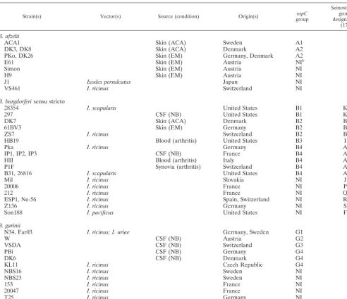
SSCP is a screening method based on the secondary struc-ture of a stranded DNA fragment. Different single-TABLE 1. Strains previously identified as invasivea(A1 to G4) or noninvasive on the basis of their sequences
Strain(s) Vector(s) Source (condition) Origin(s) groupospC
SeinostospC group designation
(17)
B. afzelii
ACA1 Skin (ACA) Sweden A1
DK3, DK8 Skin (ACA) Denmark A2
PKo, DK26 Skin (EM) Germany, Denmark A2
E61 Skin (EM) Austria NIb
Simon Skin (EM) Austria NI
H9 Skin (EM) Austria NI
J1 Ixodes persulcatus Japan NI
VS461 I. ricinus Switzerland NI
B. burgdorferisensu stricto
28354 I. scapularis United States B1 K
297 CSF (NB) United States B1 K
DK7 Skin (ACA) Denmark B2 B
61BV3 Skin (EM) Germany B2 B
ZS7 I. ricinus Switzerland B2 B
HB19 Blood (arthritis) United States B3 I
Pka I. ricinus Germany B4 A
IP1, IP2, IP3 CSF (NB) France B4 A
HII Blood (arthritis) Italy B4 A
P1F Synovia (arthritis) Switzerland B4 A
B31, 26816 I. scapularis United States B4 A
Mil I. ricinus Slovakia NI J
20006 I. ricinus France NI P
212 I. ricinus France NI Q
ESP1, Ne-56 I. ricinus Spain, Switzerland NI R
Z136 I. ricinus Germany NI S
Son188 I. pacificus United States NI F
B. garinii
N34, Far03 I. ricinus; I. uriae Germany, Sweden G1
W CSF (NB) Austria G2
VSDA CSF (NB) Switzerland G3
PBi CSF (NB) Germany G4
DK6 CSF (NB) Denmark G4
KL11 I. ricinus Czech Republic G4
NBS16 I. ricinus Sweden NI
NBS23 I. ricinus Sweden NI
153 I. ricinus France NI
20047 I. ricinus France NI
T25 I. ricinus Germany NI
BITS I. ricinus Italy NI
aA strain was considered invasive if it was isolated from secondary sites or when it belonged to a group comprising strains isolated from secondary sites. bNI, noninvasive group.
on May 15, 2020 by guest
http://jcm.asm.org/
[image:2.603.47.540.80.505.2]stranded DNA sequences result in different conformational foldings. These conformational polymorphisms can be discrim-inated by their electrophoretic mobilities on polyacrylamide gels (8, 15). This method is widely used for mutation analysis (20). Theoretically, three bands are detectable on the gel (two single DNA strands and one double DNA strand), but hetero-duplex formation causes several conformations that coexist in the gel. Under our conditions, different electrophoretic mobil-ities were indicative of sequence heterogenemobil-ities, irrespective of the genetic distance between sequences and of their muta-tion rates. This method was previously used (17, 24) to provide evidence for different groups ofospCmobility classes withinB.
burgdorferisensu stricto. The authors also demonstrated that each mobility class had a unique sequence. Our first objective
was to confirm that such data are also applicable toB.garinii
[image:3.603.43.546.81.516.2]and B. afzelii. The analysis was initiated by selecting strains from our collection (n⫽44) that belong to the three patho-genic species and whose ospC sequence is available in data banks. Therefore, these strains could be classified into either invasive (n⫽26) or noninvasive (n⫽18) groups on the basis of their sequences (Table 1). Within each ospC group, the sequences were very similar, with less than 2% nucleotide differences. SSCP patterns were determined for all of the strains. Moreover, SSCP reproducibility was checked, reveal-ing that the PCR product obtained in repeated experiments from a given DNA always yielded the same SSCP pattern and one PCR product always yielded the same SSCP pattern in different migrations.
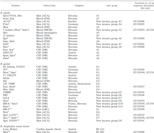
TABLE 2. Clinical strains isolated from secondary sites with unknownospCsequences
Strain(s) Clinical data Origin(s) ospCgroup sequences determined inAccession no. ofospC
this study
B. afzelii
Spet1793/01, Sbri Skin (ACA) Slovania A1
Svod, Srej Blood (EM) Slovenia A1
ACA2a Skin (ACA) Sweden New invasive group A3 AY150206
P/stoa Skin (ACA) Germany New invasive group A4 AY150205
Skoz Blood (EM) Slovenia A4
5 isolates (Skol,aSsima) Blood (MEM) Slovenia New invasive group A5 AY150203, AY150202
Shri Blood (meningitis) Slovenia A5
22 isolates Blood (EM) Slovenia A5
Spria Blood (MEM) Slovenia New invasive group A6 AY150204
Sspe, Sdol Blood (EM) Slovenia A6
Sobla Skin (ACA) Slovenia New invasive group A7 AY150201
Shrva Skin (ACA) Slovenia New invasive group A8 AY150200
Srav, Svirb CSF (NB) Slovania A1
GR4135b CSF (NB) Austria A1
Sjam, Specb CSF (NB) Slovania A5
Sabub CSF (NB) Slovania A6
B. garinii
60, Schenk, PATO3 CSF (NB) Austria G1
387a CSF (NB) Germany G1 AY150188
Skos,aSspaa CSF (NB) Slovenia G1 AY150194, AY150197
57, GR4229 CSF (NB) Austria G2
Sdom CSF (NB) Slovenia G2
Silc Blood (EM) Slovenia G2
239, VSBP CSF (NB) Austria, Switzerland G4
Scar, Stama CSF (NB) Slovenia G4 AY150187
Skle, Ssko Blood (EM) Slovenia G4
VSBMa CSF (NB) Switzerland New invasive group G5 AY150185
PBra CSF (NB) Germany New invasive group G6 AY150186
GrLil CSF (NB) Austria New invasive group G7
Sbosa CSF (NB) Slovenia New invasive group G8 AY150193
IBS-8,aSmrza CSF (NB) France, Slovenia New invasive group G10 AY150189, AY150196
Smara CSF (NB) Slovenia New invasive group G11 AY150195
IBS-9a,b Skin (LBC) France G1 AY150190
Skotb Skin (ACA) Slovenia G4
Spet 114/95a,b Skin (ACA) Slovenia G5 AY150192
Sles,a,bSsoba,b Skin (ACA) Slovenia G7 AY150199, AY150198
Spet 1058/01a,b Skin (ACA) Slovenia New invasive group G9 AY150191
B. burgdorferisensu stricto
Lenz, Holzer Cardiac muscle, blood Austria B4 (A)c
Shaja,b Skin (ACA) Slovenia B2 (B)c AY150208
Szid,a,bBRE-13a,b Skin (ACA), CSF (NB) Slovenia, France New invasive group B5 (Q)c AY150209, AY150207 aStrain whoseospCsequence was sequenced in this study.
bStrain isolated in a unusual clinical manifestation. cSeinostospCgroup designation (17).
on May 15, 2020 by guest
http://jcm.asm.org/
Within the 26 invasive strains, we identified 11 distinct pat-terns, each corresponding to 1 of the 10 invasive groups pre-viously described (A1 and -2, B1 to -4, and G1 to -4; Table 1) (3, 17). However, within invasive group G4, two different SSCP patterns were observed. From the 18 noninvasive strains, 17 different SSCP patterns were recorded, in complete accor-dance with the sequence analysis (data not shown). These results confirmed that eachospCgroup should correspond to a specific SSCP pattern, in agreement with previous studies (17, 24). Therefore, SSCP patterns could be used as references for the assignment of clinical strains to invasive or noninvasive groups.
Further, since SSCP methodology has been validated as a powerful and reproducible screening tool, we tested 80 clinical invasive European isolates from different secondary sites (Ta-ble 2). Additionally, someospCsequences were determined in order to confirm the assignment of strains to a given invasive group.
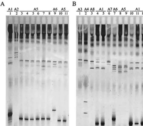
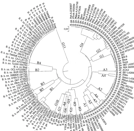
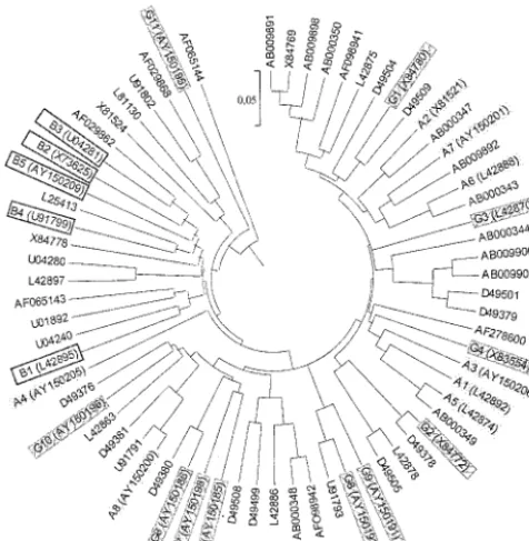
Forty-six invasiveB.afzeliistrains were analyzed by SSCP. Seven distinct SSCP patterns were recorded (Fig. 1), corre-sponding to seven differentospCgroups in accordance with the phylogenetic analysis of sequences (Fig. 2). Seven strains ex-hibited the typical pattern of invasive group A1. No pattern corresponding to previously described group A2 was observed. The 39 remaining strains belonged to six new distinct invasive
groups designated A3 to A8 (Table 2 and Fig. 2). Strains E61 and Simon, which have been previously designated noninvasive (3), had the same SSCP pattern as invasive strains ACA2 and P/sto, respectively (Table 1 and data not shown). ospC se-quencing and phylogenetic analysis confirmed that these strains should now be included in new invasive groups A3 and A4 (Table 2 and Fig. 2). Invasive group A5 comprised isolate Orth, which was not previously considered to be invasive on the basis of the sequence data (Fig. 2). Although invasive strains Spri, Sspe, and Sdol exhibited identical SSCP patterns that were different from that of noninvasive strain H9, se-quencing data allowed them to be assigned to new invasive group A6 (data not shown; Table 2 and Fig. 2). Therefore, group A6 exhibited two different patterns.
Twenty-nine invasive B. garinii strains were also tested by SSCP. Eighteen strains exhibited a pattern previously assigned to an invasive group; seven patterns referred to invasive group G1, four patterns referred to G2, and seven patterns referred to G4. Seven distinct SSCP patterns were identified among the 11 remaining strains (data not shown).ospCsequencing con-firmed that these B. garinii strains belonged to seven new distinct invasive groups designated G5 to G11 (Table 2 and Fig. 2). The SSCP patterns of groups G5 (strains VSBM, Spet 114/95, and BITS), G9 (strains Spet 1058/01 and 20047), and G10 (strains IBS-8, Smrz, and T25), which have been previ-ously designated distinct noninvasive groups (3), should be now assigned to invasive groups (data not shown; Table 1 and Fig. 2). A sequence homologous to that of group G8 was also found in data banks (Fig. 2). The three remaining invasive groups (G6, G7, and G11) constituted newospCgroups (Table 2 and Fig 2).
Finally, we tested fiveB. burgdorferi sensu stricto isolates. Two SSCP patterns referred to invasive group B4, and one pattern referred to B2. The two remaining strains, which had the same SSCP pattern as strain 212 and were previously iden-tified as noninvasive, should now be included in new invasive group B5 (data not shown; Table 1 and Fig. 2). Wang et al. (24) and Seinost et al. (17) have screened a large number ofospC
genes from North American human and tick B. burgdorferi
sensu stricto isolates. This study allowed us to list all of the
ospC groups found in North America. As additional invasive group B5 found in this study comprised only strains from Europe, it had not been identified in those previous studies. Genetic diversity studies based on the whole genome (7) or on theospCgene (13) have shown that North AmericanB. burg-dorferisensu stricto isolates are more heterogeneous than Eu-ropean ones and that some groups are restricted to either North America or Europe. Analysis of all of theB.burgdorferi
sensu strictoospCsequences found in data banks revealed that invasive groups B1 to -4 are all found in North America, whereas only groups B2, B4, and B5 are found in Europe, suggesting that groups B1 and B3 are restricted to North America and B5 is restricted to Europe.
[image:4.603.46.282.72.279.2]Previous studies based onospC sequences (3, 17) allowed the definition of 10 invasive groups within 58 groups. Within the 80 strains tested, 28 (35%) belonged to previously defined invasive groups and the 52 remaining strains (65%) belonged to the 14 new invasive ospC groups identified in this study. Nine of these 14 groups comprise isolates that, on the basis of theirospCsequences, were previously classified as noninvasive, FIG. 1. ospC SSCP patterns of 21 invasive B. afzelii isolates. A
277-bp fragment of the central variable part of theospC gene was amplified by PCR, denatured by heating and formamide, and electro-phoresed in a nondenaturing polyacrylamide gel. (A) Lanes: 1, B. afzeliiACA1; 2,B.afzeliiDK8; 3,B.afzeliiShri; 4,B.afzeliiSkol; 5:B. afzeliiSzaj (a strain of the five Slovenian isolates from blood [MEM]); 6: B.afzeliiSavb (a strain of the five Slovenian isolates from blood [MEM]); 7, B. afzelii Ssim; 8, B. afzelii Svre (a strain of the five Slovenian isolates from blood [MEM]); 9,B.afzeliiSpri; 10,B.afzelii Sper (a strain of the 22 Slovenian isolates from blood [EM] [Table 2]), 11,B.afzeliiSlet (a strain of the 22 Slovenian isolates from blood [EM] [Table 2]). (B) Lanes: 1,B.afzeliiACA2; 2,B.afzeliiP/sto; 3,B.afzelii Shrv; 4,B.afzeliiSpet 1793/01; 5,B.afzeliiSbri; 6,B.afzeliiSobl; 7,B. afzeliiSabu; 8,B.afzeliiSjam; 9,B.afzeliiSpec; 10,B.afzeliiSrav; 11
B.afzeliiSvir; 12,B.afzeliiGR4135.
on May 15, 2020 by guest
http://jcm.asm.org/
increasing the proportion of invasive groups. However, inva-siveospCgroups A7, A8, G6, G7, and G11 constituted previ-ously unobservedospC groups. At the same time, new ospC
groups were also identified and added to data banks, increasing the global ospC diversity to 69 ospC groups. Thus, ospC se-quence analysis allowed us to define 69 groups among 204 sequences, including 24 invasive groups (125 sequences) (Fig. 3 [a table listing nucleotide identities for the newly described
ospCgroups is available]). All of these results confirmed that invasive strains were not distributed within allospCgroups but belonged to restrictedospCgroups (24 [35%] of 69) (Fig. 3). Moreover, the strains additionally studied in this work were
selected for their invasiveness, thus increasing the probability of finding only new invasive groups.
Fingerprinting studies (12, 25, 26) indicate thatB.gariniiis the most heterogeneous species, whereasB.afzeliiis the most homogeneous one. We found 11 invasive groups forB.garinii. Within group G4, which was very heterogeneous, as indicated by the phylogenetic tree in Fig. 2, two different patterns were observed. These data confirmed that B. garinii is heteroge-neous. However, surprisingly, we found eight invasive groups of B. afzelii and two different patterns within the A6 group. Thus,B.afzeliiwas as heterogeneous asB.gariniiregarding the
[image:5.603.56.527.71.531.2]ospCgene.
FIG. 2. UPGMA-based tree of invasiveospCsequences from 100 isolates available in data banks (indicated by their accession numbers) and the 25ospCsequences determined in this study. B. b. ss,B.burgdorferisensu stricto; B. ga.,B.garinii; B. af.,B.afzelii; B. va.,B.valaisiana.
on May 15, 2020 by guest
http://jcm.asm.org/
Wang et al. (24) have shown that the genetic diversity ofB.
burgdorferi sensu stricto within a local population of ticks is almost equal to the worldwide genetic diversity. Our study revealed thatospCgroups identified from human disseminated forms were found in different areas, suggesting that these groups are widely distributed. Moreover, the analysis of 61 Slovenian strains showed that most invasive groups were found in a local area (Table 2). These results were in agreement with those of Wang et al. (24). However, a heterogeneous distribu-tion of the sequences of human origin was found. For instance, in data banks, 11 out of 14 sequences from humans in group G4 were from strains isolated in Germany. Regarding the Slovenian strains tested in this study, 30 (70%) out of the 43 invasiveB. afzeliistrains fell into group A5. These data sug-gested that all of theospCgroups had a wide range but some groups could be selected, in humans, in a restricted geograph-ical area.
Each pathogenic Borrelia species is predominantly associ-ated with a given late clinical manifestation: B. afzelii with cutaneous manifestations, B. gariniiwith neurologic manifes-tations, andB.burgdorferisensu stricto with articular manifes-tations (1, 22). However, sequences from strains isolated from each clinical manifestation are scattered along the tree (data not shown). Fifteen strains included in our study were respon-sible for a clinical manifestation different from that expected (sixB. afzeliistrains isolated from CSF, fiveB. gariniistrains from ACA, twoB.burgdorferisensu stricto strains from ACA, one B.garinii strain from LBC, and oneB.burgdorferi sensu
stricto strain from CSF; Table 2). Indeed, lateral transfer, which is common in theospCgene (6, 9, 12, 13), could have been responsible for such interspecific organotropism. How-ever, our results showed thatospCsequences from these par-ticular isolates clustered together with strains isolated from expected clinical manifestations. For example,B.afzeliistrains Srav and Svir, isolated from the CSF of a patient with NB, belonged to group A1, as did Spet 1793/01 and Sbri, which were isolated from a patient with ACA (Table 2). Moreover, sequence analysis revealed that B. burgdorferi sensu stricto strains P1F (arthritis) and IP1-2-3 (NB) in group B4 have exactly the sameospCsequence (Table 1 and Fig. 2). Further-more, it was found that B. afzeliistrain Shrv, which was in-volved in a genetic transfer fromB.garinii, was responsible for a case of ACA in a Slovenian patient. Our data indicate that clinical presentation is not associated with a given ospC se-quence.
Our results and those of others (17, 24) have demonstrated that SSCP can be used for epidemiological studies of tick isolates or first-stage EM isolates in order to evaluate the proportion of invasive strains and to predict evolution toward disseminated forms. Extensive geographical studies could de-termine the distribution and frequency ofospCgroups accord-ing to the area.
OspC expression is induced during tick feeding (16), and it is the major outer surface protein expressed in early infection, but its role is still unknown. Our results show that invasive strains belong to restrictedospCgroups, suggesting that OspC is one of the factors involved inBorreliainvasiveness. Further studies to determine the role of OspC inBorreliainvasiveness are in progress in our laboratory.
ACKNOWLEDGMENTS
We thank D. Dykhuizen and L. Gern for reviewing the manuscript and I. Old for reading the manuscript.
REFERENCES
1. Assous, M., D. Postic, G. Paul, P. Nevot, and G. Baranton.1993. Western blot analysis of sera from Lyme borreliosis patients according to the genomic species of theBorreliastrains used as antigens. Eur. J. Clin. Microbiol. Infect. Dis.12:261–268.
2. Baranton, G., D. Postic, I. Saint Girons, P. Boerlin, J.-C. Piffaretti, M. Assous, and P. A. D. Grimont.1992. Delineation ofBorrelia burgdorferisensu stricto,Borrelia gariniisp. nov., and group VS461 associated with Lyme borreliosis. Int. J. Syst. Bacteriol.42:378–383.
3. Baranton, G., G. Seinost, G. Theodore, D. Postic, and D. Dykhuisen.2001. Distinct levels of genetic diversity ofBorrelia burgdorferiare associated with different aspects of pathogenicity. Res. Microbiol. 149–156.
4. Canic¸a, M. M., F. Nato, L. Du Merle, J. C. Mazie, G. Baranton, and D. Postic.1993. Monoclonal antibodies for identification ofBorrelia afzeliisp. nov. associated with late cutaneous manifestations of Lyme borreliosis. Scand. J. Syst. Infect. Dis.25:441–448.
5. Dhoˆte, R., A. L. Basse-Guerineau, V. Beaumesnil, B. Christoforov, and M. Assous.2000. Full spectrum of clinical, serological, and epidemiological features of complicated forms of Lyme borreliosis in the Paris, France, area. Eur. J. Clin. Microbiol. Infect. Dis. 809–815.
6. Dykhuisen, D. E., and G. Baranton.2001. The implications of a low rate of horizontal transfer inBorrelia. Trends Microbiol.9:344–350.
7. Foretz, M., D. Postic, and G. Baranton.1997. Phylogenetic analysis of Bor-relia burgdorferisensu stricto by arbitrarily primed PCR and pulsed-field gel electrophoresis. Int. J. Syst. Bacteriol.47:11–18.
8. Hongyo, T., G. S. Buzard, R. J. Calvert, and C. Meghorst.1993. ‘Cold SSCP’: a simple, rapid and non-radioactive method for optimized single-strand conformation polymorphism analyses. Nucleic Acids Res.21:3637–3642. 9. Jauris-Heipke, S., G. Liegl, V. Preac-Mursic, D. Rossler, E. Schwab, E.
[image:6.603.45.283.73.316.2]Soutschek, G. Will, and B. Wilske.1995. Molecular analysis of genes encod-ing outer surface protein C (OspC) ofBorrelia burgdorferisensu lato: rela-tionship toospAgenotype and evidence of lateral gene exchange ofospC. J. Clin. Microbiol.33:1860–1866.
FIG. 3. ospCgenetic diversity tree (UPGMA method) drawn from one representative sequence, indicated by accession numbers, from each of 69 ospC groups of the three pathogenic species (204 se-quences). Invasive groups are identified by their coding abbreviations (A1 to G11).B.afzeliiinvasive group, plain gray shading;B.burgdorferi sensu stricto invasive group, no hatching or shading;B.gariniiinvasive group, cross-hatched and shaded gray.
on May 15, 2020 by guest
http://jcm.asm.org/
10. Johns, R. H., D. E. Sonenshine, and W. L. Hynes.2000. Enhancement of OspC expression byBorrelia burgdorferiin the presence of tick hemolymph. FEMS Microbiol. Lett.193:137–141.
11. Kumar, S., K. Tamura, and N. Masatoshi.1993. MEGA: molecular evolu-tionary genetics analysis, version 1.01. The Pennsylvania State University, University Park.
12. Livey, I., C. P. Gibbs, R. Schuster, and F. Dorner.1995. Evidence for lateral transfer and recombination in OspC variation in Lyme diseaseBorrelia. Mol. Microbiol.18:257–269.
13. Marti Ras, N., D. Postic, and G. Baranton.1997.Borrelia burgdorferisensu stricto, a bacterial species “made in the USA”? Int. J. Syst. Bacteriol.47:
1112–1117.
14. Masuzawa, T., T. Kurita, H. Kawabata, and Y. Yanagihara.1994. Relation-ship between infectivity and OspC expression in Lyme diseaseBorrelia. FEMS Microbiol. Lett.123:319–324.
15. Orita, M., Y. Suzuki, T. Sekiya, and K. Hayashi.1989. Rapid and sensitive detection of point mutations and DNA polymorphisms using the polymerase chain reaction. Genomics5:874–879.
16. Schwan, T. G., J. Piesman, W. T. Golde, M. C. Dolan, and P. A. Rosa.1995. Induction of an outer surface protein onBorrelia burgdorferiduring tick feeding. Proc. Natl. Acad. Sci. USA92:2909–2913.
17. Seinost, G., D. E. Dykhuisen, R. J. Dattwyler, W. T. Golde, J. J. Dunn, I. N. Wang, G. P. Wormser, M. E. Schriefer, and B. J. Luft.1999. Four clones of Borrelia burgdorferisensu stricto cause invasive infection in humans. Infect. Immun.67:3518–3524.
18. Sneath, P. H. A., and R. R. Sokal.1973. Numeral taxonomy. W. H. Freeman & Co., San Francisco, Calif.
19. Stevenson, B., T. G. Schwan, and P. A. Rosa.1995. Temperature-related
differential expression of antigens in the Lyme disease spirochete,Borrelia burgdorferi. Infect. Immun.63:4535–4539.
20. Tamaki, J., Y. Arimura, T. Koda, S. Fujimoto, T. Fujino, A. Wakisaka, and M. Kakinuma.1993. Heterogeneity ofHLA-Ggenes identified by polymer-ase chain reaction/single strand conformational polymorphism (PCR/SSCP). Microbiol. Immunol.37:633–640.
21. Theisen, M., B. Frederiksen, A.-M. Lebech, J. Vuust, and K. Hansen.1993. Polymorphism inospCgene ofBorrelia burgdorferiand immunoreactivity of OspC protein: implications for taxonomy and for use of OspC protein as a diagnostic antigen. J. Clin. Microbiol.31:2570–2576.
22. Van Dam, A. P., H. Kuiper, K. Vos, A. Widjojokusumo, B. M. Dejonh, L. Spanjaard, A. C. P. Ramselaar, M. D. Kramer, and J. Dankert.1993. Dif-ferent genospecies ofBorrelia burgdorferiare associated with distinct clinical manifestations of Lyme borreliosis. Clin. Infect. Dis.17:708–717. 23. Wang, G. Q., A. P. Van Dam, I. Schwartz, and J. Dankert.1999. Molecular
typing ofBorrelia burgdorferisensu lato: taxonomic, epidemiological, and clinical implications. Clin. Microbiol. Rev.12:633–653.
24. Wang, I. N., D. E. Dykhuisen, W. Qui, J. J. Dunn, E. M. Bosler, and B. J. Luft.1999. Genetic diversity ofospCin a local population ofBorrelia burg-dorferisensu stricto. Genetics151:15–30.
25. Wilske, B., S. Jauris-Heipke, R. Lobentanzer, I. Pradel, V. Preac-Mursic, D. Rossler, E. Soutschek, and R. C. Johnson.1995. Phenotypic analysis of outer surface protein C (OspC) ofBorrelia burgdorferisensu lato by monoclonal antibodies: relationship to genospecies and OspA serotype. J. Clin. Micro-biol.33:103–109.
26. Wilske, B., V. Preac-Mursic, S. Jauris, A. Hofmann, I. Pradel, E. Soutschek, E. Schwab, G. Will, and G. Wanner.1993. Immunological and molecular polymorphisms of OspC, an immunodominant major outer surface protein ofBorrelia burgdorferi. Infect. Immun.61:2182–2191.