Copyright © 2000, American Society for Microbiology. All Rights Reserved.
Evaluation of CHROMagar Staph. aureus, a New Chromogenic
Medium, for Isolation and Presumptive Identification of
Staphylococcus aureus
from Human Clinical Specimens
OLIVIER GAILLOT,* MURIEL WETSCH, NICOLAS FORTINEAU,ANDPATRICK BERCHE Laboratoire de Bacte´riologie-Virologie, Hoˆpital Necker-Enfants Malades, Paris, France
Received 27 September 1999/Returned for modification 13 November 1999/Accepted 11 December 1999
CHROMagar Staph. aureus (CSA) is a new chromogenic medium for presumptive identification of
Staph-ylococcus aureusas mauve colonies after 24 h of incubation. We conducted a preliminary study with 100 S. aureusand 45 coagulase-negativeStaphylococcus (CoNS) stock isolates plated on CSA. AllS. aureusisolates yielded mauve colonies after 24 h of incubation at 37°C, while CoNS isolates grew as blue, white, or beige colonies. Culture on CSA was then prospectively compared to a conventional laboratory method, i.e., culture on 5% horse blood agar (HBA), catalase test, and latex agglutination test (HBA-catalase-latex), for isolation
and presumptive identification ofS. aureusfrom 2,000 consecutive clinical samples. Among the 310S. aureus
isolates recovered by at least one of the two methods, 296 grew as typical mauve colonies on CSA, while only 254 yielded catalase-positive, latex-positive colonies on HBA. The sensitivity of CSA was significantly higher
than that of the conventional method (95.5 and 81.9%, respectively;P< 0.001) and allowed the recovery of
important clinical isolates that were undetected on blood agar. The specificities of the two methods were not significantly different, although that of CSA was slightly higher (99.4% versus 98.9% for HBA-catalase-latex;
Pⴝ0.08). On the basis of its excellent sensitivity and specificity, ease of identification of positive colonies, and
absence of complementary testing, CSA can be recommended as a routine plating medium for presumptive
identification ofS. aureusin clinical specimens.
Staphylococcus aureus remains one of the most frequently encountered bacterial pathogens and is responsible for a vari-ety of mild to life-threatening infections. Early isolation and identification ofS. aureusas an etiologic agent or indicator of potential health risk is essential for appropriate patient care. In the clinical laboratory routine,S. aureusis usually isolated on nonspecific media (e.g., blood agar) and then presumptively identified before definitive overnight characterization (11). Al-though immunoenzymatic and genetic assays (3, 5, 9) are avail-able, presumptive identification of suspect colonies relies mostly on the detection of specific determinants such as clump-ing factor, protein A, and specific capsular antigens by agglu-tination of sensitized latex particles (7, 17, 19). However, this method becomes costly and time-consuming when more than one suspect morphotype is present on a plate. In an attempt to achieve isolation and presumptive identification in a single step, specific selective culture media have been proposed for use to assist in the detection ofS. aureus(reviewed in reference 1). Mannitol-salt agar and related media (2, 4, 8) are widely used for specific screening ofS. aureusin potentially contam-inated samples. They can be supplemented with antibiotics in order to detect methicillin-resistantS. aureus(MRSA) isolates (13, 18). However, the average sensitivity and specificity of mannitol-salt agar (10, 12, 15) do not qualify it for use for early isolation and presumptive identification of S. aureus in all clinical specimens, although supplementation with egg yolk was shown to be of clinical interest (15). There is still a need for more specific media that would retain the sensitivity of blood agar and allow single-step isolation and presumptive identification ofS. aureus. CHROMagar Staph. aureus (CSA)
is a new selective chromogenic medium proposed for detection ofS. aureusas mauve colonies after 18 to 24 h of incubation. In this study, we first evaluated with 145 clinical stock isolates the ability of CSA to differentiateS. aureusfrom coagulase-nega-tive staphylococci (CoNS), which are frequently encountered in human clinical samples. Isolation and presumptive identifi-cation ofS. aureuson CSA were then compared to our stan-dard routine protocol, i.e., culture on 5% horse blood agar (HBA), catalase test and latex agglutination for clumping fac-tor, protein A, and/or specific capsular antigens (HBA-cata-lase-latex), with 2,000 consecutive clinical specimens in a teaching hospital.
MATERIALS AND METHODS
Culture media.CSA, a proprietary product, was provided for evaluation by CHROMagar Microbiology (Paris, France), as prepared 85-mm plates contain-ing 20 ml of opaque creamy-white medium. As indicated by the manufacturer, CSA plates were stored at 4°C in a cold room and used within 4 weeks. Columbia agar supplemented with 5% horse blood (HBA) was purchased as commercially prepared plates (Becton-Dickinson, Le Pont-de-Claix, France). In our facility, HBA was preferred to sheep blood agar for general bacterial recovery because
of its better capacity to support the growth ofStreptococcus milleri-group
strep-tococci and to grow Streptococcus agalactiaeas beta-hemolytic colonies. All
plates were incubated in air at 37°C. Brain heart infusion broth (bioMe´rieux, Marcy-l’E´toile, France) was used for cultures in liquid medium.
Stock isolates.The 145 stockStaphylococcusstrains representing 10 species (Table 1) were isolated from clinical human samples in our facility, except for
two reference strains (S. aureusATCC 25923 andStaphylococcus epidermidis
ATCC 14990). Among the 100S. aureusisolates, 26 were resistant to methicillin.
One of the MRSA isolates was a small-colony variant (auxotroph for thymidine) and was unable to grow in less than 48 h on HBA. The isolates were stored frozen
at⫺80°C; for the present study, they were thawed, seeded on blood agar, and
incubated overnight at 37°C. Suspensions in saline solution were prepared from freshly grown colonies and then streaked onto CSA plates (one isolate per plate). At this stage, the color and size of colonies were evaluated after 24 h of
incu-bation. In order to compare the output of cultures and the growth limits ofS.
aureuson each medium, comparative colony counts on HBA and CSA were
performed for eachS. aureusisolate. Suspensions were prepared from freshly
grown colonies in saline solution and were adjusted to a turbidity equivalent to that of a 0.5 McFarland unit standard suspension. Dilutions were made from
* Corresponding author. Mailing address: Laboratoire de Bacte´ri-ologie-Virologie, Hoˆpital Necker-Enfants Malades, 149, rue de Se`vres, 75730 Paris CEDEX 15, France. Phone: (33) (1) 44 49 49 61. Fax: (33) (1) 44 49 49 60. E-mail: [email protected].
1587
on May 15, 2020 by guest
http://jcm.asm.org/
these suspensions, and 100 and 5 CFU of each isolate were inoculated onto HBA
and CSA plates by spreading 100l of each dilution.
Clinical samples.The second arm of the study was carried out between March and August 1999 in the 800-bed Necker Enfants-Malades Hospital (Paris, France) on 2,000 consecutive clinical specimens of various origins (Table 2).
Midstream urine and routine stool samples were excluded, forS. aureuswas
unlikely to be isolated from such specimens. Plating on CSA was added to our routine testing regimen, and a comparison of recovery for each specimen was made. Samples were inoculated in a set order: routine medium (HBA) first and then CSA. Primary inoculation was made with a loop or a swab. A straight wire was then used to streak the material to achieve isolated colonies. Quantitative cultures were performed on bronchoalveolar lavage, protected bronchial brush,
and central veinous catheter (rinsed with 500l of saline solution) by spreading
100l of sample per plate. All plates were incubated at 37°C in air and examined
after 24 h of incubation. Incubation was occasionally prolonged to 48 h for HBA plates only, in order to enhance differences between colony morphotypes.
Presumptive identification ofS. aureus.The criteria for presumptive
identifi-cation ofS. aureuswere defined as follows: (i) on CSA, well-separated mauve
colonies after 24 h of incubation, without further testing, and (ii) on HBA, colonial appearance after 24 to 48 h of incubation (11, 12) and Gram stain morphology, positive catalase test (ID color Catalase; bioMe´rieux), and positive agglutination for Pastorex Staph Plus (Sanofi Diagnostics Pasteur, Marnes-la-Coquette, France), as previously described (14). Pastorex Staph Plus allows
simultaneous detection of clumping factor, protein A, and twoS. aureus-specific
capsular and was shown to identify mostS. aureusisolates, including MRSA
lacking clumping factor and/or protein A (17). On HBA plates with mixed staphylococcal flora, one colony of each suspected different morphotype was tested for catalase and by latex agglutination. All plates were inspected by the same technologist.
Confirmatory identification.S. aureusidentification was confirmed by a pos-itive tube-coagulase test, as previously described (11). Colonies from primary
recovery plates presumptively identified asS. aureuswere inoculated into brain
heart infusion broth and incubated overnight. The cultures were then mixed with citrated rabbit plasma (bioMe´rieux) and incubated at 37°C. Clotting was evalu-ated after 4 h, and after 24 h if negative after 4 h. When required, staphylococci and micrococci were identified by the API ID32 STAPH system (bioMe´rieux) and in-house complementary tests based on conventional phenotypic character-istics (12).
Statistical analysis.Differences in sensitivities, specificities, and predictive
values of the two methods were evaluated by the Yates-corrected2test.
RESULTS
Colonial appearance ofS. aureusand other microorganisms
on CSA. We first evaluated the colonial appearance of 145
Staphylococcusstock isolates on CSA. All 100S. aureusisolates yielded mauve (positive) colonies, which were usually 2 to 3 mm in diameter after 24 h of incubation (Fig. 1A). A mat halo surrounded theS. aureuscolonies, analogous to that observed on Tween 80-supplemented media when lipase-producing bac-teria are present. Colonies of the small-colony variant MRSA
were smaller (0.5 to 1 mm), although they were clearly visible and mauve after 24 h. Colonies of other MRSA and methicil-lin-susceptibleS. aureusisolates (n⫽25 and 74, respectively) displayed no particular difference in terms of size, shape, and color. Except for that of the small-colony variant which did not grow on HBA after 24 h, the colony counts performed on both media were identical (data not shown). When CSA was chal-lenged with low inocula (ca. 5 CFU/plate), its capacity to detect
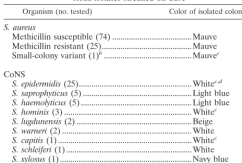
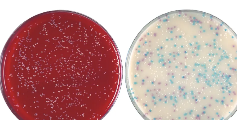
[image:2.612.307.536.79.427.2]S. aureuswas similar to that of HBA. The CoNS stock isolates tested grew as different shades of blue, white, or beige colonies (Table 1), as did most CoNS isolated from clinical specimens during the second part of the study. Figure 2 shows the com-parative growth of a mixed gram-positive flora on HBA and CSA after 24 h of incubation. Several staphylococcal morpho-types are seen on HBA, each having required catalase and latex tests, while mauve colonies readily identifiedS. aureuson CSA. SeveralS. epidermidisisolates produced small white col-onies with a mauve background in regions of denser growth, not to be confused with the actual color of individualized colonies.Micrococcus spp. grew as yellow to brown colonies, except for fiveMicrococcus luteusisolates which yielded mauve colonies (Fig. 1B). None of the numerous gram-negative or-ganisms isolated on HBA grew on CSA.Enterococcusisolates yielded blue colonies, as didListeria monocytogenes. Diphthe-roids grew on CSA as minute white colonies, except for ANF TABLE 1. Color appearance of 145Staphylococcus
stock isolates streaked on CSA
Organism (no. tested) Color of isolated coloniesa
S. aureus
Methicillin susceptible (74) ... Mauve Methicillin resistant (25)... Mauve Small-colony variant (1)b... Mauvec
CoNS
S. epidermidis(25)... Whitec,d
S. saprophyticus(5) ... Light blue
S. haemolyticus(5) ... Light blue
S. hominis(3) ... Whitec
S. lugdunensis(2) ... Beige
S. warneri(2) ... White
S. capitis(1) ... Whitec
S. schleiferi(1) ... White
S. xylosus(1) ... Navy blue
aAfter 24 h of incubation at 37°C.
bThymidine auxotroph.
cColony diameter of⬍2 mm after 24 h.
dMauve background at the inoculum site.
TABLE 2. Distribution of theS. aureusisolates (n⫽310) among the 2,000 clinical specimens
Specimen (no.) No. ofstrains isolatedS. aureus
Blood culturea(141)... 22
Vascular catheter tip (135) ... 15
Pus Wound exudate, ulcer (205)... 49
Abcess (104)... 33
Ear, tympanocentesis fluid (297)... 19
Eye (93) ... 8
Respiratory Sputum (53)b... 24
Tracheal aspirate (26)... 12
Bronchoalveolar lavage (40) ... 10
Protected bronchial brush (32)... 10
Body fluids Amniotic fluid (8)... 0
Ascites, cerebrospinal, peritoneal, and pleural fluids (38) ... 1
Drained fluid (194)... 16
High vaginal swab (152) ... 37
Neonatalc(192)... 3
Tissue sample (17)... 2
Catheterized urine (154) ... 21
Nasal, rectal, and axillary swabsd(119) ... 28
aPositive with gram-positive cocci.
bIncludes 35 samples from patients with cystic fibrosis.
cGastric aspirate, placenta, meconium, ear, and umbilical swab.
dDetection ofS. aureuscarriage.
on May 15, 2020 by guest
http://jcm.asm.org/
[image:2.612.52.294.90.253.2]groupCorynebacteriumisolates, which produced pinpoint-like red colonies that were barely visible after 24 h.Candida albi-canswas the only yeast species recovered on CSA, as white colonies.
Recovery ofS. aureusfrom clinical isolates.During the
sec-ond part of this work, the performances of CSA and HBA-catalase-latex were compared in laboratory routine. A total of 310S. aureusstrains were isolated on at least one medium from
the 2,000 consecutive clinical specimens. All 310 isolates were coagulase positive after 4 h, regardless of the medium from which they had been isolated. The distribution of the isolates in the different specimens is indicated in Table 2. On HBA, 254 isolates yielded catalase-positive, latex-positive colonies and were confirmed asS. aureus (sensitivity, 81.9%) (Table 3). A total of 1,157 latex agglutination tests were performed, repre-senting a mean of 4.55 tests for eachS. aureusisolate actually identified by the conventional method. On CSA, 296S. aureus
isolates grew as typical mauve colonies (sensitivity, 95.5%;P⬍ 0.001). The majority (n⫽53) of the 56 falsely negative isolates missed on HBA but not on CSA were either masked or inhib-ited by a gram-negative associated flora (n ⫽ 45) or were unable to grow (n⫽8) on primary plating, probably because of a low inoculum. When subcultured from CSA to HBA, those 53 isolates grew as typical staphylococcal colonies. The remain-ing three isolates falsely negative on HBA were subcultured from CSA to HBA and yielded latex-negative colonies. Among the 14 falsely negative isolates missed on CSA but identified on HBA, 1 grew as blue-violet colonies (suggesting the superpo-sition of mauve and blue colors) and proved to be coagulase positive. Six grew as creamy-white colonies with a mauve
back-FIG. 1. Colonial appearance ofS. aureusand of two false-positive isolates on
CSA. Colonies were plated out from suspensions ofS. aureus(A),M. luteus(B),
[image:3.612.58.288.70.299.2]andS. cohnii(C). CSA plates were incubated for 24 h at 37°C. Magnification,⫻2.
FIG. 2. Colonies plated out from a central venous catheter tip. (Left) Columbia agar supplemented with 5% horse blood; (right) CSA. Mauve colonies ofS. aureus
are readily differentiated from three other morphotypes on the chromogenic medium; light blue, smaller white, and larger white colonies were identified as
[image:3.612.312.553.84.146.2]Staphylococcus haemolyticus,S. epidermidis, andStaphylococcus warneri, respectively. Plates were incubated for 24 h at 37°C and are shown at their actual size.
TABLE 3. Recovery ofS. aureusfrom 2,000 clinical specimensa
Method
No. of:
Sensitivity (%) True-positive
isolates False-negativeisolates
Standardb 254 56 81.9
CSAc 296 14 95.5
aA total of 310 isolates were recovered by at least one method (sensitivity,
100%).
bSuggestive morphology on HBA, catalase, and latex agglutination.
cMauve colony on CSA after 24 h of incubation.
on May 15, 2020 by guest
http://jcm.asm.org/
[image:3.612.67.542.460.699.2]ground, and these isolates originated from mucoid sputa of patients with cystic fibrosis. Subculture of such colonies on CSA restored typical mauve morphotypes, which were then identified asS. aureusby a coagulase test. Seven isolates orig-inated as scant colonies on HBA, and noS. aureusgrowth was observed on the corresponding CSA plates, but subculture from HBA to CSA yielded typical mauve colonies.
Specificity ofS. aureuspresumptive identification.As shown
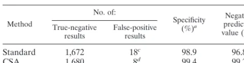
in Table 4, the specificities of both methods were high (98.9 and 99.4% for HBA-catalase-latex and CSA, respectively) and not significantly different (P ⫽ 0.08). A total of 18 and 8 false-positive strains were isolated on HBA and CSA, respec-tively. They consisted ofStaphylococcus lugdunensis(n ⫽5),
Staphylococcus schleiferi(n⫽4),Staphylococcus cohnii(n⫽4),
Staphylococcus saprophyticus (n⫽ 3),S. epidermidis(n ⫽1), andKocuria(formerlyMicrococcus)kristinae(n⫽1) on HBA (catalase- and latex-positive colonies). On CSA, false-positive isolates consisted ofM. luteus(n ⫽5) andS. cohnii(n⫽3). Falsely positive M. luteus colonies on CSA (Fig. 1B) were pink-mauve, convex, and smaller than typicalS. aureuscolonies (Fig. 1A) after 24 h of incubation, and they lacked the mat halo observed aroundS. aureuscolonies. The three false-positiveS. cohnii isolates yielded violet colonies after 24 h (Fig. 1C), which in our opinion were not different enough from typical mauve colonies of S. aureus and therefore were considered suspect. All colonies falsely positive with the conventional method were subcultured on CSA, and none of them grew as mauve colonies. Conversely, the threeS. cohniiisolates falsely positive on CSA were also falsely positive (latex-positive) on HBA. The fiveM. luteusisolates that were mauve on CSA were latex negative when grown on HBA. The negative predictive values (Table 4) of the two methods were significantly different (96.8 and 99.2% for HBA-catalase-latex and CSA, respec-tively;P⬍0.001), demonstrating a better capacity of CSA to exclude the presence ofS. aureusin clinical specimens.
DISCUSSION
The first stage of this study, performed with stock bacteria, indicated the ability of CSA to grow a variety of clinical S. aureusisolates as typical positive colonies, including MRSA. Conversely, CoNS isolates, including species frequently en-countered in clinical specimens, did not yield positive colonies. The similar yields ofS. aureusfrom cultures on HBA and CSA demonstrated that there was no inhibitory effect of CSA on the growth ofS. aureuscompared to HBA, even when the medium was challenged with very low inocula. The performance of CSA was then compared in routine analysis of clinical specimens to our routine protocol (HBA-catalase-latex) for isolation and presumptive identification ofS. aureus. Good separation added
to easy distinction of mauve from blue or white colonies (Fig. 2) was of considerable help for identification of suspect colo-nies. The difference in sensitivity observed between plating on CSA and the conventional method (Table 3) appeared to re-sult from the complete inhibition of gram-negative bacterial growth on CSA. This was demonstrated by the recovery on CSA of 53 S. aureus isolates that were unable to grow on primary plating on HBA and were associated with abundant gram-negative flora, especially Pseudomonas aeruginosa and
Escherichia coli. In such cases, the selectivity of CSA was an asset for the recovery of several clinically importantS. aureus
isolates. On HBA, only threeS. aureusisolates were negative for the Pastorex Staph Plus agglutination, confirming the ex-cellent sensitivity of this test, as previously reported (17, 19). Except for one strain which yielded atypical blue-violet colo-nies, allS. aureusisolates grew as typical mauve colonies on CSA, either on primary plating of clinical specimens (n⫽296) or by subculture of falsely negative isolates (n⫽13). Obstacles to the recovery of all isolates on primary plating on CSA appeared to be related mainly to very low inocula, possibly due to the set order of inoculation (first HBA and then CSA) and to the nature of some sputum specimens from patients with cystic fibrosis. In this setting,S. aureuscolonies were atypical on primary plating, although they were typically mauve when subcultured. We have no explanation for this phenomenon and recommend that usage of CSA should be further evaluated in the particular setting of cystic fibrosis.
In this study, both methods were shown to be highly specific. The 18 false positives with the conventional method included isolates of clumping factor-producing staphylococci (S. lug-dunensisandS. schleiferi) and other CoNS known to occasion-ally show falsely positive latex agglutination (6, 17). Only eight false-positive isolates of infrequently isolated species (S. cohnii
and M. luteus) grew on CSA. As the presence of a mat halo around S. aureus colonies on CSA was not indicated as dis-criminant by the manufacturer, we did not take it into account. However, this characteristic could be of diagnostic interest, since there was no halo around the M. luteuscolonies as op-posed toS. aureuscolonies. The white color and smaller size of individualized colonies of S. epidermidiswere clearly distinct from the color and size positive colonies despite a mauve background in the regions of dense, confluent growth. With a little experience, the difference between isolates producing a mauve background and those growing as mauve colonies was straightforward. Still, we recommend that workers who are using CSA for the first time include a mauve background-producingS. epidermidisisolate (e.g., strain ATCC 14990) for comparison withS. aureus.
The excellent negative predictive value in the absence of mauve colonies on CSA was particularly appreciated when screening for S. aureus in specimens contaminated with skin flora, which often requires several catalase and latex tests with the conventional method (Fig. 2). This suggests that CSA sup-plemented with antibiotics could be an interesting alternative to selective media currently used for screening of MRSA in hospitalized patients (13, 18, 16).
[image:4.612.53.293.92.154.2]Our results indicate that primary plating on CSA provides a convenient and time-saving method for the presumptive iden-tification of S. aureus in routine clinical microbiology. Inter-pretation of colony colors is easy and false positives are rare when the plates are incubated for 24 h. The selectivity of CSA, resulting in the absence of growth of gram-negative organisms, allows rate of recovery ofS. aureushigher than that with HBA, and definitive identification requires only a tube-coagulase or thermonuclease test. Plating on CSA is an excellent alternative TABLE 4. Specificities and negative predictive values
of the two methods
Method
No. of:
Specificity
(%)a
Negative predictive
value (%)b
True-negative
results False-positiveresults
Standard 1,672 18c 98.9 96.8
CSA 1,680 8d 99.4 99.2
a(Number of true negative results/number of negative samples)⫻100.
b[Number of true negative results/(number of true negative results⫹number
of false negative isolates)]⫻100.
cS. lugdunensis,n⫽5;S.schleiferi,n⫽4;S.cohnii,n⫽4;S.saprophyticus;n⫽
3;S.epidermidis,n⫽1,K.kristinae,n⫽1.
dM. luteus,n⫽5,S.cohnii,n⫽3.
on May 15, 2020 by guest
http://jcm.asm.org/
to conventional multistep techniques presently used for the isolation and presumptive identification ofS. aureus.
ACKNOWLEDGMENTS
We thank Alain Rambach and CHROMagar Microbiology for pro-viding us with the CHROMagar Staph. aureus medium. We are grate-ful to Claire Poyart for the kind gift of strains and to Michel Simonet and Colin R. Tinsley for their helpful comments and critical reading of the manuscript.
REFERENCES
1.Baird, R. M., and W. H. Lee.1995. Media used in the detection and
enu-meration ofStaphylococcus aureus. Int. J. Food Microbiol.26:15–24.
2.Blair, E. B., and J. S. Emerson.1967. A new medium, salt plasma agar, for
the isolation ofStaphylococcus aureus. Am. J. Clin. Pathol.47:30–39.
3.Brakstad, O. G., K. Aasbakk, and J. A. Maeland.1992. Detection of Staph-ylococcus aureusby polymerase chain reaction amplification of thenucgene.
J. Clin. Microbiol.30:1654–1660.
4.Chapman, G. H.1945. The significance of sodium chloride in studies of
staphylococci. J. Bacteriol.50:201–203.
5.Davis, T. E., and D. D. Fuller.1991. Direct identification of bacterial isolates
in blood cultures by using a DNA probe. J. Clin. Microbiol.29:2193–2196.
6.Felten, A., E. Lepage, and P. Lagrange.1995. Critical analysis of tests for
rapid detection ofStaphylococcus aureus, Pastorex Staph plus, Slidex
Staph-kit and Staph aureus in clinical isolates. Pathol. Biol.43:471–476.
7.Fournier, J. M., A. Bouvet, D. Mathieu, F. Nato, A. Boutonnier, R. Gerbal, P. Brunengo, C. Saulnier, N. Sagot, and B. Slizewicz.1993. New latex reagent using monoclonal antibodies to capsular polysaccharide for reliable
identification of both oxacillin-susceptible and oxacillin-resistant
Staphylo-coccus aureus. J. Clin. Microbiol.31:1342–1344.
8.Gunn, B. A., W. E. Dunkelberg, and J. R. Creitz.1972. Clinical evaluation of 2% LSM medium for primary isolation and identification of staphylococci.
Am. J. Clin. Pathol.57:230–240.
9.Guzma`n, C. A., M. C. Guardati, D. Fenoglio, G. Coratza, C. Pruzzo, and G. Satta.1992. A novel immunoenzymatic assay for the identification of
Staph-ylococcus aureusstrains negative for coagulase and protein A. J. Clin.
Mi-crobiol.30:1194–1197.
10. Jones, E. M., K. E. Bowker, R. Cooke, R. J. Marshall, D. S. Reeves, and A. P. MacGowan.1997. Salt tolerance of EMRSA-16 and its effect on the
sensi-tivity of screening cultures. J. Hosp. Infect.35:59–62.
11. Kloos, W. E., and T. L. Bannermann.1995.StaphylococcusandMicrococcus,
p. 282–298.InP. R. Murray, E. J. Baron, M. A. Pfaller, F. C. Tenover, and
R. H. Yolken (ed.), Manual of clinical microbiology, 6th ed. American Society for Microbiology, Washington, D.C.
12. Koneman, E. W., S. D. Allen, W. M. Janda, P. C. Schreckenberger, and W. C. Winn, Jr.1997. Staphylococci and related organisms, p. 539–576.InE. W. Koneman, S. D. Allen, W. M. Janda, P. C. Schreckenberger, and W. C. Winn, Jr. (ed.), Color atlas and textbook of diagnostic microbiology, 5th ed. Lippincott-Raven, Philadelphia, Pa.
13. Lally, R. T., M. N. Ederer, and B. F. Woolfrey.1985. Evaluation of mannitol
salt agar with oxacillin as a screening medium for methicillin-resistant
Staph-ylococcus aureus. J. Clin. Microbiol.22:501–504.
14. Luijendijk, A., A. van Belkum, H. Verbrugh, and J. Kluytmans.1996.
Com-parison of five tests for identification ofStaphylococcus aureusfrom clinical
samples. J. Clin. Microbiol.34:2267–2269.
15. Merlino, J., R. Gill, and G. J. Robertson.1996. Application of lipovitellin-salt-mannitol agar for screening, isolation, and presumptive identification of
Staphylococcus aureusin a teaching hospital. J. Clin. Microbiol.34:3012– 3015.
16. Perry, P. L., G. W. Coombs, J. D. Boehm, and J. W. Pearman.1998. A rapid
(20 h) solid screening medium for detecting methicillin-resistant
Staphylo-coccus aureus. J. Hosp. Infect.40:67–72.
17. Personne, P., M. Bes, G. Lina, F. Vandenesch, Y. Brun, and J. Etienne.1997. Comparative performances of six agglutination kits assessed by using typical
and atypical strains ofStaphylococcus aureus. J. Clin. Microbiol.35:1138–
1140.
18. van Enk, R. A., and K. D. Thompson.1992. Use of a primary isolation
medium for recovery of methicillin-resistantStaphylococcus aureus. J. Clin.
Microbiol.30:504–505.
19. Wilkerson, M., S. McAllister, J. M. Miller, B. J. Heiter, and P. P. Bourbeau.
1997. Comparison of five agglutination tests for identification of
Staphylo-coccus aureus. J. Clin. Microbiol.35:148–151.