Copyright © 2001, American Society for Microbiology. All Rights Reserved.
Role of PCR in Diagnosis and Prognosis of Visceral Leishmaniasis in
Patients Coinfected with Human Immunodeficiency Virus Type 1
MASSIMO PIZZUTO,1MANUELA PIAZZA,1DANIELA SENESE,1CHIARA SCALAMOGNA,1SARA CALATTINI,1
LAURA CORSICO,1TIZIANA PERSICO,1BEATRICE ADRIANI,2CARLO MAGNI,3GIOVANNI GUARALDI,4
GIOVANNI GAIERA,5ALESSANDRA LUDOVISI,6MARINA GRAMICCIA,6MASSIMO GALLI,1
MAURO MORONI,1MARIO CORBELLINO,1ANDSPINELLO ANTINORI1,7*
Institute of Infectious Diseases and Tropical Medicine, University of Milan,12nd Division of Infectious Diseases,2and 1st Division of Infectious Diseases,3L. Sacco Hospital Institute of Infectious Diseases, University of Modena,4and
San Luigi Center for Infectious Diseases, San Raffaele Hospital,5Milan, Department of Parasitology, Istituto Superiore di Sanita`, Rome,6and Institute of Infectious Pathology
and Virology, University of Palermo, Palermo,7Italy
Received 15 May 2000/Returned for modification 27 July 2000/Accepted 18 October 2000
A group of 76 consecutive human immunodeficiency virus (HIV)-positive patients with fever of unknown origin (nⴝ52) or fever associated with pulmonary diseases was evaluated in order to assess the usefulness of PCR with peripheral blood in the diagnosis and follow-up of visceral leishmaniasis. We identified 10 cases of visceral leishmaniasis among the 52 patients with fever of unknown origin. At the time of diagnosis, all were parasitemic by PCR with peripheral blood. During follow-up, a progressive decline in parasitemia was observed under therapy, and all patients became PCR negative after a median of 5 weeks (range, 6 to 21 weeks). How-ever, in eight of nine patients monitored for a median period of 88 weeks (range, 33 to 110 weeks), visceral leishmaniasis relapsed, with positive results by PCR with peripheral blood reappearing 1 to 2 weeks before the clinical onset of disease. EightLeishmania infantumand twoLeishmania donovaniinfections were identified by PCR-restriction fragment length polymorphism analysis. PCR with peripheral blood is a reliable method for diagnosis of visceral leishmaniasis in HIV-infected patients. During follow-up, it substantially reduces the need for traditional invasive tests to assess parasitological response, while a positive PCR result is predictive of clinical relapse.
Visceral leishmaniasis (VL) is increasingly reported in hu-man immunodeficiency virus (HIV)-positive subjects living in the countries of the Mediterranean basin, especially Spain, Italy, and France, where over 90% of the published cases have been observed (1, 20). The diagnosis of VL among HIV-pos-itive patients is hampered by the lack of specific signs and symptoms, unreliable serology, and poor sensitivity of direct microscopic diagnosis (1, 8). Furthermore, bone marrow aspi-rations and biopsies are invasive procedures, and in vitro par-asite isolation is difficult and time-consuming. Recently, PCR with peripheral blood, bone marrow aspirates, and lymph node or spleen biopsy specimens of immunocompromised patients has proved to be more rapid, sensitive, and specific than the traditional diagnostic methods (5, 9, 11, 12, 15, 18).
In the present study, VL has been diagnosed by means of
microscopic demonstration ofLeishmaniain bone marrow and/
or the buffy coat from peripheral blood and/or by culture in blood-based medium (4). We considered VL highly probable
in patients without parasitological evidence ofLeishmaniabut
with suggestive clinical signs and symptoms and/or significantly
positive indirect fluorescent-antibody test (IFAT) titers (ⱖ1:80).
Between January 1997 and August 1999, we enrolled 76
HIV-positive subjects (CD4⫹-cell counts,ⱕ200/l) with fever
of unknown origin (FUO) (n ⫽52) or fever associated with
radiological evidence of pulmonary disease (PDs) (n ⫽ 24)
(Table 1). As an additional control group, peripheral blood samples from 143 healthy blood donors attending two different transfusion centers in Milan, Italy, were also included in the study.
The DNA extracted from 300 l of EDTA-anticoagulated
peripheral blood and bone marrow aspirate was assayed by
means of aLeishmania-specific PCR. One microgram of
ex-tracted DNA was loaded into each PCR mixture.
A linearized plasmid (kindly provided by J. Eckert, Institute of Parasitology, University of Zurich, Zurich, Switzerland)
con-taining the completeLeishmania infantumsmall-subunit (SSU)
rRNA gene was used to assess PCR sensitivity. As PCR and PCR-restriction fragment length polymorphism (RFLP)
anal-ysis-positive controls, we used promastigotes of four
Leishma-niareference strains:L. infantumzymodeme MON1 (MHOM/
TN/IPT1),Leishmania donovanizymodeme MON2 (MHOM/
IN/80/DD8),Leishmania tropicazymodeme MON60 (MHOM/
SU/74/K27), andL. majorzymodeme MON4 (MHOM/SU/73/
5ASKH). The promastigote pellets were resuspended in 600l
of proteinase K (120g/ml; Sigma, St. Louis, Mo.) digestion
buffer (50 mM KCl, 10 mM Tris HCl [pH 8.0], 0.5% Tween 20, 0.5% Nonidet P-40 [all reagents were from Sigma]), and the mixture was incubated at 56°C overnight. After inactivation of the proteinase K at 95°C for 15 min, the crude lysate was
centrifuged at 12,000⫻gfor 5 min and 5l of the supernatant
was used in each PCR mixture.
* Corresponding author. Mailing address: Institute of Infectious Diseases and Tropical Medicine, University of Milan L. Sacco Hospi-tal, Via GB Grassi 74, 20157 Milan, Italy. Phone: 39 02 3567031. Fax: 39 02 3560805. E-mail: spantin@tin.it.
357
on May 15, 2020 by guest
http://jcm.asm.org/
The presence and integrity of human DNA in the extracted samples were assessed by amplifying a 252-bp fragment of the
-globin gene with the following primers: h3if (5⬘-CGGCTG
TCATCACTTAGACCTC-3⬘) and h4ir (5⬘-CTTCATCCAC
GTTCACCTTGC-3⬘). PCR for the SSU rRNA gene of
Leish-maniainvolved the use of the R223 and R333 set of primers, originally described by van Eys et al. (18), which amplify a 359-bp fragment of the SSU rRNA genes of the different
Leishmaniataxa. PCRs were performed in a final volume of
100l containing 1 g of template DNA (or 5l of crude
lysate), each primer at a concentration of 0.2 M, 200 M
deoxynucleoside triphosphates, 2.5 U of AmpliTaq Gold DNA
polymerase, 2 mM MgCl2, and 1⫻ PCR Buffer II
(Perkin-Elmer). Cycling parameters were as follows: initial denatur-ation of 9 min at 94°C; 10 cycles at 94°C for 30 s and 60°C for 1 min, with a 1-s increment per cycle; 40 cycles of 10 s at 94°C, followed by 70 s at 60°C, with a 1-s increment per cycle; and a final 7 min of incubation at 72°C, which terminated the reac-tion. PCR products were visualized by UV-light exposure after standard agarose gel electrophoresis and ethidium bromide staining.
PCR sensitivity was assessed by the limiting dilution method and signal distribution analysis as described elsewhere and was
five copies of each PCR target (i.e.,Leishmania and human
-globin) (13; Z. Wang, and J. Spadoro, Abstr. 94th Gen.
Meet. Am. Soc. Microbiol., abstr. D-256, p. 141, 1994). In order to estimate the parasite burden in peripheral blood and bone marrow samples, six 10-fold serial dilutions of the extracted DNA were performed. Each dilution sample
sepa-rately underwent amplification with human -globin and
Leishmania-specific primers (Fig. 1).
The quantification results for the positive samples by the serial dilution PCR (Fig. 1 and 2) were arbitrarily expressed as
the number ofLeishmania parasites per 5 ⫻ 106 peripheral
blood leukocytes, assuming that each parasite harbors 160 cop-ies of the SSU rRNA gene (18). Negative samples were con-sidered to have less than 1 parasite per 150,000 leukocytes (i.e.,
1g of DNA extracted from peripheral blood).
Identification of the Leishmania to the species level was
obtained by PCR-RFLP analysis of aLeishmania-specific
nu-clear repetitive genomic sequence as described by Minodier et al. (9).
Leishmania stocks isolated in vitro were characterized by means of starch gel electrophoretic analysis of 15 isoenzymes (malate dehydrogenase [EC 1.1.1.3.7], malic enzyme [EC 1.1.1.4.0], isocitrate dehydrogenase [EC 1.1.1.4.2],
6-phospho-FIG. 1.Leishmaniaburden in patient 1, estimated by semiquanti-tative PCR with peripheral blood. Six serial 10-fold dilutions of the extracted DNA separately underwent amplification with human -glo-bin andLeishmania-specific primers. Lane 1, molecular weight marker; lanes 2 to 7, serial 10-fold dilutions from 1g to 1 pg of target DNA; lane 8, negative control; lane 9, positive control. T0, time of clinical
presentation with VL (fever, pancytopenia, hepatosplenomegaly); T1,
2 weeks following successful treatment with a negative result by PCR with peripheral blood; T2, time of follow-up at 34 weeks with
reap-pearance of a positive PCR result without clinical symptoms of VL; T3,
[image:2.612.53.294.83.462.2]time of follow-up at 36 weeks with an increase in the parasite burden and the reappearance of symptoms of VL.
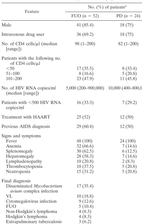
TABLE 1. Clinical and laboratory features of population studied
Feature No. (%) of patients
a
FUO (n⫽52) PD (n⫽24)
Male 41 (85.4) 18 (75)
Intravenous drug user 36 (69.2) 18 (75)
No. of CD4 cells/l (median
[range]) 98 (1–200) 82 (1–200)
Patients with the following no. of CD4 cells/l
⬍50 17 (35.5) 8 (33.4)
51–100 8 (16.6) 5 (20.8)
101–200 23 (47.9) 11 (45.8)
No. of HIV RNA copies/ml
(median [range]) 5,000 (200–900,000) 10,000 (400–800,000)
Patients with⬍500 HIV RNA
copies/ml 16 (33.3) 7 (29.2)
Treatment with HAART 25 (52) 12 (50)
Previous AIDS diagnosis 29 (60.4) 12 (50)
Signs and symptoms
Fever 48 (100) 24 (100)
Anemia 32 (66.6) 7 (14.6)
Splenomegaly 30 (62.5) 6 (12.5)
Hepatomegaly 28 (58.3) 7 (14.6)
Lymphadenopathy 10 (20.8) 2 (8.3)
Thrombocytopenia 18 (37.5) 5 (20.8)
Neutropenia 15 (31.2) 5 (20.8)
Final diagnosis
DisseminatedMycobacterium
aviumcomplex infection 17 (35.4)
VL 10 (18.8)
Cytomegalovirus infection 9 (12.6)
FUO 5 (10.4)
Non-Hodgkin’s lymphoma 4 (8.3) Hodgkin’s lymphoma 4 (8.3) Extrapulmonary tuberculosis 3 (6.2)
Bacterial pneumonia 19 (79.2)
Pulmonary tuberculosis 5 (20.8)
aUnless indicated otherwise.
on May 15, 2020 by guest
http://jcm.asm.org/
[image:2.612.315.548.374.614.2]gluconate dehydrogenase [EC 1.1.1.4.4], glucose-6-phosphate dehydrogenase [EC 1.1.1.4.9], glutamate dehydrogenase [EC 1.4.1.3], diaphorase NAD [reduced form] [EC 1.6.2.2], two isoforms of purine-nucleoside phosphorylase [EC 2.4.2.1], two isoforms of glutamate-oxoloacetate transaminase [EC 2.6.1.1], phosphoglucomutase [EC 2.7.5.1], fumarate hydratase [EC 4.2.1.2], mannose phosphate isomerase [EC 5.3.1.8], and glu-cose phosphate isomerase [EC 5.3.1.9]) as described previously
(7), using threeLeishmaniareference strains: MHOM/TN/80/
IPT1 forL. infantumzymodeme MON1, MHOM/IN/80/DD8
forL. donovani zymodeme MON2, and
MHOM/ET/93/IPB-096 forL. donovanizymodeme MON37.
TheLeishmania-specific PCR was negative for the 24 sam-ples of peripheral blood and 10 bone marrow aspirates ob-tained from the control subjects with PD and the 143 blood specimens from healthy blood donors. PCR was performed with 52 samples of peripheral blood and 31 bone marrow aspirates from the 52 patients with FUO.
Among the subjects with FUO, a definitive diagnosis of VL was obtained for nine subjects and a diagnosis of probable VL was obtained for 1 subject (patient 10) (Table 2). This patient
received anti-Leishmania treatment, and after 66 weeks he
suffered a clinical relapse, during which Leishmaniaparasites
could be microscopically demonstrated in the bone marrow.
PCR-RFLP analysis for Leishmania species identification
was performed for all patients. A single 250-bp band suggestive ofL. infantuminfection was observed for eight subjects, while
two bands of 180 and 70 bp, a characteristic pattern ofL.
do-novani, were evident for two subjects (patients 1 and 2). FourLeishmania stocks were obtained in culture, three of which underwent identification by isoenzyme analysis: two
iso-lates were identified asL. infantumzymodeme MON1 (isolates
7 and 8) and one was identified as L. donovani zymodeme
MON37 (isolate 2), thus confirming the identification obtained by the molecular technique, PCR-RFLP analysis.
All patients affected by VL received one of the following treatments at standard doses: meglumine antimoniate (four patients), liposomal amphotericin B (three patients), or am-photericin B desoxycholate (three patients).
A follow-up semiquantitative PCR with peripheral blood showed a progressive reduction in the circulating parasite bur-den while the patients were receiving therapy: 9 of the 10 patients were negative after 14 weeks, while 1 (patient 7) was negative after 21 weeks. Eight patients with HIV-VL
coinfec-FIG. 2. Graphic representation of the course of VL in patient 2. The increase in the parasite burden is followed by the reappearance of symptoms (clinical and parasitological relapse). Circles, PCR performed with peripheral blood; squares, PCR performed with bone marrow aspirates; iv, intravenous; Sb, antimony.
on May 15, 2020 by guest
http://jcm.asm.org/
[image:3.612.86.516.72.423.2]tion were monitored for a median period of 88 weeks (range, 30 to 110 weeks). Semiquantitative PCR was performed
monthly with peripheral blood samples. In all casesLeishmania
parasitemia detected by PCR was associated with clinical re-lapse but preceded the reappearance of symptoms by a mean period of 1 to 2 weeks (Fig. 2). During follow-up, 3 patients died a median of 21 weeks after the diagnosis of VL (range, 7 to 51 weeks): patient 3 died of pulmonary failure due to
Pneumocystis cariniipneumonia, patient 5 died of disseminat-ed Kaposi’s sarcoma, and patient 9 didisseminat-ed of pulmonary non-Hodgkin’s lymphoma.
At the time of the diagnosis of VL, 5 of 10 patients (patients 1, 2, 6, 7, and 8) were receiving highly active antiretroviral therapy (HAART), but sustained suppression of HIV type 1
(HIV-1) replication (i.e.,ⱕ500 copies of HIV RNA/ml) was
present in only three patients. During follow-up, HAART was initiated in two patients (patients 4 and 9) and was changed in four patients (patients 1, 2, 6, and 8). VL relapsed in patients with virological suppression of HIV-1 replication (patients 1 and 2), as well as in those showing no virological response to HAART (patients 6, 7, and 8).
In all but one patient, CD4⫹-cell counts remained below the
absolute value of 200/l. The only patient (patient 4) who
remained relapse-free and who was uninterruptedly negative as determined by PCR with peripheral blood after 114 weeks of follow-up showed progressive immune reconstitution under HAART, as demonstrated by increasing CD4 T-cell counts
(496/l after 94 weeks and 850/l at the last follow-up visit).
Previous studies of HIV-infected patients have demon-strated that the sensitivity of PCR for the diagnosis of VL ranges from 82 to 98% (3, 12, 14, 15). Our findings confirm this sensitivity for individuals coinfected with HIV-1 and also pro-vide further epro-vidence that the altered immune response in
patients with HIV-Leishmaniacoinfection not only is
respon-sible for the persistence of parasites, despite a clinical response to specific therapy, but also favors blood dissemination, as documented by a positive result by PCR with peripheral blood (7, 10, 19).
Furthermore, quantitation of parasitemia by PCR is ex-tremely useful in monitoring treatment efficacy and predicting relapse. In this regard, our data are in agreement with and complement the findings of a recent study performed in France in which qualitative PCR with peripheral blood was used in the diagnosis and follow-up of VL in both immunosuppressed and immunocompetent patients (5). Although all of our patients had a negative result by PCR with peripheral blood 6 to 21 weeks after completion of antileishmania treatment, all but one experienced a resurgence of parasitemia, and importantly, the increase in the parasite burden correlated with clinical disease relapse. From a pathogenic point of view, these results show that a parasitological cure is seldom achieved in HIV-infected patients with VL, even when a control bone marrow aspiration performed after the completion of treatment fails to
revealLeishmaniaamastigotes. From a clinical standpoint, we
confirm the findings of Lachaud et al. (5) that a positive result by PCR with peripheral blood is indicative of the presence of
viable Leishmania parasites since it correlates with clinical
disease. We observed VL relapses among the patients respond-ing well or not at all to HAART, although the only patient who remained free of relapse after 2 years of follow-up showed the
TABLE 2. Characteristics of HIV-seropositive patients with VL a Parameter Trait or result for patient no.: 12 3 b 45 c 67 8 9 d 10 Age (yrs) 45 33 32 28 31 37 38 27 29 32 Se x MF F M MM M M M M HIV risk factor IVDU IVDU Hetero IVDU IVDU IVDU IVDU IVDU Hetero IVDU HIV stage C3 C3 C3 C3 B3 B3 C3 C3 C3 C2 CD4 (no. of cells/ l) 10 78 4 158 94 82 6 62 10 190 HIV-RNA (no. of copies/ml) ⬍ 500 ⬍ 500 1,000,000 27,000 800,000 4,900 ⬍ 500 3,750 107,000 54,000 HAART at the time of VL diagnosis d4T, 3TC, IDV d4T, 3TC, IDV No No No AZT, 3TC, IDV d4T, NVP, IDV d4T, 3TC, RTV No No Primary VL diagnosis (no. of wks from primary VL to VL relapse) No (44) Yes No (76) Yes Yes Yes No (40) No (188) Yes Yes Follow-up (no. of wks) 75 88 6 94 6 30 88 100 6 110 VL relapse(s) (no. of wks) 32, 52 14, 48, 63, 84 44 73, 95 36 105 IFAT serology and titer Pos, 1:320 Pos, 1:80 Pos, 1:1,280 Neg Pos, 1:256,000 NA Neg Pos, 1:80 Neg Pos, 1:80 Microscopy result (sample[s]) Pos (Bm, Gb) Pos (Bm) Pos (Pe) Pos (Bm) Pos (Bm) Pos (Bm) Pos (Bm) ND Pos (Bm, Eb) Neg (Bm) Culture result (sample) and Leish-mania isoenzyme characterization Pos (Bm), Leish-mania spp. Pos (Bm), L. dono-vani MON37 NA Neg (Bm) Pos (Bm), Leish-mania spp. NA Pos (Bm, Wb), L. in-fantum MON1 Pos (Wb), L. infan-tum MON1 Neg (Bm) NA PCR-RFLP analysis result L. donovani L. donovani L. infantum L. infantum L. infantum L. infantum L. infantum L. infantum L. infantum L. infantum Wb PCR result Pos Pos Pos Pos Pos Pos Pos Pos Pos Pos Bm PCR result Pos Pos ND Pos Pos Pos Pos ND Pos NA Biopsy PCR (sample) Pos (Gb) Pos (Pe) Pos (Eb) aAbbreviations: M, male; F, female; IVDU, intravenous drug user; Hetero, heterosexual transmission; Bm, bone marrow; Wb, whole blood; Gb, gastric bi opsy; Eb, esophageal biopsy; Pe, pleural ef fusion; NA, not available; ND, not done; d4T, stavudine; 3TC, lamivudine; AZT, zidovudine; IDV, indinavir; RTV, ritonavir; NVP, nevirapine. bPatient died 7 weeks after VL diagnosis from P. carinii pneumonia. cPatient died 21 weeks after VL diagnosis from Kaposi’s sarcoma. dPatient died 51 weeks after VL diagnosis from non-Hodgkin’s lymphoma.
on May 15, 2020 by guest
http://jcm.asm.org/
best immunological response (a progressive increase in CD4⫹
-lymphocyte counts to above 800/l), thus indirectly
confirm-ing the fact that the cytokines produced by specific subsets of
CD4⫹ cells play a prominent role in promoting protective
immunity against parasitic infections.
Our preliminary results seem to indicate a good degree of correlation between the results of PCR-RFLP analysis and those of the traditional methods of species characterization. Although isoenzyme characterization was used only for three
Leishmaniastrains, it confirmed the identification obtained by PCR-RFLP analysis. As expected on the basis of previous
findings (1), VL was prevalently caused by L. infantum, the
common agent of VL in the Mediterranean basin, but it is
noteworthy that one patient was infected with L. donovani
(zymodeme MON37), a species that is not considered endemic in the Mediterranean area. However, the presence of this spe-cies has recently been suspected in the Middle East (2, 16, 17). Our patient had a long history of worldwide travel and drug addiction and may have acquired the infection during a trip to Turkey about 10 years before VL was diagnosed or as a result of mechanical transmission between intravenous drug users, as
suspected in the case of a Portuguese patient withL. donovani
infection (2).
In conclusion, PCR seems to be one of the most sensitive
means of detectingLeishmaniaspp. among HIV-infected
pa-tients. The presence of a positive result by PCR with peripheral blood is always associated with clinical disease. This method could be used as an alternative, noninvasive method of screen-ing individuals with suspected VL or as a tool for monitorscreen-ing the efficacy of treatment and the appearance of relapse of subclinical disease. Finally, it allows rapid decision making in the diagnostic and therapeutic management of HIV-infected patients.
REFERENCES
1.Alvar, J., C. Canavate, B. Gutierrez-Solar, M. Jime´nez, F. Laguna, R. Lopez-Ve´lez, R. Molina, and J. Moreno.1997.Leishmaniaand human immunode-ficiency virus coinfection: the first 10 years. Clin. Microbiol. Rev.10:298–319. 2.Campino, L., G. Santos-Gomes, F. Pratlong, J. P. Dedet, and P. Abranches. 1994. The isolation ofLeishmania donovaniMON18 from an AIDS patient in Portugal: possible needle transmission. Parasite1:391–392.
3.Costa, J. M., R. Durand, M. Deniau, D. Rivollet, M. Izri, R. Houin, M. Vidaud, and S. Bretagne.1996. PCR enzyme-linked immunosorbent assay for diagnosis of visceral leishmaniasis in human immunodeficiency virus-infected patients. J. Clin. Microbiol.34:1831–1833.
4.Gramiccia, M., L. Gradoni, and M. Troiani.1995. Heterogeneity among zymodemes ofLeishmania infantumfrom HIV-positive patients with visceral leishmaniasis in south Italy. FEMS Microbiol. Lett.128:33–38.
5.Lachaud, L., J. Dereure, E. Chabbert, J. Reynes, J.-M. Mauboussin, E. Oziol, J.-P. Dedet, and P. Bastien.2000. Optimized PCR using patient blood samples for diagnosis and follow-up of visceral leishmaniasis, with special reference to AIDS patients. J. Clin. Microbiol.38:236–240.
6.Looker, D., L. A. Miller, H. J. Elwood, S. Stickel, and M. L. Sogin.1998. Primary structure of theLeishmania donovanismall subunit ribosomal RNA coding region. Nucleic Acids Res.16:7198.
7.Lopez-Velez, R., F. Laguna, J. Alvar, J. A. Pe´rez-Molina, R. Molina, P. Martinez, and J. Villarrubia.1995. Parasitic culture of buffy coat for diag-nosis of visceral leishmaniasis in human immunodeficiency virus-infected patients. J. Clin. Microbiol.33:937–939.
8.Medrano, F. J., C. Canavate, M. Leal, C. Rey, E. Lissen, and J. Alvar.1998. The role of serology in the diagnosis and prognosis of visceral leishmaniasis in patients coinfected with human immunodeficiency virus type 1. Am. J. Trop. Med. Hyg.59:155–162.
9.Minodier, P., R. Piarroux, F. Gambarelli, C. Joblet, and H. Dumon.1997. Rapid identification of causative species in patients with Old World leish-maniasis. J. Clin. Microbiol.35:2551–2555.
10. Molina, R., C. Canavate, E. Cercenado, F. Laguna, R. Lopez-Velez, and J. Alvar.1994. Indirect xenodiagnosis of visceral leishmaniasis in 10 HIV-infected patients using colonizedPhlebotomus perniciosus. AIDS8:277–279. 11. Nuzum, E., F. White, C. Thakur, R. Dietze, J. Wages, M. Grogl, and J. Berman.1995. Diagnosis of symptomatic visceral leishmaniasis by use of the polymerase chain reaction on patient blood. J. Infect. Dis.171:751–754. 12. Osman, O. F., L. Oskam, E. E. Zijlistra, N. C. M. Kroon, G. J. Schoone,
E. A. G. Khalil, A. M. El-Hassan, and P. A. Kager.1997. Evaluation of PCR for diagnosis of visceral leishmaniasis. J. Clin. Microbiol.35:2454–2457. 13. Parravicini, C., E. Lauri, L. Baldini, A. Neri, F. Poli, G. Sirchia, M. Moroni,
M. Galli, and M. Corbellino.1997. Kaposi’s sarcoma herpesvirus infection and multiple myeloma. Science278:1969–1970.
14. Piarroux, R., F. Gambarelli, H. Dumon, M. Fonets, S. Duncan, C. Mary, B. Toga, and M. Quilici.1994. Comparison of PCR with direct examination of bone marrow aspiration, myeloculture, and serology for diagnosis of visceral leishmaniasis in immunocompromised patients. J. Clin. Microbiol.32:746– 749.
15. Piarroux, R., F. Gambarelli, B. Toga, H. Dumon, M. Fontes, S. Dunan, and M. Quilici.1996. Interest and reliability of a polymerase chain reaction on bone marrow samples in the diagnosis of visceral leishmaniasis in AIDS. AIDS10:452–453.
16. Qubain, H. I., E. K. Saliba, and L. Oskam.1997. Visceral leishmaniasis from Bal’a, Palestine, caused byLeishmania donovani s.l.identified through poly-merase chain reaction and restriction fragment length polymorphism anal-ysis. Acta Trop.68:121–128.
17. Rioux, J. A., N. Le`ger, and N. Haddad.1998. Infestation naturelle de Phle-botomus tobbi(Diptera,Psychodidae) parLeishmania donovani s. st.( Kineto-plastida,Trypanosomatidae), en Syrie. Parassitologia40:148.
18. Van Eys, G. J. J. M., G. J. Shoone, N. C. M. Kroon, and S. B. Ebeling.1992. Sequence analysis of small subunit ribosomal RNA genes and its use for detection and identification ofLeishmaniaparasites. Mol. Biochem. Parasi-tol.51:133–142.
19. Wolday, D., N. Berhe, H. Akuffo, and S. Britton.1998.Leishmania-HIV interaction: immunopathogenic mechanisms. Parasitol. Today15:182–187. 20. World Health Organization.1999. Leishmania-HIV coinfection south
west-ern Europe 1990–1998 retrospective cases. Wkly. Epidemiol. Rec.74:365– 375.