Journal of Chemical and Pharmaceutical Research, 2017, 9(4):191-195
Research Article
CODEN(USA) : JCPRC5
ISSN : 0975-7384
191
MRI Finding in Acute Lower Back Pain
Najat Adel Hashim
*and Raad Jawad Kadhim
Department of Surgery, College of Medicine, University of Al-Qadissiyah, Al-Diwaniyah province, Iraq _____________________________________________________________________________
ABSTRACT
Role of MRI in patients with acute lower back pain. Method: A group of 501 patients who referred unit from orthopedic were subjected to MRI examination and the MRI results were evaluated. All patients with radicular pain, neurological signs or traumatic patients were excluded from this study. Results: 501 patients complain from acute lower back pain , mean age 33.49+8.38 range (20-70), 229 (46%) male and 272 (54%) female , in combined MRI finding with age of patients , we found large number of patient have disk prolapsed (191), with mean age 36.50 , while patient with normal MRI or just muscles spasm were mean age (31.79), so the MRI not effect in outcome of young patient ( below 31 years old ) with a lower back pain. Conclusion: Lumbo-sacral MRI exam have minimal effect in outcome of young patient with acute lower back pain (less than 3wks)
Keywords: Acute low back pain; Magnetic resonance imaging
_____________________________________________________________________________
INTRODUCTION
192
MATERIALS AND METHODS
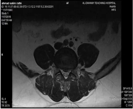
Between July 2014 to July 2016, 501 patients with acute lower back pain were referred from orthopedic outpatient to the MRI unite at AL-Diwaniya teaching hospital. The mean age 33.49+8.38 range (20-70), 229 male and 272 female. The history was taken from every one of them, the data collected include, age, sex and duration of pain (all had pain for1 to 2 wks. only). We exclude all patients with history of trauma, patients with previous attack of back pain, patients with lumbo-sacral spine diseases and patients with neurological diseases. The MRI exam was done by using Siemens Avanto (1.5 tesla) apparatus putting the patient on supine position, the time of examination (10 minute), an ordinary sequences was done, T1 and T2 weighted image sagittal views, T2 weighted image axial view ( 4mm slice thickness ) and MRI myelography.
The following criteria were evaluated in MRI examination:
1. Loss of normal lumbar lardosis, for severe muscles spasm.
2. Signal intensity of disk and it is position, to diagnose disk dehydration and prolapsed. 3. Intervertebral foramina, for nerve root compression stenosis foramina.
We divided MRI findings into three groups:
A. Negative: normal LSS MRI.
B. Muscles spasm only, without disk prolapsed.
C. Disk prolapsed in all types: central, postero-lateral and lateral.
Patients divided into 5 age groups these findings applied to patients according to their age.
The total patients classified into 5 groups with interval of 10 years 1st group (20-29 y), 2nd group (30-39 y), 3rd group (40-49 y), 4th group (50-59 y) and 5th group ≥ -60 y.
We Correlated the MRI findings with patients age group, for comparison between age groups as appropriate (P < or = 0.05) was considerable statistically significant.
RESULTS
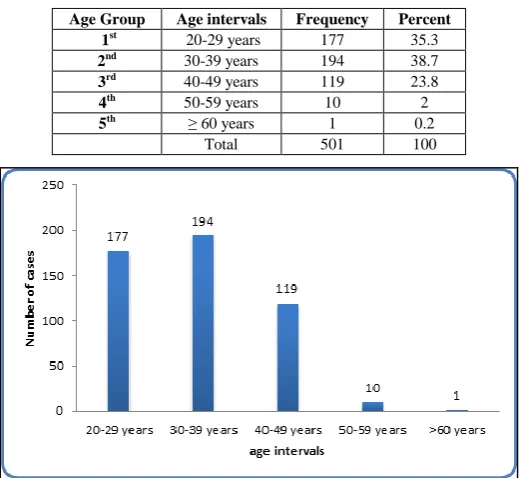
[image:2.595.168.428.445.684.2]Total number of patients was 501, with mean age 33.49+8.38 range (20-70), 229 (46%) male and 272 (54%) female. We divided the patients in to 5 age groups with 10 years interval for each group, 1st group (20-29 years), 2nd group (30-39 years), 3rd group (40-49 years), 4th group (50-59 years) and 5th group (Table 1 and Graphs 1 and 2).
Table 1: The distribution of patients according to the age group was 177 (35.3%) in the 1st group, 194 (38.7%) in 2nd age group, 119
(23.8%) in 3rd age group, 10(2%) in 4th group, and small number in 5th group 1 (0.2%)
Age Group Age intervals Frequency Percent
1st 20-29 years 177 35.3
2nd 30-39 years 194 38.7
3rd 40-49 years 119 23.8
4th 50-59 years 10 2
5th ≥ 60 years 1 0.2
Total 501 100
Graph 1: Number of patients related to age largest number are seen in 1st and 2nd age group (194 and 177), then 3rd (119), while the
193
Graph 2: showing number of female in this simple little more than male, 272 (54%) female, 229 (46%) male
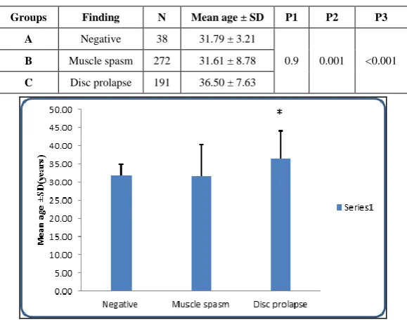
The distribution of patients according to MRI finding was as follow (Table 2 and Graph 3): Group A: 38 patients had negative results.
Group B: 272patients had only muscle spasm.
[image:3.595.153.443.322.550.2]Group C: 191 patients had disc prolapse with or without nerve root compression or spinal stenosis.
Table 2: In compared group A with B showing (P1 0.9) not significant both have same age group (mean age 31 young pat.), while comparison between group A with C we have (P2 0.001) significant that s mean these signs are seen in different age groups, and comparison between group C with B showing (P3 < 0.001 ) highly significant, means these signs also seen in different age groups,
both groups A and B are seen in young pat, while group C are seen in old age patients (mean age 36.50)
Groups Finding N Mean age ± SD P1 P2 P3
A Negative 38 31.79 ± 3.21
0.9 0.001 <0.001
B Muscle spasm 272 31.61 ± 8.78
C Disc prolapse 191 36.50 ± 7.63
Graph 3: Relation between the ages of pat. and MRI finding, showing negative and muscles spasm are seen in younger pat (Below 35 year old), while disc prolapsed are seen in older age about 40 years old
DISCUSSION
194
[image:4.595.184.410.141.309.2]pain during their 20s (Figure 1); however, LDH most frequently affects patients in their early 40s [13], (Figures 2 and 3). Only 11% of young adults with low back pain experience acute disc herniation of the lumbar region [14]. Pain has usually acute onset, flexion-related and often accompanied by back muscle spasm, tightness of hamstring muscles and, sometimes, pain in buttock region [15-18]. Radicular symptoms (paresthe-sias and muscle weakness) are uncommon [19,20]. Examination frequently demonstrates reduced flexion, positive straight leg raise and sometimes less reflexes/strength of the lower limbs.
[image:4.595.186.410.331.516.2]Figure 1: Sagittal MRI T2 weighted image showing normal disk signal intensity, no signs of prolapsed disk
Figure 2: Sagittal MRI t2 weighted image: acute prolapsed L5/S1 disk, with sever muscles spasm
[image:4.595.182.413.544.734.2]195
CONCLUSIONThe present study showed that Low back pain was more frequent in females in comparison to males. This is in accordance with the several previous studies, that showed that Low back pain symptoms were usually more common among females. Schneider et al. [21] and Hathorn et al. [22] showed that the rate of Low back pain is higher among females due to the stress of hormonal changes, problems related gynaecological disorders and also childbirth related events [23].
REFERENCES
[1] BA Casazza. Am Fam Physician. 2012, 85(4), 343-350.
[2] SJ Atlas; RA Deyo. J Gen Intern Med. 2001, 16, 120-131.
[3] S Kinkade. Am Fam Physician. 2007, 75(8), 1181-1188.
[4] RA Deyo; J Rainville; DL Kent. JAMA. 1992, 268, 760-765.
[5] S Bigos, O Bowyer, G Braen. Acute Low Back Problems in Adults. Clinical Practice Guideline No. 14,
AHCPR Publication No. 95-0642. Agency for Health Care Policy and Research, Public Health Service,
U.S. Department of Health and Human Services, Rockville, MD, 1994.
[6] E Amato; D Lizio; N Settineri; A Di Pasquale; I Salamone; I Pandolfo. Med Phys. 2010, 37, 4249-4256.
[7] R Chou; A Qaseem; V Snow; D Casey; T Cross; P Shekelle; DK Owens. Ann Intern Med. 2007, 147(7), 478-491.
[8] R Kumar; V Kumar; NK Das; S Behari; AK Mahapatra. ChildsNerv Syst. 2007, 23, 1295-1299.
[9] P Sarma; RT Thirupathi; D Srinivas; S Somanna. J Pediatr Neurosci. 2016, 11(1), 20-24.
[10] W Huang; Z Han; J Liu; L Yu; X Yu. Risk Factors for Recurrent Lumbar Disc Herniation: A Systematic Review and Meta-Analysis. Medicine. 2016, 95(2), e2378
[11] MK Kasliwal; H Deutsch. J Pediatr Neurosci. 2012, 7(2), 129-132.
[12] P Kjaer; C Leboeuf-Yde; JS Sorensen; T Bendix. An epidemiologic study of MRI and low back pain in
13-year-old children. Spine. 2005, 30, 798-806.
[13] F Strömqvist; B Strömqvist; B Jönsson; MK Karlsson. Acta Orthopaedica. 2016, 87(5), 516-521. [14] LJ Micheli; R Wood. Arch Pediatr Adolesc Med. 1995, 149, 15-18.
[15] M Zetaruk. Lumbar spine injuries. In: Micheli LJ, Purcell LK, editors. The Adolescent Athlete, Springer, New York, 2007, 109-140.
[16] TJ Trainor; MA Trainor. Curr Sport Med Reports. 2004, 3, 41-46.
[17] PA d’Hemecourt; PG Gerbino; LJ Micheli. Clin Sports Med. 2000, 19, 663-679. [18] RG Watkins. Clin Sports Med. 2002, 21, 147-165.
[19] DE Kraft. Pediatr Clin N Am. 2002, 49, 643-653.
[20] LM Simon, W Jih, JC Buller. Back pain and injuries. In: Birrer RB, Griesemer BA, Cataletto MB, editors. Pediatric Sports Medicine for Primary Care, Lippincott Williams and Wilkins, Philadelphia,
2002, 306-325.
[21] S Schneider; D Randoll; M Buchner. Clin J Pain. 2006, 22, 738-747.
[22] DC Hathorn; AK Carruth; LJ Agosta; SK Pryor. AAOHN J, 2009, 57, 232-238.