epithelial to mesenchymal cell transition
(EMT)
Kasai, H, Allen, JT, Mason, RM, Kamimura, T and Zhang, Z
http://dx.doi.org/10.1186/14659921656
Title
TGFbeta 1 induces human alveolar epithelial to mesenchymal cell
transition (EMT)
Authors
Kasai, H, Allen, JT, Mason, RM, Kamimura, T and Zhang, Z
Type
Article
URL
This version is available at: http://usir.salford.ac.uk/125/
Published Date
2005
USIR is a digital collection of the research output of the University of Salford. Where copyright
permits, full text material held in the repository is made freely available online and can be read,
downloaded and copied for noncommercial private study or research purposes. Please check the
manuscript for any further copyright restrictions.
Open Access
Research
TGF-
β
1 induces human alveolar epithelial to mesenchymal cell
transition (EMT)
Hidenori Kasai
1, Jeremy T Allen
2, Roger M Mason
3, Takashi Kamimura
4and
Zhi Zhang*
1Address: 1Teijin Biomedical Laboratory, Medical Research Council Technology, 1–3 Burtonhole Lane, London, NW7 1AD, UK, 2Biosciences Research Institute, University of Salford, Greater Manchester M5 4WT, UK, 3Renal Medicine Section, Faculty of Medicine, Imperial College London, Hammersmith Hospital, London, W12 0NN, UK and 4Institute for Bio-Medical Research, Teijin Pharma Ltd, Tokyo, 191-8512, Japan Email: Hidenori Kasai - hidenori.kasai@tech.mrc.ac.uk; Jeremy T Allen - J.T.Allen@salford.ac.uk; Roger M Mason - roger.mason@imperial.ac.uk; Takashi Kamimura - t.kamimura@teijin.co.jp; Zhi Zhang* - zzhang@tech.mrc.ac.uk
* Corresponding author
Abstract
Background: Fibroblastic foci are characteristic features in lung parenchyma of patients with idiopathic pulmonary fibrosis (IPF). They comprise aggregates of mesenchymal cells which underlie sites of unresolved epithelial injury and are associated with progression of fibrosis. However, the cellular origins of these mesenchymal phenotypes remain unclear. We examined whether the potent fibrogenic cytokine
TGF-β1 could induce epithelial mesenchymal transition (EMT) in the human alveolar epithelial cell line,
A549, and investigated the signaling pathway of TGF-β1-mediated EMT.
Methods: A549 cells were examined for evidence of EMT after treatment with TGF-β1. EMT was assessed by: morphology under phase-contrast microscopy; Western analysis of cell lysates for expression of mesenchymal phenotypic markers including fibronectin EDA (Fn-EDA), and expression of epithelial phenotypic markers including E-cadherin (E-cad). Markers of fibrogenesis, including collagens and connective tissue growth factor (CTGF) were also evaluated by measuring mRNA level using RT-PCR, and protein by immunofluorescence or Western blotting. Signaling pathways for EMT were characterized by Western analysis of cell lysates using monoclonal antibodies to detect phosphorylated Erk1/2 and Smad2
after TGF-β1 treatment in the presence or absence of MEK inhibitors. The role of Smad2 in TGF-β
1-mediated EMT was investigated using siRNA.
Results: The data showed that TGF-β1, but not TNF-α or IL-1β, induced A549 cells with an alveolar epithelial type II cell phenotype to undergo EMT in a time-and concentration-dependent manner. The process of EMT was accompanied by morphological alteration and expression of the fibroblast phenotypic markers Fn-EDA and vimentin, concomitant with a downregulation of the epithelial phenotype marker E-cad. Furthermore, cells that had undergone EMT showed enhanced expression of markers of fibrogenesis
including collagens type I and III and CTGF. MMP-2 expression was also evidenced. TGF-β1-induced EMT
occurred through phosphorylation of Smad2 and was inhibited by Smad2 gene silencing; MEK inhibitors failed to attenuate either EMT-associated Smad2 phosphorylation or the observed phenotypic changes.
Conclusion: Our study shows that TGF-β1 induces A549 alveolar epithelial cells to undergo EMT via Smad2 activation. Our data support the concept of EMT in lung epithelial cells, and suggest the need for further studies to investigate the phenomenon.
Published: 09 June 2005
Respiratory Research 2005, 6:56 doi:10.1186/1465-9921-6-56
Received: 08 December 2004 Accepted: 09 June 2005 This article is available from: http://respiratory-research.com/content/6/1/56
© 2005 Kasai et al; licensee BioMed Central Ltd.
Background
Idiopathic pulmonary fibrosis (IPF), the most common pulmonary fibrotic disorder, is a progressive and lethal disease of unknown etiology whose pathogenesis uniquely features the presence of fibroblastic foci in the parenchyma of the lungs [1]. These are comprised of aggregates of mesenchymal cells including fibroblasts and cells which exhibit phenotypic features of myofibroblasts, α-smooth muscle actin (αSMA) expression, increased mitogenic capacity, and enhanced extracellular matrix (ECM) production. The number of fibroblastic foci corre-lates with worsening lung function, progression of IPF and a poor prognosis [2]. According to the recent epithe-lial/fibroblastic model of IPF pathogenesis it is considered that fibroblastic foci underlie areas of unresolved epithe-lial injury and are sites where activated fibroblasts and myofibroblasts migrate, proliferate and synthesize ECM proteins [3]. However, the cellular origins of the mesen-chymal phenotypes in fibroblast foci remain unclear.
It is now well recognized from many studies that a number of key growth factors are responsible for driving the process of fibrogenesis [4]. For example, transforming growth factor-beta1 (TGF-β1), interleukin-1 beta (IL-1β), and tumor necrosis factor-alpha (TNF-α) are able to induce the characteristic motility, proliferation and ECM synthesis observed in mesenchymal cells with a myofi-broblast-like phenotype from fibroblastic foci. In general though, it is levels of TGF-β1 that best correlate with the extent of fibrosis and myofibroblast-like cell induction [5] and TGF-β1 continues to be regarded as the most impor-tant of the growth factors involved in pulmonary fibro-genesis [6]. For example, the biologically active form of TGF-β1 was aberrantly expressed in the epithelial cells lin-ing honeycomb cysts within the lung of patients with IPF [7,8]. An increased level of TGF-β1 was found in BAL fluid derived from patients suffering from IPF [8]. Furthermore, overexpression of TGF-β1 in lung tissue induced pro-longed pulmonary fibrosis in an animal model [9].
Recent evidence from studies of other fibrotic disorders, including renal [10,11] and liver fibrosis [12], supports a view that TGF-β1 may play a novel role in pulmonary fibrogenesis by promoting alveolar epithelial cell transi-tion to form mesenchymal cells with a myofibroblast-like phenotype [10-14]. This process, termed epithelial-mes-enchymal transition (EMT), occurs widely under both physiologic and pathologic conditions, for example dur-ing normal wound healdur-ing [13] and renal fibrosis [10,11]. Very recently it was reported that TGF-β1 induced type II alveolar epithelial cells isolated from rat lung to undergo EMT [15]. Epithelial cells are polarised, and display cytok-eratin filaments and membrane-associated junctions. During EMT membrane-associated adherens junctions and desmosomes are dissociated, whilst at the same time
or shortly after, cytoskeletal rearrangement takes place and mRNA for intermediate filament proteins is increased, facilitating the cell adopting a mesenchymal phenotype [14]. E-cadherin (E-cad) is an epithelial cell transmembrane protein whose extracellular domain inter-acts with that of an E-cad molecule expressed by an adja-cent cell. It has a critical role in establishing firm adhesion, maintaining cell polarity and epithelial tight-ness [16]. The cadherin complex suppresses the dissocia-tion of epithelial cells, and thus, the crucial step of EMT is the downregulation of E-cad [14].
To begin to understand the role of EMT in the develop-ment of fibroblastic foci in IPF, we have examined whether TGF-β1 can induce EMT in a human lung epithe-lial cell line (A549). A549 cells retain important character-istics of alveolar type II epithelial cells and have been employed in numerous studies as a valuable tool for stud-ying promoter activity [17], apoptosis [18], and alveolar epithelial cell DNA damage [19]. In this study we demon-strate that TGF-β1 induces EMT in human type II alveolar epithelial cells through the activation of Smad2, and that this transition is accompanied by functional changes that are relevant to the progression of lung fibrosis.
Methods
MaterialsRecombinant human TGF-β1, human IL-1β; and TNF-α were purchased from R&D systems (Minneapolis, MN). A mouse monoclonal antibody against human E-cad was from BD Transduction Laboratory (Oxford, UK). Mouse monoclonal anti-human fibronectin EDA+ splice form
Cell culture
Human type II alveolar epithelial cells (A549) were sup-plied from ATCC/LGC Promochem (Middlesex, UK). Cells were maintained in low glucose-DMEM containing 10% FBS, 2 mM L-glutamine, 100 U/ml penicillin and 100 ug/ml streptomycin at 37°C in a humidified 5% CO2 atmosphere. Confluent cultures of cells were maintained in serum-free DMEM containing 0.1% BSA for 24 h prior to stimulation with cytokines. The cells were incubated with several concentrations of the cytokines for the peri-ods indicated. In experiments using inhibitors of MAPK/ Erk kinase (MEK), the cells were preincubated for 1 h with PD-098059 or U-0126 (up to 10 µM) before treatment
with exogenous TGF-β1. In small interfering RNA
(siRNA)-dependent gene silencing experiments, after transfection the cells were stimulated with 5 ng/ml of TGF-β1 in serum free 0.1% BSA/DMEM for 48 h. The cells were then harvested and lysed. In experiments testing col-lagen expression, the cells were incubated in the presence of 5 ug/ml of L-ascorbic acid. For immunocytochemical staining, the cells were maintained throughout the exper-iment in culture medium without serum deprivation.
SDS-PAGE and Western blot
The cells were scraped and lysed in M-PER (Pierce, Chesh-ire, UK) containing a protease inhibitor cocktail (Roche Diagnostic, East Sussex, UK). Cell suspensions were cleared by centrifugation at 13,000 g for 15 min at 4°C. Total protein concentration was measured using the BCA protein assay kit (Pierce) with bovine serum albumin as the standard protein. Equal amounts of protein were loaded for each lane of 10% SDS PAGE gels, followed by electrophoresis, and protein transfers to Hybond ECL membranes (Amersham, Buckinghamshire, UK), as described previously [14]. After the transfer, membranes were blocked with 5% skimmed milk and then probed with primary antibodies for 1 h at room temperature. After washing, the membranes were probed with appro-priate peroxidase-conjugated secondary antibodies. After further extensive washing, the immunoblots were visual-ized by ECL (Amersham) and the band densities for each phenotype marker were quantified using QuantityOne Software (Bio-Rad) after scanning with a GS-710 Cali-brated Imaging Densitometer (Bio-Rad). Results were expressed as a ratio of band density to total actin. For Western blotting of phosphorylated proteins, the cells were scraped and lyzed in RIPA buffer containing a pro-tease inhibitor cocktail (Roche), plus 1 mM sodium-orthovanadate and 1 mM NaF. Western blot was per-formed according to manufacturer's instructions for each antibody. Changes in levels of phosphorylated proteins were assessed with reference to the respective non-phos-phorylated proteins.
Reverse Transcription PCR (RT-PCR)
Total RNA was isolated using Trizol (Invitrogen, Carlsbad, CA). A one-step RT-PCR was performed using the Super-Script One-Step RT-PCR kit (Invitrogen). The target tran-script was reverse transcribed at 50°C for 30 min. The PCR products for type III collagen and GAPDH were amplified using 35 cycles (initial denaturation at 94°C/2 min fol-lowed by PCR amplification, 94°C/15 s, 60°C/30 s, and 68°C/1 min). For type I collagen, the PCR product was amplified using 35 cycles (initial denaturation at 94°C/2 min followed by PCR amplification, 94°C/30 s, 58°C/30 s, and 68°C/1 min). PCR products were visualized on a GelDoc 1000 system (Bio-Rad) and semi-quantified using a scanning densitometer. The following primers were used for amplification of target transcripts; human collagen type I forward; 5'-ACGTCCTGGTGAAGTTGGTC-3', human collagen type I reverse; 5'-ACCAG-GGAAGCCTCTCTCTC-3', human collagen type III for-ward; 5'-AGCCTCATTAGTCCTGATGGTTCTCG-3', human collagen type III reverse; CTTCTCAGCACTA-GAATCTGTCCACC-3', human GAPDH forward; 5'-GGGCTGCTTTTAACTCTGGT-3', human GAPDH reverse; 5'-TGGCAGGTTTTTCTAGACGG-3'.
RNA Interference and Transfection
Dharmacon's SMARTpool SMAD2 siRNA reagent includes a pool of 4 SMARTselection-designed synthetic Smad2 siRNA duplexes, together with a non-specific con-trol pool of siRNA as a negative concon-trol. Transfection of these pooled 21-nucleotide siRNA duplexes was carried out using Lipofectamine 2000 (Invitrogen), following the manufacturer's instructions. Cells were seeded at 5 × 10⁴
cells/well of 24 wells plate and then incubated in normal medium without antibiotics for overnight to reach 80– 90% confluence. siRNA-transfection reagent complexes were prepared as described by the manufacturer. Briefly 1 µl/well Lipofectamine 2000 was mixed with either 50 pmol/well Smad2 or negative control siRNA. FITC conju-gated dsRNA (Block-iT, Invitrogen) was used as positive control. The cells were then transfected with siRNA/Lipo-fectamine complexes in Opti-MEM (Invitrogen) and incu-bated for 24 h at 37°C in a CO2 incubator. Following incubation, transfection efficiency was evaluated under fluorescence microscopy.
Immunocytochemistry
Cells were plated into a Lab-TeK Chamber Slide at a den-sity of 2 × 10⁴ cells/well and grown until approximately
appropriate FITC-conjugated secondary antibodies (1:50). Images were collected using an Eclipse TE2000-S microscope system (Nikon UK Ltd, Surrey) and Image-Pro Plus (Media Cybernetics UK, Berkshire).
Gelatin zymography for matrix metalloproteinases (MMPs) expression
Conditioned media were concentrated approximately 10-fold using ultra-centrifugation. After measuring protein concentration, equal amounts of samples were mixed with an equal volume of 2 × non-reducing SDS PAGE sample buffer. The samples were applied to a 10% (w/v) polyacrylamide gel impregnated with 2 mg/ml gelatin (Sigma). After electrophoresis, SDS was removed from the gel by washing 3 times for 10 min in 2.5% Triton X-100 solution. Then the gels were incubated overnight with gentle shaking at 37°C in buffer (50 mM Tris-HCl (pH7.6), 10 mM CaCl2, 50 mM NaCl, 0.05% Brij35), after which, the gel was stained with 0.25% Coomassie blue R250 in 40% methanol and 10% acetic acid for 2 h at room temperature, and subsequently destained with a 40% methanol-10% acetic acid solution until the bands became clear.
ELISA
Culture media were analyzed for collagens type I, type III and type IV using sandwich ELISA [14]. Each purified human collagen was used as a standard. Culture media were incubated in ELISA plates in which wells had been coated with non-biotinylated primary antibodies. Follow-ing addition of biotinylated antibodies, the plates were washed and reacted with alkaline phosphatase-conju-gated anti-Extravidin. P-nitrophenyl phosphate substrate tablets were used to detect alkaline phosphatase activity and the product was measured at 405 nm using a micro-plate reader (Bio-Rad).
Statistical analysis
Data are presented as the mean ± SD of at least three inde-pendent experiments. For statistical analysis, an unpaired t-test was used for pair-wise comparisons and ANOVA with Dunnett's post test for other data. Statistical analysis was performed using commercial statistical software Prism Version 4.0 (GraphPad, Software Inc., San Diego, CA). P values less than 0.05 were considered as statisti-cally significant.
Results
TGF-β1 induces alveolar epithelial cells to undergo EMT
We first determined the optimum concentrations and time required for TGF-β1 to initiate EMT in cultures of the alveolar type II epithelial cell line, A549. The expression of the epithelial phenotype markers, E-cad and cytokeratin 19, and of the mesenchymal phenotype markers, Fn-EDA and vimentin, were determined following treatment of
A549 cells with various concentrations (0.01–10 ng/ml) of TGF-β1 for 24, 48 and 72 h (Figure 1). Changes in cell morphology were also assessed under phase contrast light microscopy (Figure 2).
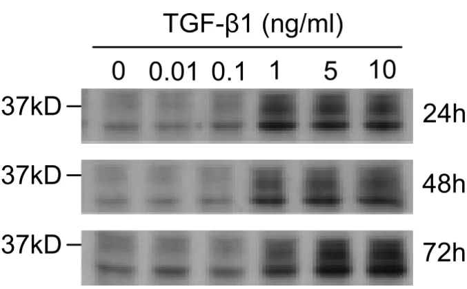
TGF-β1 significantly (P < 0.01) decreased E-cad expres-sion in a concentration-and time-dependent manner (Fig-ure 1A). Concentrations as low as 1 ng/ml of TGF-β1 induced up to 70% loss of E-cad expression within 48 h (Figure 1B). However, the extent of cytokeration 19 sup-pression was not as profound as for E-cad. Only high con-centrations of TGF-β1 resulted in a measurable decrease (Figure 1B). In parallel with the marked decrease in the E-cad epithelial marker, TGF-β1 significantly (P < 0.01) induced expression of the mesenchymal marker Fn-EDA, in a concentration-and time-dependent manner (Figure 1A). But the level of vimentin expression was not as pro-found as for Fn-EDA. Our data also suggested that the de
novo expression of Fn-EDA might occur earlier than E-cad
suppression (Figure 1A).
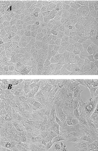
In addition to the changes in the phenotypic markers expressed in A549 cells after TGF-β1 stimulation, the cells also underwent morphological changes on exposure to the growth factor (Figure 2). A549 cells cultured in the absence of TGF-β1 maintained a classic cobblestone epi-thelial morphology and growth pattern (Figure 2A), but after stimulation with 5 ng/ml of TGF-β1 for 48 h, the cells adopted a more fibroblast-like morphology and reduced their cell-cell contact (Figure 2B).
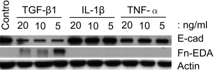
Both IL-1β and TNF-α have been suggested to play a role in fibroblast/myofibroblast motility, proliferation and ECM synthesis [4], and IL-1β induces kidney epithelial cells to undergo EMT [20,21]. We therefore tested whether these cytokines had similar properties to TGF-β1 in induc-ing lung alveolar epithelial cells to form mesenchymal-like cells. Up to 20 ng/ml of IL-1β had no effect on the expression of any of the molecular markers examined, whilst TNF-α, at 20 ng/ml concentration, resulted in a ~20% fall in the expression of E-cad (Figure 3). Neither TNF-α nor IL-1β induced the expression of Fn-EDA. These results suggested that alveolar EMT might be induced by aberrant expression and activation of TGF-β1 rather than by IL-1β or TNF-α.
TGF-β1-induced EMT is accompanied by other changes relevant to fibrogenesis
Expression changes of EMT-related markers in A549 cells
Figure 1
Expression changes of EMT-related markers in A549 cells. (A) A549 cells were incubated with up to 10 ng/ml of
TGF-β1 in the absence of serum for up to 72 h. Expression of the epithelial marker E-cadherin is down-regulated by TGF-β1
stimu-lation in a concentration-and time-dependent manner. Expression of Fn-EDA, which is a mesenchymal marker, is up-regulated
by TGF-β1 in parallel with the down regulation in the epithelial marker. The same amounts of total protein are loaded in each
lane. (B) Densitometric analysis of band intensities for each EMT related marker was performed at 48 h. Each bar represents mean ± SD of three independent experiments. * P < 0.05 and ** P < 0.01.
A
TGF-ȕ1 (ng/ml)0 0.01 0.05 0.1 0.2 0.5 1 2 5 10
24h E-cad 48h 72h 24h 48h 72h 24h Fn-EDA 48h Actin 72h
B
0 0.01 0.1 1 5 10
** ** * Fn-EDA 0.0 0.2 0.4 0.6 0.8 1.0 B a nd i n te nsi ty
0 0.01 0.1 1 5 10
0.0 0.5 1.0 1.5 Vimentin B a nd i n te nsi ty
0 0.01 0.1 1 5 10
0.0
Cytokeratin19
0 0.01 0.1 1 5 10
E-cadherin
0 0.01 0.1 1 5 10
** ** *
Fn-EDA
0 0.01 0.1 1 5 10
0.0 0.5 1.0 1.5 Vimentin B a nd i n te nsi ty
0 0.01 0.1 1 5 10
0.0
Cytokeratin19
0 0.01 0.1 1 5 10
E-cadherin 0.0 0.2 0.4 0.6 0.8 1.0 B a nd i n te nsi ty 0.0 0.5 1.0 1.5 2.0 ** ** ** B a nd i n te nsi ty 0.0 0.5 1.0 1.5 2.0 ** ** ** B a nd i n te nsi ty 0.5 1.0 1.5 2.0 0 0.01 0.1 1 5 10
TGF-E1 (ng/ml)
B a nd i n te nsi ty 0.5 1.0 1.5 2.0 0 0.01 0.1 1 5 10
TGF-E1 (ng/ml)
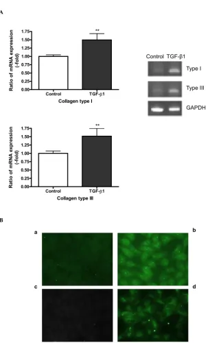
with that of type IV collagen which is characteristically synthesized by epithelial cells making basement mem-brane. Cells were treated with several concentrations of TGF-β1, and the effects on collagen expression assessed by RT-PCR; protein synthesis of collagens was also examined by ELISA and immunocytochemical staining. Figure 4 shows that TGF-β1 significantly (P < 0.01) stimulated the expression of collagens type I and type III, as detected by both RT-PCR (Figure 4A) and immuno-staining of the cell layer (Figure 4B). Protein levels of collagens type I and type III secreted into the culture medium were too low to be measured quantitatively by ELISA. Interestingly, the secretion of collagen type IV into the medium was increased in the presence of TGF-β1 (Figure 5). Concen-trations of TGF-β1 as low as 0.1 ng/ml significantly induced secretion of collagen type IV as compared with
control cells, the increased secretion level reaching a pla-teau at 1 ng/ml of TGF-β1 (Figure 5).
CTGF acts in concert with TGF-β1 and is thought to have a significant role in promoting and maintaining fibrogen-esis [22]. Thus, we investigated whether the expression of CTGF in A549 was affected by TGF-β1 treatment. Western analysis of both A549 cell lysates (Figure 6) and culture medium (data not shown) indicated that treatment with TGF-β1 at concentrations >1 ng/ml upregulated the expression of native CTGF (36–38 kDa) in a time-depend-ent manner. An additional smaller immunoreactive CTGF species was also detected and may correspond to a CTGF breakdown product which is frequently found in cell cul-tures [23].
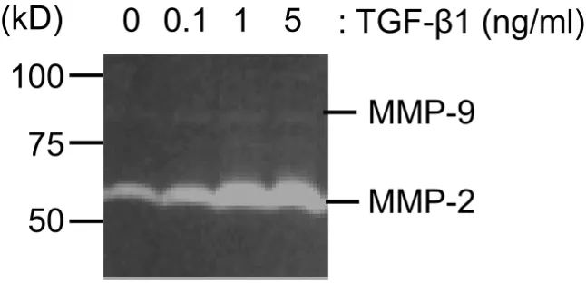
MMPs expression was examined using gelatin zymogra-phy to check whether A549 cells adopt characteristics nec-essary for cell migration in response to TGF-β1 treatment. As shown in Figure 7, A549 cells express gelatinases with molecular weights consistent with an identity of MMP-2 and MMP-9. MMP-2 was the main gelatinase expressed and TGF-β1 treatment up-regulated MMP-2 expression in a concentration-dependent manner. Basal MMP-9 expres-sion was low and TGF-β1 had almost no effect on it.
EMT is associated with TGF-β1 signalling through the Smad pathway rather than via MAP Kinases
TGF-β1 signaling involves both the Smads and MAP
kinases pathways [24]. The phosphorylation of Erk1/2 and Smad2 was examined at various time points after add-ing TGF-β1 (5 ng/ml) to A549 cells. The phosphorylation of Erk1/2 was increased slightly at 5 min after stimulation, and this effect lasted for at least 4 h without alteration of total Erk1/2 protein (Figure 8A). The TGF-β1-induced phosphorylation of Erk1/2 was completely suppressed in the presence of the MEK inhibitors, PD98059 (data not shown) or U0126 (Figure 8A). However MEK inhibitors had little or no effect on TGFβ1-induced changes in the expression of EMT markers over 48 h (Figure 8B).
Interestingly, 5 ng/ml of TGF-β1 induced phosphoryla-tion of Smad2 within 5 min of stimulaphosphoryla-tion, and the level of Smad2 phosphorylation reached a maximum between 30–60 min after treatment and remained elevated for the duration of the experiment without affecting total Smad2 expression (Figure 8A). Co-incubation with either of the MEK inhibitors, PD98059 (data not shown) or U0126, had no effect on the TGF-β1 mediated Smad2 phosphor-ylation (Figure 8A). Taken together, these data indicate that rapid and sustained phosphorylation of Smad2 is associated with TGF-β1-induced EMT events and that TGF-β1-induced Erk1/2 signalling pathways are less likely to be involved in the EMT of A549 cells.
[image:7.612.90.252.121.369.2]Morphological changes induced by TGF-β1
Figure 2
Morphological changes induced by TGF-β1. A549 cells
were incubated with 5 ng/ml of TGF-β1 for 48 h. (A)
Untreated A549 cells show a pebble-like shape and cell-cell
adhesion is clearly observed. (B) TGF-β1-treated cells show a
decrease in cell-cell contacts and adopt a more elongated morphological shape (magnification of 200×).
A
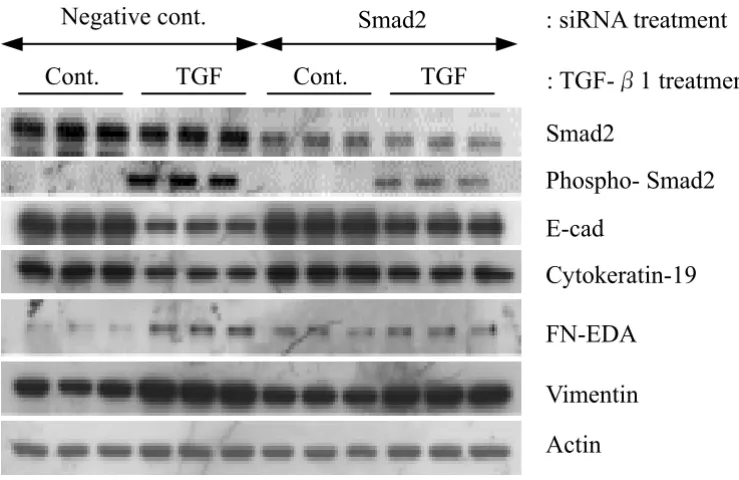
siRNA-mediated Smad2 gene silencing inhibits TGF-β -mediated EMT
In order to confirm whether Smad2 is involved in TGF-β 1-mediated EMT, siRNAs were used to silence Smad2 gene expression in A549 cells. Transfection efficiency, using FITC-conjugated dsRNA oligomers, was approximately 100 % (data not shown). Western analysis revealed that total Smad2 protein expression was significantly (P < 0.01) depleted by Smad2 siRNA gene silencing, while pooled negative control siRNA had no detectable effect on Smad2 protein expression (Figure 9). Using a phospho-specific antibody against Smad2, we also tested for the effect of Smad2 siRNA on phosphorylated Smad2 levels after TGF-β1 treatment. In the presence of TGF-β1 phos-phorylated Smad2 levels were significantly (P < 0.01) diminished by Smad2 siRNA (Figure 9). Most impor-tantly, Smad2 siRNA restored the decreased expression of E-cad and cytokeration 19 induced by TGF-β1 treatment. However, the expression of Fn-EDA and vimentin were only slightly suppressed by Smad2 siRNA (Figure 9). Nev-ertheless, our data suggest that the activation of Smad2 signaling pathway is involved in TGF-β1-mediated EMT in A549 cells.
Discussion
Fibroblastic foci in the IPF lung parenchyma are character-ized by vigorous replication of mesenchymal cells, includ-ing fibroblasts and myofibroblasts, and subsequent abnormal deposition of ECM proteins [25]. However, pre-cise mechanisms responsible for the formation of fibrob-lastic foci are unknown and their cellular origins are unclear. It is recognized though, that injury to the alveolar epithelium precedes their formation and that epithelial cells may therefore contribute to their formation, perhaps by their responses to, or production of, key fibrogenic mediators including TGF-β1 [3]. Thus, one possible role of TGF-β1 in IPF is to induce or promote epithelial cells in injured alveolar epithelium to undergo EMT [15], as is evi-dent from studies of other fibrotic disorders such as renal fibrosis [10,11,14]. Cells that have undergone EMT would then contribute to the development of fibroblastic foci and to their abnormal ECM production. The results of our present in vitro study support the concept underpinning this hypothesis; namely that TGF-β1 can induce alveolar EMT in human lung epithelial cells via the Smad2 pathway.
[image:8.612.131.478.150.266.2]Comparison of EMT-related marker expression in response to TGF-β1, IL-1β and TNF-α treatments
Figure 3
Comparison of EMT-related marker expression in response to TGF-β1, IL-1β and TNF-α treatments. A549 cells
were incubated with TGF-β1, IL-1β and TNF-α at the indicated concentrations for 48 h. Only TGF-β1 decreases E-cadherin
expression concomitant with increasing Fn-EDA expression. In contrast, TNF-α only slightly decreases E-cadherin expression
and IL-1β has no influence on EMT-related marker expression. Equal amounts of total protein are loaded in each lane.
20 10 5
Control
20
10
5
20
10
5
TGF-
ȕ
1
IL-1
ȕ
TNF-
ǩ
TGF-β1 induces the expression of collagens type I and type III
Figure 4
TGF-β1 induces the expression of collagens type I and type III. A549 cells were incubated with 5 ng/ml of TGF-β1 in
the presence of 5 µg/ml of L-ascorbic acid for 72 h. (A) mRNA expression of collagens type I and type III was detected using
RT-PCR. Densitometric analysis was performed. The changes of expression level are expressed as fold increase compared to the control. Each bar represents the mean ± SD of three independent experiments. ** P < 0.01. (B) Protein expression of col-lagens type I and type III was detected by immunocytochemical staining. Panels (a) and (b) represent collagen type I and panels
(c) and (d) represent collagen type III expression, respectively. TGF-β1 induces collagen type I and type III expression in A549
as shown in panel (b) and (d).
A
Control TGF-E1 0.00
0.25 0.50 0.75 1.00 1.25 1.50 1.75
es
si
o
n **
Collagen type I
Rat
io
of
m
R
N
A
ex
pr
(-fo
ld
)
Control TGF-ȕ1 Type I
Type III
GAPDH
Control TGF-E1 0.00
0.25 0.50 0.75 1.00 1.25 1.50
1.75 **
Collagen type III
Ra
ti
o
of
m
RNA e
x
p
re
s
s
ion
(-fo
ld
)
B
b a
Epithelial phenotype is determined by the specific range of proteins expressed by the epithelial cell and the nature of its environment [14,26,27]. E-cad is an epithelial cell transmembrane protein with conserved cadherin repeats in the extracellular domain. In the presence of Ca2+, the
extracellular domain binds to that of E-cad on an adjacent epithelial cell to form tight cell-cell adhesion and to sup-press the dissociation of epithelial cells from their location. Certain morphogenic and/or environmental cues, such as local expression of TGF-β1, result in the loss of epithelial cell polarity, adherens junctions, tight junc-tions, desmosomes, cytokeratin intermediate filaments and, subsequently, rearrangement of F-actin stress fibers and the development of filopodia and lamelopodia [10,11]. Several in vitro studies have demonstrated that
addition of TGF-β1 to cultured human epithelial cells from organs other than lung induces them to downregulate E-cad expression and to become mesenchy-mal cells, resembling myofibroblasts, via EMT [27-29].
In the present study, we investigated the potential for alve-olar EMT by examining the expression of phenotypic markers in A549 cells. Although submerged monolayer culture of A549 may not completely mimic the pulmo-nary epithelium, these cells are still widely used for studies on the role of human alveolar type II epithelial cells as they retain features and metabolic properties characteris-tic of type II cells [17,19,30]. The present study demonstrates that low doses of TGF-β1 induce A549 cells to lose expression of epithelial phenotypic markers, such
[image:10.612.108.489.142.406.2]TGF-β1 increases collagen type IV secretion in A549
Figure 5
TGF-β1 increases collagen type IV secretion in A549. A549 cells were incubated with several concentrations of TGF-β1
in the presence of 5 µg/ml of L-ascorbic acid for 72 h. Concentrations of collagen type IV in conditioned media were
deter-mined using ELISA. TGF-β1 increases collagen type IV in a concentration-dependent manner. Each bar was expressed as the
mean ± SD of four independent experiments. ** P < 0.01.
0
0.01
0.1
1
5
10
0
25
50
75
100
125
150
175
ty
p
e
IV
ml
)
**
**
**
**
TGF-
E
1 concentration (ng/ml)
C
o
lla
g
e
n
as E-cad and cytokeratin 19 expression, similar to our pre-viously reported findings in kidney epithelial EMT [14]. Concurrently A549 cells gain a mesenchymal phenotype with de novo expression of Fn-EDA and increased expres-sion of vimentin. In addition to these classic mesenchy-mal markers, these newly formed mesenchymesenchy-mal cells expressed the fibrillar collagens type I and type III which are likely to contribute to excessive accumulation of ECM in fibrotic tissue. Moreover, these cells expressed an ele-vated level of MMP-2 after EMT which could contribute to breakdown of basement membranes and facilitate migra-tion. Our data is consistent with the findings made in in kidney tubular epithelial cells where MMP-2 and MMP-9 were up-regulated by TGF-β1 in parallel with changes in EMT markers [27,31]. It remains uncertain whether these fibroblast-like cells possess migratory and/or invasive capabilities, but TGF-β1 induced MMP-2 expression in A549 supports this notion. Clearly further investigation of cell migration in TGF-β1-mediated alveolar EMT in lung fibrosis is warranted.
We have yet to confirm our results in primary human type II alveolar epithelial cells because cultures of these cells rapidly adopt fibroblast-like morphology, even in the absence of TGF-β1. Therefore, we cannot rule out at this stage the possibility that the responses of A549 cells to TGF-β1 are unique to this human cell line. However, a recent report indicates that rat primary type II alveolar epithelial cells undergo EMT in vitro in response to treat-ment of TGF-β1 [15], suggesting EMT is likely to be a phe-nomenon common to all type II alveolar epithelial cells.
In addition to TGF-β1, many other cytokines, including IL-1β and TNF-α, have been suggested to play a role in IPF [4]. We examined whether, like TGF-β1, IL-1β and TNF-α also convert A549 to fibroblast-like cells. It is widely accepted that IL-1β induces EMT of renal epithelial cells through a TGF-β1-dependent mechanism [20,21]. How-ever, our results showed that both TNFα and IL-1β failed to induce alveolar epithelial cells to undergo EMT, possi-bly due to the differences in the cell types investigated. Similarly A549 cells failed to respond to stimulation with 30% (v/v) activated PBMC-conditioned medium
[image:11.612.133.472.110.320.2]TGF-β1 induces CTGF expression in A549
Figure 6
TGF-β1 induces CTGF expression in A549. A549 cells were incubated for the times indicated with the concentrations of
TGF-β1 shown. Cell lysates were used as a source for Western blot analysis of CTGF expression. Up-regulation of CTGF
expression is observed with 1 ng/ml and higher concentrations of TGF-β1. This phenomenon parallels the altered expression
of EMT related markers. Representative blots are shown from three independent experiments.
TGF-
ȕ
1 (ng/ml)
0
0.01
0.1
1
5
10
37kD
37kD
37kD
24h
48h
(aPBMC-CM) (H. Kasai, unpublished data), a stimulus which induces renal epithelial cells to undergo EMT-mediated conversion to myofibroblasts [14,32,33].
We observed that A549 EMT was not accompanied by expression of αSMA, even during longer periods (7 days) of exposure to TGF-β1 (data not shown). Although αSMA, together with vimentin and desmin, is often used to clas-sify myofibroblasts [34,35], the expression of these cellu-lar markers is varied and dependent on cell types and culture conditions [36].
TGF-β1 regulates various cell functions, such as cell prolif-eration, cell differentiation, apoptosis, cell adhesion/ motility, ECM production, and its association with pulmonary fibrosis is well known [37]. In vivo studies have demonstrated increased TGF-β1 gene expression and protein secretion in the lungs of animals [38] and humans with fibrotic diseases [7,8,39]. Furthermore, transient overexpression of active TGF-β1 in rat lung resulted in severe interstitial and pleural fibrosis characterized by extensive deposition of ECM proteins, and by the emer-gence of cells with the myofibroblast phenotype [9]. A time-dependent production of endogenous TGF-β1 from rat alveolar epithelial cells after exogenous TGF-β1 treat-ment was also noted by Yao et al [15], suggesting initial
EMT induced by TGF-β1 may result in further EMT
induced by endogenous TGF-β1 production in an
auto-crine or paraauto-crine manner. Based on these studies and our data it is tempting to speculate about a role for TGF-β 1-mediated EMT in pulmonary fibrogenesis. Indirectly, our data showing TGF-β1-mediated induction of collagen type I and type III expression, which are present in fibrotic lesions in vivo [40], and the expression of Fn-EDA and vimentin which represent cellular markers for myofibrob-lasts, supports such a role for EMT.
TGF-β1 exerts its effects through heteromeric receptor complexes composed of type I and type II serine/threo-nine receptors. Upon ligand binding, the type II receptor phosphorylates the type I receptor inducing its kinase activity [41]. TGF-β1 activity may be transduced along the Smads pathway immediately downstream of the receptor complex in a variety of cell systems and also via JNK, p38 MAPK and Erk pathways [37,41,42]. Thus, we examined the intracellular signalling pathway involved in TGF-β 1-mediated alveolar EMT. Although phosphorylation and activation of p38 MAPK and Erk1/2 were observed in some lung fibroblasts [43], our data suggested that the sig-nalling pathway involved in alveolar EMT was likely to be a Smad2-dependent pathway since the MEK inhibitors, PD98059 and U0126, failed to reverse TGF-β1-induced phenotypic modulation of A549 cells, whereas Smad2 siRNA attenuated the loss of E-cad and cytokeratin 19
induced by TGF-β1. Smad2 phosphorylation has been
[image:12.612.154.477.120.276.2]Effect of TGF-β1 on MMPs expression in A549
Figure 7
Effect of TGF-β1 on MMPs expression in A549. Gelatin zymography was performed using the conditioned media that
were harvested after 48 h TGF-β1 treatment (0.1 to 5 ng/ml). The samples were applied without reduction to a 10%
polyacry-lamide gel containing gelatin, and proteolytic activity was demonstrated by digestion of the gelatin and clearing of the gel.
(kD)
0 0.1 1
5
: TGF-
ȕ
1 (ng/ml)
100
MMP-2
MMP-9
noted in EMT processes for several cell types including breast and renal epithelial cells [44-46].
Transition from the alveolar epithelial phenotype to the mesenchymal phenotype initiated by TGF-β1 was accom-panied by elevated expression of CTGF. CTGF induction is
mediated through a TGF-β1 response element in the
CTGF promoter and its mediation of at least some of the activities attributed to TGF-β1 is well recognized [4,22], as is its involvement in the maintenance of fibrogenesis. In lung for example, Allen et al reported that the mRNA level
of CTGF in BALF cells of patients with IPF was signifi-cantly higher than that in healthy control subjects [46]. Furthermore, expression of CTGF was found in both interstitial fibroblasts and type II alveolar epithelial cells in patients with IPF [47]. CTGF secreted by alveolar epi-thelial cells and myofibroblasts responding to TGF-β1 may also act as a paracrine factor for lung fibroblasts and is known to be a critical intermediate for the synthesis of connective tissue proteins stimulated by TGF-β1, but not by other fibrogenic cytokines [48,49]. CTGF may therefore play a role in mediating the expression of
colla-Activation of Smad and ERK1/2 pathways by TGF-β1
Figure 8
Activation of Smad and ERK1/2 pathways by TGF-β1. A549 cells were pre-incubated in the presence or the absence of
10 µM of U0126, a potent MEK inhibitor, for 1 h prior to TGF-β1 stimulation. 5 ng/ml of TGF-β1 was used as a stimulus.
TGF-β1 activates Erk1/2 and Smad2 pathways within 5 min after stimulation. The MEK inhibitor blocks Erk1/2 phosphorylation, but
does not influence Smad2 phosphorylation. Equal amounts of total protein are loaded in each lane.
0
5 15 30 60 240 0
5 15 30 60 240
U0126
DMSO
min
A
phospho-Erk1/2
total-Erk1/2
phospho-Smad2
total-Smad2
+ + +
+
㧙
㧙
:TGF-
ȕ
1 (5 ng/ml
㧕
B
0.1 1 10 10
㧙
㧙
:U0126 (
P
M)
E-cad
Fn-EDA
gens by EMT cells. Most recently, investigations with mesangial cells showed that CTGF interacts with the TrkA receptor, triggering events which lead to the induction of a transcriptional repressor, TIEG [50]. It was proposed that since this suppresses negative regulation of the TGF-β-Smad signaling pathway by repressing Smad7 expres-sion, it leads to enhanced TGF-β1 signaling [51]. It is pres-ently unclear whether CTGF plays a similar role in alveolar epithelial responses to TGF-β1, or in EMT cells derived from them. However, in spite of uncertainties sur-rounding its precise role, CTGF remains implicated in the pathogenesis of many fibrotic disorders [4], and is likely to contribute significantly to fibrogenesis in the lung.
Conclusion
Our findings show that TGF-β1 induces an EMT-like proc-ess in A549 alveolar epithelial cells, most likely by activa-tion of the Smad2 signaling pathway. These data provide evidence to support the concept that human lung epithe-lial cells can undergo EMT and indicate a need for further studies. In particular, the expression profile associated with alveolar epithelial cells that have undergone EMT indicates a potential role for EMT in pulmonary fibrogenesis.
List of abbreviations
IPF = Idiopathic pulmonary fibrosis; αSMA = α-smooth muscle actin; ECM = extracellular matrix; TGF-β1 =
[image:14.612.122.492.153.393.2]trans-Effect of Smad2 siRNA on TGF-β1 induced EMT in A549 cells
Figure 9
Effect of Smad2 siRNA on TGF-β1 induced EMT in A549 cells. Pooled synthetic siRNA duplexes targeting different regions of Smad2 were transfected into A549 cells at 50 pmol per well. 24 h after transfection, cells were stimulated with 5 ng/
ml of TGF-β1 in serum free 0.1% BSA/DMEM for a further 48 h prior to harvest. Equal amounts of lysates were resolved by
SDS-PAGE and analyzed by Western blotting for expression of proteins.
Negative cont.
Smad2
: siRNA treatment
TGF
Cont.
Cont.
TGF
: TGF-
Ǫ
1 treatment
Smad2
Phospho- Smad2
E-cad
Cytokeratin-19
FN-EDA
Vimentin
forming growth factor-beta1; IL-1β = interleukin-1 beta; TNF-α = tumor necrosis factor-alpha; EMT = epithelial mesenchymal transition; E-cad = E-cadherin; Fn-EDA = fibronectin EDA+ splice form; CTGF = connective tissue
growth factor; MEK = MAPK/Erk kinase; RT-PCR = Reverse Transcription PCR; MMP = matrix metalloproteinase; siRNA = small interfering RNA
Authors' contributions
HK carried out the cellular and biochemical studies and participated in drafting the manuscript. JA, RM and TK participated in the design of the study and drafted the manuscript. ZZ conceived the study, and participated in its design and coordination, and in drafting and finalizing the manuscript. All authors read and approved the final manuscript.
Acknowledgements
This work was supported by the MRC Technology and Teijin Pharma Ltd.
References
1. Selman M, King TE, Pardo A: Idiopathic pulmonary fibrosis: pre-vailing and evolving hypotheses about its pathogenesis and implications for therapy. Ann Intern Med 2001, 134:136-151. 2. King TE Jr, Schwarz MI, Brown K, Tooze JA, Colby TV, Waldron JA
Jr, Flint A, Thurlbeck W, Cherniack RM: Idiopathic pulmonary fibrosis: relationship between histopathologic features and mortality. Am J Respir Crit Care Med 2001, 164:1025-1032. 3. Selman M, Pardo A: Idiopathic pulmonary fibrosis: an epithelial/
fibroblastic cross-talk disorder. Respir Res 2002, 3:3.
4. Allen JT, Spiteri MA: Growth factors in idiopathic pulmonary fibrosis: relative roles. Respir Res 2002, 3:13.
5. Gauldie J, Kolb M, Sime PJ: A new direction in the pathogenesis of idiopathic pulmonary fibrosis? Respir Res 2002, 3:1. 6. Kelly M, Kolb M, Bonniaud P, Gauldie J: Re-evaluation of
fibro-genic cytokines in lung fibrosis. Curr Pharm Des 2003, 9:39-49. 7. Khalil N, O'Connor RN, Unruh HW, Warren PW, Flanders KC,
Kemp A, Bereznay OH, Greenberg AH: Increased production and immunohistochemical localization of transforming growth factor-beta in idiopathic pulmonary fibrosis. Am J Respir Cell Mol Biol 1991, 5:155-162.
8. Khalil N, Parekh TV, O'Connor R, Antman N, Kepron W, Yehaulaeshet T, Xu YD, Gold LI: Regulation of the effects of TGF-beta 1 by activation of latent TGF-beta 1 and differen-tial expression of TGF-beta receptors (T beta R-I and T beta R-II) in idiopathic pulmonary fibrosis. Thorax 2001, 56:907-915. 9. Sime PJ, Xing Z, Graham FL, Csaky KG, Gauldie J: Adenovector-mediated Gene Transfer of Active Transforming Growth Factor-β1 Induces Prolonged Severe Fibrosis in Rat Lung. J Clin Invest 1997, 100:768-776.
10. Kalluri R, Neilson EG: Epithelial-mesenchymal transition and its implications for fibrosis. J Clin Invest 2003, 112:1776-1784. 11. Liu Y: Epithelial to mesenchymal transition in renal
fibrogen-esis: pathologic significance, molecular mechanism, and therapeutic intervention. J Am Soc Nephrol 2004, 15:1-12. 12. Desmouliere A, Darby IA, Gabbiani G: Normal and pathologic
soft tissue remodeling: role of the myofibroblast, with spe-cial emphasis on liver and kidney fibrosis. Lab Invest 2003,
83:1689-1707.
13. Desmouliere A: Factors influencing myofibroblast differentia-tion during wound healing and fibrosis. Cell Biol Int 1995,
19:471-476.
14. Nightingale J, Patel S, Suzuki N, Buxton R, Takagi KI, Suzuki J, Sumi Y, Imaizumi A, Mason RM, Zhang Z: Oncostatin M, a cytokine released by activated mononuclear cells, induces epithelial cell-myofibroblast transdifferentiation via Jak/Stat pathway activation. J Am Soc Nephrol 2004, 15:21-32.
15. Yao HW, Xie QM, Chen JQ, Deng YM, Tang HF: TGF-beta1 induces alveolar epithelial to mesenchymal transition in vitro. Life Sci 2004, 76:29-37.
16. Vleminckx K, Kemler R: Cadherins and tissue formation: inte-grating adhesion and signaling. Bioessays 1999, 21:211-220. 17. Higashimoto Y, Keicho N, Elliott WM, Hogg JC, Hayashi S: Effect of
adenovirus E1A on ICAM-1 promoter activity in human alve-olar and bronchial epithelial cells. Gene Expr 1999, 8:287-297. 18. Maeyama T, Kuwano K, Kawasaki M, Kunitake R, Hagimoto N,
Mat-suba T, Yoshimi M, Inoshima I, Yoshida K, Hara N: Upregulation of Fas-signalling molecules in lung epithelial cells from patients with idiopathic pulmonary fibrosis. Eur Respir J 2001,
17:180-189.
19. Upadhyay D, Bundesmann M, Panduri V, Correa-Meyer E, Kamp DW:
Fibroblast growth factor-10 attenuates H2O2-induced
alveo-lar epithelial cell DNA damage: role of MAPK activation and DNA repair. Am J Respir Cell Mol Biol 2004, 31:107-113.
20. Vesey DA, Cheung CWY, Cuttle L, Endre Z, Gobe G, Johnson DW:
Interleukin-1β induces human proximal tubule cell injury, α -smooth muscle actin expression and fibronectin production. Kidney Int 2002, 62:31-40.
21. Fan J, Huang X, Ng Y, Nikolic-Paterson DJ, Mu W, Atkins RC, Lan HY:
Interleukin-1 induces tubular epithelial-myofibroblast transdifferentiation through a transforming growth
factor-β1-dependent mechanism in vitro. Am J Kidney Dis 2001,
37:820-831.
22. Bonniaud P, Margetts PJ, Kolb M, Haberberger T, Kelly M, Robertson J, Gauldie J: Adenoviral gene transfer of connective tissue growth factor in the lung induces transient fibrosis. Am J Respir Crit Care Med 2003, 168:770-778.
23. Wahab NA, Yevdokimova N, Weston BS, Roberts T, Li XJ, Brinkman H, Mason RM: Role of connective tissue growth factor in the pathogenesis of diabetic nephropathy. Biochem J 2001,
35:77-87.
24. Bottinger EP, Bitzer M: TGF-beta signaling in renal disease. J Am Soc Nephrol 2002, 13:2600-2610.
25. Gross TJ, Hunninghake GW: Idiopathic pulmonary fibrosis. N Engl J Med 2001, 345:517-525.
26. Ewing CM, Ru N, Morton RA, Robinson JC, Wheelock MJ, Johnson KR, Barrett JC, Isaacs WB: Chromosome 5 suppresses tumori-genicity of PC3 prostate cancer cells: correction with re-expression of alpha-catenin and restoration of E-cadherin function. Cancer Res 1995, 55:4813-4817.
27. Yang J, Liu Y: Dissection of key events in tubular epithelial to myofibroblast transition and its implications in renal intersti-tial fibrosis. Am J Pathol 2001, 159:1465-1475.
28. Miettinen PJ, Ebner R, Lopez AR, Derynck R: TGF-beta induced transdifferentiation of mammary epithelial cells to mesen-chymal cells: involvement of type I receptors. J Cell Biol 1994,
127:2021-2036.
29. Bhowmick NA, Ghiassi M, Bakin A, Aakre M, Lundquist CA, Engel ME, Arteaga CL, Moses HL: Transforming growth factor-beta1 mediates epithelial to mesenchymal transdifferentiation through a RhoA-dependent mechanism. Mol Biol Cell 2001,
12:27-36.
30. Foster KA, Oster CG, Mayer MM, Avery ML, Audus KL: Character-ization of the A549 cell line as a type II pulmonary epithelial cell model for drug metabolism. Exp Cell Res 1998, 243:359-366. 31. Strutz F, Zeisberg M, Ziyadeh FN, Yang CQ, Kalluri R, Muller GA, Neilson EG: Role of basic fibroblast growth factor-2 in epithe-lial-mesenchymal transformation. Kidney Int 2002,
61:1714-1728.
32. Sharmila P, Takagi K, Suzuki J, Imaizumi A, Kimura T, Mason RM, Kamimura T, Zhang Z: RhoGTPase activation is a key step in renal epithelial mesenchymal transdifferentiation. J Am Soc Nephrol 2005, 16(7):1977-1984.
33. Healy E, Leonard M, Madrigal-Estebas L, O'Farrelly C, Watson AJ, Ryan MP: Factors produced by activated leukocytes alter renal epithelial cell differentiation. Kidney Int 1999,
56:1266-1269.
34. Mermall V, Post PL, Mooseker MS: Unconventional myosins in cell movement, membrane traffic, and signal transduction. Science 1998, 279:527-533.
35. Powell DW, Mifflin RC, Valentich JD, Crowe SE, Saada JI, West AB:
Publish with BioMed Central and every scientist can read your work free of charge "BioMed Central will be the most significant development for disseminating the results of biomedical researc h in our lifetime."
Sir Paul Nurse, Cancer Research UK
Your research papers will be:
available free of charge to the entire biomedical community peer reviewed and published immediately upon acceptance cited in PubMed and archived on PubMed Central yours — you keep the copyright
Submit your manuscript here:
http://www.biomedcentral.com/info/publishing_adv.asp
BioMedcentral
36. Powell DW, Mifflin RC, Valentich JD, Crowe SE, Saada JI, West AB:
Myofibroblasts. II. Intestinal subepithelial myofibroblasts. Am J Physiol 1999, 277:C183-C201.
37. Derynck R, Feng XH: TGF-β receptor signaling. Biochim Biophys Acta 1997, 1333:F105-F150.
38. Hoyt DG., Lazo JS: Alterations in pulmonary mRNA encoding procollagens, fibronectin, and transforming growth factor-β precedes bleomycin-induced pulmonary fibrosis in mice. J Pharmacol Exp Ther 1988, 246:765-771.
39. Khalil N, O'Conner RN, Flanders KC, Umruh H: TGF-β1, but not TGF-β2 or TGF-β3, is differentially present in epithelial cells of advanced pulmonary fibrosis: an immunohistochemical study. Am J Respir Cell Mol Biol 1996, 14:131-138.
40. Raghu G, Masta S, Meyers D, Narayanan AS: Collagen synthesis by normal and fibrotic human lung fibroblasts and the effect of transforming growth factor-beta. Am Rev Respir Dis 1989,
140:95-100.
41. Whitman M: Smads and early developmental signaling by the TGFβ superfamily. Genes Dev 1998, 12:2445-2462.
42. Atfi A, Djellou S, Chastre E, Davis R, Gespach C: Evidence for a role of Rho-like GTPase and stress-activated protein kinase/ c-Jun-N-terminal kinase (SAPK/JNK) in transforming growth factorβ-mediated signaling. J Biol Chem 1997, 272:1429-1432. 43. Hashimoto S, Gon Y, Takeshita I, Matsumoto K, Maruoka S, Horie T:
Transforming growth factor-β1 induces phenotypic modula-tion of human lung fibroblasts to myofibroblast through a c-Jun-NH2-terminal kinase-dependent pathway. Am J Respir Crit
Care Med 2001, 163:152-157.
44. Piek E, Moustakas A, Kurisaki A, Heldin CH, ten Dijke P: TGF-(beta) type I receptor/ALK-5 and Smad proteins mediate epithelial to mesenchymal transdifferentation in NMuMG breast epi-thelial cells. J Cell Sci 1999, 112:4557-4568.
45. Rhyu DY, Yang Y, Ha L, Lee GT, Song JS, Uh ST, Lee HB: Role of reactive oxygen species in TGF-beta1-induced mitogen-acti-vated protein kinase activation and epithelial-mesenchymal transition in renal tubular epitheliasl cells. J Am Soc Nephrol
2005, 16:667-675.
46. Allen JT, Knight RA, Bloor CA, Spiteri MA: Enhanced insulin-like growth factor binding protein-related protein 2 (Connective tissue growth factor) expression in patients with idiopathic pulmonary fibrosis and pulmonary sarcoidosis. Am J Respir Cell Mol Biol 1999, 21:693-700.
47. Pan LH, Yamauchi K, Uzuki M, Nakanishi T, Takigawa M, Inoue H, Sawai T: Type II alveolar epithelial cells and interstitial fibrob-lasts express connective tissue growth factor in IPF. Eur Respir J 2001, 17:1220-1227.
48. Grotendorst GR: Connective tissue growth factor: a mediator of TGF-beta action on fibroblasts. Cytokine Growth Factor Rev
1997, 8:171-179.
49. Moussad EE, Brigstock DR: Connective tissue growth factor: what's in a name? Mol Genet Metab 2000, 71:276-292.
50. Wahab NA, Weston BS, Mason RM: Connective tissue growth factor (CTGF, CCN2) interacts with and activates the tyro-sine kinase receptor TrkA. J Am Soc Nephrol 2005, 16:340-351. 51. Wahab NA, Mason RM: Connective tissue growth factor and