0095-1137/96/$04.0010
Copyrightq1996, American Society for Microbiology
Analysis of HL and O Serotypes of Campylobacter Strains by
the Flagellin Gene Typing System
IRVING NACHAMKIN,1* HUONG UNG,1
ANDCHARLOTTE M. PATTON2
Department of Pathology and Laboratory Medicine, University of Pennsylvania School of Medicine, Philadelphia, Pennsylvania 19104,1and Foodborne Diseases Laboratory Section, Centers for Disease Control
and Prevention, Atlanta, Georgia 303332
Received 27 July 1995/Returned for modification 4 October 1995/Accepted 26 October 1995
We recently developed a molecular typing system forCampylobacter jejuniandCampylobacter colibased on
restriction fragment length polymorphism analysis of the flagellin gene,flaA(I. Nachamkin, K. Bohachick, and
C. M. Patton, J. Clin. Microbiol. 31:1531–1536, 1993). We extended the typing system to 83 flagellin types
(designatedflaA-1,flaA-2, etc.) on the basis of analysis of 404 isolates ofC. jejuniandC. coliincluding common
serotypes isolated in the United States, a selection of less common serotypes, and serotype reference strains. Of the 295 strains previously shown to belong to common HL and O serotypes (C. M. Patton, M. A. Nicholson,
S. M. Ostroff, A. A. Ries, I. K. Wachsmuth, and R. V. Tauxe, J. Clin. Microbiol. 31:1525–1530, 1993), sixflaA
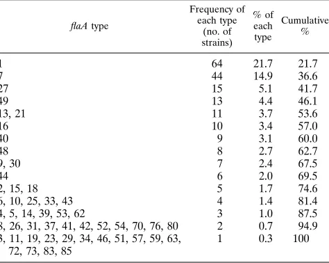
types accounted for 53.6% of strains as follows:flaA-1, 21.7%; flaA-7, 14.9%; flaA-27, 5.1%; flaA-49, 4.4%;
flaA-13, 3.7%; andflaA-21, 3.7%. Seventy-five percent of the strains were within 15flaAtypes, 90% were within
30flaAtypes, and all 295 strains were contained within 52flaAtypes. Within each HL or O serotype, there
usually were multipleflaAtypes. For 12 common HL serotypes and 7 common O serotypes, more than 50% of
these isolates were a singleflaAtype. A database was developed by using commercially available restriction
fragment length polymorphism analysis software (ProRFLP; DNA ProScan, Inc., Nashville, Tenn.) that should allow other investigators to perform typing with this system.
Campylobacter jejuni subsp. jejuni (referred to as C. jejuni) is
one of the most common causes of bacterial gastroenteritis in the United States and worldwide, with an estimated annual incidence of 2 million cases in the United States (15). C. jejuni typically causes an acute enterocolitis accompanied by fever and abdominal cramping lasting 3 to 5 days. Although compli-cations are infrequent, campylobacters may cause reactive ar-thritis and infections may be confused with acute appendicitis; resulting in unnecessary surgery, and there is increasing evi-dence that Campylobacter infection is associated with the
de-velopment of Guillain-Barre´ syndrome (5, 6). Although deaths
associated with campylobacter infection are uncommon, the morbidity due to the disease is significant and results in sub-stantial economic costs (13).
Most C. jejuni cases in the United States occur sporadically; however, common-source outbreaks do occur. Campylobacter infections are acquired by ingestion of contaminated food, water, or milk, and although numerous vehicles for the trans-mission of Campylobacter infection have been described, chicken appears to be the single most important vehicle for sporadic cases in the United States and other developed na-tions (14, 15). Other reservoirs for the organism include tur-keys, cattle, swine, goats, ducks, and sheep (14). Poultry can become contaminated with Campylobacter strains at various stages of production by endogenous or exogenous sources (12, 14). A system for tracking specific strains or serotypes of
Campylobacter strains would be useful for studying the
epide-miology of human infections, analyzing modes of transmission, and devising and assessing intervention strategies important in agriculture.
Serotyping and other means of typing Campylobacter spp.
have played an important role in understanding the epidemi-ology and identifying the sources of Campylobacter infection. Numerous typing schemes have been developed, and the use-fulness of these methods has been reviewed (9). The most commonly used methods today include serotyping to detect heat-labile (HL) antigens (4) or O (formerly called heat-sta-ble) antigens (11). These methods have been applied widely in epidemiologic investigations (9, 10), but they require appro-priate serotyping reagents that are expensive and extraordi-narily time-consuming to produce. Serotyping methods are moderately complex, yet they have an excellent ability to dis-criminate among unrelated strains (9). However, only a few reference laboratories can provide serotyping, and such ser-vices are not readily available to investigators. A widely avail-able alternative method that would be simple to perform, that would not require specialized reagents (i.e., antisera), and that could be used for many applications is highly desirable (3, 9). Molecular methods have increasingly been used as alterna-tives to serologic methods to type Campylobacter strains. We recently developed a typing system based on restriction frag-ment length polymorphism (RFLP) analysis of the flagellin gene, flaA, in C. jejuni and C. coli (7). This system is referred to as flagellin gene typing, and each RFLP pattern has a des-ignated flaA type (flaA-1, flaA-2, etc). We showed that flagellin gene typing correlated with the HL and the O serotypes when analyzing outbreak-related strains and also discriminated be-tween strains within a particular serotype (7). The purpose of the current study was (i) to extend the initial flagellin typing system by using a large collection of isolates, (ii) to examine previously analyzed strains to identify the most common sero-types of C. jejuni and C. coli isolated in the United States, and (iii) to establish the distributions of common flaA types in the United States. We further developed a database using com-mercially available image analysis software that can be used by other investigators to type strains.
* Corresponding author. Mailing address: University of Pennsylva-nia, Department of Pathology and Laboratory Medicine, 3400 Spruce St., Philadelphia, PA 19104-4283. Phone: (215) 662-6651. Fax: (215) 662-6655. Electronic mail address: [email protected].
277
on May 15, 2020 by guest
http://jcm.asm.org/
MATERIALS AND METHODS
Bacterial strains.All 404 Campylobacter isolates were obtained from the Campylobacter Reference Laboratory at the Centers for Disease Control and Prevention. Isolates included C. jejuni (n5322), hippurate-negative C. jejuni/C.
coli (n521), C. coli (n514), C. lari (n53), and 44 C. jejuni, C. coli, and C. lari HL reference strains. The hippurate-negative group of C. jejuni/C. coli strains are grouped separately since they could not be differentiated on the basis of pheno-typic studies. The majority of strains were from human sources; 32 isolates were from animals, and 1 was from cow’s milk. Two-hundred ninety-five of the strains were characterized previously and represented a random sample of strains from humans with sporadic cases of infection in the United States (8). Briefly, these strains were collected between July 1989 and June 1990 and came from individ-uals with diarrheal illness who resided in 1 of 19 counties representing all nine standard census regions of the United States plus California. Additional strains represented less common HL and O serotypes and serotype reference strains. Sixty-five of the isolates, previously typed with the flagellin typing system (7), were reanalyzed by the modified method described below.
Serotyping.Serotyping was performed by using both O and HL typing systems (4, 11) as described previously (8).
Flagellin gene typing.Flagellin gene typing was performed as described pre-viously, with some modifications (7). Bacteria were grown on Trypticase soy agar with 5% sheep blood (Becton Dickinson) at 428C overnight under microaerobic conditions in a Tri-Gas incubator (Forma Scientific, Marietta, Ohio). Using a 10-ml loop, a small loopful of bacteria was resuspended in 0.2 ml of sterile water, vortexed, and heated for 5 min in a boiling water bath. After centrifugation at 14,0003g for 5 min (Eppendorf Microfuge, model 5415C), the clear
superna-tant was used as a template for further testing. If a yellow tinge was observed in the supernatant, presumably from bacterial proteins, this usually represented too much inoculum and a new extract was prepared.
PCR was performed with the following reagents at the indicated final concen-trations (total final volume, 100ml): PCR buffer (13), MgCl2(1.5 mM), forward and reverse primers (1mM), deoxynucleoside triphosphates (200mM); Ultrapure dNTP set; Pharmacia Biotech), 27-2033-01; and Taq polymerase (2.5 U; 5 U/ml; 17038-018; Gibco BRL). For each PCR, 5ml of template was used.
The forward primer sequence was 59-GGATTTCGTATTAACACAAAT GGTGC-39, and the reverse primer sequence was 59-CTGTAGTAATCTTAA AACATTTTG-39. PCR was performed with a Gene AMP PCR System 9600 (Perkin-Elmer Cetus) with the following cycling conditions: 948C for 1 min and then 948C for 15 s, 558C for 45 s, and 728C for 1 min and 45 s for 35 cycles. After the last cycle, the sample was held at 728C for 5 min. Five microliters from the PCR mixture was first checked for the presence of the expected 1.7-kb amplicon representing the Campylobacter flagellin gene by electrophoresis on a 0.8% agarose minigel. If a strong band was obtained, 8ml of the PCR product (con-taining ca. 6mg of DNA based on the basis of the optical density at 260 nm) was digested with the restriction enzyme DdeI (175S; New England Biolabs), accord-ing to the manufacturer’s instructions, with 1.5ml of 103DdeI buffer, 5ml of sterile water, and 0.5 ofml of DdeI enzyme, and the mixture was incubated at 378C for 4 to 6 h. The DdeI-digested product was analyzed by agarose gel electrophoresis by using 4% Nusieve GTG Agarose (FMC Bioproducts, Rock-land, Maine) in 13Tris-acetate (TAE) buffer (0.04 M Tris-acetate, 0.001 M EDTA). One-hundred milliliters of gel was prepared by the boiling method as
described by the manufacturer. After dissolving the gel, 100mg of ethidium bromide (10 mg/ml of stock) was added and poured into a gel casting tray (10 by 14 cm) with a 16-well comb (slots of 1 by 3 mm). The gel was cooled for at least 30 min at room temperature, and then enough 13TAE buffer was used to cover the gel and the gel was placed at 48C for 30 min prior to use. Samples were electrophoresed at 50 V for 16 h at room temperature by using 13TAE running buffer containing 0.5mg of ethidium bromide per ml. A 123-bp ladder (5613SA; Gibco BRL) was used as a molecular size standard. Under these conditions, the 123-bp marker ran within 1 cm of the gel bottom. The gels were examined with a UV transilluminator and were photographed with Polaroid type 57 high-speed 4-by-5 sheet film.
Image analysis.Computer analysis of the images was performed by using the software program Pro-RFLP Macintosh, version 2.0 (DNA ProScan, Inc., Nash-ville, Tenn.). Briefly, the photographs were scanned into the computer (Macin-tosh Quadra 800) by using a flatbed scanner (HP Scanjet IICX; Hewlett-Packard, Inc., Palo Alto, Calif.) and DeskScan II, version 2.0, software. The images were imported into Adobe Photoshop (LE version 2.5; Adobe Systems), where the images were adjusted for contrast and labeled. The photographs were analyzed by the Pro-RFLP software program and were processed according to the man-ufacturer’s instructions. During this process, lanes containing samples and stan-dards were manually identified and the bands were marked, and band sizes were automatically calculated by the software. Each unique pattern was saved in a Pro-RFLP image database file whereby new images could be compared with existing patterns (Campylobacter RFLP Database version 1.0;q1994 Trustees of the University of Pennsylvania). Pattern comparisons were performed at the 5% stringency level, with one-to-one matching; i.e., the pattern being searched had to have the same number of bands and each band had to be within65% of the band size stored in the database. If a match occurred, the image in the database was recalled to visually compare the two patterns to ensure matching. If a complete match did not occur, the unknown pattern was given a new type designation and was stored in the image database file for future comparisons.
RESULTS
Database.We analyzed 404 Campylobacter strains using the
flagellin gene typing system and identified 83 distinct flagellin types, designated flaA-1, flaA-2, etc. Sixty-seven flaA types were detected among C. jejuni strains, 17 flaA types were de-tected among C. coli strains, and 12 flaA types were dede-tected among C. jejuni/C. coli strains. None of the flaA types was found to be unique among the different species; however,
flaA-59 accounted for 15% of C. coli strains and 21.1% of C. jejuni/C. coli strains but was observed in only 1 (0.28%) C. jejuni strain.
Identification of common flaAtypes in the United States.
We analyzed 295 of the original 298 O and HL serotype strains isolated in the United States to determine the most common
flaA types (8). Six common flaA types emerged from the
anal-ysis of these strains; flaA-1, flaA-7, flaA-27, flaA-49, flaA-13, and flaA-21 accounted for 53.6% of the strains (Table 1). Seventy-five percent of the strains fell into 15 flaA types, and all 295 strains were typed within 52 flaA types. Thirty-seven
flaA types were infrequently encountered (fewer than five
oc-currences each) and accounted for approximately 25% of the strains tested. Within the 52 flaA types, there were 71 different HL serotypes and 74 different O serotypes.
The distribution of flaA types was correlated with the O serotype for each strain (Table 2). Within the O serotypes tested, in nine O serotypes at least 10 strains were represented. Among these strains, a single flaA type usually predominated, but other flaA types were represented. Some predominant flaA types occurred within multiple O serotypes, such as flaA-1 (O1, O1,8, O8, O8,17, O4 complex) and flaA-7 (O1, O1,8, O2). Within O19 strains, a single flaA type, flaA-21, occurred in 80% of the isolates. A predominant flaA type was not apparent in O:3 and O6,7,25 strains.
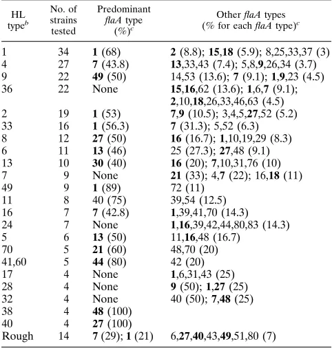
[image:2.612.60.297.102.291.2]The distribution of flaA types was also correlated with the HL serotype for each strain (Table 3). flaA-1 occurred with several HL serotypes (HL1, H2, HL33, HL49), as did flaA-7 (HL4, HL16). flaA-27 occurred in both HL8 and HL40 sero-types. A predominant flaA type was not observed in the HL36, HL7, HL24, HL17, HL28, and HL32 serotypes.
TABLE 1. Distributions of flaA types among 295
Campylobacter strains isolated in the United States containing
common HL and O serotypes
flaA type
Frequency of each type
(no. of strains)
% of each type
Cumulative %
1 64 21.7 21.7
7 44 14.9 36.6
27 15 5.1 41.7
49 13 4.4 46.1
13, 21 11 3.7 53.6
16 10 3.4 57.0
40 9 3.1 60.0
48 8 2.7 62.7
9, 30 7 2.4 67.5
44 6 2.0 69.5
2, 15, 18 5 1.7 74.6
6, 10, 25, 33, 43 4 1.4 81.4
4, 5, 14, 39, 53, 62 3 1.0 87.5
8, 26, 31, 37, 41, 42, 52, 54, 70, 76, 80 2 0.7 94.9 3, 11, 19, 23, 29, 34, 46, 51, 57, 59, 63,
72, 73, 83, 85
1 0.3 100
on May 15, 2020 by guest
http://jcm.asm.org/
We analyzed the distributions of the flaA types in the United States from a sample of strains obtained from different U.S. census regions. The most common flaA types were seen in every region of the country. Some unique flaA types were observed in particular regions; however, the numbers of strains with uncommon flaA types were small. In New England
(rep-resented by strains from Massachusetts; n5 17), flaA-3 was
observed. flaA types 11 and 34 were observed in the Mid-Atlantic region (represented by strains from New York State;
n 530), flaA types 29 and 46 were seen in the East
North-Central region (represented by strains from Illinois and
Wis-consin n543), flaA-80 was observed in the Mountain region
(represented by strains from Colorado; n533), flaA types 41
and 57 were observed in the Pacific region (represented by
strains from Washington and Oregon; n573), and flaA-19 was
observed in California (n517). Several regions did not show
any unique types, such as the West North-Central region
(Min-nesota and Missouri; n532), the East South-Central region
(Alabama; n 5 19), and the Southeast region (Virginia and
Georgia; n57).
DISCUSSION
In our initial description of the flagellin gene typing system, 24 flaA types were identified among non-outbreak-related and outbreak-related strains (7). In that study, flagellin gene typing showed a complete correlation with conventional serotyping in distinguishing outbreak-related from non-outbreak-related strains obtained from four different outbreaks. We have now identified 83 flaA types among a set of 404 isolates that in-cluded the originally described strains, additional reference serotype strains, and 295 strains representing the O and HL serotypes isolated in the United States (8). The results of the present study establish the distribution of common flaA types among strains of C. jejuni and some C. coli strains isolated in the United States.
Several important pieces of information emerged from our analysis of U.S. strains with this new typing system. First, a reference point for further epidemiologic studies of
Campy-lobacter infections by flagellin gene typing has been
estab-lished. Among the selection of serotypes in the United States, 52 flaA types were identified, and of these types, 15 accounted for 75% of the strains tested. flaA types 1 and 7 accounted for approximately one-third of the total number of isolates.
Second, the present study also confirms the heterogeneity of
Campylobacter strains isolated in the United States (1). Both
[image:3.612.60.559.82.232.2]common and uncommon flaA types were observed in all re-gions of the United States. We did note that certain flaA types were restricted to certain regions; however, the number of strains of each of these types was small. Surveys of isolates within particular geographic regions using flagellin gene typing would be of help in defining whether unique types circulate in particular parts of the country. Most O and HL serotypes were represented by multiple flaA types, but within a particular O or
TABLE 2. Distributions of flaA types among Campylobacter strains isolated in the United Statesacontaining common O serotypes
O typeb
No. of strains tested Predominant flaA type (%)c
Other flaA types (% for each flaA type)c
1 or 1,8 34 1(47); 7 (26) 5,9,52 (6); 26,27,51 (3)
4,13,16,43,50 33 1(30.3) 1,21(11.8); 2,6,7,15,18,21 (6); 4,16 (9.1); 8,9,39,41,83 (3)
8 or 8,17 29 1(58.6) 7(17); 9 (7); 3,5,30,62,72 (3.5)
4 27 1(44) 2(11); 18 (7.4); 6,15,25,31,33,37,39,42,44,80 (3.7)
5152 22 49(54) 14,53 (13.6); 7 (9); 1,23 (4.5)
2 20 7(65) 43 (15); 33 (10); 8,34 (5)
3 17 None 7,15,16,62 (12); 1,6,10,13,26,33,43,46,63 (6)
6,7,25(29)d 14 None 13(43); 25 (21); 48 (36)
19 10 21(80) 4,70 (10)
15,38 9 30(33); 16 (22) 7,10,31,76 (11)
11 9 27(55.6) 1,7,10,16 (11)
10,17 8 7(38) 1,30,37,70,80 (12.5)
21 or 21,29 7 27(85.7) 13(14.3)
23,36 6 13(50) 11,16,48 (16.7)
a
The strains were previously characterized by Patton et al. (8). b
O:52and O:25 are C. coli serotypes. The other serotypes listed are C. jejuni serotypes. c
Numbers in boldface type represent the 15 most common flaA types in the United States. d
Parentheses indicate that the presence of the antigen varies.
TABLE 3. Distribution of flaA types among Campylobacter isolates isolated in the United Statesacontaining common HL serotypes
HL typeb
No. of strains tested
Predominant
flaA type
(%)c
Other flaA types (% for each flaA type)c
1 34 1(68) 2(8.8); 15,18 (5.9); 8,25,33,37 (3) 4 27 7(43.8) 13,33,43 (7.4); 5,8,9,26,34 (3.7) 9 22 49(50) 14,53 (13.6); 7 (9.1); 1,9,23 (4.5) 36 22 None 15,16,62 (13.6); 1,6,7 (9.1);
2,10,18,26,33,46,63 (4.5) 2 19 1(53) 7,9 (10.5); 3,4,5,27,52 (5.2) 33 16 1(56.3) 7(31.3); 5,52 (6.3) 8 12 27(50) 16(16.7); 1,10,19,29 (8.3) 6 11 13(46) 25 (27.3); 27,48 (9.1) 13 10 30(40) 16(20); 7,10,31,76 (10) 7 9 None 21(33); 4,7 (22); 16,18 (11)
49 9 1(89) 72 (11)
11 8 40 (75) 39,54 (12.5)
16 7 7(42.8) 1,39,41,70 (14.3) 24 7 None 1,16,39,42,44,80,83 (14.3)
5 6 13(50) 11,16,48 (16.7)
70 5 21(60) 48,70 (20)
41,60 5 44(80) 42 (20)
17 4 None 1,6,31,43 (25)
28 4 None 9(50); 1,27 (25)
32 4 None 40 (50); 7,48 (25)
38 4 48(100)
40 4 27(100)
Rough 14 7(29); 1 (21) 6,27,40,43,49,51,80 (7)
a
The strains were previously characterized by Patton et al. (8). b
The HL serotype includes strains that reacted in two or more antisera; for example, HL1 includes HL1,2; 1,7; 1,24; etc.
c
Numbers in boldface type represent the 15 most common flaA types in the United States.
on May 15, 2020 by guest
http://jcm.asm.org/
[image:3.612.57.298.433.686.2]HL serotype, there was some strain similarity by flaA typing. Among the strains for which a sufficient number of represen-tative serotypes were tested, at least one flaA type occurred in 30% of the strains within a particular HL or O serotype.
Of particular interest were the results of flaA typing of O:19 strains. Of 10 strains belonging to this serotype, 8 (80%) were
flaA-21. O:19 strains are strongly associated with the
develop-ment of Guillain-Barre´ syndrome (5, 6). O:19 strains appear to
have a unique lipopolysaccharide structure that may be
in-volved in the pathogenesis of Guillain-Barre´ syndrome and
suggests a clonal nature to these particular strains (2, 16). The high percentage of flaA-21 types among these isolates further suggests such a clonal origin. Analysis of other O:19 strains
from patients with and without Guillain-Barre´ syndrome by
flagellin gene typing and other molecular methods will help to further clarify this type association. It is also interesting that in our previous analysis of strains from one water-associated out-break, epidemiologically linked isolates were all O:19 strains and also had the flaA-21 type (7, 10).
With regard to some of the technical issues, the boiling method used to prepare chromosomal templates is simple to perform, and approximately 85% of the strains prepared in this manner could be amplified with the flaA primer set. However, inoculum size appears to be important, because too much inoculum inhibits the PCR. We found that when too large an inoculum was used, the supernatant had a yellow appearance after boiling. Thus, this color seems to be a good marker for a poorly prepared sample. Repreparing the template with a smaller amount of organism usually resulted in successful am-plification. Further studies to standardize the inoculum are in progress.
We (I.N.) used a commercially available software package to analyze the RFLP patterns and to create an image-based da-tabase for comparing unknown strains. The Campylobacter strains characterized in the present study had between three and seven bands resulting from digestion with the restriction enzyme DdeI. While certain flaA types were easily recognizable by visual inspection, manual matching of the patterns would be a daunting task for less common flaA types. The band sizes observed for a particular RFLP pattern do vary from one gel to another; however, under the conditions used in our study, the variation was usually less than 5%. Pro-RFLP software is de-signed to take into account variations in band sizes, and the stringency of matching is controlled by the user. Thus, one can perform matching searches with as little as 1% variation among patterns. For routine purposes, we used a 5% strin-gency for matching patterns and occasionally used stringencies of between 5 and 10%. The software is relatively easy to use and can be used on either Macintosh or personal computer platforms; our database created in Macintosh format can be converted to the personal computer format. With a modest investment in software and equipment (i.e., flatbed scanner), typing of Campylobacter strains by the flagellin gene typing system should be achievable by virtually any laboratory inter-ested in molecular typing. The images for each type, however, can be used in other similarly designed software packages.
Flagellin gene typing is relatively easy to perform, and the ability to type strains is not limited by the availability of spe-cialized antisera. Patton et al. (8) used 57 unabsorbed O anti-serum specimens and 55 HL antianti-serum specimens to serotype the strains used in our study, and some strains were not type-able or rough with the HL antisera. Serotyping by these meth-ods is also complicated by the weak cross-reactions with certain antigens and the variability of detecting certain antigens.
In contrast, there is no limit to the number of strains that can be typed by flagellin gene typing. For example, 14 strains that
were characterized as rough and untypeable with the HL sys-tem were successfully typed by flagellin gene typing. If a strain does not match a given type in the type database, the pattern is assigned a new designation and is available for future typing. The current study predominantly evaluated C. jejuni strains. Although only a small number of C. coli serotypes was ana-lyzed, flaA-59 occurred frequently in these strains as well as in
C. jejuni/C. coli strains (hippurate-negative strains), but was
observed in only 1 (0.28%) of the hippurate-positive C. jejuni strains. Whether this flaA type is a specific marker for C. coli serotypes deserves further investigation. Only three C. lari strains were included in the study, and thus, it is not known whether this system will be useful for typing C. lari strains. Further studies evaluating C. lari and other Campylobacter species are planned for the future.
Finally, multicenter studies evaluating the software and in-terlaboratory accuracy of typing unknown strains are ongoing. With the modification, if needed, and validation of methods, flagellin typing should be a useful adjunct to serotyping
Campylobacter strains.
ACKNOWLEDGMENTS
This study was supported in part by a grant from the Cooperative State Research Service, U.S. Department of Agriculture (93-37201) (to I.N.).
We thank Mabel Ann Nicholson and Bala Swaminathan at the Centers for Disease Control and Prevention for assistance. The sup-port of Norman Stern in initiating these studies is gratefully appreci-ated.
REFERENCES
1. Aeschbacher, M., and J.-C. Piffaretti. 1989. Population genetics of human and animal enteric Campylobacter strains. Infect. Immun. 57:1432–1437. 2. Aspinall, G. O., S. Fujimoto, A. G. McDonald, H. Pang, L. A. Kurjanczyk,
and J. L. Penner.1994. Lipopolysaccharides from Campylobacter jejuni as-sociated with Guillain-Barre´ syndrome patients mimic human gangliosides in structure. Infect. Immun. 62:2122–2125.
3. Cowden, J. 1992. Campylobacter: epidemiological paradoxes. Br. Med. J. 305:132–133.
4. Lior, H., D. L. Woodward, J. A. Edgar, L. J. Laroche, and P. Gill. 1982. Serotyping of Campylobacter jejuni by slide agglutination based on heat-labile antigenic factors. J. Clin. Microbiol. 15:761–768.
5. Mishu, B., and M. J. Blaser. 1993. Role of infection due to Campylobacter
jejuni in the initiation of Guillain-Barre syndrome. Clin. Infect. Dis. 17:104–108.
6. Mishu, B., A. A. Ilyas, C. L. Koski, F. Vriesendorp, S. D. Cook, F. A. Mithen, and M. J. Blaser.1993. Serologic evidence of previous Campylobacter jejuni infection in patients with the Guillain-Barre syndrome. Ann. Intern. Med. 118:947–953.
7. Nachamkin, I., K. Bohachick, and C. M. Patton. 1993. Flagellin gene typing of Campylobacter jejuni by restriction fragment length polymorphism analy-sis. J. Clin. Microbiol. 31:1531–1536.
8. Patton, C. M., M. A. Nicholson, S. M. Ostroff, A. A. Ries, I. K. Wachsmuth, and R. V. Tauxe.1993. Common somatic O and heat-labile serotypes among
Campylobacter strains from sporadic infections in the United States. J. Clin.
Microbiol. 31:1525–1530.
9. Patton, C. M., and I. K. Wachsmuth. 1992. Typing systems: are current methods useful?, p. 110–128. In I. Nachamkin, M. J. Blaser, and L. S. Tompkins (ed.), Campylobacter jejuni: current status and future trends American Society for Microbiology, Washington, D.C.
10. Patton, C. M., I. K. Wachsmuth, G. M. Evins, J. A. Kiehlbauch, B. D. Plikaytis, N. Troup, L. Tompkins, and H. Lior.1991. Evaluation of 10 methods to distinguish epidemic-associated Campylobacter strains. J. Clin. Microbiol. 29:680–688.
11. Penner, J. L., and J. N. Hennessy. 1980. Passive hemagglutination technique for serotyping Campylobacter fetus subsp. jejuni on the basis of soluble heat-stable antigens. J. Clin. Microbiol. 12:732–737.
12. Shane, S. M. 1991. Environmental factors associated with Campylobacter
jejuni colonization of poultry, p. 29–46. In L. C. Blankenship, J. S. Bailey,
N. A. Cox, S. E. Craven, R. J. Meinersmann, and N. J. Stern (ed.), Coloni-zation control of human bacterial enteropathogens in poultry. Academic Press, Inc., San Diego, Calif.
13. Skirrow, M. B., and M. J. Blaser. 1992. Clinical and epidemiologic consid-erations, p. 3–8. In I. Nachamkin, M. J. Blaser, and L. S. Tompkins (ed.),
Campylobacter jejuni: current status and future trends. American Society for
Microbiology, Washington, D.C.
on May 15, 2020 by guest
http://jcm.asm.org/
14. Stern, N. J. 1992. Reservoirs for Campylobacter jejuni and approaches for intervention in poultry, p. 49–60. In I. Nachamkin, M. J. Blaser, and L. S. Tompkins (ed.), Campylobacter jejuni: current status and future trends. American Society for Microbiology, Washington, D.C.
15. Tauxe, R. V. 1992. Epidemiology of Campylobacter jejuni infections in the United States and other industrialized nations, p. 9–19. In I. Nachamkin,
M. J. Blaser, and L. S. Tompkins (ed.), Campylobacter jejuni: current status and future trends American Society for Microbiology, Washington, D.C. 16. Yuki, N., T. Taki, F. Inagaki, T. Kasama, M. Takahashi, K. Saito, S. Handa,
and T. Miyatake.1993. A bacterium lipopolysaccharide that elicits Guillain-Barre syndrome has a GM1 ganglioside-like structure. J. Exp. Med. 178: 1771–1775.