Leydig cell–derived heme oxygenase-1
regulates apoptosis of premeiotic germ cells in
response to stress
Nobuaki Ozawa, … , Yasunori Yoshimura, Makoto Suematsu
J Clin Invest.
2002;
109(4)
:457-467.
https://doi.org/10.1172/JCI13190
.
Stress-induced downregulation of spermatogenesis remains poorly understood. This study
examined the induction of heme oxygenase-1 (HO-1), a carbon monoxide–generating
inducible enzyme, in modulation of spermatogenesis. Rats were exposed to cadmium
chloride (CdCl
2), a stressor causing oligozoospermia, and HO-1–induction was monitored
by following HO isozyme expression. CdCl
2-treated testes increased HO-1 activity and
suppressed microsomal cytochromes P450, which are required for steroidogenesis. CdCl
2-elicited HO-1 occurred mostly in Leydig cells and coincided with CO generation, as judged
by bilirubin-IX
a
immunoreactivity. Under these circumstances, germ cells in peripheral
regions of seminiferous tubules exhibited apoptosis; laser flow cytometry revealed that
these apoptotic cells involve diploid and tetraploid germ cells, suggesting involvement of
spermatogonia and primary spermatocytes in CdCl
2-elicited apoptosis. Pretreatment with
zinc protoporphyrin-IX, an HO inhibitor, but not copper protoporphyrin-IX, which does not
block the enzyme, attenuated the CdCl
2-induced apoptosis. Such antiapoptotic effects of
zinc protoporphyrin-IX were repressed by supplementation of dichloromethane, a CO donor.
Upon CdCl
2-treatment, both Sertoli cells and the germ cells upregulated Fas ligand; this
event was also suppressed by zinc protoporphyrin-IX and restored by dichloromethane.
Thus, Leydig cells appear to use HO-1–derived CO to trigger apoptosis of premeiotic germ
cells and thereby modulate spermatogenesis under conditions of stress.
Article
Find the latest version:
http://jci.me/13190/pdf
Introduction
Testes execute spermatogenesis and steroidogenesis. These two events occur in distinct compartments of the organ, seminiferous tubules and the interstitial space, respectively (1). Functional integrity of both compart-ments has thus been considered necessary to maintain quantity and quality of the sperm production. Leydig cells constitute a majority of interstitial cells generating testosterone for spermatogenesis through cytochromes P450 reactions (1). The cells exposed to luteinizing hor-mone use cholesterol to generate pregnenolone through the cholesterol side-chain cleavage enzyme (cytochrome P450scc) in mitochondria. Pregnenolone is converted in endoplasmic reticulum to testosterone through other P450 enzymes such as 17α-hydroxylase. A variety of stressors have been shown to inhibit sper-matogenesis: hyperthermia, microorganisms, exposure to heavy metals, and environmental hormones are involved in such stressors (2). Recent studies revealed that mechanisms for decreased population of germ cells involve apoptosis (3, 4). The stress-inducible reduction of a quantity of cells through apoptosis results in testic-ular atrophy, often leading to irreversible pathologic changes. Considering the necessity of cytochromes P450
in maintaining steroidogenesis and spermatogenesis, it could be hypothesized that regulatory mechanisms for the reaction of the monooxygenases in Leydig cells and their functional link to germ cell apoptosis play crucial roles in controlling spermatogenesis. However, such mechanisms involved in Leydig cells for regulation of spermatogenesis have not been fully investigated. This study examined roles of heme oxygenase-1 (HO-1) in Leydig cells for such a quality-controlling mechanism under stress conditions. HO-1 is the stress-inducible HO isozyme generating biliverdin-IXα, divalent iron, and carbon monoxide (CO) through oxidative cleavage of protoheme-IX, the prosthetic group of cytochromes P450 (5). The testis is characterized as one of the organs with high activity of HO, the majority of which has been thought to derive from the constitutive isozyme HO-2 (6). We herein tested effects of cadmium chloride (CdCl2), a heavy metal stressor inducing
oligozoo-spermia upon overdose (7), on activities of these HO isozymes and microsomal P450 cytochromes and exam-ined their functional link to apoptosis of testicular germ cells. Current results suggest that Leydig cells serve as a metal stress sensor that primarily induces HO-1 and causes germ cell apoptosis through mechanisms
Leydig cell–derived heme oxygenase-1 regulates apoptosis
of premeiotic germ cells in response to stress
Nobuaki Ozawa,
1Nobuhito Goda,
2Nobuya Makino,
2Tokio Yamaguchi,
3Yasunori Yoshimura,
1and Makoto Suematsu
21Department of Obstetrics and Gynecology, School of Medicine, and
2Department of Biochemistry and Integrative Medical Biology, Keio University, Tokyo, Japan
3Department of Molecular Genetics and Biochemistry, Tokyo Medical and Dental University, Tokyo, Japan
Address correspondence to: Makoto Suematsu, Department of Biochemistry and Integrative Medical Biology, School of Medicine, Keio University, 35 Shinanomachi, Shinjuku-ku, Tokyo 160-8582, Japan.
Phone: 81-3-5363-3753; Fax: 81-3-3358-8138; E-mail: msuem@sc.itc.keio.ac.jp.
Received for publication May 3, 2001, and accepted in revised form January 7, 2002.
Stress-induced downregulation of spermatogenesis remains poorly understood. This study examined the induction of heme oxygenase-1 (HO-1), a carbon monoxide–generating inducible enzyme, in mod-ulation of spermatogenesis. Rats were exposed to cadmium chloride (CdCl2), a stressor causing
oligo-zoospermia, and HO-1–induction was monitored by following HO isozyme expression. CdCl2
-treat-ed testes increas-treat-ed HO-1 activity and suppress-treat-ed microsomal cytochromes P450, which are requir-treat-ed for steroidogenesis. CdCl2-elicited HO-1 occurred mostly in Leydig cells and coincided with CO
gen-eration, as judged by bilirubin-IXαimmunoreactivity. Under these circumstances, germ cells in periph-eral regions of seminiferous tubules exhibited apoptosis; laser flow cytometry revealed that these apop-totic cells involve diploid and tetraploid germ cells, suggesting involvement of spermatogonia and primary spermatocytes in CdCl2-elicited apoptosis. Pretreatment with zinc protoporphyrin-IX, an HO
inhibitor, but not copper protoporphyrin-IX, which does not block the enzyme, attenuated the CdCl2
-induced apoptosis. Such antiapoptotic effects of zinc protoporphyrin-IX were repressed by supple-mentation of dichloromethane, a CO donor. Upon CdCl2-treatment, both Sertoli cells and the germ
cells upregulated Fas ligand; this event was also suppressed by zinc protoporphyrin-IX and restored by dichloromethane. Thus, Leydig cells appear to use HO-1–derived CO to trigger apoptosis of pre-meiotic germ cells and thereby modulate spermatogenesis under conditions of stress.
involving CO. Furthermore, such CO-mediated apop-totic processes appear to contribute to a quality control of spermatogenesis under stress conditions.
Methods
Experimental protocols. The present protocols for animal experiments were approved by Institutional Guidelines in Keio University School of Medicine. Male Wistar rats (280 to 320 g) obtained from Nippon Bio-Material Inc. (Tokyo, Japan) were allowed free access to laboratory chow and tap water. The rats were treated with a subcu-taneous injection of CdCl2 at a dose of 10 or 20 µmol/kg
under ether anesthesia. As shown later in Results, 20
µmol/kg was the minimal dose to cause notable apop-tosis at 12 hours after administration. At greater doses of CdCl2, testes were exposed to remarkable edema
and/or hemorrhagic changes (data not shown). Thus, the dose of 20 µmol/kg was used throughout the exper-iments, unless otherwise mentioned. With varied lengths of time after the CdCl2 administration, testes
were removed to collect samples for determination of enzyme activities and immunohistochemistry. Rats in different groups were pretreated with an intraperitoneal injection of zinc protoporphyrin-IX (ZnPP), a potent HO inhibitor, or copper protoporphyrin-IX (CuPP), which did not block the enzyme activity, at a dose of 40 mg/kg 4 hours prior to CdCl2 administration. When
necessary, dichloromethane was also given intraperi-toneally at a dose of 6 mmol/kg simultaneously with an injection of CdCl2, and the same dose of the reagent was
administered again 6 hours later. Dichloromethane is converted to CO in vivo through the reaction of P450 cytochromes (8, 9). We examined temporal changes in percentages of carboxyhemoglobin (HbCO) contents in venous blood samples collected from rats undergoing the current protocols for the dichloromethane admin-istration according to the previous method (8). The testes removed 12 hours after the CdCl2 administration
was used to determine contents of microsomal P450 cytochromes and to examine apoptosis. Dispersed tes-ticular cells, including germ cells, obtained from control and CdCl2-treated rats were used to measure DNA
con-tents by flow cytometry (10).
HO activities in testicular microsomes. Microsomal HO activities were determined by measuring formation of bilirubin as described previously (11). To determine the HO-1–specific activity, we used an anti-rat HO-1 mAb GTS-3 prepared and purified in our laboratory. When added in cell lysates, it blocks the HO-1 enzyme activi-ty with its relatively low dose versus another anti-rat HO-1 mAb GTS-1 (12). Such HO-1–specific inhibitory effects of GTS-3 were shown (see Results) in experi-ments using lysates collected from the rat HO-1– or HO-2–overexpressing WR19L cells (12). Accordingly, the HO-1–specific enzyme activity was determined by calculating differences between the values measured in the absence and presence of GTS-3 at 0.5 mg/ml. The HO-1–independent activity was determined by meas-uring bilirubin production in the presence of GTS-3.
Immunohistochemistry of HO isozymes and their specific reac-tion product bilirubin-IXα.Frozen sections were immunos-tained with anti-rat HO-1 (GTS-1) and HO-2 (GTS-2) mAb’s through indirect immunoperoxidase methods as described previously (11). In dual-color immunohisto-chemistry, GTS-1 was used as the initial primary Ab, while either an anti-rat macrophage mAb KiM2R (BMA Biomedicals AG., Augst, Switzerland) or an anti-Ad4BP polyclonal Ab (a generous gift from Ken-ichirou Moro-hashi, National Institute for Basic Biology, Okazaki, Japan) was used as a second primary Ab. Ad4BP is a tran-scriptional factor constitutively expressed in nuclei of steroidogenic cells and serves as a reliable marker to identify Leydig cells and Sertoli cells in testis (13). Micro-topographic distribution of HO-derived CO generation was examined immunohistochemically by an anti–bilirubin-IXα mAb 24G7 (14, 15). Contents of microsomal P450 monooxygenases in testicular tissues were determined as described previously (16).
Analyses of CdCl2-induced apoptosis in testes. Histochem-ical detection of apoptosis-associated DNA fragmen-tation in testes was performed by the TUNEL method as described elsewhere (17). To count TUNEL-positive cells in the tissues, more than 200 different tubules (four to five tubules were contained in 1 mm2) were
observed in different microscopic fields covering approximately a total of 50 mm2for an individual
CdCl2: testosterone propionate (Sigma Chemical Co.,
St. Louis, Missouri, USA) at 30 mg/kg intramuscular-ly and desferrioxamine mesylate (Sigma Chemical Co.), an iron chelator, at 100 mg/kg intraperitoneally. These reagents were administered simultaneously with 20 µmol/kg CdCl2. We also attempted to investigate if
Sertoli cells and/or germ cells change their expression of Fas and Fas ligand (FasL) upon the CdCl2exposure
under circumstances with or without ZnPP and dichloromethane. To this end, the testicular expres-sion of Fas and FasL was examined immunohisto-chemically using polyclonal Ab’s sc-1023 and sc-834, respectively (Santa Cruz Biotechnology Inc., Santa Cruz, California, USA), according to previous methods (17). As described previously (18), sc-834 recognized an intracellular domain mapping at the terminus of FasL of rat origin and was used to examine its expression through FACS analyses using dispersed testicular cells collected from the experimental groups. In these experiments, cells were suspended in 2% formaldehyde, treated with saponin, and incubated with the Ab as described elsewhere (18).
Analyses of DNA contents in testicular germ cells. Het-erogeneity in DNA contents among testicular germ cells was examined by propidium iodide–associated (PI-associated) fluorescence flow cytometry (10). This procedure allowed us to distinguish germ cells with different phases of meiosis by judging DNA contents; haploid (spermatids and spermatozoa), diploid (rest-ing spermatogonia, secondary spermatocytes, and resting nongerminal cells), and tetraploid (primary spermatocytes and spermatogonia in G2 and M phas-es) cells constitute a series of three histograms with the specific ratio of 1:2:4 in their DNA contents, respectively. Under normal conditions, these three histograms were characterized by their peaks with small SDs, indicating homogeneous DNA contents among individual cells with the same differentiation phase. Such an integrity of DNA contents has been reported to be disrupted through multiple mecha-nisms involving DNA fragmentation by apoptosis, mechanical or inflammatory injury, and aneuploidy in the cells (19, 20). To examine quantitatively such a disruption of DNA contents in germ cells, we calcu-lated the values of coefficient of variation (CV) according to the formula shown in the previous stud-ies: CV = 100 ×SD/peak channel of the histogram (%). When necessary, two-color analysis was carried out by flow cytometry using dispersed cells stained with GTS-2 and PI. We also examined if the presence of CdCl2might affect the quality of PI staining; so far, as
determined in a range between 1 nM and 100 µM, CdCl2did not alter the PI labeling in germ cells in
vitro. Data in the present study were expressed as mean plus or minus standard error of measurements. Differences in statistical significance among groups were analyzed by one-way ANOVA combined with Fisher’s-type multiple comparison test. P values less than 0.05 were considered statistically significant.
Results
CdCl2administration elicits an induction of HO-1 concurrent-ly with a reduction of HO-2. Figure 1 illustrates time-dependent alterations in protein expression of HO isozymes and their activities in testes of the CdCl2
-treat-ed rats. Western blot analyses in Figure 1a show-treat-ed a slight expression of the HO-1 protein under unstimu-lated conditions. Upon the CdCl2administration, the
protein increased time dependently and peaked at 24 hours, followed by a decline. On the other hand, HO-2 occurred constitutively in testes as described previous-ly from our and other laboratories (11, 21) and exhibit-ed a significant decline upon CdCl2administration.
GTS-3, another mAb against rat HO-1, turned out to block the HO-1 enzyme activities. As seen in Figure 1b, an addition of the mAb at 0.5 mg/ml, but not that of the same dose of IgG, to the cell lysate of the HO-1– overexpressing WR19L cells attenuated the catalytic activity almost completely. Such inhibitory actions of GTS-3 were not observed in the HO-2–overexpressing WR19L cells. Using this mAb, the HO-1–dependent and –independent fractions of the enzyme activities were determined, and the results are summarized in Figure 1, c and d. The HO-1–specific activity was markedly ele-vated at 6 hours with a peak at 24 hours and decreased downward to the baseline level. By contrast, the HO-1– independent activity was significantly reduced at 24 hours and further decreased time dependently. Consid-ering findings of Western blot analyses, the decrease in the HO-1–independent enzyme activity elicited by CdCl2appeared to result from that in HO-2.
Accord-ingly, the total HO activities reached a peak at 12 hours followed by a decrease backward to the baseline level around 24 hours, and then became approximately 25% of the initial constitutive level at 48 hours.
Leydig cells as a sensor inducing HO-1 upon the CdCl2 administration. Upper and lower panels of Figure 2 illustrate representative pictures of immunohisto-chemistry showing CdCl2-induced alterations in
pro-tein expression of HO-1 and HO-2 in testes, respec-tively. As seen in Figure 2a, HO-1 was expressed in cells in the interstitial space and those located in per-itubular regions characterized by dendritic processes extending toward the tubular center; these cells turned out to be Sertoli cells, as shown later in Figure 3. On the other hand, HO-2 immunoreactivities were mostly observed in a subpopulation of germ cells in the central regions of tubules and their residual bod-ies, while the expression was evident neither in those distributing in peritubular regions nor in the inter-stitial space, if any. At 12 hours after the CdCl2
patterns of HO-2–positive cells observed in the con-trol tubules were severely disrupted at 24 hours after the CdCl2exposure (Figure 2c, lower).
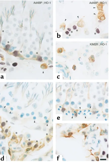
Cell types responsible for the HO-1 induction were examined through double-color immunohistochem-istry (Figure 3). In the CdCl2-untreated controls, the
HO-1–positive cells in the peritubular regions (asterisks in Figure 3a) were Sertoli cells that were characterized by nuclear expression of Ad4BP with dendritic process-es for docking spindle-shaped germ cells, while those in the interstitial space were Ad4BP negative as shown by arrowheads in Figure 3b. On the other hand, the Ad4BP-positive cells in the interstitium (asterisks in Fig-ure 3b), that is, Leydig cells, did not display any notable immunoreactivities of HO-1. Double immunostaining with GTS-1 and KiM2R indicated that macrophages
but not Leydig cells constitute a major component for the interstitial HO-1 expression under unstimulated conditions (Figure 3c). By contrast, in testes of the 12-hour CdCl2-treated rats, cells markedly inducing HO-1
were Leydig cells, which were recognized as Ad4BP-pos-itive cells in the interstitium (asterisks in Figure 3d). Another important feature of the CdCl2-exposed testes
was apoptosis of germ cells in peritubular regions: This change was characterized by nuclear condensation (arrowheads in Figure 3d) as well as fragmentation of their nuclei (arrowheads in Figure 3e). Cell-phase speci-ficity of such germ cell apoptosis is examined in Results. At the same time, the interstitial macrophages that expressed HO-1 under the unstimulated conditions changed their shapes with pseudopod formation, sug-gesting activation of phagocytosis (Figure 3f).
[image:5.576.60.268.54.437.2]Leydig cells primarily display HO-dependent heme degra-dation upon the CdCl2administration. Observation that the CdCl2exposure triggers a marked HO-1 induction
Figure 1
Temporal alterations in expression of HO isozymes in testicular micro-somes of CdCl2–treated and control rats. (a) Western blot analyses of
HO-1 and HO-2 protein expression, using mAb’s GTS-1 and GTS-2, as a function of time after the CdCl2administration. M, molecular
markers; Sp, spleen lysate as a positive control for HO-1. (b) Inhibito-ry action of GTS-3, an anti-rat HO-1 mAb, on HO activities. WR19L-rHO-1, cell transfectant overexpressing rat HO-1; WR19L-rHO-2, cell transfectant overexpressing rat HO-2. HO activities were expressed as relative values versus parent WR19L cells (P). The mAb or mouse IgG was added at 0.5 mg/ml to the lysates of the rat HO-1 or HO-2 cDNA-transfected WR19L cells. V, vehicle. Mean ± SE of three separate exper-iments. *P ≤0.05 as compared with the vehicle. (c) Temporal
alter-ations in the HO-1–specific enzyme activity in testicular microsomes collected from the CdCl2-treated (filled circles) and vehicle-treated
(open circles) rats. (d) Time course of the HO-1–independent enzyme activities in the same animals. Data in cand drepresent mean ± SE of measurements from five to eight testes from different rats. *P < 0.05 as
[image:5.576.315.534.507.729.2]compared with the data measured at 0 hours.
Figure 2
Immunohistochemistry illustrating temporal alterations in HO-1 and HO-2 protein expression in the CdCl2-treated and control rats.
Upper and lower panels denote time-dependent alterations in GTS-1–associated (anti–HO-1) and GTS-2–associated (anti–HO-2) immunoreactivities, respectively. (a) Vehicle-treated control at 12 hours; (b) 12 hours after the CdCl2treatment; (c) 24 hours after the
preferentially in Leydig cells led us to examine further whether the enzyme induction could cause actual degradation of heme in situ. Immunohistochemistry using the anti–bilirubin-IXαmAb 24G7 was thus per-formed (Figure 4). In control rats, bilirubin-IXα –asso-ciated immunoreactivities were evident in Sertoli cells, as judged by those in their dendritic structures and also faintly noted in interstitial macrophages, while germ cells displayed few reactivities, if any (Figure 4a). On the other hand, the 12-hour CdCl2treatment
elicit-ed a markelicit-ed elevation of the immunoreactivities in a majority of the interstitial cells (Figure 4b). Double-color immunohistochemistry using anti–bilirubin-IXαand Ad4BP Ab’s confirmed that the interstitial cells responsible for bilirubin generation are Leydig cells (data not shown). These results provided evidence that CdCl2evokes a bilirubin-IXαoverproduction
site-specifically in Leydig cells, while HO-2–expressing
germ cells did not exhibit any notable levels of the bilirubin generation under both control and CdCl2
-stimulated conditions. As seen in Figure 4c, ZnPP, an HO blocker, attenuated the bilirubin immunoreactiv-ities in Leydig cells. Since ZnPP might affect function of other enzymes besides HO, such as soluble guany-late cyclase (22, 23), we examined effects of CuPP, a metalloprotoporphyrin that does not block HO as a negative control reagent. This reagent did not sup-press the CdCl2-induced increase in bilirubin-IXα
immunoreactivities (Figure 4d), suggesting that inhibitory effects of ZnPP is HO dependent. We also confirmed that the present dose of ZnPP for the HO blockade could sufficiently block the microsomal HO activities in the testicular samples, while the same dose of CuPP did not change the activities (data not shown). The CdCl2-induced and HO-dependent
eleva-tion of the bilirubin generaeleva-tion appeared to coincide with reduced amounts of intact cytochrome P450 in the testicular microsome. The 12-hour CdCl2
[image:6.576.64.280.56.377.2]treatment caused approximately a 40% reduction of the enzyme contents as compared with the control
Figure 3
Characterization of cell types expressing HO-1 in testes of the CdCl2
-untreated (a–c) and -treated (d–f) rats by double-color immunohis-tochemistry. (aand b) Distribution of GTS-1–positive (anti–HO-1– positive) cells and its topographic relationship to Ad4BP-positive steroidogenic cells. Arrowheads; macrophages characterized with positive HO-1 immunoreactivities (brown) lacking in nuclear Ad4BP expression. Asterisks indicate Sertoli cells (peritubular cells in a) and Leydig cells (interstitial cells in b), which were characterized by the Ad4BP expression (purple). The former cells were also characterized by their dendritic processes for docking spindle-shaped spermatids. (c) Double immunostaining with HO-1 (brown) and KiM2R (pur-ple). Note that in control testes, HO-1 is expressed mostly in the interstitial macrophages, but not in Leydig cells. (d) Distribution of GTS-1 (anti–HO-1) immunoreactivities and its topographic rela-tionship to Ad4BP-positive steroidogenic cells in CdCl2-treated rats.
[image:6.576.310.537.563.728.2]Note that HO-1–associated immunoreactivities (brown) markedly increase in Leydig cells (asterisks, purple nuclei). (e) Nuclear con-densation and fragmentation in peritubular germ cells (arrowheads in dand e). (f) A representative picture of pseudopod formation in macrophages in testes of CdCl2-treated rats. Bar, 10 µm.
Figure 4
In situ demonstration of the HO-dependent heme degradation by the anti–bilirubin-IXαmAb–assisted immunohistochemistry in testes of the CdCl2-treated rats. (a) Control. (b) Elevation of bilirubin-IXα
generation in the interstitium in the CdCl2-treated rats (arrows).
Note marked immunoreactivities in the interstitial cells, while germ cells remained unstained, even after exposure to the metal stressor. (cand d) Effects of pretreatment with ZnPP and CuPP on the CdCl2
(15.5 ± 0.4 vs. 26.2 ± 2.8 pmol/mg microsomal protein; mean ± SE of four experiments; P < 0.05), while the group pretreated with ZnPP did not exhibit such a sig-nificant reduction (20.5 ± 1.8 pmol/mg microsomal protein, n = 4). These results suggest that the increase in HO activities plays a role in the CdCl2-induced
reduction of microsomal contents of functionally intact cytochrome P450, at least in part.
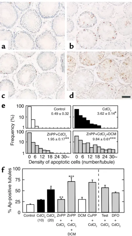
CO generated by CdCl2-induced HO activity induces apop-tosis of germ cells. Results shown in Figure 4 led us to examine whether the CdCl2-elicited HO-1 induction
could play an inhibitory or stimulatory role in apopto-sis of testicular germ cells. As seen in Figure 5, the TUNEL staining in the 12-hour CdCl2-treated groups
confirmed that the initial apoptotic changes occur mostly in germ cells distributing in peritubular regions, being consistent with results shown in Figure 3. Fur-thermore, the severity of apoptotic changes appeared to vary among individual seminiferous tubules. The CdCl2-induced apoptotic changes appeared to be
atten-uated by pretreatment with ZnPP, as shown in Figure 5c. We then examined effects of supplementation of
CO by administering dichloromethane. A single dose of dichloromethane at 6 mmol/kg in the control rats markedly increased percentages of HbCO in blood samples: the values peaked at 3 hours (23.7% ± 3.4% vs. 1.9% ± 1.0%, mean ± SE of three rats, P < 0.05) and were kept at significantly greater levels until 6 hours (10.6% ± 2.8% vs. 2.6% ± 0.3%, mean ± SE of three rats,
P < 0.05). These results suggested that the protocol for administration of dichloromethane twice at 6-hour intervals stimulated endogenous CO generation for at least 12 hours until the start of tissue sampling. As seen in Figure 5d, the current protocol for CO supplemen-tation markedly restored CdCl2-induced apoptosis of
the germ cells even under blockade of HO by ZnPP. Differences in the magnitude of apoptosis among the groups were examined by calculating both densi-ty of TUNEL-positive germ cells among individual seminiferous tubules and percentages of the TUNEL-positive tubules. The CdCl2 administration at 20
µmol/kg increased both the density of apoptotic cells per a single tubule and percentages of apoptosis-pos-itive tubules (Figure 5, e and f), while its dose of 10
µmol/kg did not cause a significant change. As seen in Figure 5f, the apoptotic change elicited by 20
µmol/kg CdCl2was inhibited significantly by
block-ing HO with ZnPP and restored by donatblock-ing CO with administration of dichloromethane. Furthermore, the present protocol for the CO supplementation by itself did not cause any notable apoptosis, despite the fact that HbCO levels markedly increased during the 12-hour period for the dichloromethane supplementa-tion. On the other hand, CuPP did not suppress the CdCl2-induced apoptosis. Differences in percentages
[image:7.576.61.287.53.455.2]of apoptosis-positive tubules among the groups were
Figure 5
Demonstration of apoptotic cells in seminiferous tubules in the 12-hour CdCl2–treated rats assessed by in situ 3′end labeling of
sin-gle-strand DNA. (aand b) Representative pictures of testes in the CdCl2-untreated and -treated groups, respectively. Note that
per-itubular germ cells constitute a major cellular component displaying apoptosis. (c) Effects of pretreatment with ZnPP on the CdCl2
-elicit-ed apoptosis of germ cells. (d) Restoration of CdCl2-induced germ
cell apoptosis by CO supplementation with dichloromethane under blockade of the HO reaction by ZnPP. Bar, 100 µm. (e) Differences in histograms showing the density of apoptotic cells in individual seminiferous tubules among groups. Data were indicated by relative frequency (%) as a function of the density of apoptotic cells per a sin-gle tubule. (f) Differences in percentages of TUNEL-positive seminif-erous tubules (percentage of apoptosis-positive tubules) among groups. Two different doses of CdCl2(10 and 20 µmol/kg) were
examined. Effects of other interventions were examined in rats treat-ed with 20 µmol/kg CdCl2. Tubules containing at least one
TUNEL-positive cell were considered to be TUNEL-positive in this analysis. Bars rep-resent the mean ± SE of measurements from four to eight separate experiments. In an individual experiment, at least 200 different tubules were examined to calculate a single data set. DCM, dichloromethane; Test, testosterone, DFO, desferrioxamine.
*P < 0.05 as compared with the control. **P < 0.05 as compared with
the group treated with 20 µmol/kg CdCl2. ***P < 0.05 as compared
unlikely to result from possible heterogeneity of the density of testicular cells in the areas observed, since the density of Sertoli cells, i.e., Ad4BP-positive per-itubular cells, in the same regions did not differ sig-nificantly among the groups; the relative percentage densities of Sertoli cells versus controls (100% ± 4%) were 110% ± 5%, 108% ± 5%, and 102% ± 8% in groups treated with CdCl2, ZnPP + CdCl2, and ZnPP + CdCl2
+ dichloromethane, respectively (mean ± SE of data collected from four to five separate rats). These data also convinced us that the viability of Sertoli cells was not altered by the CdCl2exposure under the current
experimental conditions. The increased HO activity could degrade the prosthetic heme group for steroidogenic cytochromes P450 and provide divalent iron as a cytotoxic factor. Thus, effects of supplemen-tation of testosterone or iron chelation by desferriox-amine mesilate were exdesferriox-amined, but without any notable suppression of apoptosis (Figure 5f). These results collectively suggest that the CdCl2-induced
apoptosis of testicular germ cells is an event mediat-ed by CO derivmediat-ed from inducible HO.
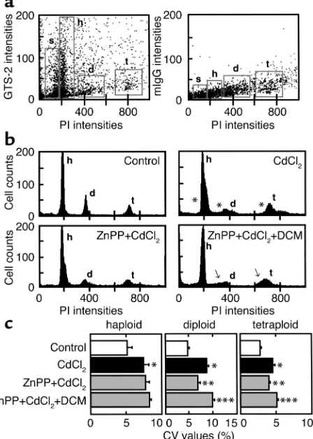
Diploid and tetraploid germ cells exhibit CO-mediated apop-tosis. Cell phase–specific alterations and variations of DNA contents were determined in individual germ cells by FACS analyses in the 24-hour CdCl2-treated rats. As
seen in the two-color dot-plot diagrams (Figure 6a), three main populations of the cells expressing distinct levels of HO-2 protein were identified as a function of their DNA contents, denoting haploid (h), diploid (d), and tetraploid (t), with increasing PI fluorescence. With careful evaluation of the haploid peak, the small cell population with moderate HO-2 expression with the smallest amounts of DNA (by PI fluorescence) was notable and corresponded to elongated spermatids and spermatozoa, being designated as subhaploid (s). Among these subgroups, the cells in groups s and h exhibited greater expression of HO-2 than those in d and t. These results were in good agreement with immuno-histochemistry showing GTS-2 immunoreactivities in relatively differentiated cells in centritubular regions.
[image:8.576.68.292.57.371.2]As seen in the control data in Figure 6b, haploid, diploid, and tetraploid cells constituted the specific ratio of DNA contents, such as 1:2:4, respectively, peak-ing with small SDs. The germ cells collected from the CdCl2-treated rats also displayed three peaks, but with
Figure 6
CdCl2-induced disruption of cell phase–specific DNA contents in
tes-ticular germ cells and effects of the HO blockade by ZnPP and CO supplementation by dichloromethane (DCM). (a) Characterization of HO-2 expression in testicular germ cells. s, subhaploid; h, haploid; d, diploid; t, tetraploid. Note that the cells in subgroups s and h exhib-ited greater expression of HO-2 than those in d and t. The right panel exhibits two-color dot-plot analysis using mouse IgG as a negative control. (b) Differences in histogram showing DNA contents in spe-cific cell phases among groups. The ratio of DNA contents as judged by PI fluorescence is 1:2:4 for subgroups h, d, and t, respectively. Note that variations of DNA contents become evident in the CdCl2-treated
group (asterisks in b). Such variations of DNA contents were also evi-dent in the ZnPP + CdCl2–treated group supplemented with DCM
(arrows in b). (c) Differences in CV values of each cell population among groups. *P < 0.05 as compared with the control. Data indicate
mean ± SE of six to seven separate experiments for each group.
**P < 0.05 as compared with the CdCl
2-treated group. ***P < 0.05 as
[image:8.576.308.536.573.730.2]compared with the group treated with ZnPP and CdCl2.
Figure 7
Immunohistochemical analyses of CdCl2-induced alterations in
expression of Fas and FasL. (a and b) Representative pictures for Fas immunoreactivities. (c and d) Representative pictures for FasL immunoreactivities in the control and 12-hour CdCl2-treated groups,
enlarged widths of histograms (asterisks in Figure 6b), indicating enhanced heterogeneity of DNA contents. Among them, diploid cells markedly reduced the peak height upon CdCl2exposure. When pretreated with
ZnPP in the absence or presence of dichloromethane, the heights and SD values of each cell type appeared to change notably (arrows in Figure 6b). We thus com-pared differences in CV values among groups. The CdCl2exposure significantly increased CV values of
haploid, diploid, and tetraploid cells (Figure 6c). The HO blockade by ZnPP attenuated the CdCl2-elicited
elevation of CV values preferentially in diploid and tetraploid cells, but not in haploid cells. On the other hand, the CuPP administration did not alter these CdCl2-induced changes in CV values (data not shown).
Furthermore, supplementation of dichloromethane, a CO donor, significantly restored the ZnPP-induced CV attenuation in these cell types. As shown in Figure 3, the CdCl2-induced apoptosis appeared to occur in
germ cells in peritubular regions involving spermato-gonia (diploid) and primary spermatocytes (tetraploid) rather than in Sertoli cells, as judged by the fact that Ad4BP-positive cells did not exhibit nuclear
fragmen-tation and condensation. Since apoptosis accounts for a factor causing increased CV values (19), these results collectively suggest that spermatogonia and primary spermatocytes exhibit CO-mediated apoptosis in the current experimental model. On the other hand, an increase in CV values in haploid germ cells was unlike-ly to result from apoptosis, but presumabunlike-ly from non-apoptotic damage of the cells by CdCl2, inasmuch as
apoptosis could hardly be observed in central regions of tubules under current experimental conditions.
[image:9.576.62.292.55.282.2]CO appears to serve as a factor responsible for triggering apoptosis. Fas-mediated signal transduction was shown previously to participate in apoptosis of testicular germ cells and to be implicated in maintaining immune priv-ilege in testes (17, 24). Considering this fact, we attempted to examine immunohistochemically if
Figure 8
Effects of ZnPP and dichloromethane on the CdCl2-induced
alter-ations in the FasL expression in peritubular regions. (aand b) Rep-resentative pictures in the control and CdCl2-treated groups,
respec-tively. (candd) Representative pictures of the CdCl2+ ZnPP–treated
groups in the absence and presence of dichloromethane, respective-ly. Note that CdCl2increases FasL-associated immunoreactivities in
Sertoli cells (arrowheads in b) and peritubular germ cells (asterisks in b). Such changes were also reproduced in the ZnPP + CdCl2
–treat-ed group supplement–treat-ed with dichloromethane (arrowheads and asterisks in d). The dose of CdCl2is 20 µmol/kg. Bar, 10 µm.
Figure 9
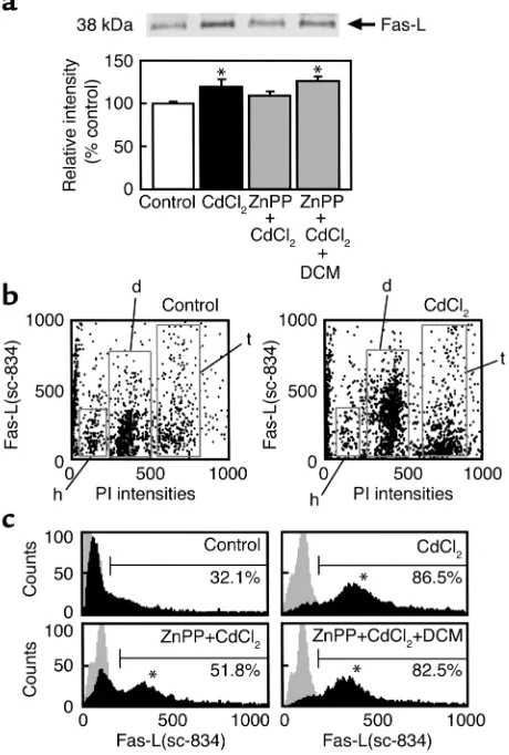
Overexpression of FasL in testicular diploid cells elicited by CdCl2. (a)
Western blot analyses in whole testicular lysates using the anti-FasL Ab sc-834. Densitometric analyses indicate relative gray levels versus controls (mean ± SE of four separate experiments). CdCl2at 20 µmol/kg was administered 12 hours before experiments. *P ≤0.05
as compared with the control. (b) Dual-color analyses between DNA contents (PI) and FasL expression (sc-834). (c) Histograms showing alterations in the FasL expression in the diploid cells among groups. A representative set of three experiments was shown. Histograms with gray backgrounds indicate controls stained by rabbit IgG. Note that the CdCl2 exposure markedly upregulates FasL expression, and
[image:9.576.310.540.393.734.2]CdCl2could cause alterations in expression of Fas and
FasL in testes. As shown in Figure 7a, Fas was expressed in a majority of germ cells in the control, and the CdCl2
exposure did not greatly change such an immunos-taining pattern (Figure 7b). On the other hand, FasL-associated immunoreactivities were faintly observed over germ cells in peritubular regions (Figure 7c), sup-porting recent results showing their FasL expression (25). When exposed to CdCl2, however, the FasL
immunoreactivities became enhanced in different types of cells in the peritubular regions as indicated by arrow-heads and asterisks in Figure 7d. Careful observation by high magnification in the peritubular regions of the CdCl2-untreated and -treated (Figure 8, a and b) testes
revealed that Sertoli cells (arrowheads) and premeiotic germ cells, including spermatocytes and spermatogo-nia (asterisks), were responsible for the CdCl2-induced
enhancement of FasL immunoreactivities. It should be noted that such an enhancement of FasL staining appeared to be attenuated by blockade of HO by ZnPP (Figure 8c). Furthermore, CO supplementation by dichloromethane caused a restoration of FasL immunoreactivities in both Sertoli cells (arrowheads in Figure 8d) and germ cells (asterisks in Figure 8d), even under the HO-inhibiting conditions.
Such cell phase–specific alterations in the FasL expression upon CdCl2exposure were confirmed by
FACS analyses of the dispersed testicular cells. West-ern blot analyses using lysates of the whole testes (Fig-ure 9a) revealed the stimulus-elicited increase in the FasL overexpression. This increase was partially atten-uated by ZnPP and was restored significantly with CO donation by dichloromethane, although the change appeared small when assessed in the whole organ. Considering the immunohistochemical analyses that alterations in the FasL expression seemed to be cell type–specific events, dual-color FACS analyses using PI (DNA contents) and sc-834 (FasL) were carried out. As seen in Figure 9b, the CdCl2-induced
overexpres-sion of FasL occurred mainly in diploid cells and par-tially in tetraploid cells. On the other hand, postmei-otic cells such as haploid cells did not show any notable overexpression of FasL. Further examination of the diploid cells gated with their DNA contents showed that greater than 80% of these cells exhibited a marked elevation of the FasL expression. The CdCl2
-elicited elevation of the FasL expression was attenuat-ed markattenuat-edly by ZnPP and restorattenuat-ed almost completely by CO supplementation with dichloromethane (aster-isks in histograms of Figure 9c). These results were in agreement with aforementioned results in immuno-histochemistry and suggest that CO plays a role in CdCl2-induced alterations in FasL expression in both
Sertoli cells and germ cells.
Discussion
The testis is a major organ that abundantly expresses HO activities, though cell types expressing the HO isozymes and the alterations in their activities under
stress conditions have yet to be well characterized. This study provided evidence that HO-1 and HO-2 occur in distinct cell types in testes under control and stress conditions. Under unstimulated control states, HO-1 is expressed in interstitial macrophages and Sertoli cells, whereas it was expressed in germ cells and Leydig cells little, if at all. On the other hand, HO-2 occurs in subpopulation of the matured germ cells and their residual bodies: this finding was also confirmed by FACS analyses of these cells in vitro. Activities and dis-tribution of the isozymes are dramatically altered as a function of time after exposure to CdCl2. HO-1 is
markedly induced in Leydig cells, while HO-2 is reduced as a result of the stressor-induced reduction of germ cells. Such an inverse relationship between the two isozymes was also well demonstrated by our novel method to determine the isozyme-specific enzyme activities. The current results indicate that testis use Leydig cells as a sensor of metal stress and induces HO-1, and a reduction of the testicular HO activities as a whole appears to result merely from the reduction of intact testicular germ cells that express HO-2. Previous studies showing that CdCl2 exposure did not increase
but decrease total HO activities in the testis suggested that this organ might exhibit stress responses distinct from those in the liver and spleen (26), being obvious-ly inconsistent with our results.
Another important finding in the current study is identification of cells responsible for actual degrada-tion of heme in this HO-enriched organ. As judged by immunohistochemical localization of bilirubin-IXα, the HO-derived end product of the degradation, Sertoli cells appeared to constitute a major cellular compo-nent responsible for HO-mediated heme degradation, while Leydig cells displayed only small amounts of the product under unstimulated conditions. By contrast, upon exposure to CdCl2, Leydig cells exhibited marked
bilirubin-IXα–positive reactivities, while the change in Sertoli cells was not notable. It should be noted in both conditions that germ cells displayed few immunoreac-tivities of bilirubin-IXα, despite considerable expres-sion of HO-2 in these cells. Considering that genera-tion of bilirubin-IXα occurs in parallel with CO production, these results suggest that Leydig cells con-stitute a major cellular component for active HO reac-tion and CO generareac-tion upon CdCl2exposure.
induction could decrease the reductant available for the testosterone synthesis in Leydig cells. Third, CO generated by HO-1 in Leydig cells could be bound directly to the prosthetic heme of P450 cytochromes and thereby inhibit testosterone synthesis in the cells. Such a topographic correlation between micro-somal cytochromes P450s and HO-1 induction shed light on a crucial role of the cell-specific heme degra-dation in testicular steroidogenesis and is also con-sistent with previous observation that the CdCl2
administration actually reduced testosterone pro-duction in Leydig cells (27).
Current observation that the stress-inducible CO generation occurs preferentially in Leydig cells raised an important question as to mechanisms through which the stress response occurring in the interstitial space (e.g., Leydig cells) could transfer signal(s) for apoptosis of germ cells in the seminiferous tubules, considering the presence of the blood-testicular barri-er that limits access of signaling molecules from the interstitium into the tubules (1). CO appears to serve as such a barrier-permeable signaling molecules for the germ cell apoptosis. Extrapolation of previous studies provided several mechanisms for cell death triggered by HO-1. First, the HO-1 induction could reduce cytochrome P450 monooxygenases in Leydig cells and secondarily suppress synthesis of testosterone required for spermatogenesis. However, as judged so far by neg-ative effects of exogenous supplementation of testos-terone, a shortage of the hormone synthesis is unlikely to play a crucial role directly in the germ cell apoptosis at the initial phase of the CdCl2toxicity. Second,
diva-lent iron, another HO product, could cause the Fenton reaction that results in propagation of oxygen radical toxicity when it is generated in excess. However, this scenario appears to be implausible, since administra-tion of the iron chelator did not attenuate the apopto-sis in the CdCl2-exposed testes. A possible secondary
effect of CO on downregulation of the testosterone synthesis could also be excluded because of the inef-fectiveness of the hormone supplementation.
Of greatest importance is the direct effect of CO on germ cell apoptosis. Recent observations show that CO is able to trigger apoptosis at nanomolar concentra-tions in certain types of cells, such as vascular endothe-lial cells in culture (28), though the gas-reception mechanisms are still unknown. Restoration effects of CO supplementation by dichloromethane on germ cell apoptosis fully support a concept that CO overpro-duced through the HO-1 reaction in Leydig cells could trigger germ cell apoptosis. The current immunohisto-chemical analyses using Ab’s against Fas and FasL led us to consider that at least two pathways could be involved in mechanisms through which CO derived from Leydig cells triggers apoptosis of spermatogonia and primary spermatocytes, Sertoli cell-dependent and -independent pathways. Under the current experimen-tal conditions, Sertoli cells neither exhibited apoptosis nor displayed loss of viability, as judged by the density
of Ad4BP-positive cells in seminiferous tubules. These cells notably upregulated FasL expression in response to the CdCl2exposure, while the response was
attenu-ated by the HO blockade and restored by the CO dona-tion. These results suggest that Sertoli cells actively respond to CO without altering their viability for trig-gering Fas/FasL-mediated apoptosis in germ cells adja-cent to them (e.g., spermatogonia and primary sper-matocytes). On the other hand, the present study did not exclude a possibility that CO generated in Leydig cells could directly stimulate germ cell apoptosis, inas-much as the CdCl2 exposure as well as
dichloro-methane supplementation under the HO blockade causes a notable increase in FasL immunoreactivities in spermatogonia and/or primary spermatocytes, as seen in Figure 8. Such an apoptogenic effect of stress-induced CO deserves further studies given the evidence for the gas-reception mechanisms at peritubular regions of seminiferous tubules involving Sertoli cells and/or germ cells as the effector apparatus.
Acknowledgments
The authors thank Hitomi Irisawa and Mieko Kondo for their expert technical support in immunohisto-chemistry and manuscript preparation. This study was supported by Advanced Medical Technology in Health Sciences Research Grants from Ministry of Health and Welfare in Japan.
1. Weinbauer, G.F., Gromoll, J., Simoni, M., and Nieschlag, E. 2000. Physi-ology of testicular function. In Andrology. E. Nieschlag and H.M. Behre, editors. Springer. Berlin, Germany. 23–61.
2. Rowe, P.J., Comhaire, F.H., Hargreave, T.B., and Mahmoud, A.M.A. 2000. History-taking. In WHO manual for the standardized investigation, diagnosis and management of the infertile male. Cambridge University Press. New York, New York, USA. 5–16.
3. Shikone, T., Billig, H., and Hsueh, A.J. 1994. Experimentally induced cryptorchidism increases apoptosis in rat testis. Biol. Reprod. 51:865–872. 4. Richburg, J.H. 2000. The relevance of spontaneous- and chemically-induced alterations in testicular germ cell apoptosis to toxicology. Toxi-col. Lett. 112:79–86.
5. Maines, M.D. 1988. Heme oxygenase: function, multiplicity, regulatory mechanisms, and clinical applications. FASEB J. 2:2557–2568. 6. Trakshel, G.M., Kutty, R.K., and Maines, M.D. 1986. Purification and
characterization of the major constitutive form of testicular heme oxy-genase. The noninducible isoform. J. Biol. Chem. 261:11131–11137. 7. Chia, S.E., Xu, B., Ong, C.N., Tsakok, F.M., and Lee, S.T. 1994. Effect of
cadmium and cigarette smoking on human semen quality. Int. J. Fertil. Menopausal. Stud. 39:292–298.
8. Kim, Y.C. 1997. Dichloromethane potentiation of carbon tetrachloride hepatotoxicity in rats. Fundam. Appl. Toxicol. 35:138–141.
9. Jiang, Y., Kuo, C.L., Pernecky, S.J., and Piper, W.N. 1998. The detection of cytochrome P450 2E1 and its catalytic activity in rat testis. Biochem.
Biophys. Res. Commun. 246:578–583.
10. Scott, C.A., et al. 1996. Effects of cis-platinum and luteinizing hormone releasing hormone analogues on rat spermatogenesis. A morphologic and flow cytometric study. Anal. Quant. Cytol. Histol. 18:361–373. 11. Goda, N., et al. 1998. Distribution of heme oxygenase isoforms in rat
liver. Topographic basis for carbon monoxide-mediated microvascular relaxation. J. Clin. Invest. 101:604–612.
12. Makino, N., et al. 2001. Altered expression of heme oxygenase-1 in the livers of patients with portal hypertensive diseases. Hepatology. 33:32–42. 13. Hatano, O., et al. 1994. Sex-dependent expression of a transcription fac-tor, Ad4BP, regulating steroidogenic P-450 genes in the gonads during prenatal and postnatal rat development. Development. 120:2787–2797. 14. Nakayama, M., et al. 2001. Increased expression of heme oxygenase-1 and bilirubin accumulation in foam cells of rabbit atherosclerotic lesions.Arterioscler. Thromb. Vasc. Biol. 21:1373–1377.
15. Kyokane, T., et al. 2001. Carbon monoxide from heme catabolism
pro-tects against hepatobiliary dysfunction in endotoxin-treated rat liver.
Gastroenterology. 120:1227–1240.
16. Omura, T., and Sato R. 1964. The carbon monoxide-binding pigment of liver microsome. J. Biol. Chem. 239:2370–2378.
17. Lee, J., Richburg, J.H., Younkin, S.C., and Boekelheide, K. 1997. The Fas system is a key regulator of germ cell apoptosis in the testis. Endocrinol-ogy. 138:2081–2088.
18. Kiener, P.A., et al. 1997. Human monocyte cells contain high levels of intracellular Fas ligand. J. Immunol.159: 1594–1598.
19. Darzynkiewicz, Z., et al. 1997. Cytometry in cell necrobiology: analysis of apoptosis and accidental cell death (necrosis). Cytometry. 27:1–20. 20. Rabinovitch, P.S. 1993. Practical considerations for DNA content and cell cycle analysis. In Clinical flow cytometry. K.D. Bauer, R.E. Duque, and T. Vincent Shankey, editors. Williams & Wilkins. Baltimore, Maryland, USA. 117–142.
21. Ewing, J.F., and Maines, M.D. 1995. Distribution of constitutive (HO-2) and heat-inducible (HO-1) heme oxygenase isozymes in rat testes: HO-2 displays stage-specific expression in germ cells. Endocrinology.
136:2294–2302.
22. Agarwal, A., and Nick, H.S. 2000. Renal response to tissue injury: lessons from heme oxygenase-1 gene ablation and expression. J. Am. Soc. Nephrol.
11:965–973.
23. Hayashi, S., et al. 1999. Induction of heme oxygenase-1 suppresses venu-lar leukocyte adhesion elicited by oxidative stress: role of bilirubin gen-erated by the enzyme. Circ. Res.85: 663–671.
24. Bellgrau, D., et al. 1995. A role for CD95 ligand in preventing graft rejec-tion. Nature. 377:630–632.
25. D’Alessio, A., et al. 2001. Testicular FasL is expressed by sperm cells. Proc. Natl. Acad. Sci. USA. 98:3316–3321.
26. Maines, M.D., Chung, A.S., and Kutty, R.K. 1982. The inhibition of tes-ticular heme oxygenase activity by cadmium. A novel cellular response.
J. Biol. Chem. 257:14116–14121.
27. Laskey, J.W., and Phelps, P.V. 1991. Effect of cadmium and other metal cations on in vitro Leydig cell testosterone production. Toxicol. Appl.
Pharmacol. 108:296–306.
28. Thom, S.R., Fisher, D., Xu, Y.A., Notarfrancesco, K., and Ischiropoulos, H. 2000. Adaptive responses and apoptosis in endothelial cells exposed to carbon monoxide. Proc. Natl. Acad. Sci. USA. 97:1305–1310. 29. Rodriguez, I., Ody, C., Araki, K., Garcia, I., and Vassalli, P. 1997. An early
and massive wave of germinal cell apoptosis is required for the develop-ment of functional spermatogenesis. EMBO J. 16:2262–2270. 30. Stephenson, W.T., Poirier, S.M., Rubin, L., and Einhorn, L.H. 1995.
Evalu-ation of reproductive capacity in germ cell tumor patients following treat-ment with cisplatin, etoposide, and bleomycin. J. Clin. Oncol. 13:2278–2280. 31. Kelly, M. 1988. Case reports of individuals with oligospermia and
meth-ylene chloride exposures.Reprod. Toxicol. 2:13–17.
32. Tielemans, E., et al. 1999. Occupationally related exposures and reduced semen quality: a case-control study. Fertil. Steril. 71:690–696. 33. Maines, M.D., and Ewing, J.F. 1996. Stress response of the rat testis: in