Thyrocyte-specific G
q
/G
11
deficiency impairs
thyroid function and prevents goiter
development
Jukka Kero, … , Günther Schütz, Stefan Offermanns
J Clin Invest.
2007;
117(9)
:2399-2407.
https://doi.org/10.1172/JCI30380
.
The function of the adult thyroid is regulated by thyroid-stimulating hormone (TSH), which
acts through a G protein–coupled receptor. Overactivation of the TSH receptor results in
hyperthyroidism and goiter. The G
s-mediated stimulation of adenylyl cyclase–dependent
cAMP formation has been regarded as the principal intracellular signaling mechanism
mediating the action of TSH. Here we show that the G
q/G
11-mediated signaling pathway
plays an unexpected and essential role in the regulation of thyroid function. Mice lacking the
a
subunits of G
qand G
11specifically in thyroid epithelial cells showed severely reduced
iodine organification and thyroid hormone secretion in response to TSH, and many
developed hypothyroidism within months after birth. In addition, thyrocyte-specific
G
a
q/G
a
11-deficient mice lacked the normal proliferative thyroid response to TSH or
goitrogenic diet, indicating an essential role of this pathway in the adaptive growth of the
thyroid gland. Our data suggest that G
q/G
11and their downstream effectors are promising
targets to interfere with increased thyroid function and growth.
Research Article
Endocrinology
Find the latest version:
Thyrocyte-specific G
q
/G
11
deficiency
impairs thyroid function and prevents goiter
development
Jukka Kero,1 Kashan Ahmed,1 Nina Wettschureck,1 Sorin Tunaru,1 Tim Wintermantel,2
Erich Greiner,2 Günther Schütz,2 and Stefan Offermanns1
1Institute of Pharmacology, University of Heidelberg, Heidelberg, Germany. 2Division of Molecular Biology of the Cell I,
German Cancer Research Center, Heidelberg, Germany.
The function of the adult thyroid is regulated by thyroid-stimulating hormone (TSH), which acts through a
G protein–coupled receptor. Overactivation of the TSH receptor results in hyperthyroidism and goiter. The
G
s-mediated stimulation of adenylyl cyclase–dependent cAMP formation has been regarded as the principal
intracellular signaling mechanism mediating the action of TSH. Here we show that the G
q/G
11-mediated
sig-naling pathway plays an unexpected and essential role in the regulation of thyroid function. Mice lacking the
α
subunits of G
qand G
11specifically in thyroid epithelial cells showed severely reduced iodine organification
and thyroid hormone secretion in response to TSH, and many developed hypothyroidism within months after
birth. In addition, thyrocyte-specific G
α
q/G
α
11-deficient mice lacked the normal proliferative thyroid response
to TSH or goitrogenic diet, indicating an essential role of this pathway in the adaptive growth of the thyroid
gland. Our data suggest that G
q/G
11and their downstream effectors are promising targets to interfere with
increased thyroid function and growth.
Introduction
Thyroid hormone plays a central role in maintaining the basal level of metabolism in the body. It regulates O2 consumption as
well as lipid and carbohydrate metabolism and is required for normal growth and maturation (1, 2). The primary regulator of thyroid gland growth and function in the adult organism is the thyroid-stimulating hormone (TSH). Lack of TSH or TSH action results in a reduced weight of the adult thyroid gland and almost abolishes thyroid function, leading to hypothyroidism (3–5). Conversely, pathologically elevated serum TSH levels stimulate thyroid hormone production and thyroid growth, leading to hyperthyroidism and goiter (6).
TSH regulates thyroid function through a G protein–coupled receptor on thyrocytes (7–9). TSH receptor–dependent activation of the Gs/adenylyl cyclase–mediated pathway has been suggested
to account for most of the biological effects of TSH on thyroid cells, such as the stimulation of iodine uptake, hormone secre-tion, and proliferation (7). Consistent with this, thyroid glands of mice lacking the TSH receptor have defects in producing iodinat-ed thyroglobulin, but the ability to take up iodine and to organify it can be restored by the adenylyl cyclase activator forskolin (4). Nongoitrous hypothyroidism has also been observed in patients with one defective allele of the gene encoding Gαs (GNAS) and
pseudohypoparathyroidism type 1a (10) as well as in at least one mouse model with Gαs deficiency (11). In addition, constitutive
activation of the thyrocyte cAMP cascade in humans carrying acti- vating somatic mutations of GNAS or in transgenic mice overex-pressing the Gs-coupled adenosine A2 receptor, a constitutively
active mutant of Gαs
, or cholera toxin in thyroids causes hyper-functioning thyroid adenomas (12–17).
In various species, including humans, TSH can also induce the Gq/G11-mediated stimulation of phospholipase C–β (PLC-β),
leading to mobilization of intracellular Ca2+ ([Ca2+]i) by inositol
1,4,5-trisphosphate and formation of diacyl glycerol (18–20). How-ever, the role of the Gq/G11-mediated signaling pathway in thyroid
function is unclear. There is evidence that the constitutive activa-tion of the Gq/G11/PLC-β
pathway in thyrocytes of mice overex-pressing an active mutant of the α1B adrenergic receptor further
promotes malignant transformation of the thyroid gland (21), but it is unclear whether Gq/G11 mediate a growth-promoting effect
under more physiological conditions.
In order to understand the role of Gq/G11-mediated signaling
in thyroid follicular cells, we have generated mice lacking the α subunits of Gq/G11 selectively in thyrocytes. Because Gαq/Gα11
-deficient mice die in utero (22), we used a floxed allele of the gene encoding Gαq (gnaq), which can be used to inactivate Gαq function
in a Gα11-deficient background (23), using the Cre/loxP system.
Our data indicate that the Gq/G11-mediated signaling pathway
is dispensable for thyroid development but is required for TSH-induced thyroid hormone synthesis and release in the adult and that the lack of Gq/G11 leads to hypothyroidism. In addition,
Gq/G11 deficiency prevented the development of goiter induced by
blockade of thyroid function or TSH treatment.
Results
Generation of thyroid-specific Gαq/Gα11 deficiency. In order to inactivate
the Gq/G11-mediated signaling pathway in thyrocytes, we generated
a transgenic mouse line expressing Cre recombinase under the con- trol of the thyrocyte-specific thyroglobulin promoter using a P1- derived bacterial artificial chromosome (PAC) harboring the thyro-globulin gene (see Methods). After crossing with the Gt(ROSA)26Sor Cre reporter mouse line (24), 3 of the 4 tested transgenic founder
Nonstandard abbreviations used: [Ca2+]i, intracellular Ca2+
; PAC, P1-derived bacteri-al artificial chromosome; Tc-Gαq/Gα11–KO, thyrocyte-specific Cre transgenic crossed
with Gnaqflox/floxGna11–/–; TSH, thyroid-stimulating hormone.
research article
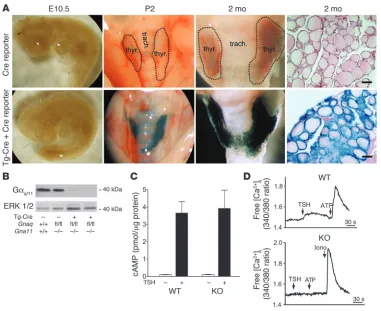
lines showed Cre-mediated recombination exclusively in the thy-roid gland (see Supplemental Figure 1, available online with this article; doi:10.1172/JCI30380DS1). Cre activity was observed in vir-tually all thyrocytes, but not in other cells of the thyroid gland, like parathyroid cells or stromal cells (Figure 1A and data not shown). As expected from the time course of thyroglobulin promoter activ-ity, no recombination was seen on E10.5 (Figure 1A). However, Cre-mediated recombination was observed by P2. There was no indication of altered thyroid histology or serum TSH and thyroid hormone levels in mice expressing Cre compared with wild-type lit-termates (Figure 1A and data not shown).
The thyrocyte-specific Cre transgenic mouse line (Tg-Cre) was crossed with mice lacking the Gα11 gene (Gna11–/–) and carrying
the floxed Gαq allele (Gnaqflox/flox), resulting in the generation of
Tg-Cre;Gnaqflox/floxGna11–/– mice (Tc-Gα
q/Gα11–KO). As shown in
Figure 1B, the amount of Gαq/Gα11 proteins was strongly reduced
in thyroids from Tc-Gαq/Gα11–KO mice compared with wild-type
littermates. The small amount of remaining Gαq/Gα11 protein was
most likely derived from cells other than thyrocytes. We also ana-lyzed Gαq/Gα11 expression in
nonthyroid tissues including brain, liver, spleen, and plate-lets of Tc-Gαq/Gα11–KO mice,
but did not observe any dif-ference compared with levels in wild-type mice (data not shown). No significant differ-ence in basal or TSH-induced cAMP levels was seen between thyrocytes prepared from Tc-Gαq/Gα11–KO mice and
wild-type littermates at 1–2 months of age, indicating no significant alteration in the Gs
-mediated signaling path-way in the absence of Gαq/
Gα11 (Figure 1C). In contrast,
thyrocytes from Tc-Gαq/Gα11–
KO mice lacked a functional Gq/G11-mediated signaling
pathway, as demonstrated by determination of [Ca2+]i
con- centrations using the fluores-cent calcium indicator Fura-2. While in wild-type thyrocytes, TSH, ATP, and other stimuli acting via Gq/G11-coupled
receptors induced an increase in [Ca2+]
i (Figure 1D and data
not shown), in thyrocytes from Tc-Gαq/Gα11–KO mice,
only the Ca2+
ionophore ion-omycin induced a response (Figure 1D).
Hypothyroidism in thyrocyte-specific Gαq/Gα11-deficient mice.
The development of the thy-roid gland in the absence of the Gq/G11
-mediated signal-ing pathway was normal, as indicated by normal thyroid histology and normal thyroid hor-mone and TSH plasma levels during the first 2 months of age (Figures 2 and 3). Similarly, no change in the size or form of thy-roid follicles was observed in mice up to 2 months of age (Figure 3 and data not shown). There was also no difference in body weight or reproductive performance in Tc-Gαq/Gα11
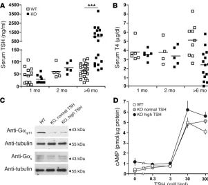
–KO mice com-pared with littermate controls (data not shown). However, after 2 months of age, the TSH plasma levels in the thyrocyte-specific Gαq/Gα11–KO mice gradually increased, differing significantly at
6 months of age. Eventually, about half of the Tc-Gαq/Gα11–KO
animals developed overt hypothyroidism, with low T4 levels and
strongly increased TSH levels, at 6 months of age or older (Fig-ure 2 and data not shown). The proportion of males to females was very similar in groups with normal and altered TSH and T4
plasma levels (data not shown), which indicates that there were no sex-specific differences. There was also no difference in thyroid weights of Tc-Gαq/Gα11–KO mice with normal (<150 ng/ml) and
[image:3.585.41.422.81.392.2]elevated TSH levels (>500 ng/ml) (0.12 ± 0.01 and 0.11 ± 0.01 mg/g body weight, respectively).
Figure 1
Validation of thyrocyte-specific deletion of the genes encoding Gαq and Gα11. (A) Gt(ROSA)26Sor Cre
report-er animals (Cre reportreport-er) carrying no Cre gene (top row) or carrying the Cre gene undreport-er the control of the thyroglobulin promoter (Tg-Cre; bottom) were analyzed at the indicated ages, and β-galactosidase staining was performed on whole-mount preparations as well as on sections (far right panels). trach., trachea; thyr.,
thyroid gland. Scale bars: 50 μm. Original magnification, ×12. (B) Western blot of thyroid gland lysates from
4-week-old wild-type (Gnaq+/+Gna11+/+), Gα11-deficient (Gnaqfl/flGna11–/–) and Tc-Gαq/Gα11–KO mice
(Tg-Cre;Gnaqfl/flGna11–/–) probed with antibodies recognizing Gαq/Gα11 (Gαq/11) or ERK1/2. (C) cAMP levels in
primary thyrocytes from wild-type and Tc-Gαq/Gα11–KO animals treated (+) or not (–) with 50 mU/ml TSH.
Values are mean ± SEM of experiments performed in triplicate. (D) Effect of TSH (10 mU/ml), ATP (10 μM),
and ionomycin (Iono; 1 μM) on [Ca2+]i in thyrocytes prepared from wild-type or Tc-Gαq/Gα11–KO animals.
To test whether the slowly progressing hypothyroidism in some animals was due to incomplete recombination of the floxed Gαq
allele or a defect in the Gαs-mediated regulation of cAMP levels,
we compared thyrocytes from 5- to 6-month-old Tc-Gαq/Gα11–KO
mice with normal and elevated TSH levels. As shown in Figure 2C, Gαq/Gα11 as well as Gαs protein levels were indistinguishable
between the groups. Also, the ability of TSH to induce an increase in cAMP levels in wild-type mice was similar to that in Tc-Gαq/
Gα11–KO mice with normal and elevated TSH levels (Figure 2D).
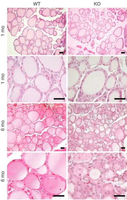
The histology of the Tc-Gαq/Gα11–KO thyroid glands at 1 month
of age showed no obvious difference in follicle size or form or staining of the colloid compared with control mice. However, at 6 months of age, concomitant with elevated TSH and reduced thyroid hormone levels, the thyroid histology of the thyroid-specific Gαq/
Gα11–KO mice was altered (Figure 3). In these animals the normal
thyroid follicular structure was disturbed, with few normal follicles left. Follicle cells were often enlarged and columnar and had large nuclei. Despite the long-term elevation of TSH levels and the fol-licular cell changes, the thyroids of the Tc-Gαq/Gα11–KO mice were
not significantly larger than those of controls. Nor was the thyroid weight increased: thyroid weights were 1.65 ± 0.09 mg in control mice (n = 14) and 1.58 ± 0.15 mg in Tc-Gαq/Gα11–KO mice (n = 8).
Defect of TSH-induced regulation of thyroid function in the absence of Gαq/Gα11. To analyze potential defects in thyrocytes resulting
from Gαq/Gα11
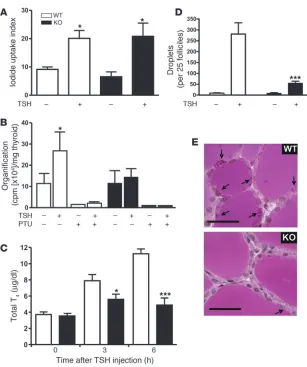
deficiency, we determined several cellular func-tions required for thyroid hormone formation, storage, and release. The functional studies were performed at the age of 1–2 months, when thyroid morphology was still normal and animals were euthyroid with normal TSH levels. TSH has previously been shown to increase iodine uptake in thyrocytes (6). As shown in Figure 4A, there was no significant difference between basal and TSH-stimulated iodine uptake between 6-week-old control and thyrocyte-specific Gαq/Gα11-deficient mice, indicating that the
Gαq/Gα11-mediated signaling pathway is not
required for TSH-stimulated iodine uptake. To test whether the stimulation of iodine cou-pling to thyroglobulin by TSH is dependent on Gαq/Gα11, we measured the amount of iodine
incorporated into thyroid proteins. While basal levels of iodine incorporation were the same in wild-type and Tc-Gαq/Gα11
–KO animals, stimu-lation of incorporation by TSH was abrogated in the absence of Gαq/Gα11 (Figure 4B).
In order to evaluate the role of Gq/G11
-medi-ated signaling for thyroid hormone release, TSH was administered to 4-week-old mice, which are still euthyroid with no apparent alteration in thyroid histology. At this stage, there was also no difference in the T4 content of thyroids
from wild-type and Tc-Gαq/Gα11–KO mice (7.5 ± 0.6 and 8.05 ± 2.1 μg/dl, respectively). As expected, TSH led to an increase in total T4
plasma levels in wild-type mice, with a maximal effect after 6 hours (Figure 4C). However, in Tc-Gαq/Gα11–KO mice, the TSH-induced
thyroid hormone release was almost completely abrogated (Figure 4C). There was no difference between wild-type and Tg-Cre mice (data not shown).
To test whether the impaired thyroid hormone release in Tc-Gαq/
Gα11
–KO mice was due to an impaired pinocytotic uptake of col-loid, we challenged control and euthyroid Tc-Gαq/Gα11–KO mice
with TSH and measured the formation of intracellular pinocytotic vesicles by counting the colloid droplets in the thyrocytes. In wild- type animals, TSH stimulation resulted in the formation of multi-ple pinocytotic vesicles in thyroid follicular epithelium cells within 5 hours (Figure 4, D and E). However, the number of pinocytotic vesicles after TSH treatment was severely reduced in thyrocytes from Tc-Gαq/Gα11–KO mice and amounted to less than 20% that
of wild-type animals.
Lack of goiter development in the Gαq/Gα11-deficient thyroid. To study
the role of the Gq/G11-mediated signaling pathway in thyroid gland
growth, weights of thyroid glands were determined. At the age of 1 month, when there is no significant difference in TSH levels between Tc-Gαq/Gα11
–KO and control mice, there was no signifi-cant difference in thyroid weight either. Interestingly, at the age of 1 year, despite the highly elevated serum TSH levels in the thy-rocyte-specific Gαq/Gα11-deficient mice, there was no significant
increase in thyroid weight compared with control animals (data not shown). This suggested that thyroids from Tc-Gαq/Gα11–KO
[image:4.585.42.343.81.349.2]mice did not respond to elevated levels of TSH by growing. To test the acute effects of TSH on thyroid growth, we treated 6- to 8-week-old wild-type and Tc-Gαq/Gα11–KO animals for 1 week
Figure 2
Physiological consequences of the
thyrocyte-spe-cific Gαq/Gα11 deficiency. Serum TSH (A) and T4
levels (B) in wild-type and Tc-Gαq/Gα11–KO
ani-mals at the indicated ages. ***P < 0.001. (C)
West-ern blot of thyroid gland lysates from wild-type and
Tc-Gαq/Gα11–KO mice with normal (<150 ng/ml)
and elevated (>500 ng/ml) TSH levels probed with
antibodies recognizing Gαq/Gα11, Gαs or tubulin. (D)
Effect of increasing concentrations of TSH on cAMP formation in thyrocytes prepared from wild-type or
Tc-Gαq/Gα11–KO mice with normal or high TSH
research article
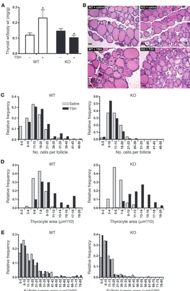
with TSH. While the weight of wild-type thyroid increased by about 100%, no increase was observed in Gαq/Gα11
-deficient thy-roids in response to TSH (Figure 5, A and B). Instead, thyroids of Tc-Gαq/Gα11–KO mice showed decreased weight, caused by
a slight reduction in follicle lumen size (see below). In wild-type animals, TSH treatment resulted in an increase in cell number as well as in cell size, while in Tc-Gαq/Gα11–KO mice, no increase
in cell proliferation was observed (Figure 5C). While the average number of thyrocytes per follicle increased from 13.3 to 17.2 after treatment of wild-type animals with TSH, the number of thyroids per follicle found under basal conditions in Tc-Gαq/Gα11
–KO ani-mals did not increase after treatment with TSH (11.8 and 10.9, respectively). However, Gαq/Gα11-deficient thyrocytes still showed
a hypertrophic response to TSH (Figure 5D). In addition, unlike wild-type thyroids, the colloid area in Tc-Gαq/Gα11–KO mice was
reduced after TSH treatment (Figure 5E).
To test the response of Gαq/Gα11
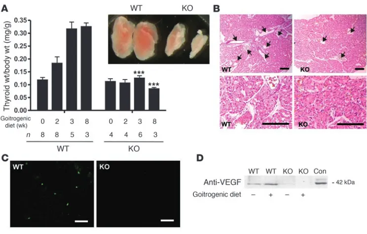
-deficient thyrocytes to inhi-bition of thyroid function, 1- to 2-month-old animals were fed a goitrogenic diet containing sodium perchlorate and methima-zole. After 3 weeks of goitrogenic diet, the thyroid weight of the wild-type animals increased more than 2-fold compared with ani-mals that received control diet (Figure 6A). Interestingly, in the Tc-Gαq/Gα11
–KO mice, there was no significant difference in thy-roid weight between the treated and untreated groups, indicating that Gαq/Gα11 proteins are required for goiter development. TSH
values increased 20-fold in wild-type and about 30-fold in Tc-Gαq/
Gα11–KO mice 2–3 weeks after starting the diet (data not shown).
The failure of Tc-Gαq/Gα11–KO mice to respond to goitrogenic diet
with an enlargement of the thyroid was accompanied by a lack of corresponding changes in the morphology of the thyroid (Figure 6B). While wild-type mice showed hyperplastic goiters characterized by colloid-depleted follicles, large vessels, and varying epithelial cell size, thyroids of Tc-Gαq/Gα11
–KO animals did not show cell pro-liferation and showed much less vascularization than did thyroids from wild-type animals fed the goitrogenic diet (Figure 6B). The lack of cell proliferation in Tc-Gαq/Gα11
–KO mice fed the goitro-genic diet was verified by the determination of BrdU incorporation (Figure 6C). While wild-type animals showed 10 ± 2.1 BrdU-positive cells per visual field in thyroid sections, less than 1 BrdU-positive cell per field was observed in Tc-Gαq/Gα11–KO mice and in mice
fed normal diet. Because goiter-associated angiogenesis requires the synthesis of various vascular growth factors by thyrocytes (25), we determined the amount of VEGF in thyroids from wild-type and Tc-Gαq/Gα11–KO mice before and 3 weeks after starting the
goitrogenic diet. As seen in Figure 6D, wild-type thyroids showed an increase in VEGF levels after treatment with goitrogenic diet. In contrast, in the absence of Gαq/Gα11 there was already a lower basal
level of VEGF in thyroid lysates, and treatment with goitrogenic diet did not induce any change in VEGF levels.
Discussion
In this study we have addressed the role of Gq/G11
-mediated signal- ing in the regulation of thyroid function by generating a condi-tional genetic mouse model lacking both Gαq and Gα11 proteins
in thyrocytes. While the deletion of genes encoding Gαq and Gα11
proteins occurred perinatally, the lack of Gq/G11 did not lead to
any obvious defect in postnatal development of the thyroid gland, as indicated by normal thyroid histology and normal serum TSH and thyroid hormone levels for up to 2 months. However, by that age, the thyroid-specific Gαq/Gα11-deficient mice showed impaired
thyroid hormone formation and secretion when acutely challenged with TSH. Starting at 2 months of age, most of the Tc-Gαq/Gα11–
KO animals slowly developed hypothyroidism, with elevated serum TSH levels and alteration in thyroid histology appearing later in life. Despite the highly elevated TSH levels at 6 months of age, the thyroid weight of the Gαq/Gα11-deficient mice was not increased.
In addition, the lack of Gαq/Gα11 proteins in thyrocytes prevented
thyroid growth when challenged with TSH or goitrogenic diet. While basal and TSH-stimulated iodine uptake was normal, the incorporation of iodine into thyroid proteins in response to TSH was impaired in Tc-Gαq/Gα11–KO mice. This indicates that the
rapid TSH-dependent stimulation of iodine uptake via the Na+/I–
symporter is not mediated by the Gq/G11-dependent pathway, but
rather involves Gs-mediated cAMP formation (26). In contrast to
the TSH-dependent regulation of iodine uptake, iodination in response to TSH requires signaling through the Gq/G11-mediated
pathway. This is consistent with earlier reports suggesting a regu-lation of peroxidase primarily through Ca2+ and PKC (27, 28).
Mice lacking the Gq/G11-mediated signaling pathway show
impaired thyroid hormone secretion in response to TSH. TSH-Figure 3
Histological analysis of thyroids from wild-type and Tc-Gαq/Gα11–KO
[image:5.585.49.263.81.418.2]induced thyroid hormone secretion is initiated by internalization of thyroglobulin via macropinocytosis (29–31). There is evidence that TSH-induced pinocytotic uptake of thyroglobulin and thy- roid hormone release are mediated by the cAMP-dependent path-way (7, 32). However, other mediators have also been suggested to play a role in processes leading to thyroid hormone secretion. In sheep thyroid cells, for example, [Ca2+]
i
has been shown to regu-late hormone secretion in vitro (33). Our results indicate that the Gq/G11-mediated signaling pathway in mice is required for the
macropinocytotic uptake of thyroglobulin in response to TSH. In addition to macropinocytosis, endocytotic processes can contribute to the uptake of thyroglobulin by thyrocytes (34). The recently described endocytosis of thyroglobulin via megalin is fol-lowed by the apical to basolateral transcytosis of thyroglobulin with low hormone content (35, 36). The transcytotic removal of hormone-poor thyroglobulin is believed to increase lysosomal deg- radation of hormone-rich thyroglobulin and hence hormone secre-tion. There is, however, no evidence that megalin or other endocytic receptors are regulated via G protein–mediated signaling pathways (37). During and after pinocytotic uptake of thyroglobulin, thy-roid hormone is released via enzymatic digestion by cathepsins (38, 39). The effect of the Gαq/Gα11-mediated signaling pathway on
proteolysis was not studied here, but our data indicate that the lack of Gαq/Gα11 proteins in thyrocytes impairs the process of secretion
already at the level of colloid uptake via pinocytosis.
The fact that serum TSH levels in 2-month-old animals were nor-mal while their response to TSH treatment was impaired suggests that under normal in vivo conditions the thyroid can fully com-pensate the partial defect in TSH responsiveness for a variable time period. However, challenge with high, supraphysiological TSH concentrations revealed the defect even when thyroid function in vivo was completely normal and the defect was compensated.
Abnormal thyrocyte cell proliferation underlies a variety of dis-eases, including various forms of goiter and thyroid neoplasia. In many cases thyroid proliferation has been shown to be under the control of TSH, and the cAMP-mediated signaling pathway is believed to play a predominant role in the mitogenic effects exerted by TSH (6, 40, 41). The induction of thyroid growth by a goitrogen-ic diet consisting of thyrostatic drugs is thought to be initiated by increased pituitary secretion of TSH, which results in thyroid cell hyperplasia accompanied by hypervascularization caused by angio- genesis within 1–2 weeks (42, 43). The lack of thyrocyte prolifera-tion in Tc-Gαq/Gα11–KO mice in response to TSH or goitrogenic
diet indicates that downstream mediators of the Gq/G11-mediated
signaling pathway are required for TSH-induced thyroid cell pro-liferation as well as for thyroid growth in response to goitrogenic diet. This is consistent with previous data indicating that Gq/G11
can mediate mitogenic effects in different cell types (44, 45). Other than the follicular cell hyperplasia, thyroid enlargement
caused by nontoxic goiter is also characterized by an early vascu-Figure 4
Cellular effects of TSH in wild-type and Tc-Gαq/
Gα11–KO mice. (A–C) Iodine uptake (A),
organi-fication (B), and thyroid hormone release (C) were
determined as described in Methods. TSH was administered i.p. at a dose of 100 mIU/100 g body weight. As a control, organification was suppressed by treatment of animals with propylthiouracil (PTU; 0.4 mg/10 g body weight i.p. –24, –12, and 0 h
rela-tive to TSH administration). *P < 0.05, ***P < 0.001
versus non–TSH-treated (A and B) or wild-type
(C). Results in A–C are representative of
experi-ments performed at least 3 times with 3–4 animals
per group. (D) Periodic acid-Schiff–stained colloid
droplets in follicle epithelial cells of thyroid from
wild-type and Tc-Gαq/Gα11–KO animals treated
or not with TSH (100 mIU/10 g body weight) for
5 hours (n = 6). ***P < 0.001 versus wild-type. (E)
Representative sections of wild-type and Tc-Gαq/
Gα11–KO animals treated for 5 hours with TSH
[image:6.585.43.350.77.444.2]research article
lar response resulting in hypervascularization and abnormally enlarged blood vessels. There is good evidence that the goiter-asso-ciated angiogenesis is initiated by the production of endothelial growth factors including VEGF by thyrocytes exposed to increased TSH levels (25, 46, 47). The fact that no increase in VEGF levels in response to goitrogenic diet was observed in Tc-Gαq/Gα11–KO
mice indicates that induction of VEGF expression requires an intact Gq/G11-mediated signaling pathway.
The analysis of the intracellular signaling mechanisms regulating thyroid follicular cell function and growth has led to the concept that the Gs-dependent cAMP-mediated signaling pathway is the principal
mechanism through which TSH and other factors acting via G pro-Figure 5
Role of Gαq/Gα11 in
TSH-induced thyroid growth.
Wild-type and Tc-Gαq/Gα11–KO
mice were treated or not with TSH (50 mIU/10 g body weight)
twice daily for 7 days. (A)
Ani-mals were sacrificed and thy-roid weights were determined.
Values are mean ± SEM (n = 4
per group). (B) Thyroids were
fixed, sectioned, and stained with hematoxylin and eosin.
Scale bars: 50 μm. (C–E) In
stained sections, the number
of cells per follicle (C), the
indi-vidual thyrocyte area (D), and
the follicle lumen area (E) were
morphometrically analyzed. See
Methods for details. *P < 0.05
[image:7.585.42.414.78.646.2]tein–coupled receptors increase thyroid function and growth. Our data, based on a thyrocyte-specific knockout of the Gq/G11
-medi-ated signaling pathway, reveal an essential role of these G proteins in mediating the regulation of thyroid function. We show that Gq/G11
- mediated signaling processes are required for thyroid hormone for-mation and release as well as for the adaptive growth of the thyroid. Thus, inhibition of Gq/G11-mediated signaling processes or their
downstream effectors may be a promising strategy to interfere with diseases characterized by increased thyroid function and/or growth.
Methods
Experimental animals and treatments. Procedures of animal care and use in this study were approved by the Regierungspräsidium Karls-ruhe (Karls-ruhe, Germany). For the analysis of serum hormone levels, 100 μl of blood was drawn from the retroorbital plexus under anesthesia with xylazine hydro-chloride (3 mg/kg body weight) and ketamine hydrochloride (100 mg/kg body weight). For collecting tissue samples, mice were sacrificed with CO2. To induce goiter, a group of mice was treated with 5 g/l sodium perchlorate and 0.05 g/l methimazole (Sigma-Aldrich) in the drinking water.
Generation of mice expressing Cre recombinase in thyrocytes . In order to gener- ate a mouse line expressing Cre recombinase specifically in thyroid epithe- lial cells, a 100-kb PAC containing the mouse thyroglobulin locus was iso-lated from the mouse genomic RPCI21 library using a mouse thyroglobulin EST probe (48). The coding sequence of the iCre recombinase (49), followed by the bovine growth hormone polyadenylation signal and an ampicillin resistance cassette (bla) flanked by frt sites (50), was amplified twice using nested PCR to fuse sequences from the proximal promoter and first intron
of the gene encoding thyroglobu-lin to the iCre-frt-bla-frt construct. By homologous recombination in E. coli (51, 52), this fragment was introduced into the Tg PAC in such a way that the ATG of the iCre matched the Tg ATG. After removal of the selection cassette by Flp-mediated recombination, the recombined PAC containing the Tg-Cre gene and the Cre cDNA was injected into pronuclei of fertilized mouse oocytes. Mice were analyzed for PAC insertion by Southern blot and genomic PCR amplification and backcrossed into the C57BL/6 mouse genomic background.
Histology, tissue staining, and BrdU labeling. Cre recombinase activity was evaluated by crossing Tg-Cre mice with the Gt(ROSA)26Sor Cre reporter mouse line (The Jackson Laboratory) (24). Animals were histologically analyzed for lacZ expression in different tissues using whole mount β-galactosidase staining as described previously (24). For the evaluation of thyroid histology, thyroids were fixed in 4% paraformaldehyde and stained with hematoxylin and eosin. For the counting of droplets, fixed and sec-tioned thyroids were treated with periodic acid-Schiff stain as previously described by Gerber et al. (53). Histo-logical morphometry was performed using NIH ImageJ software (http://rsb. info.nih.gov/ij/) on 5 randomly selected slides from 3 thyroids of each group. From each slide, 30 thyroid follicles were analyzed, and the following param-eters were determined and quantified: colloid-containing area, whole follicle area, thyrocyte area (whole follicle area minus colloid area), number of visible nuclei, and average thyrocyte size (thyrocyte cell area divided by number of visible nuclei). For BrdU labeling, mice were injected i.p. with 10 μg BrdU per kg body weight and sacrificed 1 hour after injection. Paraffin sections of the thyroid glands were stained with anti-BrdU antibody (BD Biosciences).
Determination of TSH, thyroid hormones, and cAMP levels . Serum TSH con- centrations were measured using a specific mouse TSH radioimmunoas-say provided by A.F. Parlow (Pituitary and Antisera Center, Harbor-UCLA Medical Center, Los Angeles, California, USA). The serum thyroid hor-mones T4 and T3 were measured with a commercial ELISA (Trinity Biotech) according to the manufacturer’s instructions.
Culture of mouse primary thyrocytes and determination of [Ca2+]i and cAMP
[image:8.585.43.415.80.318.2]levels. Mouse primary thyrocytes were prepared as described previously (54). For cAMP determinations, the cells were washed with PBS, and the medium was replaced with modified Coon’s F12 medium containing 0.1% bovine serum albumin and 0.5 mM 3-isobutyl-1-methylxanthine in the absence or presence of the indicated concentrations of bovine TSH. For cAMP mea-surements, media were collected after 1 or 3 hours, boiled for 5 minutes, and stored at –80°C until measurement. The cAMP concentration were determined using an ELISA system from Cayman Europe. Cells grown on 10-mm-diameter coverslips were washed twice with HBSS (pH 7.4) and incubated in the same buffer with 3 mM fura-2/AM containing 0.04% (w/v)
Figure 6
Role of Gαq/Gα11 in goiter development. (A) Wild-type and Tc-Gαq/Gα11–KO mice were treated with
goi-trogenic diet 0–8 weeks, and thyroid weights were determined thereafter. Inset: thyroids of wild-type and
Tc-Gαq/Gα11–KO mice after 8 weeks of treatment. Values are mean ± SEM. ***P < 0.001 versus wild-type.
(B) Sections of thyroids from wild-type and Tc-Gαq/Gα11–KO mice after 2 weeks of goitrogenic treatment.
Arrows indicate blood vessels. (C) After 20 days of goitrogenic treatment, wild-type and Tc-Gαq/Gα11–KO
mice were injected with BrdU, and thyroid sections were prepared and stained with anti-BrdU antibodies
to determine cell proliferation. Scale bars: 125 μm. (D) Thyroid homogenates from wild-type and Tc-Gαq/
Gα11–KO mice treated with normal or goitrogenic diet for 8 weeks were separated on SDS-PAGE and
research article
Pluronic F-127. After a 30-minute incubation at 37°C, the cells were washed twice and incubated for another 30 minutes. To monitor the changes in [Ca2+]i, the coverslips were mounted in a holder and the fluorescence at 510 nm was measured. The excitation wavelength alternated between 340 and 380 nm in intervals of 500 ms. Changes in [Ca2+]i are given as the ratio of 340-nm to 380-nm intensities.
Western blotting. For Western blot analyses, thyroid gland lysates were analyzed by Western blot using anti-Gαq/Gα11 and anti-VEGF antibod-ies (Santa Cruz Biotechnology Inc.) and antibodies against ERK1/2 (Cell Signaling Technology).
Iodine uptake and iodination. For iodine uptake experiments, Na 125I was administered i.p. (0.1 mCi/10 g body weight) with or without TSH (100 mIU/100 g body weight) 3 hours before the mice were sacrificed. To analyze incorporation of iodine, animals were first treated with TSH (100 mIU/10 g body weight) twice, 24 and 12 hours before administration of 125I, together with a third dose of TSH; 5 hours later, animals were sacrificed. The whole thyroid gland and a piece of liver were dissected, weighed, and counted in a γ counter. An uptake index was calculated as counts of thyroid per weight of thyroid tissue divided by counts of liver per weight of liver. Organification was determined after trichloroacetic acid (10% w/v) precipitation of thyroid homogenates as previously described (55). Statistics. The Statview program (Windows version 4.57, Abacus Concepts Inc.) was used for ANOVA and for Fisher’s protected least-significant-dif-ference post-hoc tests. A P value less than 0.05 was considered significant. Results are presented as mean ± SEM. Acknowledgments The skillful technical assistance of Karin Meyer and Rose LeFau-cheur is gratefully acknowledged. This study was supported by a grant from the Serono Foundation to J. Kero. Radioimmunoas- say kits for thyroid hormone determinations were kindly sup- plied by A.F. Parlow through the National Institute of Diabe-tes and Digestive and Kidney Diseases National Hormone and Peptide Program. Received for publication September 18, 2006, and accepted in revised form May 29, 2007. Address correspondence to: Stefan Offermanns, Institute of Phar-macology, University of Heidelberg, Im Neuenheimer Feld 366, 69120 Heidelberg, Germany. Phone: 49-6221-54-8246; Fax: 49-6221-54-8549; E-mail: [email protected]. Sorin Tunaru’s present address is: Department of Molecular Biol-ogy, The Scripps Research Institute, La Jolla, California, USA. Tim Wintermantel’s present address is: Schering AG, Berlin, Germany. Erich Greiner’s present address is: Evotec OAI AG, Hamburg, Germany. 1. Larsen, P.R., Davies, T.F., Schlumberger, M.-J., and Hay, I.D. 2003. Thyroid physiology and diagnostic evaluation of patients with thyroid disorders. In
Williams textbook of endocrinology. P.R. Larsen, H.M. Kronenberg, S. Melmed, and K.S. Polonsky, editors. W.B. Saunders. Philadelphia, Pennsylvania, USA. 331–373.
2. Spaulding, S.W. 2005. Biological actions of thyro-tropin. In Werner and Ingbar’s the thyroid: a fundamen-tal and clinical text. L.E. Bravermen and R.D. Utiger, editors. Lippincott Williams & Wilkins. Philadel-phia, Pennsylvania, USA. 183–197.
3. Jemec, B. 1980. Studies of the goitrogenic and tumorigenic effect of two goitrogens in combina-tion with hypophysectomy or thyroid hormone treatment. Cancer. 45:2138–2148.
4. Marians, R.C., et al. 2002. Defining thyrotropin- dependent and -independent steps of thyroid hor-mone synthesis by using thyrotropin receptor-null mice. Proc. Natl. Acad. Sci. U. S. A. 99:15776–15781.
5. Postiglione, M.P., et al. 2002. Role of the thyroid- stimulating hormone receptor signaling in devel-opment and differentiation of the thyroid gland.
Proc. Natl. Acad. Sci. U. S. A. 99:15462–15467. 6. Dumont, J.E., Lamy, F., Roger, P., and Maenhaut, C.
1992. Physiological and pathological regulation of thyroid cell proliferation and differentiation by thy-rotropin and other factors. Physiol. Rev. 72:667–697.
7. Vassart, G., and Dumont, J.E. 1992. The thyrotro- pin receptor and the regulation of thyrocyte func-tion and growth. Endocr. Rev. 13:596–611. 8. Parmentier, M., et al. 1989. Molecular cloning of
the thyrotropin receptor. Science. 246:1620–1622. 9. Davies, T.F., Ando, T., Lin, R.Y., Tomer, Y., and Latif,
R. 2005. Thyrotropin receptor-associated diseases: from adenomata to Graves disease. J. Clin. Invest. 115:1972–1983. doi:10.1172/JCI26031.
10. Weinstein, L.S., Chen, M., Xie, T., and Liu, J. 2006. Genetic diseases associated with heterotrimeric G proteins. Trends Pharmacol. Sci. 27:260–266. 11. Germain-Lee, E.L., et al. 2005. A mouse model of albright hereditary osteodystrophy generated by targeted disruption of exon 1 of the Gnas gene. Endocrinology. 146:4697–4709. 12. Ledent, C., Dumont, J.E., Vassart, G., and Parmentier, M. 1992. Thyroid expression of an A2 adenosine receptor transgene induces thyroid hyperplasia and hyperthyroidism. EMBO J. 11:537–542.
13. Michiels, F.M., et al. 1994. Oncogenic potential of guanine nucleotide stimulatory factor alpha subunit in thyroid glands of transgenic mice. Proc. Natl. Acad. Sci. U. S. A. 91:10488–10492.
14. Zeiger, M.A., et al. 1997. Thyroid-specific expres-sion of cholera toxin A1 subunit causes thyroid hyperplasia and hyperthyroidism in transgenic mice. Endocrinology. 138:3133–3140.
15. Spiegel, A.M. 1996. Defects in G protein-coupled signal transduction in human disease. Annu. Rev. Physiol. 58:143–170.
16. Farfel, Z., Bourne, H.R., and Iiri, T. 1999. The expanding spectrum of G protein diseases. N. Engl. J. Med. 340:1012–1020.
17. Weinstein, L.S., Yu, S., Warner, D.R., and Liu, J. 2001. Endocrine manifestations of stimulatory G protein alpha-subunit mutations and the role of genomic imprinting. Endocr. Rev. 22:675–705. 18. Laurent, E., Mockel, J., Van Sande, J., Graff, I., and
Dumont, J.E. 1987. Dual activation by thyrotropin of the phospholipase C and cyclic AMP cascades in human thyroid. Mol. Cell. Endocrinol. 52:273–278. 19. Van Sande, J., et al. 1992. Thyroid stimulating
immunoglobulins, like thyrotropin activate both the cyclic AMP and the PIP2 cascades in CHO cells expressing the TSH receptor. Mol. Cell. Endocrinol. 88:R1–R5.
20. Allgeier, A., et al. 1994. The human thyrotropin receptor activates G-proteins Gs and Gq/11. J. Biol. Chem. 269:13733–13735. 21. Ledent, C., et al. 1997. Costimulation of adenylyl cyclase and phospholipase C by a mutant alpha 1B-adrenergic receptor transgene promotes malignant transformation of thyroid follicular cells. Endocri-nology. 138:369–378. 22. Offermanns, S., et al. 1998. Embryonic cardiomyo-cyte hypoplasia and craniofacial defects in G alpha q/G alpha 11-mutant mice. EMBO J. 17:4304–4312. 23. Wettschureck, N., et al. 2001. Absence of pressure
overload induced myocardial hypertrophy after
conditional inactivation of Galphaq/Galpha11 in cardiomyocytes. Nat. Med. 7:1236–1240. 24. Soriano, P. 1999. Generalized lacZ expression with the
ROSA26 Cre reporter strain. Nat. Genet. 21:70–71. 25. Ramsden, J.D., et al. 2005. Complete inhibition
of goiter in mice requires combined gene therapy modification of angiopoietin, vascular endothelial growth factor, and fibroblast growth factor signal-ing. Endocrinology. 146:2895–2902.
26. Dohan, O., and Carrasco, N. 2003. Advances in Na(+)/I(–) symporter (NIS) research in the thyroid and beyond. Mol Cell. Endocrinol. 213:59–70. 27. Lippes, H.A., and Spaulding, S.W. 1986. Peroxide
formation and glucose oxidation in calf thyroid slices: regulation by protein kinase-C and cytosolic free calcium. Endocrinology. 118:1306–1311. 28. Takasu, N., Yamada, T., Shimizu, Y., Nagasawa, Y.,
and Komiya, I. 1989. Generation of hydrogen per-oxide in cultured porcine thyroid cells: synergistic regulation by cytoplasmic free calcium and protein kinase C. J. Endocrinol. 120:503–508.
29. Nadler, N.J., Sarkar, S.K., and Leblond, C.P. 1962. Origin of intracellular colloid droplets in the rat thyroid. Endocrinology. 71:120–129.
30. van den Hove, M.F., Couvreur, M., de Visscher, M., and Salvatore, G. 1982. A new mechanism for the reabsorption of thyroid iodoproteins: selective fluid pinocytosis. Eur. J. Biochem. 122:415–422. 31. Wetzel, B.K., Spicer, S.S., and Wollman, S.H. 1965.
Changes in fine structure and acid phosphatase localization in rat thyroid cells following thyrotro-pin administration. J. Cell Biol. 25:593–618. 32. Dumont, J.E., Willems, C., Van Sande, J., and Neve, P.
1971. Regulation of the release of thyroid hormones: role of cyclic AMP. Ann. N. Y. Acad. Sci. 185:291–316. 33. Eggo, M.C., Lippes, H., and Burrow, G.N. 1992.
Control of thyroid secretion: effects of stimulators of protein kinase C, thyrotropin, and calcium mobi-lization on secretion of iodinated compounds from sheep thyroid cells. Endocrinology. 130:2274–2283. 34. Marino, M., and McCluskey, R.T. 2000. Role of
35. Lisi, S., et al. 2003. Preferential megalin-mediated transcytosis of low-hormonogenic thyroglobulin: a control mechanism for thyroid hormone release.
Proc. Natl. Acad. Sci. U. S. A. 100:14858–14863. 36. Lisi, S., et al. 2005. Thyroid dysfunction in megalin
deficient mice. Mol. Cell. Endocrinol. 236:43–47. 37. May, P., Herz, J., and Bock, H.H. 2005. Molecular
mechanisms of lipoprotein receptor signalling.
Cell. Mol. Life Sci. 62:2325–2338.
38. Brix, K., Lemansky, P., and Herzog, V. 1996. Evi- dence for extracellularly acting cathepsins mediat- ing thyroid hormone liberation in thyroid epithe-lial cells. Endocrinology. 137:1963–1974.
39. Friedrichs, B., et al. 2003. Thyroid functions of mouse cathepsins B, K, and L. J. Clin. Invest. 111:1733–1745. doi:10.1172/JCI200315990.
40. Kimura, T., et al. 2001. Regulation of thyroid cell pro- liferation by TSH and other factors: a critical evalua-tion of in vitro models. Endocr. Rev. 22:631–656. 41. Rivas, M., and Santisteban, P. 2003. TSH-activated
signaling pathways in thyroid tumorigenesis. Mol. Cell. Endocrinol. 213:31–45.
42. Wollman, S.H., Herveg, J.P., Zeligs, J.D., and Eric-son, L.E. 1978. Blood capillary enlargement during the development of thyroid hyperplasia in the rat.
Endocrinology. 103:2306–2314.
43. Redmond, O., and Tuffery, A.R. 1981. Thyroid pro-liferation, body weight, thyrotropin and thyroid hormones in chronic antithyroid (carbimazole) treatment in rats. J. Anat. 133:37–47.
44. van Biesen, T., Luttrell, L.M., Hawes, B.E., and Lefkowitz, R.J. 1996. Mitogenic signaling via G pro-tein-coupled receptors. Endocr. Rev. 17:698–714. 45. Gudermann, T., Grosse, R., and Schultz, G. 2000.
Contribution of receptor/G protein signaling to cell growth and transformation. Naunyn Schmiede-bergs Arch. Pharmacol. 361:345–362.
46. Sato, K., et al. 1995. Stimulation by thyroid-stimu-lating hormone and Grave’s immunoglobulin G of vascular endothelial growth factor mRNA expres-sion in human thyroid follicles in vitro and flt mRNA expression in the rat thyroid in vivo. J. Clin. Invest. 96:1295–1302.
47. Viglietto, G., et al. 1997. Upregulation of the angio-genic factors PlGF, VEGF and their receptors (Flt-1, Flk-1/KDR) by TSH in cultured thyrocytes and in the thyroid gland of thiouracil-fed rats suggest a TSH-dependent paracrine mechanism for goiter hypervascularization. Oncogene. 15:2687–2698. 48. Vente, A., Korn, B., Zehetner, G., Poustka, A., and
Lehrach, H. 1999. Distribution and early develop-ment of microarray technology in Europe. Nat. Genet. 22:22.
49. Shimshek, D.R., et al. 2002. Codon-improved Cre
recombinase (iCre) expression in the mouse. Gen-esis. 32:19–26.
50. Wintermantel, T.M., Mayer, A.K., Schutz, G., and Greiner, E.F. 2002. Targeting mammary epithelial cells using a bacterial artificial chromosome. Gen-esis. 33:125–130.
51. Muyrers, J.P., Zhang, Y., Testa, G., and Stewart, A.F. 1999. Rapid modification of bacterial artificial chromosomes by ET-recombination. Nucleic Acids Res. 27:1555–1557.
52. Zhang, Y., Buchholz, F., Muyrers, J.P., and Stewart, A.F. 1998. A new logic for DNA engineering using recombination in Escherichia coli. Nat. Genet. 20:123–128.
53. Gerber, H., Peter, H.J., Bachmeier, C., Kaempf, J., and Studer, H. 1987. Progressive recruitment of follicular cells with graded secretory responsive-ness during stimulation of the thyroid gland by thyrotropin. Endocrinology. 120:91–96.
54. Jeker, L.T., Hejazi, M., Burek, C.L., Rose, N.R., and Caturegli, P. 1999. Mouse thyroid primary culture.
Biochem. Biophys. Res. Commun. 257:511–515.