How does blood glucose control with insulin
save lives in intensive care?
Greet Van den Berghe
J Clin Invest.
2004;
114(9)
:1187-1195.
https://doi.org/10.1172/JCI23506
.
Patients requiring prolonged intensive care are at high risk for multiple organ failure and
death. Insulin resistance and hyperglycemia accompany critical illness, and the severity of
this “diabetes of stress” reflects the risk of death. Recently it was shown that preventing
hyperglycemia with insulin substantially improves outcome of critical illness. This article
examines some potential mechanisms underlying prevention of glucose toxicity as well as
the effects of insulin independent of glucose control. Unraveling the molecular mechanisms
will provide new insights into the pathogenesis of multiple organ failure and open avenues
for novel therapeutic strategies.
Science in Medicine
Find the latest version:
Science in medicine
How does blood glucose control with insulin
save lives in intensive care?
Greet Van den Berghe
Department of Intensive Care Medicine, Catholic University of Leuven, Leuven, Belgium.
Patients requiring prolonged intensive care are at high risk for multiple organ failure and death.
Insulin resistance and hyperglycemia accompany critical illness, and the severity of this “diabetes
of stress” reflects the risk of death. Recently it was shown that preventing hyperglycemia with
insulin substantially improves outcome of critical illness. This article examines some potential
mechanisms underlying prevention of glucose toxicity as well as the effects of insulin independent
of glucose control. Unraveling the molecular mechanisms will provide new insights into the
patho-genesis of multiple organ failure and open avenues for novel therapeutic strategies.
Historical introduction
The 1952 Scandinavian epidemic of poliomyelitis necessitated mechanical ventilation of a large number of patients with respi-ratory failure, an intervention that reduced mortality from 80% to 40% (1). Since then, development of sophisticated mechanical devices to support all vital organ functions, a wide array of pow-erful drugs, and high-tech monitoring systems have revolution-ized modern intensive-care medicine. This evolution improved short-term survival of previously lethal conditions such as mul-tiple trauma, extensive burns, major surgery, and severe sepsis. Many patients nowadays indeed survive the initial shock phase of such conditions but often subsequently enter a chronic phase of critical illness. Mortality among such patients requiring inten-sive care for more than a few days has remained around 20% worldwide, to a large extent irrespective of the initial disease or trauma for which they were admitted to the intensive care unit (ICU). Most deaths in the ICU occurring beyond the first few days of critical illness are attributable to nonresolving failure of multiple organ systems, either due to or coinciding with sepsis. An increased susceptibility to infectious complications and the functional and structural sequelae of the systemic inflammatory response to infection and cellular injury play a role (2). Several lines of evidence support the concept that disturbed cellular energy metabolism contributes to organ failure (3, 4). This was originally ascribed exclusively to inadequate tissue perfusion and cellular hypoxia. Recent studies, however, also point to a distur-bance in oxygen utilization rather than delivery, which has been termed cytopathic hypoxia (3, 5, 6). Although extensive research efforts during the last decade focused on strategies to prevent or reverse the potentially lethal multiple organ failure, only few of them revealed positive results (7–10). One of these strategies is tight blood glucose control with insulin (10).
Insulin resistance and hyperglycemia in the critically ill
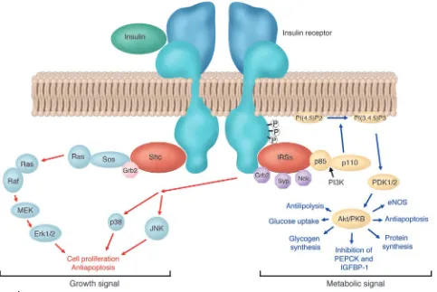
One hundred fifty years ago, Reyboso observed glucosuria, a con-dition induced by ether anesthesia, in which glucose is discharged in the urine, and in 1877 Claude Bernard described hyperglycemia during hemorrhagic shock (11). Today, it is well known that any type of acute illness or injury results in insulin resistance, glucose intolerance, and hyperglycemia, a constellation termed “diabetes of injury” (12, 13). Illness or trauma increases hepatic glucose production with ongoing gluconeogenesis despite hyperglycemia and abundantly released insulin. Hepatic insulin resistance is fur-ther characterized by elevated circulating levels of IGF–binding protein–1 (IGFBP-1) (14, 15). Also, in skeletal muscle and heart, insulin-stimulated glucose uptake is impaired (16, 17). Overall, glucose uptake in critically ill patients, however, is increased but takes place mainly in the tissues that are not dependent on insulin for glucose uptake, such as, among others, the nervous system and the blood cells (13, 18). The most severe cases of stress-induced hyperglycemia (13) and highest levels of circulating IGFBP-1 (14, 15) are observed in patients with the highest risk of death. Orchestrated “counterregulatory” hormonal responses, cytokine release, and signals from the nervous system, all affecting glucose metabolic pathways, bring about the diabetes of injury. The hor-mones involved include catecholamines, cortisol, glucagon, and growth hormone (GH). Proinflammatory cytokines affect glucose homeostasis indirectly, by stimulating counterregulatory hor-mone secretion, and directly, by altering insulin receptor signal-ing (Figure 1) (13, 19, 20). Although insulin receptor signalsignal-ing is still incompletely understood, generation of SOCS-1 and SOCS-3 may be involved. Indeed, IL-6–stimulated SOCS-3 generation has been shown to inhibit insulin receptor tyrosine phosphorylation and downstream signal transduction (21), and both SOCS-1 and SOCS-3 have been shown to degrade insulin receptor substrate–1 (IRS-1) and IRS-2 (22). Furthermore, both endogenous and exog-enous catecholamines promptly inhibit insulin secretion from β
cells, and catecholamines as well as angiotensin II exert anti-insu-lin effects. Abnormalities in insuanti-insu-lin signaanti-insu-ling have been described in a rat model of critical illness resulting from prolonged admin-istration of a nonlethal dose of endotoxin and concomitant star-vation partly mimicking the condition of human sepsis (23). In the liver, abundance and tyrosine phosphorylation of the insu-lin receptor, IRS-1, and IRS-2 were reduced. Furthermore, there was reduced association of PI3K with IRS-1. In skeletal muscle,
Nonstandard abbreviations used: CRP, C-reactive protein; GABA, γ-aminobutyric acid; GH, growth hormone; GHBP, GH-binding protein; GIK, glucose, insulin, and potassium; HXK-II, hexokinase II; ICU, intensive care unit; IGFBP-1, IGF–binding protein–1; IRS-1, insulin receptor substrate–1; MnSOD, manganese superoxide dismutase; PEPCK, phosphoenolpyruvate carboxykinase; VDAC, voltage-dependent anion channel.
Conflict of interest: The author holds an unrestrictive Catholic University of Leuven — Novo Nordisk Chair of Research.
science in medicine
similar abnormalities were observed, except that the number of insulin receptors and the abundance of the IRSs were normal. Furthermore, the insulin signaling defects in the muscle were not seen when rats were studied shortly after injection of endotoxin or under conditions of adequate nutrition (23). Apart from these insights generated from in vitro or animal models, little is known about the exact molecular basis of insulin resistance in critically ill patients. Type 2 diabetes mellitus, and to a lesser extent obesity, are also characterized by hyperglycemia, reduced glucose uptake and oxidation, unsuppressed gluconeogenesis, and impaired gly-cogen and NO synthesis. Here, the metabolic consequences of insulin resistance are mediated predominantly by abnormalities along the IRS-1–PI3K pathway of insulin signaling (Figure 1). However, a disrupted PI3K pathway does not necessarily mean that the other insulin signaling pathways are equally unresponsive. Indeed, signaling through the Ras-MAPK cascade, for example, via Erk 1 and Erk 2, may retain normal sensitivity. Compensatory hyperinsulinemia may thus still exert mitogenic actions in certain cell types, while the PI3K-dependent metabolic actions of insulin are suppressed (24–28). This discrepancy may occur in vascular smooth muscle cells and in specific capillary endothelial cells of
patients with type 2 diabetes and obesity. Proliferation of retinal capillary endothelial cells results in microaneurysms and neovas-cularization. Excessive proliferation of arterial smooth muscle cells and increased extracellular matrix lead to atherosclerosis. Hence, compensatory hyperinsulinemia, due to metabolic insulin resistance, may contribute to some of the vascular complications of type 2 diabetes and obesity through overstimulation of the mitogenic, insulin-sensitive MAPK signaling pathway. Whether or not insulin resistance is similarly “selective” during critical illness, and whether hyperinsulinemia exerts deleterious effects through this pathway in the acute setting of severe illness, is at present unknown. The diabetes of injury used to be interpreted as an adaptive stress response and as such important for survival. Par-ticularly, the overall increase in glucose turnover and the fact that hyperglycemia persists despite abundantly released insulin were considered arguments in favor of tolerating moderately elevated blood glucose levels during critical illness. Indeed, if one considers hyperglycemia of injury as beneficial in promoting cellular glu-cose uptake in non–insulin-dependent tissues, tolerating modest degrees of hyperglycemia is beneficial. Consequently, blood glu-cose concentrations of 160–200 mg/dl were recommended to
max-Figure 1
[image:3.585.54.537.83.406.2]science in medicine
imize cellular glucose uptake while avoiding hyperosmolarity (18). In addition, moderate hyperglycemia was often viewed as a buffer against hypoglycemia-induced brain damage. In 2001, however, the critical care community was forced to reconsider this dogma (29), as a large, randomized, controlled, clinical study — hereaf-ter referred to as “the Leuven study” — showed that preventing even moderate hyperglycemia during critical illness substantially improved outcome (10).
Intensive insulin therapy in critical illness: clinical benefits
The Leuven study of critically ill patients, the majority of whom did not previously have diabetes, showed that titrating insulin infusion during intensive care to strict normoglycemia (below 110 mg/dl) strikingly reduced mortality when compared with the con-ventional insulin treatment (Figure 2). The latter comprised insu-lin infusion only when blood glucose exceeded 200 mg/dl, leading to average blood glucose levels of 150–160 mg/dl (10). Although the Diabetes Mellitus, Insulin Glucose Infusion in Acute Myocar-dial Infarction study previously showed that avoiding excessive hyperglycemia (blood glucose >200 mg/dl) after acute myocardial infarction in patients with diabetes mellitus improved long-term outcome (30), the Leuven study (10) on nondiabetic ICU patients aimed for a much lower level of blood glucose. The benefit of inten-sive insulin therapy in the ICU was particularly apparent among patients with prolonged critical illness, requiring intensive care for more than 5 days, with mortality reduced from 20.2% to 10.6%. Besides saving lives, intensive insulin therapy also prevented com-plications such as severe nosocomial infections, acute renal failure, liver dysfunction, critical illness polyneuropathy, muscle weak-ness, and anemia and thus reduced the time that patients were dependent on intensive care. Although intensive insulin therapy induced a slightly higher incidence of hypoglycemia as compared with the conventional approach, these episodes were never associ-ated with clinically relevant sequelae. Indeed, the use of an insulin titration algorithm guaranteed that when hypoglycemia occurred, it was always quickly resolved. Although the Leuven study includ-ed a large number of patients recovering from cardiac surgery, the beneficial effects of strict glucose control were equally present in most other diagnostic subgroups. Furthermore, Krinsley recently confirmed the survival benefit of implementing tight blood glu-cose control with insulin in a mixed medical-surgical intensive care population (31).The substantial improvement of outcome with such a simple measure was considered major progress in the mod-ern era of intensive care.
Mechanisms explaining the acute life-saving effects of intensive insulin therapy in the ICU
One immediately wonders how such a simple intervention — pre-venting a moderate degree of hyperglycemia with insulin — dur-ing the relatively short time a patient is in intensive care was able to prevent the most feared complications such as sepsis, multiple organ failure, and death. Normal cells are relatively protected from deleterious effects of brief exposure to moderate hyperglycemia by a downregulation of glucose transporters (32). In diabetes melli-tus, prolonged untreated hyperglycemia contributes to the devel-opment of chronic debilitating complications. However, except in embryonic development, where hyperglycemia causes acute toxicity, the time required for hyperglycemia to cause disorders in patients with diabetes is several orders of magnitude longer than
the time it took to prevent life-threatening complications with insulin therapy in the ICU. In order to understand how metabolic control with insulin proved to be so acutely protective in the criti-cally ill, the following key questions must be answered. Is glycemic control crucial in bringing about the clinical benefits of intensive insulin therapy, or is blood glucose control epiphenomenal to the other metabolic and nonmetabolic effects of insulin? Are there fac-tors predisposing the critically ill to hyperglycemia-induced toxic-ity? What are the common pathways mediating the plethora of clinical benefits of intensive insulin therapy?
Preventing direct glucose toxicity plays a crucial role
Clinical evidence. A post hoc analysis of the Leuven study (33) revealed a linear correlation between the degree of hyperglycemia and the risk of death, which persisted after correction for insulin dose and sever-ity of illness scores. Patients in the conventional insulin treatment group who showed only moderate hyperglycemia (110–150 mg/dl or 6.1–8.3 mmol/l) had a lower risk of death than those with frank hyperglycemia (150–200 mg/dl or >8.3 mmol/l) but a higher risk of death than those who were intensively treated with insulin to restore blood glucose levels to below 110 mg/dl (6.1 mmol/l) (33). Similarly, for the prevention of morbidity effects such as acute renal failure, bacteremia, and anemia, it appeared crucial to reduce blood glucose to below 110 mg/dl. The risk of developing critical illness polyneu-ropathy in particular correlated linearly with blood glucose levels (34). Multivariate logistic regression analysis confirmed the indepen-dent role of blood glucose control in achieving most of the clinical benefits of intensive insulin therapy and underlined the importance of lowering the blood glucose level to strict normoglycemia.
[image:4.585.307.528.458.619.2]Target tissue responsiveness to insulin in the critically ill. Since critically ill patients suffer from hepatic and skeletal muscle insulin resis-tance, the mechanism by which insulin lowered blood glucose in
Figure 2
Intensive insulin therapy saves lives in the intensive care unit. Kaplan-Meier curves show cumulative survival of 1,548 patients from the Leuven study who received intensive insulin treatment (blood glucose maintained below 110 mg/dl; yellow) or conventional insulin treatment (insulin only given when blood glucose exceeded 200 mg/dl, result-ing in mean blood glucose levels of 150–160 mg/dl; green) durresult-ing their ICU or hospital stay. The upper panels display results from all patients; the lower panels display results for long-stay (>5 days) ICU patients only. P values were determined with the use of the Mantel-Cox log-rank test. Adapted with permission from the New England
science in medicine
these patients is not obvious. Analysis of liver and skeletal muscle biopsies, obtained immediately after death from nonsurvivors in the Leuven study, indicated that the classical insulin-regulated metabolic pathways in the liver did not respond to insulin (35, 36). For example, expression of IGFBP-1, normally under the inhibitory control of insulin, was unaltered by insulin in the critically ill (35). Circulating IGFBP-1 levels in both survivors and nonsurvivors were also unaltered by insulin at all times, and circulating IGFBP-1 levels correlated positively with the mRNA levels in the liver (35). Despite the fact that insulin did not effect IGFBP-1 in the criti-cally ill patients, a high and rising serum IGFBP-1 concentration still predicted nonsurvival as early as 3 weeks prior to death. Fur-thermore, mRNA levels of phosphoenolpyruvate carboxykinase (PEPCK), the rate-limiting enzyme for hepatic gluconeogenesis, was unaffected by intensive insulin therapy (35). Together these findings may indicate that controlling gluconeogenesis was not the major factor responsible for the normalization of blood glu-cose levels with exogenous insulin in the critically ill. Expression of hepatic glucokinase, which controls glycogen synthesis, was also unaltered by intensive insulin therapy in critically ill patients, which, along with previous findings, confirms severe hepatic insu-lin resistance. In contrast, analysis of snap-frozen skeletal muscle biopsies of nonsurvivors in the Leuven study showed that skel-etal muscle steady-state mRNA levels of the glucose transporter GLUT-4 and hexokinase II (HXK-II) were upregulated by insulin (36). Since GLUT-4 controls insulin-stimulated glucose uptake in muscle and since HXK-II is the rate-limiting step in intracellular insulin-stimulated glucose metabolism, the data suggest that in the critically ill patient, insulin lowers blood glucose
predomi-nantly through increased skeletal muscle glucose uptake (36). However, true glucose kinetics can only be estimated from glucose turnover studies. Such a study, using a well-designed canine model of critical illness, recently confirmed a more severe insulin resis-tance in the liver as compared with other peripheral tissues (37). The transcription factor FOXO-1 is a forkhead protein expressed in liver and β cells but not in adipose tissue or skeletal muscle (38). Dephosphorylated FOXO-1 translocates to the nucleus, where it stimulates transcription of PEPCK and glucose-6-phopha-tase. Insulin, via the PI3K/Akt branch of its signaling cascade, phosphorylates FOXO-1, whereby it is retained in the cytosol, blocking transcription of PEPCK or glucose-6-phosphatase. In the critically ill patient, adipose tissue and skeletal muscle remain relatively responsive to insulin, whereas the liver is much more resistant, and the β cells appear unable to compensate fully for hyperglycemia. Although impaired phosphorylation of FOXO-1 could theoretically explain the difference among tissues, this pos-sibility remains to be investigated.
Mechanisms of accelerated glucose toxicity in the critically ill
There are two possible explanations, not mutually exclusive, for hyper-glycemia to be more acutely toxic in ICU patients than in healthy individuals or patients with diabetes mellitus. The first is accentu-ated cellular glucose overload, and the second is more pronounced toxic side effects of glycolysis and oxidative phosphorylation.
[image:5.585.46.538.88.311.2]Cellular glucose overload in the critically ill. The central and periph-eral nervous system, hepatocytes, and endothelial, epithelial, and immune cells are cellular compartments that take up glucose
Figure 3
A diagrammatic representation of energy production in mitochondria and the mechanism of peroxynitrite generation. Excessive glycolysis and oxidative phosphorylation may result in more peroxynitrite generation in the critically ill. The ensuing nitration of mitochondrial complexes I and IV, MnSOD, GAPDH, and VDAC may suppress the activity of the mitochondrial electron transfer chain, impair detoxification of superoxide, shuttle glucose into toxic pathways, and increase apoptosis, respectively. These toxic effects may explain organ and cellular system failure related to adverse outcome in the critically ill.Proteins that are nitrated are indicated by the letter N in a yellow circle. Figure adapted with permission from
American journal of physiology. Heart and circulatory physiology (44). TCA, tricarboxylic acid cycle; CoQ, coenzyme Q; Cyt c, cytochrome c;
mtNOS, mitochondrial NO synthase; ANT, adenine nucleotide translocase; SCOT, succinyl-CoA:3-oxoacid CoA-transferase, ONOO–,
science in medicine
independently of insulin. Three glucose transporters, GLUT-1, GLUT-2, and GLUT-3, facilitate insulin-independent glucose transport in these tissues. In virtually all cell types, the GLUT-1 transporter ensures basal glucose uptake due to its low Km (≈2
mM). In hepatocytes, renal tubular cells, pancreatic β cells, and the gastrointestinal mucosa, the high Km (≈66 mM) and high Vmax
of the GLUT-2 transporter allow glucose to enter the cell directly in equilibrium with the level of extracellular glucose. In neurons, GLUT-3 transporters (Km≈ 9 mM) are predominant. In normal
cells, hyperglycemia downregulates GLUT-1 transporters, thereby protecting cells against glucose overload (32).
Cytokines, angiotensin II, endothelin-1, VEGF, TGF-β, and hypoxia have been shown to upregulate expression and membrane localization of GLUT-1 and GLUT-3 in different cell types, such as the endothelium, neurons, astrocytes, alveolar epithelial cells, and vascular smooth muscle cells (39–43). This “stress response” may overrule the normal protection of the cells against hyperglycemia, thus allowing cellular glucose overload. Hence, particularly in criti-cal illness, characterized by high circulating levels of all these regula-tors, all organ systems that take up glucose passively may theoreti-cally be at high risk for direct glucose toxicity. In contrast, skeletal muscle and the myocardium, which normally take up glucose pre-dominantly via the insulin-dependent GLUT-4 transporter, may be relatively protected against toxic effects of circulating glucose.
More pronounced toxic side effectsof oxidative phosphorylation in the critically ill. Besides cellular glucose overload, vulnerability to glu-cose toxicity may be due to increased generation of and/or defi-cient scavenging systems for ROS produced by activated glycolysis and oxidative phosphorylation (Figure 3). Glucose in the cytosol undergoes glycolysis, and its metabolite pyruvate is further trans-formed into acetyl-CoA, after which, in the presence of O2,
oxida-tive phosphorylation generates ATP. Along with the generation of ATP by the mitochondrial respiratory chain complexes I–V, a small amount of superoxide is concomitantly produced. Normally, 2–5% of O2 used in the mitochondria is metabolized into
super-oxide, which is subsequently detoxified by manganese superox-ide dismutase (MnSOD). When more glucose enters the cell and more pyruvate is being used for oxidative phosphorylation, more superoxide will be generated. Superoxide interacts with NO to form peroxynitrite, which nitrates proteins, such as mitochondrial complexes I and IV, MnSOD, GAPDH, and the voltage-depen-dent anion channel (VDAC). During critical illness as compared with the non–critically ill condition, more peroxynitrite may be generated due to cytokine-induced iNOS activation and hypoxia/ reperfusion-associated superoxide production (44). Hence, when cells of critically ill patients are overloaded with glucose, more superoxide and peroxynitrite production is to be expected. The ensuing nitration of mitochondrial complexes I and IV, MnSOD, GAPDH, and VDAC may theoretically suppress the activity of the mitochondrial electron transfer chain; impair detoxification of superoxide; shuttle glucose into toxic pathways — such as the polyol and hexosamine pathways — or induce advanced glycation end-product formation; and increase apoptosis, respectively.
Singer et al. showed that tyrosine nitration relates to suppressed activity of complex I in a cellular model of cytokine overload (45). Brownlee reported GAPDH inhibition linked to vascular damage of organs and tissues of patients with diabetes (46). We recently showed that preventing hyperglycemia with intensive insulin therapy beneficially affects the hepatocytic mitochondrial com-partment of critically ill patients (47). Liver biopsies revealed
pro-found ultrastructural abnormalities in hepatocytic mitochondria of patients to whom conventional insulin therapy was randomly allocated and thus with moderate hyperglycemia, whereas these abnormalities were virtually absent when normoglycemia was maintained during intensive care. The prevention or reversal of these morphological abnormalities had a functional correlate, as reflected by a higher activity of respiratory chain complex I and complex IV. In contrast to what was observed in the liver, electron microscopy revealed no major abnormalities in the mitochondria of skeletal muscle, and morphology as well as respiratory chain activity were not detectably affected by intensive insulin therapy in this tissue (47). The lack of effect on skeletal muscle mitochondria suggests a direct effect of glucose control, rather than of insulin, as the likely explanation. Whether these mitochondrial changes shown in critically ill patients are associated with alterations in protein nitration is currently being investigated.
Prevention of hyperglycemia-induced mitochondrial dysfunction in other cellular systems that allow glucose to enter passively, such as immune and endothelial cells, and in the central and peripheral nervous system would theoretically explain some of the protective effects of insulin therapy in critically ill patients. Increased blood glucose levels indeed have previously been shown to be associated with an increased risk of postoperative infection in patients with diabetes (48, 49). The Leuven study of nondiabetic ICU patients showed that maintaining normoglycemia with insulin prevents severe nosocomial infections and lethal sepsis (10). Polymorphonu-clear neutrophil dysfunction (50), decreased intracellular bactericid-al (51, 52) and opsonic activity (50, 53) following exposure to high concentrations of glucose, as well as nonenzymatic glycosylation of immunoglobulins (54) may play a role in the increased incidence of infections in patients with hyperglycemia. In an animal model of prolonged critical illness induced by trauma (55), it was recently shown that maintaining normoglycemia with insulin indeed rap-idly affected innate immunity by preserving phagocytosis and oxi-dative burst function of monocytes (56). Ongoing studies will dif-ferentiate between the direct impact of preventing hyperglycemia and that of hyperinsulinemia on innate immunity.
science in medicine
Other metabolic effects of insulin in the critically ill
Improvement of dyslipidemia. Insulin exerts other metabolic effects besides the control of blood glucose. As in patients with diabetes mellitus (59), abnormal serum lipid profiles are observed in criti-cally ill patients (60–62). Most characteristicriti-cally, circulating tri-glyceride levels are elevated, whereas the levels of HDL and LDL cholesterol are very low (63). On the other hand, the numbers of circulating small, dense LDL particles, which presumably are more proatherogenic than the medium and large LDL particles (64), are increased (65). Interestingly, this dyslipidemia could be par-tially restored by intensive insulin therapy, with almost complete reversal of the hypertriglyceridemia and a substantial increase in, but not normalization of, the serum levels of HDL and LDL (36). The roles of triglycerides in energy provision and of lipoproteins in coordinating transportation of lipid components (cholesterol, triglycerides, phospholipids, lipid-soluble vitamins) are well estab-lished (66). In addition, it has recently been shown that lipopro-teins can scavenge endotoxins and by doing so are able to prevent death in animal models (67, 68). Multivariate logistic regression analysis demonstrated that the improvement of dyslipidemia with insulin in the Leuven study explained a significant part of the ben-eficial effect on mortality and organ failure and, surprisingly, its effects surpassed those of glycemic control and insulin dose (36). The latter was an important observation, because when control-ling for all metabolic effects of insulin, including the lipid effect, the risk associated with a high dose of exogenously administered insulin (32, 69) disappeared (36). This finding negates the notion that high doses of insulin would be deleterious in the acute setting of critical illness (69). Rather, the data provide a strong argument in favor of titrating insulin to the doses that are required in order to achieve its metabolic effects. Evidently, blood glucose level is the easiest value to measure, and when insulin is titrated to normogly-cemia, the other metabolic effects occur concomitantly. A molecu-lar explanation for the dominant effect of serum lipid correction, however, still needs to be delineated.
Anabolic effects. In view of the catabolic state of critically ill patients with loss of lean body mass, despite adequate enteral or parenteral nutrition, the beneficial effects of insulin administra-tion to the critically ill may have been mediated in part by its ana-bolic actions. Indeed, the binding of insulin to its receptor nor-mally suppresses proteolysis and activates protein synthesis, both through the PI3K signaling pathway, and evokes cell proliferation through the MAPK pathway. Although in the Leuven study, no clinically obvious anabolic effects were observed, analysis of the skeletal muscle biopsies revealed a higher protein content with intensive insulin therapy (47). Also, in a rabbit model of prolonged critical illness, intensive insulin therapy appeared to prevent weight loss (56). Poor blood glucose control in patients with type 1 diabetes mellitus has been associated with low serum IGF-I levels, which can be increased by insulin therapy (70). Thus, the anabolic effects of insulin could be partly mediated by a rise in serum IGF-I. However, contrary to expectations, in the critically ill patients, serum IGF-I, acid-labile subunit, IGFBP-3, and GH-binding pro-tein (GHBP) levels were further suppressed instead of increased by intensive insulin therapy, and circulating growth hormone (GH) levels were elevated instead of lowered (71). A molecular explana-tion for this transformaexplana-tion to a more “GH-resistant” state with insulin therapy in the critically ill is still lacking. Growth-promot-ing and anabolic effects of insulin are presumably mediated largely by its suppressive effect on IGFBP-1 (72, 73), whereby bioavailable IGF-I increases. Hence, the fact that intensive insulin therapy did not affect IGFBP-1 generation and serum levels in the critically ill patient may explain why such anabolic effects of insulin did not appear to play a major role in its beneficial effect on outcome.
Other nonmetabolic effects of insulin in the critically ill
Anti-inflammatory effects. Critical illness also resembles diabetes mel-litus in the activation of the inflammatory cascade, although the inflammation, as reflected by a high circulating level of C-reactive protein (CRP), in the critically ill is several times more pronounced than in diabetic patients. Intensive insulin therapy prevented exces-sive inflammation in critically ill patients, as indicated by lowered serum CRP and mannose-binding lectin levels (10, 74). The anti-inflammatory effects of intensive insulin therapy were present independently of its preventive effect on infections. This finding was recently confirmed in an experimental rabbit model of pro-longed critical illness (56). The exact mechanisms explaining the anti-inflammatory effects of intensive insulin therapy have not yet been unraveled. Insulin may exert direct anti-inflammatory effects through its suppression of NF-κB–regulated pathways, including the production of inflammatory cytokines such as TNF-α, macro-phage migration–inhibitory factor, and the generation of superox-ide (75, 76). Alternatively, prevention of hyperglycemia may also play a role. The effect of insulin-titrated maintenance of normo-glycemia on inflammation in the critically ill (74) was no longer independently related to the outcome benefit when the changes in lipid metabolism were taken into account (36). This observation is suggestive of a link between the anti-inflammatory effect of inten-sive insulin therapy and its amelioration of the lipid profile.
[image:7.585.48.282.86.195.2]Preventing endothelial dysfunction and hypercoagulation. Diabetes mellitus and critical illness are both hypercoagulable states (77, 78). In the critically ill, this may contribute to the risk of organ failure. Putative causes in diabetes include vascular endothelium dysfunction (79), increased circulating levels of several clotting fac-tors (80, 81), elevated platelet activation (82, 83), and inhibition
Figure 4
science in medicine
of the fibrinolytic system (84). Levels of the anticoagulant protein C are also decreased (85). The similarities between critical illness and diabetes (86, 87); the powerful preventive effect of intensive insulin therapy on septicemia, multiple organ failure, and mortal-ity (10); the influence of intensive insulin therapy on endothelial activation; and the balance between coagulation and fibrinolysis in the critically ill should be investigated in detail. On the one hand, insulin has been shown to activate Ca2+-independent eNOS
genera-tion in endothelial cells (88). On the other hand, the recent concept of “selective insulin resistance,” involving unresponsiveness of the PI3K post-receptor signaling pathway but responsiveness of the Ras-MAP kinase pathways in endothelial cells, raises the possibility of insulin-induced aggravation of endothelial activation, the lat-ter evoked by hyperglycemia, angiotensin II, and vascular growth factors such as VEGF. Since VEGF is known to be highly expressed in the inflammatory phase of critical illness, this hypothesis was recently tested in samples obtained in the Leuven study. In line with the clinical benefits observed, circulating levels of the adhe-sion molecules soluble ICAM-I and E-selectin were downregulated by intensive insulin therapy (G. Van den Berghe, unpublished data). This suggests that in the critically ill, the PI3K pathway in the endothelium, as in skeletal muscle, remains at least partially responsive, in contrast to the metabolic pathways in the liver, which are highly insulin resistant.
Anti-apoptotic effects. More than 4 decades ago the concept was introduced that glucose, insulin, and potassium (GIK), admin-istered concomitantly, could protect the ischemic myocardium (89). Opie suggested that the mechanism behind this cardiopro-tective effect of GIK is ATP generation when O2 supply is limited
by promoting glycolysis instead of FFA oxidation (90). Currently, experimental data support the hypothesis that insulin itself has direct cardioprotective effects during reperfusion, mainly via anti-apoptotic properties that are independent of glucose uptake (91–93). The insulin signaling pathways involved include PI3K, Akt, and eNOS phosphorylation (92). Whether such direct insulin-induced cell survival plays a role in mediating the observed protec-tion of organ funcprotec-tion in the critically ill remains unclear.
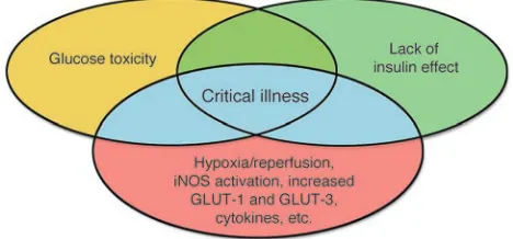
Future directions
It is clear that several mechanisms are involved and interrelated in explaining the clinical benefits of intensive insulin therapy in the critically ill (Figure 4). Accelerated toxicity of hyperglycemia and lack of insulin effect during critical illness explain why the con-sequences are so rapidly deleterious in this condition. The direct effects of preventing hyperglycemia as well as the distinct meta-bolic and nonmetameta-bolic effects of insulin that occur concomitant-ly with the gconcomitant-lycemic control are likeconcomitant-ly to play a role. The relative contribution of those different mechanisms, however, is presently unknown. Furthermore, several questions await an answer. The exact molecular basis for the increased susceptibility for these toxic
effects in the critically ill patient remains to be explored. It also remains unclear which of the insulin signal transduction pathways in different target tissues respond to insulin treatment and how these effects mediate the protection on organ function and overall outcome. Various hypotheses have been advanced, and these can be tested by analysis of tissue samples obtained from patients and from experimental animals in carefully designed studies. Further investigation of the mitochondrial abnormalities in tissues other than the liver and skeletal muscle, such as neurons, kidneys, and endothelial and epithelial cells, as well as the selective impact of insulin and glucose control on this cellular compartment should be investigated in great detail. Furthermore, exploring the molecu-lar link between improved glycemic and lipid control and insulin, endothelial function, inflammation, and clinical outcome will be required. Finally, manipulation of those pathways that are linked with outcome but appear unresponsive to intensive insulin ther-apy may point to the potential for other therapeutic strategies to further improve survival in the ICU.
Conclusion
The results of the clinical study performed in Leuven demonstrat-ed that a simple metabolic intervention, maintaining normogly-cemia with insulin, improved survival and reduced morbidity of critically ill patients. This reflected major clinical progress in the modern era of intensive care. Furthermore, this clinical study has opened a whole new area of basic research besides the ongoing search for additional clinical applications. Indeed, only some of the underlying mechanisms have been studied, and many more pathways need to be investigated in great detail. Results from fun-damental research carefully designed to elucidate how the clini-cal benefits were brought about are likely to set off development of new strategies for further improving outcome of critically ill patients and perhaps also of other target populations within and outside the hospital setting.
Acknowledgments
I thank Roger Bouillon, Ilse Vanhorebeek, and Lies Langouche for critically reviewing the manuscript. This work was supported by the Fund for Scientific Research, Flanders, Belgium (G.0278.03), the Research Council of the Catholic University of Leuven (OT 03/56), and the Belgian Foundation for Research in Congenital Heart Diseases. The author is a Fundamental Clinical Research Investigator (G.3C05.95N) for the Fund for Scientific Research, Flanders, Belgium, and holds an unrestrictive Catholic University of Leuven — Novo Nordisk Chair of Research.
Address correspondence to: Greet Van den Berghe, Department of Intensive Care Medicine, Catholic University of Leuven, B-3000 Leuven, Belgium. Phone: 32-16-34-40-21; Fax: 32-16-34-40-15; E-mail: greta.vandenberghe@med.kuleuven.ac.be.
1. Lassen, H.C.A. 1953. A preliminary report on the 1952 epidemic of poliomyelitis in Copenhagen with special reference to the treatment of respira-tory insufficiency. Lancet. i:37–40.
2. Fink, M.P., and Evans, T.W. 2002. Mechanisms of organ dysfunction in critical illness: report from a round table conference held in Brussels. Intensive Care Med. 28:369–375.
3. Fink, M.P. 2001. Cytopathic hypoxia. Mitochondrial dysfunction as mechanism contributing to organ dysfunction in sepsis. Crit. Care Clin. 17:219–237. 4. Brealey, D., et al. 2002. Association between
mitochondrial dysfunction and severity and out-come of septic shock. Lancet. 360:219–223. 5. Singer, M., and Brealey, D. 1999. Mitochondrial
dys-funtion in sepsis. Biochem. Soc. Symp. 66:149–166. 6. Crouser, E.D., Julian, M.W., Blaho, D.V., and
Pfei-ffer, D.R. 2002. Endotoxin-induced mitochondrial damage correlates with impaired respiratory activ-ity. Crit. Care Med. 30:276–284.
7. Amato, M.B., et al. 1998. Effect of a protective-venti-lation strategy on mortality in the acute respiratory distress syndrome. N. Engl. J. Med. 338:347–354. 8. Rivers, E., et al. 2001. Early goal-directed therapy
in the treatment of severe sepsis and septic shock. N. Engl. J. Med. 345:1368–1377.
9. Bernard, G.R., et al. 2001. Efficacy and safety of recombinant human activated protein C for severe sepsis. N. Engl. J. Med. 344:699–709.
10. Van den Berghe, G., et al. 2001. Intensive insulin therapy in critically ill patients. N. Engl. J. Med. 345:1359–1367.
11. Bernard, C. 1878. Leçons sur les phénomènes de la vie communs aux animaux et aux végétaux. Volume 1. J.B. Baillière et Fils. Paris, France. 564 pp.
science in medicine
Insulin resistance: a marker of surgical stress. Curr. Opin. Clin. Nutr. Metab. Care. 21:69–78.
13. McCowen, K.C., Malhotra, A., and Bistrian, B.R. 2001. Stress-induced hyperglycaemia. Crit. Care Clin. 17:107–124.
14. Van den Berghe, G., et al. 1999. Reactivation of pituitary hormone release and metabolic improve-ment by infusion of growth hormone-releasing peptide and thyrotropin-releasing hormone in patients with protracted critical illness. J. Clin. Endocrinol. Metab. 84:1311–1323.
15. Van den Berghe, G., et al. 2000. A paradoxical gen-der dissociation within the growth hormone/insu-lin-like growth factor I axis during protracted criti-cal illness. J. Clin. Endocrinol. Metab. 85:183–192. 16. Wolfe, R.R., Durkot, M.J., Allsop, J.R., and Burke,
J.F. 1979. Glucose metabolism in severely burned patients. Metabolism. 28:210–220.
17. Wolfe, R.R., Herndon, D.N., Jahoor, F., Miyoshi, H., and Wolfe, M. 1987. Effects of severe burn injury on substrate cycling by glucose and fatty acids. N. Engl. J. Med. 317:403–408.
18. Mizock, B.A. 1995. Alterations in carbohydrate metabolism during stress: a review of the literature. Am. J. Med. 98:75–84.
19. Grimble, R.F. 2002. Inflammatory status and insu-lin resistance. Curr. Opin. Cinsu-lin. Nutr. Metab. Care. 5:551–559.
20. Marette, A. 2002. Mediators of cytokine-induced insulin resistance in obesity and other inflamma-tory settings. Curr. Opin. Clin. Nutr. Metab. Care. 5:377–383.
21. Senn, J.J., et al. 2003. Suppressor of cytokine signal-ing-3 (SOCS-3), a potential mediator of interleukin-dependent insulin resistance in hepatocytes. J. Biol. Chem. 278:13740–13746.
22. Rui, L., Yuan, M., Frantz, D., Shoelson, S., and White, M.F. 2002. SOCS-1 and SOCS-3 block insu-lin signainsu-ling by ubiquitin-mediated degradation of IRS1 and IRS2. J. Biol. Chem. 277:42394–42398. 23. McCowen, K.C., et al. 2001. Sustained endotoxemia
leads to marked down-regulation of early steps in the insulin-signaling cascade. Crit. Care Med. 29:839–846.
24. Jiang, Z.Y., et al. 1999. Characterization of selective resistance to insulin signaling in the vasculature of obese Zucker (fa/fa) rats. J. Clin. Invest. 104:447–457. 25. Cusi, K., et al. 2000. Insulin resistance differential-ly affects the PI 3-kinase-and MAP kinase-medi-ated signaling in human muscle. J. Clin. Invest. 105:311–320.
26. Draznin, B., et al. 2000. Effects of insulin on pre-nylation as a mechanism of potential detrimen-tal influence of hyperinsulinemia. Endocrinology. 141:1310–1316.
27. Golovchenko, I., Goalstone, M.L., Watson, P., Brown-lee, M., and Draznin, B. 2000. Hyperinsulinemia enhances transcriptional activity of Nuclear Factor-kB induced by angiotensin II, hyperglycemia, and advanced glycosylation end products in vascular smooth muscle cells. Circ. Res. 87:746–752. 28. Montagnani, M., et al. 2002. Inhibition of
phosphatidylinositol 3-kinase enhances mitogenic actions of insulin in endothelial cells. J. Biol. Chem. 277:1794–1799.
29. Mizock, B.A. 2001. Alterations in fuel metabolism in critical illness: hyperglycaemia. Best Pract. Res. Clin. Endocrinol. Metab. 15:533–551.
30. Malmberg, K., et al. 1995. Randomized trial of insulin-glucose infusion followed by subcutaneous insulin treatment in diabetic patients with acute myocardial infarction (DIGAMI study): effects on mortality at 1 year. J. Am. Coll. Cardiol. 26:57–65. 31. Krinsley, J.S. 2004. Effect of an intensive glucose
management protocol on the mortality of critically ill adult patients. Mayo Clin. Proc. 79:992–1000. 32. Klip, A., Tsakiridis, T., Marette, A., and Ortiz, P.A.
1994. Regulation of expression of glucose
trans-porters by glucose: a review of studies in vivo and in cell cultures. FASEB J. 8:43–53.
33. Van den Berghe. G., et al. 2003. Outcome benefit of intensive insulin therapy in the critically ill: insu-lin dose versus glycemic control. Crit. Care Med. 31:359–366.
34. Van den Berghe, G., Schoonheydt, K., Becx, P., Bruyninckx, F., and Wouters, P.J. 2004. Intensive insulin therapy protects the central and peripheral nervous system of intensive care patients. In: Pro-gram and abstracts of the 86th Annual Meeting of the Endocrine Society. New Orleans, Louisiana, USA. June 16–19, 2004. Abstract OR8-2, p. 79. 35. Mesotten, D., et al. 2002. Regulation of
insulin-like growth factor binding protein-1 during pro-tracted critical illness. J. Clin. Endocrinol. Metab. 87:5516–5523.
36. Mesotten, D., Swinnen, J., Vanderhoydonc, F., Wouters, P.J., and Van den Berghe, G. 2004. Con-tribution of circulating lipids to the improved outcome of critical illness by glycemic control with intensive insulin therapy. J. Clin. Endocrinol. Metab. 89:219–226.
37. Donmoyer, C.M., et al. 2003. Infection impairs insulin-dependent hepatic glucose uptake during total parenteral nutrition. Am. J. Physiol. Endocrinol. Metab. 284:E574–E582.
38. Puigserver, P., et al. 2003. Insulin-regulated hepatic gluconeogenesis through FOXO1-PGC-1alpha interaction. Nature. 423:550–555.
39. Pekala, P., Marlow, M., Heuvelman, D., and Con-nolly, D. 1990. Regulation of hexose transport in aortic endothelial cells by vascular permeability factor and tumor necrosis factor-alpha, but not by insulin. J. Biol. Chem. 265:18051–18054. 40. Shikhman, A.R., Brinson, D.C., Valbracht, J., and
Lotz, M.K. 2001. Cytokine regulation of facilitated glucose transport in human articular chondrocytes. J. Immunol. 167:7001–7008.
41. Quinn, L.A., and McCumbee, W.D. 1998. Regula-tion of glucose transport by angiotensin II and glucose in cultured vascular smooth muscle cells. J. Cell. Physiol. 177:94–102.
42. Clerici, C., and Matthay, M.A. 2000. Hypoxia regu-lates gene expression of alveolar epithelial trans-port proteins. J. Appl. Physiol. 88:1890–1896. 43. Sanchez-Alvarez, R., Tabernero, A., and Medina, J.M.
2004. Endothelin-1 stimulates the translocation and upregulation of both glucose transporter and hexokinase in astrocytes: relationship with gap junc-tional communication. J. Neurochem. 89:703–714. 44. Aulak, K.S., Koeck, T., Crabb, J.W., and Stuehr, D.J.
2004. Dynamics of protein nitration in cells and mitochondria. Am. J. Physiol. Heart Circ. Physiol. 286:H30–H38.
45. Frost, M., Wang, Q., Moncada, S., and Singer, M. 2004. Bi-phasic, oxygen- and NO-dependent modulation of complex I activity in activated macrophages by S-nitrosylation and nitration. Am. J. Physiol. In press.
46. Brownlee, M. 2001. Biochemistry and molecu-lar cell biology of diabetic complications. Nature. 414:813–820.
47. Vanhorebeek, I., et al. 2004. Strict blood glucose control with insulin in critically ill patients pro-tects hepatocytic mitochondrial ultrastructure and function. Lancet. In press.
48. Pozzilli, P., and Leslie, R.D. 1994. Infections and diabetes: mechanisms and prospects for preven-tion. Diabet. Med. 11:935–941.
49. Funari, A.P., Zerr, K.J., Grunkemeier, G.L., and Starr, A. 1999. Continuous intravenous insulin infusion reduces the incidence of deep sternal wound infection in diabetic patients after cardiac surgical procedures. Ann. Thorac. Surg. 67:352–360. 50. Rassias, A.J., et al. 1999. Insulin infusion improves
neutrophil function in diabetic cardiac surgery patients. Anesth. Analg. 88:1011–1016.
51. Nielson, C.P., and Hindson, D.A. 1989. Inhibition of polymorphonuclear leukocyte respiratory burst by elevated glucose concentrations in vitro. Diabe-tes. 38:1031–1035.
52. Perner, A., Nielsen, S.E., and Rask-Madsen, J. 2003. High glucose impairs superoxide production from isolated blood neutrophils. Intensive Care Med. 29:642–645.
53. Rayfield, E.J., et al. 1982. Infection and diabetes: the case for glucose control. Am. J. Med. 72:439–450. 54. Black, C.T., Hennessey, P.J., and Andrassy, R.J. 1990.
Short-term hyperglycemia depresses immunity through nonenzymatic glycosylation of circulat-ing immunoglobulin. J. Trauma. 30:830–832. 55. Weekers, F., et al. 2002. A novel in vivo rabbit model
of hypercatabolic critical illness reveals a bi-pha-sic neuroendocrine stress response. Endocrinology. 143:764–774.
56. Weekers, F., et al. 2003. Metabolic, endocrine and immune effects of stress hyperglycemia in a rabbit model of prolonged critical illness. Endocrinology. 144:5329–5338.
57. Bruning, J.C., et al. 2000. Role of brain insulin receptor in control of body weight and reproduc-tion. Science. 289:2122–2155.
58. Vincent, A.M., Brownlee, M., and Russell, J.W. 2002. Oxidative stress and programmed cell death in dia-betic neuropathy. Ann. N. Y. Acad. Sci. 959:368–383. 59. Taskinen, M.R. 2001. Pathogenesis of dyslipidemia
in type 2 diabetes. Exp. Clin. Endocrinol. Diabetes. 109:S180–S188.
60. Lanza-Jacoby, S., Wong, S.H., Tabares, A., Baer, D., and Schneider, T. 1992. Disturbances in the composition of plasma lipoproteins during gram-negative sepsis in the rat. Biochim. Biophys. Acta. 1124:233–240.
61. Khovidhunkit, W., Memon, R.A., Feingold, K.R., and Grunfeld, C. 2000. Infection and inflamma-tion-induced proatherogenic changes of lipopro-teins [review]. J. Infect. Dis. 181:S462–S472. 62. Carpentier, Y.A., and Scruel, O. 2002. Changes
in the concentration and composition of plasma lipoproteins during the acute phase response. Curr. Opin. Clin. Nutr. Metab. Care. 5:153–158.
63. Gordon, B.R., et al. 1996. Low lipid concentrations in critical illness: implications for preventing and treating endotoxemia. Crit. Care Med. 24:584–589. 64. Kwiterovich, P.O. 2002. Lipoprotein
heterogene-ity: diagnostic and therapeutic implications. Am. J. Cardiol. 90:1i–10i.
65. Feingold, K.R., et al. 1993. The hypertriglyceri-demia of acquired immunodeficiency syndrome is associated with an increased prevalence of low density lipoprotein subclass pattern B. J. Clin. Endocrinol. Metab. 76:1423–1427.
66. Tulenko, T.N., and Sumner, A.E. 2002. The physi-ology of lipoproteins. J. Nucl. Cardiol. 9:638–649. 67. Harris, H.W., Grunfeld, C., Feingold, K.R., and
Rapp, J.H. 1990. Human very low density lipo-proteins and chylomicrons can protect against endotoxin-induced death in mice. J. Clin. Invest. 86:696–702.
68. Harris, H.W., et al. 1993. Chylomicrons alter the fate of endotoxin, decreasing tumor necrosis fac-tor release and preventing death. J. Clin. Invest. 91:1028–1034.
69. Finney, S.J., Zekveld, C., Elia, A., and Evans, T.W. 2003. Glucose control and mortality in critically ill patients. JAMA. 290:2041–2047.
70. Brismar, K., Fernqvist-Forbes, E., Wahren, J., and Hall, K. 1994. Effect of insulin on the hepatic production of insulin-like growth factor-binding protein-1 (IGFBP-1), IGFBP-3, and IGF-I in insu-lin-dependent diabetes. J. Clin. Endocrinol. Metab. 79:872–878.
science in medicine
89:3105–3113.
72. Suikkari, A.M., et al. 1988. Insulin regulates the serum levels of low molecular weight insulin-like growth factor-binding protein. J. Clin. Endocrinol. Metab. 66:266–272.
73. Suwanichkul, A., Morris, S.L., and Powell, D.R. 1993. Identification of an insulin-responsive ele-ment in the promoter of the human gene for insu-lin-like growth factor binding protein-1. J. Biol. Chem. 268:17063–17068.
74. Hansen, T.K., Thiel, S., Wouters, P.J., Christian-sen, J.S., and Van den Berghe, G. 2003. Intensive insulin therapy exerts antiinflammatory effects in critically ill patients and counteracts the adverse effect of low mannose-binding lectin levels. J. Clin. Endocrinol. Metab. 88:1082–1088.
75. Das, U.N. 2001. Is insulin an antiinflammatory molecule? Nutrition. 17:409–413.
76. Dandona, P., et al. 2001. Insulin inhibits intra-nuclear factor kappaB and stimulates IkappaB in mononuclear cells in obese subjects: evidence for an anti-inflammatory effect? J. Clin. Endocrinol. Metab. 86:3257–3265.
77. Carr, M.E. 2001. Diabetes mellitus: a hypercoagu-lable state. J. Diabetes Complicat. 15:44–54. 78. Calles-Escandon, J., Garcia-Rubi, E., Mirza, S., and
Mortensen, A. 1999. Type 2 diabetes: one disease, multiple cardiovascular risk factors. Coron. Artery Dis. 10:23–30.
79. Williams, E., Timperley, W.R., Ward, J.D., and Duckworth, T. 1980. Electron microscopical stud-ies of vessels in diabetic peripheral neuropathy. J. Clin. Pathol. 33:462–470.
80. Patrassi, G.M., Vettor, R., Padovan, D., and Girola-mi, A. 1982. Contact phase of blood coagulation in diabetes mellitus. Eur. J. Clin. Invest. 12:307–311. 81. Carmassi, F., et al. 1992. Coagulation and
fibrinolytic system impairment in insulin depen-dent diabetes mellitus. Thromb. Res. 67:643–654. 82. Hughes, A., et al. 1983. Diabetes, a hypercoagulable
state? Hemostatic variables in newly diagnosed type 2 diabetic patients. Acta Haematol. 69:254–259. 83. Garcia Frade, L.J., et al. 1987. Diabetes mellitus
as a hypercoagulable state: its relationship with fibrin fragments and vascular damage. Thromb. Res. 47:533–540.
84. Carmassi, F., et al. 1992. Coagulation and fibrinolytic system impairment in insulin depen-dent diabetes mellitus. Thromb. Res. 67:643–654. 85. Vukovich, T.C., and Schernthaner, G. 1986.
Decreased protein C levels in patients with insu-lin-dependent type I diabetes mellitus. Diabetes. 35:617–619.
86. Garcia Frade, L.J., et al. 1987. Changes in fibrinolysis in the intensive care patient. Thromb. Res. 47:593–599.
87. Mavrommatis, A.C., et al. 2001. Activation of the fibrinolytic system and utilization of the
coagulation inhibitors in sepsis: comparison with severe sepsis and septic shock. Intensive Care Med. 27:1853–1859.
88. Montagnani, M., Chen, H., Barr, V.A., and Quon, M.J. 2001. Insulin-stimulated activation of e-NOS is independent of Ca2+ but requires phosphorylation by Akt at Ser(1179). J. Biol. Chem. 276:30392–30398.
89. Sodi-Pallares, D., Testelli, M.R., and Fishleder, B. 1962. Effects of an intravenous infusion of a potas-sium-insulin-glucose solution on the electrocar-diographic signs of myocardial infarction. A pre-liminary clinical report. Am. J. Cardiol. 9:166–181. 90. Opie, L. 1970. The glucose hypothesis: relation
to acute myocardial ischemia. J. Mol. Cell. Cardiol. 1:107–114.
91. Jonassen, A., Aasum, E., Riemersma, R., Mjos, O., and Larsen, T. 2000. Glucose-insulin-potassium reduces infarct size when administered during reperfusion. Cardiovasc. Drugs Ther. 14:615–623. 92. Gao, F., et al. 2002. Nitric oxide mediates the
anti-apoptotic effect of insulin in myocardial ischemia-reperfusion: the role of PI3-kinase, Akt, and eNOS phosphorylation. Circulation. 105:1497–1502. 93. Jonassen, A., Sack, M., Mjos, O., and Yellon, D. 2001.