Activated human protein C prevents
thrombin-induced thromboembolism in mice. Evidence
that activated protein c reduces intravascular
fibrin accumulation through the inhibition of
additional thrombin generation.
P Gresele, … , N Semeraro, M Colucci
J Clin Invest.
1998;
101(3)
:667-676.
https://doi.org/10.1172/JCI575
.
Activated protein C (APC) is a potent physiologic anticoagulant with profibrinolytic
properties, and has been shown to prevent thrombosis in different experimental models. We
investigated the effect of human APC on thrombin-induced thromboembolism in mice, a
model of acute intravascular fibrin deposition leading to death within minutes. APC given
intravenously (i.v.) as a bolus 2 min before thrombin challenge (1,250 U/kg) reduced
mortality in a dose-dependent manner despite the lack of thrombin inhibitor activity.
Significant inhibition of thrombin-induced death was observed at the dose of 0.05 mg/kg,
and maximal protection was obtained with 2 mg/kg (> 85% reduction in mortality rate).
Histology of lung tissue revealed that APC treatment (2 mg/kg) reduced significantly
vascular occlusion rate (from 89.2 to 46.6%, P < 0.01). The protective effect of APC was due
to the inhibition of endogenous thrombin formation as indicated by the fact that (a) the
injection of human thrombin caused a marked decrease in the coagulation factors of the
intrinsic and common pathways (but not of Factor VII), suggesting the activation of blood
clotting via the contact system; (b) APC pretreatment reduced markedly prothrombin
consumption; (c) the lethal effect of thrombin was almost abolished when the animals were
made deficient in vitamin K-dependent factors by warfarin treatment, and could be restored
only by doubling the dose of thrombin, indicating that […]
Research Article
Find the latest version:
J. Clin. Invest.
© The American Society for Clinical Investigation, Inc. 0021-9738/98/02/0667/10 $2.00
Volume 101, Number 3, February 1998, 667–676 http://www.jci.org
Activated Human Protein C Prevents Thrombin-induced Thromboembolism in Mice
Evidence that Activated Protein C Reduces Intravascular Fibrin Accumulation through the Inhibition of
Additional Thrombin Generation
Paolo Gresele,* Stefania Momi,* Mauro Berrettini,* Giuseppe G. Nenci,* Hans P. Schwarz,‡ Nicola Semeraro,§
and Mario Colucci§
*Institute of Internal and Vascular Medicine, University of Perugia, 06126 Perugia, Italy; ‡Immuno AG, 1220 Vienna, Austria; and §Department of Biomedical Sciences, Section of General Pathology, University of Bari, 70124 Bari, Italy
Abstract
Activated protein C (APC) is a potent physiologic anticoag-ulant with profibrinolytic properties, and has been shown to prevent thrombosis in different experimental models. We investigated the effect of human APC on thrombin-induced thromboembolism in mice, a model of acute intravascular fibrin deposition leading to death within minutes. APC given intravenously (i.v.) as a bolus 2 min before thrombin challenge (1,250 U/kg) reduced mortality in a dose-depen-dent manner despite the lack of thrombin inhibitor activity. Significant inhibition of thrombin-induced death was ob-served at the dose of 0.05 mg/kg, and maximal protection was obtained with 2 mg/kg (. 85% reduction in mortality rate). Histology of lung tissue revealed that APC treatment (2 mg/kg) reduced significantly vascular occlusion rate (from 89.2 to 46.6%, P, 0.01). The protective effect of APC was due to the inhibition of endogenous thrombin formation as indicated by the fact that (a) the injection of human thrombin caused a marked decrease in the coagulation fac-tors of the intrinsic and common pathways (but not of Fac-tor VII), suggesting the activation of blood clotting via the contact system; (b) APC pretreatment reduced markedly prothrombin consumption; (c) the lethal effect of thrombin was almost abolished when the animals were made deficient in vitamin K–dependent factors by warfarin treatment, and could be restored only by doubling the dose of thrombin, in-dicating that the generation of endogenous thrombin con-tributes significantly to death; and (d) APC failed to protect warfarin-treated animals, in which mortality is entirely due to injected thrombin, even after protein S supplementation. Other results suggest that APC protects from thrombin-induced thromboembolism by rendering the formed fibrin more susceptible to plasmin degradation rather than by re-ducing fibrin formation: in thrombin-treated mice, fibrino-gen consumption was not inhibited by APC; and inhibition of endogenous fibrinolysis by e-aminocaproic or tranexamic acid resulted in a significant reduction of the protective
ef-fect of APC. Since APC did not enhance plasma fibrinolytic activity, as assessed by the measurement of plasminogen ac-tivator (PA) or PA inhibitor (PAI) activities, PAI-1 antigen, or 125I-fibrin degrading activity, we speculate that the
inhi-bition of additional (endogenous) thrombin formation by APC interrupts thrombin-dependent mechanisms that make fi-brin clots more resistant to lysis, so that the intravascular deposited fibrin can be removed more rapidly by the endog-enous fibrinolytic system. (J. Clin. Invest. 1998. 101:667– 676.) Key words: bleeding time• coagulation activation •
fi-brinolysis • protein S • vessel occlusion
Introduction
In current models of hemostasis, coagulation is initiated at sites of vessel wall damage by exposure of blood to tissue fac-tor produced constitutively by cells beneath the endothelium (1). Factor VII/VIIa present in plasma then binds to this tissue factor, and the ensuing Factor VIIa–tissue factor complex acti-vates definite amounts of Factors X and IX. Subsequent prop-agation and amplification of coagulation pathways required to ensure hemostasis are dependent on a series of positive feed-back mechanisms that are regulated mainly by thrombin. In-deed, the initial burst of Factor Xa generation will provide suf-ficient thrombin to activate the cofactors VIII and V, thus enhancing Factor Xa and thrombin formation. In addition, thrombin may activate Factor XI (2), which in turn would sus-tain the coagulation process by activating additional Factor IX. The latter concept is supported by recent evidence that activa-tion of the intrinsic clotting pathway occurs in rabbits after in-jection of Factor Xa/phospholipid (3). Blood coagulation is controlled by various inhibitors acting at different levels of the enzymatic cascade. Protein C (PC)1 is one of the main
physio-logical anticoagulants in blood (4); it is a vitamin K–dependent factor which, upon activation by thrombin–thrombomodulin complex at the endothelial surface, is converted to a serine protease, activated protein C (APC), that inhibits blood coag-ulation through the proteolytic inactivation of the cofactors VIIIa and Va. The latter reaction is strongly accelerated by negatively charged phospholipids and protein S (PS), another vitamin K–dependent protein devoid of enzymatic activity. In this way, APC interrupts two crucial steps of the coagulation cascade, thereby regulating both the generation of thrombin Address correspondence to Paolo Gresele, M.D., Ph.D., Institute of
Internal and Vascular Medicine, University of Perugia, Via Enrico dal Pozzo, 06126 Perugia, Italy. Phone: 572-2905; FAX: 39-75-572-2011; E-mail: grespa@unipg.it
Received for publication 7 May 1997 and accepted in revised form 24 November 1997.
induced by the triggering agent (e.g., VIIa/tissue factor) and the generation of additional thrombin brought about by the positive feedback mechanisms. The relevance of the PC sys-tem in the maintenance of a normal hemostatic balance is in-ferred from the elevated incidence of thrombotic episodes in patients with an impairment of the PC-dependent anticoagu-lant mechanism due to PC or PS deficiency (5, 6) or to APC re-sistance (7), a condition caused by a point mutation of the Fac-tor V gene (8).
Purified APC has been shown to be an effective therapy in various experimental models of thrombosis (9, 10), in septic shock in baboons (11), and has been evaluated in pilot studies in humans (12, 13). Importantly, APC does not significantly af-fect the hemostatic function or produce an increased bleeding tendency. The interest for APC as an antithrombotic drug is further strengthened by the observation that it may exert profibrinolytic functions. In dogs (14) and cats (15), APC was shown to induce an elevation of circulating plasminogen acti-vator (PA) activity, though this effect could not be confirmed in squirrel monkeys (16) or humans (12). Moreover, evidence has been provided in experimental endotoxemia in rabbits and mice (17, 18) that APC may promote fibrinolysis by reducing the blood levels of PA inhibitor 1 (PAI-1). This mechanism was considered important for the enhancement of the thera-peutic efficacy of recombinant tissue PA (t-PA) in endotoxin-treated rabbits (19). An alternative mechanism through which APC may stimulate fibrinolysis has been suggested by several in vitro studies and relates to the ability of the anticoagulant to depress the generation of thrombin (20–22). Indeed, the latter enzyme might inhibit the fibrinolytic process by rendering fi-brin more resistant to lysis via activation of Factor XIII and TAFI (thrombin-activatable fibrinolysis inhibitor). Factor XIIIa catalyzes the cross-linking of fibrin a and g chains as well as the cross-linking of a2-antiplasmin (23), while activated
TAFI, a carboxypeptidase identical to carboxypeptidase B, at-tenuates fibrinolysis through the removal of carboxy-terminal lysines from partially degraded fibrin (24, 25). Interestingly, Bajzar and co-workers have reported recently that enhance-ment of in vitro clot lysis by APC in a plasma system was to-tally dependent on the inhibition of the activation of TAFI by thrombin (26).
Thrombin-induced thromboembolism in mice is a model of acute and massive intravascular fibrin deposition, mainly within the pulmonary arteries, that leads to death of the ani-mals within minutes (27–29). The model is sensitive not only to thrombin inhibitors but also to thrombolytic agents, such as t-PA, or to drugs with profibrinolytic activity, such as defi-brotide (29–31). We hypothesized that APC, although devoid of thrombin inhibitor activity, might attenuate the lethal effect of thrombin by interfering with the postulated feedback activa-tion of blood coagulaactiva-tion induced by thrombin injecactiva-tion and/ or by stimulating blood fibrinolytic activity either directly or as a consequence of reduced generation of endogenous throm-bin. Therefore, exploiting the fact that human APC retains its anticoagulant activity in mice (18), we investigated the effect of purified human APC in this model of thrombin-induced thromboembolism. We report that APC prevents the lethal ef-fect of injected thrombin by inhibiting the in vivo generation of endogenous thrombin, thereby reducing the accumulation of intravascular fibrin. This effect does not appear to be related to the reduction in fibrin formation but rather to an enhanced susceptibility of the formed fibrin to endogenous fibrinolysis.
Moreover, our observation confirms the existence and the pathophysiological relevance of feedback activation of the in-trinsic coagulative pathway by thrombin in vivo.
Methods
Chemicals.Human thrombin, warfarin (3-[acetonylbenzyl]-4-hydrox-ycumarin), and PMSF were purchased from Sigma Chemical Co. (Mi-lan, Italy); PC and APC were from human plasma (32, 33) and were provided by Immuno AG, Vienna, Austria; active site–blocked APC was prepared by incubating APC (0.5 mg/ml) with PMSF (1023 M) for
30 min at 378C, and dialyzing overnight against TBS (75 mM Tris, 75 mM NaCl, pH 7.4); unfractionated sodium heparin was from Novo Nordisk (Bagsvaerd, Denmark); human PS was a gift of Drs. A. D’Angelo and S. Viganò D’Angelo (S. Raffaele Hospital, Milan, It-aly); e-aminocaproic acid (EACA, Caprolysin) and tranexamic acid (AMCA, Tranex) (Malesci, Florence, Italy), and the chromogenic substrates S-2238 and S-2366 (Ortho Diagnostic Systems, Inc., Milan, Italy) were purchased from commercial sources.
In vivo thrombosis model. This study was approved by the Com-mittee on Ethics of Animal Experiments, Faculty of Medicine, Uni-versity of Perugia. Thrombin-induced pulmonary thromboembolism in mice was induced by a method described previously (29, 34). Briefly, male CD-1 mice (Charles River Italia S.P.A., Calco, Como, Italy) weighing 20–25 g were caged and fed a regular diet for at least 1 wk before use. The drugs to be tested, or their respective vehicles, were administered intravenously (i.v.) in a fixed volume of 0.1 ml in one of the tail veins 2 min before the injection of 1,250 U/kg human throm-bin. This dose was selected from a concentration–response curve as the minimal dose giving a reproducible 80–90% mortality, generally within 5 min. In each experimental session, at least five animals per treatment group were tested; control groups were run at the begin-ning and end of every experimental session. Mice were accustomed to handling by the investigators, and the injections were carried out by skilled investigators with minimal disturbance to the animals. The to-tal duration of the experiment was 15 min, and all surviving animals were killed by exposure to ether vapors. No anesthesia was used dur-ing the experiment because of the short duration and because anes-thesia interferes with thromboembolism in this model (35). The eval-uation of the effect of APC on the i.v. challenge with thrombin was carried out as described previously (29, 34): the cumulative end point to be overcome by drug treatment was death of the animal or pro-longed (15 min) paralysis of the hind limbs.
In selected experiments, mice were made deficient in vitamin K–dependent proteins by prolonged treatment with sodium warfarin dissolved in drinking water (7.5 mg/liter on the first day and 2.5 mg/li-ter on the following days), leaving free access to wamg/li-ter to the animals. After 5–7 d an animal from each cage was killed, blood was collected immediately by cardiac puncture in 4% trisodium citrate (1:10 vol), and anticoagulation was checked by prothrombin time (PT). The data are presented as number of animals dead to total number of animals or as a percentage of the total. Protection against thrombin-induced mortality was expressed as (1 2TAPC/Tsal) 3 100, where TAPC is the
mortality rate in APC-treated mice, and Tsal is the mortality rate in
controls treated with thrombin only.
for every specimen. The total number of identifiable lung vessels per field was counted, and the percentage of them occluded by fibrin plugs was annotated.
Bleeding time. Bleeding time was assessed by a tail transection method adapted from a method described previously for rats (37). Briefly, mice pretreated with the tested drugs or TBS were positioned in a special immobilization cage that keeps the tail steady and im-mersed in saline thermostated at 378C. After 2 min, the tip of the tail was transected with a razor blade, at z 2 mm from its end. The tail was reimmersed immediately in warm saline, and the bleeding time was recorded. The end point was an arrest of bleeding lasting . 30 s.
Assays. Blood was collected from ether-anesthetized mice by car-diac puncture and anticoagulated with 4% trisodium citrate (1:10 vol). Anticoagulated blood was centrifuged immediately for 5 min at 12,000 g in an Eppendorf microfuge, and the supernatant platelet-poor plasma was separated and transferred on melting ice until tested (generally within 1 h) or frozen at 2208C. Activated partial thrombo-plastin time (APTT), PT, and thrombin clotting time were measured by standard assays in an automatic coagulometer (ACL 300R; Instru-mentation Laboratory, Inc., Milan, Italy) using reagents from the manufacturer. Plasma fibrinogen was measured by the Clauss method in a Coagulab MJ coagulometer (Ortho Diagnostic Systems, Inc.) us-ing bovine thrombin (Behrus-ing, Scoppito, Italy). Plasma levels of Fac-tors XII, XI, IX, VII, X, and V and prothrombin were assayed by one-stage clotting methods using commercially available human plas-mas deficient in the respective coagulation factor (Hemolab; bio-Mérieux, Marcy-l’Etoile, France). Mouse plasma was diluted in cold Tris-buffer (0.05 M Tris-HCl, 0.15 M NaCl, pH 7.5) containing 1 mg/ml BSA. Factors XII, XI, and IX were assayed using optimized thrombo-fax (Ortho Diagnostic Systems, Inc.) as APTT reagent; briefly, 0.1 ml human deficient plasma was incubated for 5 min at 378C with 0.1 ml 1:20 or 1:40 (for Factor XII) mouse plasma and 0.1 ml thrombofax, and clotting was started by the addition of 0.025 M CaCl2. For Factor
VII, X, V, and II determination, 0.1 ml of the appropriate human de-ficient plasma was incubated for 1 min at 378C with 0.1 ml of 1:20 or 1:40 (for Factor V) diluted mouse plasma, and clotting was trig-gered by the addition of 0.2 ml CaCl2/thromboplastin mixture
(Throm-borel-S; Behring). In all cases, time to clot formation was recorded visually with the tilting method. The concentration of coagulation fac-tors was expressed as percentage of normal by reference to a stan-dard curve constructed with different dilutions of pooled mouse plasma obtained from at least 10 normal mice. The determination of PA and PAI activities in mouse plasma was carried out by spectrophoto-metric assays using commercially available kits (Spectrolyse; Biopool AB, Umea, Sweden). PAI-1 antigen was measured with an ELISA method using mAbs (kindly provided by Prof. H.R. Lijnen, Univer-sity of Leuven, Belgium) raised against murine PAI-1 in PAI-1 gene– deficient mice (38). Global plasma fibrinolytic activity was evaluated by a 125I-fibrin solid-phase assay (39) as reported (40). Briefly, 0.1 ml
of mouse plasma was transferred in a 0.5 3 5 cm tube, coated with
125I-fibrin (z 100,000 cpm), and incubated at 378C. Aliquots of 0.01 ml
were withdrawn at 1-h intervals up to 4 h for g-counting; fibrinolytic activity was expressed as percentage of radioactivity released in solu-tion. Blank samples, consisting of phosphate buffer (pH 7.4) contain-ing 3 mg/ml BSA, were run in parallel to correct for the spontaneous leakage of radioactivity (which never exceeded 5% over 4 h).
Human APC activity was measured by an ELISA capture assay, as described previously (41). Briefly, three dilutions were made for each plasma sample or reference APC preparation in a dilution mi-croplate coated with anti–human PC antibody. Measurement of the amidolytic activity of immobilized APC was carried out using the syn-thetic chromogenic substrate S-2366, and performing multiple read-ings on different days. As the test does not detect mouse APC, the measured values reflect the infused or generated human APC activity in plasma. Total human PC antigen (PC 1 APC) in plasma samples was assayed by a commercial ELISA (Asserachrom PC; Boehringer Mannheim, Mannheim, Germany).
Statistical analysis. The x2 test was applied to the studies on
mor-tality, using Bonferroni’s correction (statistically significant P value , 0.05/number of comparisons). ANOVA, followed by Student-Newman-Keuls’ test for multiple comparisons, was used for the other studies. Data are expressed as mean6SD.
Results
Anticoagulant activity of human APC in mouse plasma in vitro. Human APC prolonged the APTT of mouse plasma in a concentration-dependent way. However, the anticoagulant ef-fect in the murine system was somewhat lower than in human plasma (Fig. 1), the concentration of APC needed to double the APTT amounting to 23 nM for mouse plasma and 12 nM for human plasma.
Prevention of thrombin-induced thromboembolism. Bolus injection of 1,250 U/kg of human thrombin caused death in
[image:4.612.316.555.483.658.2]. 80% of mice. A single i.v. injection of APC, given 2 min be-fore thrombin, prevented mortality in a dose-dependent man-ner, with a significant effect at a dose of 0.05 mg/kg (52% mor-tality, P5 0.005), and a survival of 88% at a dose of 2 mg/kg (Fig. 2 A). Injection of native PC had a weak protective effect on thrombin-induced thromboembolism, with a significant re-duction in mortality observed at a dose of 2 mg/kg (55% mor-tality, P5 0.006). Specific assay of human APC in plasma from thrombin-treated mice indicated that 17.262.5% of the in-jected PC was in the active form. This percentage was much higher than that present in the native PC preparation (0.08%) or in plasma samples from mice given PC only (0.038%) (Ta-ble I), indicating that the reduction in mortality by native PC was due to the partial in vivo activation of the proenzyme by injected thrombin. APC (2 mg/kg) whose active site had been blocked by PMSF was virtually inactive in protecting animals from thrombin-induced death (80% mortality). Heparin also produced a dose-dependent inhibition of mortality; however, the range of antithrombotic dosages was rather narrow, be-tween 10 and 50 U/kg (Fig. 2 B).
Figure 1. Anticoagulant activity of human APC in mouse plasma
(circles) and human plasma (diamonds) in vitro, assessed by APTT prolongation. APC (20 ml) was added to the system immediately be-fore CaCl2. x-axis, Final concentration of the anticoagulant in the test
Anticoagulant and hemorrhagic effect of APC. The antico-agulant and hemorrhagic effects of APC and heparin were studied in mice not treated with thrombin. The changes in APTT caused by three different doses of the two anticoagu-lants, selected on the basis of their antithrombotic activity, are shown in Fig. 3 A. APC caused a markedly less pronounced prolongation of clotting time compared with heparin: at the in-termediate dose, the mean APTT ratio was 1.5 for APC (0.5 mg/kg) and 7.5 for heparin (25 U/kg). Interestingly, the mini-mum dose of APC capable of reducing thrombin-induced mor-tality (0.05 mg/kg) had no anticoagulant effect (mean APTT ratio, 0.96) (Fig. 3 A). Thrombin clotting time was dose-depen-dently prolonged by heparin but it was not affected by APC (2 mg/kg), excluding the presence of thrombin inhibitory activity in our APC preparation (not shown). The latter point was fur-ther supported by in vitro experiments showing that APC (up to 300 nM) had no antithrombin activity as assessed by clotting test, amidolytic assay (with the chromogenic substrate S-2238), and thrombin-induced platelet aggregation (not shown). In line with the APTT results, the tail transection bleeding time
was markedly less affected by APC than by heparin. At dos-ages causing 60% reduction in mortality, APC and heparin prolonged the bleeding time by two- and fourfold, respectively (Fig. 3 B).
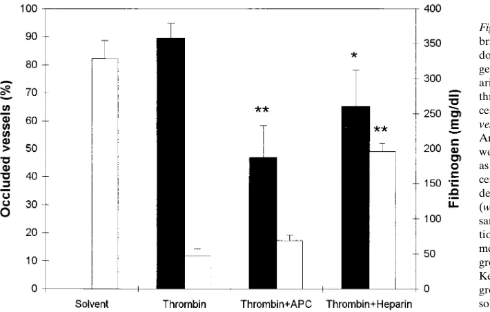
Prevention of intravascular fibrin accumulation. Thrombin injection in normal mice caused the formation of widespread fibrin-rich microthrombi at the pulmonary level, where 89.26 5.8% of the vessels were found to be occluded on histologic ex-amination, and a very pronounced fibrinogen consumption (from 329625.6 to 47.569.7 mg/dl) (Fig. 4). Pretreatment with 2 mg/kg of APC resulted in a marked inhibition of fibrin depo-sition, with the percentage of lung vessel occlusion reduced to 46.6611.8% (P, 0.01). Notably, APC had virtually no effect on the consumption of plasma fibrinogen. In contrast, heparin (50 U/kg) reduced both pulmonary microthrombosis (64.96 13.2% occlusion, P, 0.05) and fibrinogen consumption (Fig. 4). To explain the protective effect of APC on thrombin-induced thromboembolism, two hypotheses were considered: (a) the acceleration of fibrin removal by stimulation of the endoge-nous fibrinolytic system; and (b) the inhibition of a positive feedback mechanism triggered by thrombin, leading to blood clotting activation and additional (endogenous) thrombin gen-eration.
Profibrinolytic effect of APC. PA and PAI activity levels in plasma samples taken 3 min after thrombin injection were similar in APC- (2 mg/kg) and vehicle-treated mice (Fig. 5). PAI-1 antigen was very low in both groups, indicating that it accounts for only a minimal part of the plasma PAI activity. Pretreatment with APC also failed to enhance global plasma fibrinolytic activity as assessed by 125I-fibrin solid-phase assay
(Fig. 5). To evaluate the effect of APC over a longer time in-terval, experiments were performed in mice not treated with thrombin. Under these conditions too, all measured parame-ters were unchanged in plasma samples collected up to 20 min after APC injection (not shown).
[image:5.612.58.553.61.252.2]To explore the in vivo relevance of the fibrinolytic system in the prevention of thrombin-induced mortality by APC, ex-periments were performed in mice pretreated (5 min before starting the experiment) with the fibrinolytic inhibitors EACA and AMCA. As shown in Fig. 6, EACA (1 g/kg) and AMCA (100 mg/kg) reduced the protective effect of APC by 32 and
Figure 2. APC and
hep-arin dose-dependently prevent thrombin-induced death in mice. Drugs were given as an i.v. bolus 2 min before in-jection of human throm-bin (1,250 U/kg), and the mortality rate was evalu-ated as reported in Meth-ods. The number of dead to tested animals for each dose is reported. *Mini-mum dose of anticoagu-lant producing a sta-tistically significant reduction in mortality (P , 0.008 by x2 and
Bon-ferroni’s correction).
Table I. APC Activity in Plasma from Mice Treated with PC and Thrombin
Treatment
PC Thrombin PC 1 Thrombin
APC activity (mg/ml) 0.01260.006 , 0.001 4.160.7 PC/APC antigen (mg/ml) 29.962.3 , 0.001 24.364.3 % APC activity 0.03860.016 — 17.262.5
[image:5.612.57.299.572.654.2]86%, respectively. As expected, both inhibitors attenuated markedly the effect of t-PA (2.5 mg/kg) on thrombin-induced mortality (Fig. 6). These findings suggest that endogenous fi-brinolysis, even if not enhanced by APC, contributes to fibrin removal and prevention of thrombin-induced mortality in APC-treated mice. A role of endogenous fibrinolysis in modu-lating thrombin-induced mortality in this model has been re-ported previously (29).
Role of anticoagulation in the prevention of thrombin-induced mortality. Functional levels of coagulation Factors XII, XI, IX, VII, and X and prothrombin in mouse plasma 3 min after injection of thrombin or saline are shown in Fig. 7. In the thrombin-treated group, all factors were reduced signifi-cantly compared with controls, except for Factor VII, which fell only by 10%. On average, the decreases in clotting factors amounted to 40–50%, with the extreme values of 67% for Fac-tor XI and 28% for FacFac-tor X. To rule out that the clotting
as-says had been biased by the presence of anticoagulant factors eventually generated by thrombin treatment (such as, for ex-ample, fibrin/fibrinogen degradation products), clotting tests were carried out using mixtures of normal human and mouse plasma, in proportions similar to those used for factor assays. PT and APTT determinations of these mixtures showed that plasma from thrombin-treated mice behaved similarly to con-trol mouse plasma, even when the reagents used to trigger co-agulation (thrombofax and thromboplastin) were diluted in order to have clotting times similar to those obtained with defi-cient plasmas, thus excluding the interference of thrombin-generated anticoagulants in the factor assays (not shown). The effect of APC pretreatment on the consumption of coagula-tion factors could not be determined 3 min after thrombin in-jection because of the high concentration of APC in plasma samples, which markedly affected the clotting-based assays. To overcome this problem, we collected blood samples from
APC-Figure 3. Anticoagulant
and hemorrhagic effects of equipotent doses of APC and heparin. Three doses of the anticoagu-lants were chosen, pro-ducing 30–40, 60–70, and . 85% protection from thrombin-induced mor-tality, respectively. The doses were 0.05, 0.5, and 2 mg/kg for APC (white
bars), and 10, 25, and 50
U/kg for heparin (striped
bars). Experiments were
[image:6.612.59.546.59.218.2]performed in mice not treated with thrombin. APTT determinations (A) were carried out on plasma samples collected 2 min after treatment with APC or heparin. Results are expressed as APTT ratios. Tail transection bleeding time (B) was performed, as reported in Methods, 2 min after drug injection. Results are the mean1SD of 12–22 experiments. Dotted lines, Mean6SD of control values. *P , 0.001 between APC and heparin by ANOVA.
Figure 4. APC prevents intravascular
fi-brin deposition at the pulmonary level but does not inhibit thrombin-induced fibrino-gen consumption. APC (2 mg/ml) or hep-arin (50 U/kg) were injected 2 min before thrombin (1,250 U/kg). Control groups re-ceived either two injections of solvent
(Sol-vent) or TBS and thrombin (Thrombin).
[image:6.612.58.413.514.740.2]treated mice 20 min after thrombin injection, when , 10% of the injected APC was still in the circulation as determined in separate experiments in mice not treated with thrombin. This amount of APC in plasma had no influence on clotting factor assay. The concentration of clotting factors in plasma samples
of APC-treated mice is illustrated in Fig. 7. All factors of the intrinsic pathway as well as Factor X were reduced to a level fairly comparable to that observed in animals treated with thrombin alone. However, at variance with these factors, pro-thrombin levels were significantly less reduced in mice receiv-ing APC, despite the fact that blood was collected at a longer interval after thrombin challenge, suggesting that blood clot-ting was inhibited mainly at the prothrombinase level via deg-radation of Factor Va. Functional assay revealed that throm-bin treatment caused a very marked fall in Factor V activity both in control (21.465.6%, at 3 min) and APC-pretreated mice (17.863.2%, at 20 min). This finding is likely due to the fact that most of the circulating Factor V is activated by exoge-nous thrombin, and thus its consumption cannot be prevented but rather is exacerbated by APC.
[image:7.612.59.416.56.286.2]To assess the importance of blood clotting activation in thrombin-induced death, we determined the mortality rate in mice on warfarin treatment. Administration of warfarin for 5 consecutive days caused a . 95% reduction in vitamin K–depen-dent proteins (PT ratio . 12) without affecting fibrinogen con-centration or platelet count (not shown). The injection of a usually lethal dose of thrombin in these animals caused a very low mortality rate (21%) (Fig. 8 A) which could be brought to a level comparable to that observed in untreated mice only by increasing the thrombin dose above 2,000 U/kg. Interestingly, in warfarin-treated mice, APC pretreatment (2 mg/kg) had no effect on the mortality rate induced by 2,250 U/kg of thrombin (Fig. 8 B). Since the latter result might have been due to the deficiency of PS caused by warfarin treatment, human PS (0.6 mg/kg) was given to mice immediately before APC adminis-tration. As illustrated in Fig. 8 B, APC failed to prevent death induced by 2,250 U/kg of thrombin in warfarin-treated animals even after human PS supplementation. The efficacy of human PS as a cofactor of human APC in a murine system was con-firmed by in vitro experiments showing that human PS at the concentrations of 35, 70, and 140 nM enhanced the anticoagu-lant response of mouse plasma to APC (40 nM) by raising the APTT ratio from 2.9 to 3.3, 3.5, and 3.9, respectively.
Figure 5. APC administration does not
en-hance plasma fibrinolytic activity in throm-bin-treated mice. APC (2 mg/kg, striped
bars) or solvent (white bars) was given 2
min before thrombin injection, and blood was collected 3 min thereafter for plasma preparation. PA and PAI activities were de-termined by spectrophotometric methods using purified reagents, and PAI-1 antigen was measured by an ELISA method. Glo-bal plasma fibrinolytic activity was evalu-ated with a 125I-fibrin solid-phase method
and expressed as percent release of
125I-fibrin split products (Fibrin lysis). Data
are the mean1SD of 6–12 experiments.
Figure 6. The antifibrinolytic agents EACA and AMCA attenuate
the protective effect of APC on thrombin-induced mortality in mice. Three groups of mice were studied, one (controls) not receiving anti-fibrinolytics (white bars), one receiving EACA (1 g/kg, i.v., striped
bars), and one receiving AMCA (100 mg/kg, i.v., black bars). 5 min
[image:7.612.57.298.426.611.2]Discussion
This study shows that APC dose-dependently protects from thrombin-induced thromboembolism in mice. The effect is re-stricted to the active enzyme, as indicated by the poor protec-tive action exerted by naprotec-tive PC, which is dependent on the partial in vivo activation of the proenzyme by the injected thrombin, and by the lack of activity of APC, whose active site has been blocked by PMSF. The doses of APC that signifi-cantly inhibit thrombin-induced mortality have little effect on hemostasis, as indicated by a minor prolongation of APTT and tail transection bleeding time. In this respect, heparin, which is also active in preventing mortality, causes a marked prolonga-tion of APTT and bleeding time.
The interesting finding in our study is that APC prevents thrombinduced mortality despite the lack of thrombin in-hibitor activity. Indeed, our APC preparation failed to affect the thrombin clotting time when injected in mice and in in
vitro experiments, did not inhibit thrombin activity as mea-sured by chromogenic assay and platelet aggregation.
In our animal model, intravascular fibrin deposition, partic-ularly at the pulmonary level, is an important intermediary mechanism of organ failure and death. Indeed, histologic ex-amination of lung tissue showed that thrombin injection causes widespread fibrin deposition within the microvasculature. There-fore, it is conceivable that the prevention of fibrin deposition by APC treatment reported in this study represents one of the main mechanisms whereby the anticoagulant protects animals from death. APC is known to affect fibrin formation and accu-mulation by at least two different mechanisms, one related to the inhibition of blood coagulation via degradation of Factors Va and VIIIa, the other involving stimulation of the fibrino-lytic system (4, 42). Both hypotheses were considered in this study.
[image:8.612.60.432.59.272.2]As to the effect of APC on fibrinolysis, no evidence of en-hanced plasma PA activity or reduced PAI-1 was obtained.
Figure 7. The injection of human thrombin
in mice causes the reduction of the intrinsic and common pathway coagulation factors but does not affect Factor VII levels. APC pretreatment prevents prothrombin con-sumption. Three groups of animals were tested. The first received two injections of solvent (white bars), the second received TBS and thrombin (1,250 U/kg) (striped
bars), and the third received APC (2 mg/
kg) and thrombin (black bars). In all groups, injections were given 2 min apart. Blood samples for plasma preparation were taken 3 min after the second injection in the first two groups, and 20 min after thrombin injection in the third group. The assay of coagulation factors was carried out by one-stage clotting methods using human plasmas deficient in the respective factor. The concentration of the coagulation fac-tors in mouse plasma is expressed as per-centage of normal by reference to a pooled mouse plasma preparation. The results are the mean1SD of 6–10 experiments. *P , 0.01, #P , 0.05 vs. solvent-treated mice; §P , 0.01 vs.
thrombin-treated mice, as assessed by ANOVA followed by Student-Newman-Keuls’ post hoc comparisons test. The concentration of the coag-ulation factors in samples taken 20 min after saline injections was similar to that recorded in samples taken after 3 min (not shown). F, Factor.
Figure 8. Induction of vitamin K
[image:8.612.60.433.571.710.2]Moreover, APC injection did not affect a global and sensitive assay of plasma fibrinolytic activity based on the degradation of 125I-fibrin, thus making stimulation of systemic fibrinolysis
rather unlikely.
To evaluate the relevance of blood clotting inhibition by APC in the protection from thrombin-induced death, we first addressed the question of whether injection of thrombin leads to activation of the blood clotting cascade. To that purpose, we measured the plasma levels of coagulation factors and found that both the intrinsic and common pathway factors were de-creased markedly 3 min after thrombin injection, whereas Fac-tor VII was virtually unchanged. This result is the consequence of a true reduction in clotting factors and not of the presence of an anticoagulant activity generated by thrombin injection, which might have influenced the clotting assays, since no evi-dence of inhibitor activity was obtained by testing mixtures of normal human plasma and plasma derived from thrombin-treated animals. These data indicate that thrombin injection triggers the coagulation cascade by activating the early phase of the intrinsic pathway, and are in agreement with the results of Warn-Cramer and Rapaport (3), who showed that the ad-ministration of Factor Xa and phospholipids in rabbits is asso-ciated with the activation of the contact phase of coagulation, likely via a thrombin-dependent mechanism. Under these con-ditions, APC, by degrading Factors VIIIa and Va, should re-duce the consumption of Factor X and prothrombin. Actually, 20 min after APC-thrombin treatment, we found that all the intrinsic pathway factors and Factor X were reduced mark-edly, whereas prothrombin was only slightly lower than nor-mal. However, these results cannot be compared directly with those obtained in mice treated with thrombin alone, in which blood was collected as early as 3 min after thrombin challenge. Therefore, it is difficult to say whether or not the consumption of the intrinsic factors and Factor X is influenced by APC. It is apparent, however, that prothrombin consumption is inhibited markedly by the anticoagulant. Indeed, the possibility that newly synthesized prothrombin had been released in the circu-lation to compensate for consumption is unlikely because of the relatively short time involved (20 min). Moreover, if this were the case, other coagulation factors should have been found less consumed too.
In our experimental model, evidence that the feedback ac-tivation of blood clotting contributes significantly to organ fail-ure and death comes from the very low mortality rate ob-served in warfarin-treated mice after the injection of a usually lethal thrombin dose (i.e., 1,250 U/kg). The observation that a high mortality rate could be restored in these animals by dou-bling the dose of exogenous thrombin indicates that oral an-ticoagulation prevents mortality by inhibiting endogenous thrombin generation. Interestingly, APC did not protect war-farin-treated mice from thrombin-induced death, even when PS deficiency was corrected by the injection of human PS. Taken together, these data indicate that in our model, APC prevents mortality by limiting the thrombin-induced feedback activation of coagulation and subsequent formation of addi-tional thrombin. The thrombin generated in vivo by endoge-nous clotting activation may increase intravascular fibrin accu-mulation not only by enhancing the amount of fibrin formed but also by modifying fibrin structure via activation of Factor XIII or TAFI (23–25), thus making thrombi more resistant to endogenous fibrinolysis. The observation that in thrombin-treated mice fibrinogen consumption is not inhibited by APC
suggests that the anticoagulant does not prevent fibrinogen to fibrin conversion, probably because the latter phenomenon is largely dependent on exogenous thrombin. Thus, the finding that fibrin deposits in lungs of APC-pretreated mice are strik-ingly reduced suggests that fibrin is probably laid down but is then rapidly cleared by endogenous fibrinolysis. This interpre-tation is in accordance with the observation that inhibition of endogenous fibrinolysis by EACA or AMCA attenuates sig-nificantly the protective effect of APC. Therefore, it is con-ceivable that inhibition of endogenous thrombin generation by APC protects from thrombin-induced thromboembolism by rendering the formed fibrin more susceptible to plasmin deg-radation rather than by reducing fibrin formation. This conten-tion is supported by the observaconten-tion that a low molecular weight heparin, with a high anti-Xa/anti-IIa activity ratio, was also able to reduce thrombin-induced mortality but failed to inhibit fibrinogen consumption. Interestingly, low molecular weight heparin, like APC, inhibited thrombin-induced mortal-ity at doses prolonging the bleeding time and the APTT signif-icantly less than unfractionated heparin (our unpublished data).
Several reports indicate that acceleration of clot lysis by APC in vitro may be a consequence of the inhibition of throm-bin generation (20–22). More recently, von Dem Borne et al. (43) have demonstrated that after addition of tissue factor or thrombin to human plasma, a Factor XI–dependent feedback activation of coagulation takes place that protects fibrin clots from fibrinolysis via generation of additional thrombin. Inter-estingly, the feedback enhancement of thrombin generation inhibited fibrinolysis even in conditions in which the rate of fi-brin formation could not be further enhanced (43). In this ex-perimental setting, activation of TAFI by thrombin was shown to be the main mechanism leading to clot resistance (44), and evidence has been produced that the profibrinolytic effect of APC is dependent on the inhibition of thrombin formation and the subsequent TAFI activation (26). It is conceivable that a similar mechanism occurs in vivo in our model, and the pre-liminary observation that pretreatment of mice with iodoacet-amide (10 mg, i.v.), a sulphydryl reagent that inhibits Factor XIIIa, or with potato tuber inhibitor (5 mg, i.v.), an inhibitor of carboxypeptidases A and B, including TAFI (26), reduced thrombin-induced mortality by 50% (P , 0.003) and 80% (P , 0.001), respectively, supports this hypothesis.
Thrombin also exerts proinflammatory activities (45, 46), including leukocyte activation and extravasation, increase in vascular permeability, and pulmonary vasoconstriction, that might contribute to organ failure in our model, as suggested by the observation that vasodilatory agents (e.g., nicardipine) may reduce thrombin-induced mortality (our unpublished ob-servations). Since APC has been shown to have antiinflamma-tory activity (47, 48), the possibility that prevention of mortal-ity is in part related to this function cannot be excluded. Two observations make unlikely a substantial contribution of the antiinflammatory effect of APC to its protective activity in our model: (a) similar protection can be obtained by other antico-agulants with poor anti-IIa activity (e.g., low molecular weight heparin); and (b) when the anticoagulant effect of APC is masked, as in warfarin-treated mice, the inhibitory effect on thrombin-induced mortality is lost.
thrombin. Several data also suggest that the inhibition of en-dogenous thrombin formation by APC results in enhanced fi-brinolysis, presumably because the fibrin formed intravascu-larly is more susceptible to lysis. A schematic representation of the proposed mechanism of action of APC is illustrated in Fig. 9. If one considers that under certain conditions, such as endotoxemia, APC may also enhance systemic fibrinolysis by reducing the levels of circulating PAI-1 (17–19), it can be hy-pothesized that the antithrombotic effect of this natural antico-agulant may be the result of multiple mechanisms, all concur-ring to limit intravascular thrombus formation. The fact that APC prevents thrombin-induced mortality in mice with only a minor impairment of hemostasis adds to previous experimen-tal evidence that APC may represent a useful and safe anti-thrombotic agent.
Acknowledgments
We are grateful to Prof. H.R. Lijnen, for providing monoclonal anti– murine PAI-1 antibodies; to Drs. A. D’Angelo and S. Viganò D’An-gelo, for their kind gift of human PS; to Dr. C. Cerletti, for her kind assistance in the standardization of tail transection bleeding time measurements; and to Dr. P. Alberti, for his help with the histologic studies.
This work was supported in part by grants from Italian Ministero dell’Universitá e della Ricerca Scientifica e Tecnologica (60%).
References
1. Rapaport, S.I., and L.V.M. Rao. 1992. Initiation and regulation of tissue factor-dependent blood coagulation. Arterioscler. Thromb. 12:1111–1121.
2. Gailani, D., and G.J. Broze. 1991. Factor XI activation in a revised model of blood coagulation. Science. 253:909–912.
3. Warn-Cramer, B.J., and S.I. Rapaport. 1995. Evidence suggestive of acti-vation of the intrinsic pathway of blood coagulation after injection of FXa/ phospholipid into rabbits. Arterioscler. Thromb. Vasc. Biol. 15:133–139.
4. Esmon, C.T. 1992. The protein C anticoagulant pathway. Arterioscler.
Thromb. 12:135–145.
5. Esmon, C.T., and H.P. Schwarz. 1995. An update on clinical and basic as-pects of the protein C anticoagulant pathway. Trends Cardiovasc. Med. 5:141–146. 6. Lane, D.A., P.M. Mannucci, K.A. Bauer, R.M. Bertina, N.P. Bochkov, V. Boulyjenkov, M. Chandy, B. Dahlback, E.K. Ginter, J.P. Miletich, et al. 1996. Inherited thrombophilia: Part 1. Thromb. Haemost. 76:651–662.
7. Dahlback, B., M. Carlsson, and P.J. Svensson. 1993. Familial thrombo-philia due to a previously unrecognized mechanism characterized by poor anti-coagulant response to activated protein C: prediction of a cofactor to activated protein C. Proc. Natl. Acad. Sci. USA. 90:1004–1008.
8. Bertina, R.M., B.P.C. Koeleman, T. Koster, F.R. Rosendaal, R.J. Dirven., H. de Ronde, P. van der Veiden, and P.H. Reltsma. 1994. Mutation in blood coagulation factor V associated with resistance to activated protein C.
Nature. 369:64–67.
9. Gruber, A., J.H. Griffin, L.A. Harker, and S.R Hanson. 1989. Inhibition of platelet-dependent thrombus formation by human activated protein C in a primate model. Blood. 73:639–642.
10. Yamashita, T., A. Matsuoka, A. Funatsu, and J. Yamamoto. 1994. The antithrombotic effect of human activated protein C on He-Ne laser-induced thrombosis in rat mesenteric microvessels. Thromb. Res. 75:33–40.
11. Taylor, F.B., Jr., A. Chang, C.T. Esmon, A. D’Angelo, S. Viganò-D’An-gelo, and K.E. Blick. 1987. Protein C prevents the coagulopathic effects of
Es-cherichia coli infusion in the baboon. J. Clin. Invest. 79:918–925.
12. Okajima, K., S. Koga, M. Kaji, M. Inoue, T. Nakagaki, A. Funatsu, H. Okabe, K. Takatsuki, and N. Aoki. 1990. Effect of protein C and activated pro-tein C on coagulation and fibrinolysis in normal human subjects. Thromb.
Hae-most. 63:48–53.
13. Okajima, K., H. Imamura, S. Koga, M. Inoue, K. Takatsuki, and N. Aoki. 1990. Treatment of patients with disseminated intravascular coagulation by protein C. Am. J. Cardiol. 33:277–278.
14. Comp, P.C., and C.T. Esmon. 1981. Generation of fibrinolytic activity by infusion of activated protein C into dogs. J. Clin. Invest. 68:1221–1228.
15. Burdick, M.D., and R.G. Schaub. 1987. Human protein C induces anti-coagulation and increased fibrinolytic activity in the cat. Thromb. Res. 45:413–419. 16. Colucci, M., J.M. Stassen, and D. Collen. 1984. Influence of protein C activation on blood coagulation and fibrinolysis. J. Clin. Invest. 74:200–204.
17. Colucci, M., R. Triggiani, L.G. Cavallo, and N. Semeraro. 1989. Throm-bin infusion in endotoxin-treated rabbits reduces the plasma levels of plasmino-gen activator inhibitor. Evidence for a protein C-mediated mechanism. Blood. 74:1976–1982.
18. Colucci, M., S. Momi, N. Nasimi, G.G. Nenci, N. Semeraro, and P. Gre-sele. 1995. Activated human protein C (APC) reduces the plasma levels of type 1 plasminogen activator inhibitor (PAI-1) activity in mice. Thromb. Haemost. 73: 1351. (Abstr.)
19. Krishnamurti, C., G.D. Young, C.F. Barr, A. Colleton, and B.M. Alving. 1991. Enhancement of tissue plasminogen activator-induced fibrinolysis by acti-vated protein C in endotoxin-treated rabbits. J. Lab. Clin. Med. 118:523–530.
20. Bajzar, L., J.C. Fredenburgh, and M.E. Nesheim. 1990. The activated protein C-mediated enhancement of tissue-type plasminogen activator-induced fibrinolysis in a cell-free system. J. Biol. Chem. 265:16948–16954.
21. de Fouw, N.J., F. Haverkate, and R.M. Bertina. 1990. Protein C and fi-brinolysis: a link between coagulation and fibrinolysis. Adv. Exp. Med. Biol. 281:235–243.
22. Gruber, A., E. Mori, G.J. del Zoppo, L. Waxman, and J.H. Griffin. 1994. Alteration of fibrin network by activated protein C. Blood. 83:2541–2548. 23. Sakata, Y., and N. Aoki. 1980. Cross-linking of a2-plasmin inhibitor to fibrin by fibrin-stabilizing factor. J. Clin. Invest. 65:290–297.
24. Bajzar, L., J. Morser, and M.E. Nesheim. 1996. TAFI, or plasma procar-boxypeptidase B, couples the coagulation and fibrinolytic cascades through the thrombin-thrombomodulin complex. J. Biol. Chem. 271:16603–16608.
25. Sakharov, D.V., E.F. Plow, and D.C. Rijken. 1997. On the mechanism of the antifibrinolytic activity of plasma carboxypeptidase B. J. Biol. Chem. 272: 14477–14482.
26. Bajzar, L., M.E. Nesheim, and P.B. Tracy. 1996. The profibrinolytic ef-fect of activated protein C in clots formed from plasma is TAFI-dependent.
Blood. 88:2093–2100.
27. Kumada, T., W.A. Dittman, and P.W. Majerus. 1988. A role for throm-bomodulin in the pathogenesis of thrombin-induced thromboembolism in mice.
Blood. 71:728–733.
28. Komakazu, G., M. Zushi, G. Honda, S. Kawahara, O. Matsuzaki, T. Kanabayashi, S. Yamamoto, I. Maruyama, and K. Suzuki. 1990. Antithrom-botic effect of recombinant human thrombomodulin on thrombin-induced thrombembolism in mice. Blood. 75:1396–1399.
[image:10.612.57.297.57.227.2]29. Paul, W., P. Gresele, S. Momi, G. Bianchi, and C.P. Page. 1993. The ef-fect of defibrotide on thromboembolism in the pulmonary vasculature of mice and rabbits and in the cerebral vasculature of rabbits. Br. J. Pharmacol. 110:
Figure 9. Schematic representation of the proposed mechanism of
ac-tion of APC in the prevenac-tion of thrombin-induced thromboembo-lism in mice. Injected thrombin promotes intravascular fibrin accu-mulation (a) via a direct effect on fibrinogen, leading to fibrin formation and deposition, and (b) via the feedback activation of blood clotting, leading to additional (endogenous) thrombin forma-tion that would play a major role in making intravascularly deposited fibrin more resistant to fibrinolysis (likely via activation of Factor XIII and TAFI). APC by abating the generation of endogenous thrombin through the degradation of the cofactors VIIIa and Va would prevent the formation of lysis-resistant fibrin, thereby allowing the endogenous fibrinolytic system to work more efficiently. Black
1565–1571.
30. Momi, S., G.G. Nenci, and P. Gresele. 1992. Thrombin-induced throm-boembolism in mice: a model for testing antithrombotic agents acting on coagu-lation/fibrinolysis. Thromb. Res. 65:S162. (Abstr.)
31. Gresele, P., M. Nasimi, and G.G. Nenci. 1994. Comparison between PEG-Hirudin (PEG-hir) and unfractionated heparin (UFH) in a thrombin-induced pulmonary thromboembolism model in mice. Haemostasis. 24(Suppl.1): 260. (Abstr.)
32. Dreyfus, M., M. Masterson, M. David, G.E. Rivard, F.-M. Muller, W. Kreuz, T. Beeg, A. Minford, J. Hallgrove, J.D. Cohen, et al. 1995. Replacement therapy with a monoclonal antibody purified protein C concentrate in new-borns with severe congenital protein C deficiency. Semin. Thromb. Hemost. 21: 371–381.
33. Varadi, K., A. Philapitsch, T. Santa, and H.P. Schwarz. 1994. Activation and inactivation of human protein C by plasmin. Thromb. Haemost. 71:615–621.
34. Gresele, P., C. Corona, P. Alberti, and G.G. Nenci. 1990. Picotamide protects mice from death in a pulmonary embolism model by a mechanism in-dependent from thromboxane suppression. Thromb. Haemost. 64:80–86.
35. Di Minno, G., and J.M. Silver. 1983. Mouse antithrombotic assay: a sim-ple method for the evaluation of antithrombotic agents in vivo. Potentiation of antithrombotic activity by ethyl alcohol. J. Pharmacol. Exp. Ther. 225:57–60.
36. Everson Pearse, A.G. 1968. Histochemistry. Theoretical and Applied. Vol. 1. Churchill Livingstone, London. 759 pp.
37. Dejana, E., S. Villa, and G. de Gaetano. 1982. Bleeding time in rats: a comparison of different experimental conditions. Thromb. Haemost. 48:108–111.
38. Declerck, P.J., M. Verstreken, and D. Collen. 1995. Immunoassay of murine t-PA, u-PA and PAI-1 using monoclonal antibodies raised in gene-inac-tivated mice. Thromb. Haemost. 74:1305–1309.
39. Moroz, L.A., and N.J. Gilmore. 1975. A rapid and sensitive 125I-fibrin solid-phase fibrinolytic assay for plasmin. Blood. 46:543–553.
40. van Giezen, J.J., J.E. Chung-A-Hing, C.B. Vegter, B.N. Bouma, and J.W. Jansen. 1994. Fibrinolytic activity in blood is distributed over a cellular and the plasma fraction which can be modulated separately. Thromb. Haemost. 72: 887–892.
41. Gruber, A., and J.H. Griffin. 1992. Direct detection of activated protein C in blood from human subjects. Blood. 79:2340–2348.
42. Sakata, Y., D.J. Loskutoff, C.L. Gladson, C.M. Hekman, and J.H. Grif-fin. 1986. Mechanism of protein C-dependent clot lysis: role of plasminogen ac-tivator inhibitor. Blood. 68:1218–1223.
43. von dem Borne, P.A.K., J.C.M. Meijers, and B.N. Bouma. 1995. Feed-back activation of factor XI by thrombin in plasma results in additional forma-tion of thrombin that protects fibrin clots from fibrinolysis. Blood. 86:3035– 3042.
44. von dem Borne, P.A.K., L. Bajzar, J.C.M. Meijers, M.E. Nesheim, and B.N. Bouma. 1997. Thrombin-mediated activation of factor XI results in a thrombin-activatable fibrinolysis inhibitor–dependent inhibition of fibrinolysis.
J. Clin. Invest. 99:2323–2327.
45. Rabiet, M.J., J.L. Plantier, and E. Dejana. 1994. Thrombin-induced en-dothelial cell dysfunction. Br. Med. Bull. 50:936–945.
46. Griendling, K.K., and R.W. Alexander. 1996. Endothelial control of the cardiovascular system: recent advances. FASEB (Fed. Am. Soc. Exp. Biol.) J. 10:283–292.
47. Uchiba, M., K. Okajima, K. Murakami, K. Naua, H. Okabe, and K. Takatsuki. 1995. Recombinant human soluble thrombomodulin reduces endo-toxin-induced pulmonary vascular injury via protein C activation in rats.
Thromb. Haemost. 74:1265–1270.