Vol. 47, No. 3, pp. 293 - 306, 2006
Skin, as the outermost organ in the human body, continu-ously confronts the external environment and serves as a pri-mary defense system. The protective functions of skin include UV-protection, anti-oxidant and antimicrobial functions. In addition to these protections, skin also acts as a sensory organ and the primary regulator of body temperature. Within these important functions, the epidermal permeability barrier, which controls the transcutaneous movement of water and other electrolytes, is probably the most important. This permeability barrier resides in the stratum corneum, a resilient layer com-posed of corneocytes and stratum corneum intercellular lipids. Since the first realization of the structural and biochemical diversities involved in the stratum corneum, a tremendous amount of work has been performed to elucidate its roles and functions in the skin, and in humans in general. The pertur-bation of the epidermal permeability barrier, previously spec-ulated to be just a symptom involved in skin diseases, is currently considered to be a primary pathophysiologic factor for many skin diseases. In addition, much of the evidence provides support for the idea that various protective functions in the skin are closely related or even co-regulated. In this review, the recent achievements of skin researchers focusing on the functions of the epidermal permeability barrier and their importance in skin disease, such as atopic dermatitis and psoriasis, are introduced.
Key Words: Epidermal calcium gradient, epidermal permea-bility barrier, keratinocyte differentiation, lamellar body, nuclear hormone receptors, protease-activated receptor-2, skin pH
INTRODUCTION
The skin, as an interface between the organism and the external environment, plays a major role
in protecting and supporting the life it encloses. The functions of human skin include the main-tenance of body temperature, recognition of the outer environment, defense against microor-ganisms and protection from harmful materials in the external environment. Among these important functions, defensive functions can be further clas-sified as physical, thermal, immune, ultra- violet, oxidant, racial, antimicrobial and the permea-bility barrier.
The epidermis is the outermost viable layer of the skin and expresses various proteins and other molecules that perform the above mentioned protective functions. Inflammatory mediators such as prostaglandins, eicosanoids, leukotrienes, hista-mines and cytokines are synthesized and secreted from keratinocytes and regulate the skin's im-mune responses.1 UV-absorbing molecules, in-cluding melanin, trans-urocanic acid, vitamin D and C metabolites, and heat-shock proteins are also expressed in keratinocytes and play impor-tant roles in thermal and UV-barrier functions.2-5 The antimicrobial systems in skin are primarily mediated through the surface lipids, skin surface acidification, iron-binding proteins and antimi-crobial peptides.6,7 The permeability barrier func-tion, which impedes the transcutaneous move-ment of water and other important electrolytes, is the most important defensive functions for ter-restrial life.
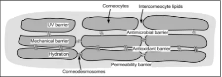
The outermost layer of the skin, the stratum corneum (SC), primarily mediates this permea-bility barrier function (Fig. 1). SC is composed of two different structural components: the corneo-cytes and intercorneocyte lipids. Both components are derived from keratinocytes through the ter-minal differentiation process. Considerable efforts
An Update of the Defensive Barrier Function of Skin
Seung Hun Lee,1,2 Se Kyoo Jeong,1 and Sung Ku Ahn3
1Department of Dermatology, 2Cutaneous Biology Research Institute, Yonsei University College of Medicine, Seoul, Korea; 3Department of Dermatology, Yonsei University Wonju College of Medicine, Wonju, Korea
Received May 18, 2006
Reprint address: requests to Dr. Seung Hun Lee, Department of Dermatology, Yongdong Severance Hospital, Yonsei University College of Medicine, 146-92 Dogok-dong, Kangnam-gu, Seoul 135-720, Korea. Tel: 82-2-2019-3360, Fax: 82-2-3463-6136, E-mail: [email protected]
have been made to elucidate the structure, func-tion and biochemistry of the SC and, about two decades ago, Elias proposed the 'brick and mortar model', in which the SC is composed of flat cells (bricks) surrounded by a lipid matrix (mortar).8 Since then, many studies have been performed to elucidate the role of the SC at the organismal, bio-chemical and molecular biological levels. The corneocytes, as terminal differentiation forms of keratinocytes, provide structural supports for the SC and act as hydrating reservoirs for adequate enzymatic processes in the SC. The cornified en-velope (CE), which encapsulates the corneocytes in SC, is a 15-20 nm-thick structure comprising defined structural proteins.9A 5nm-thick structure of specialized lipids, identified as the cornified lipid envelope (CLE), encloses the CE and this membrane-bound lipid monolayer provides hy-drophobic interfaces between the hydrophilic surface of the CE and the highly hydrophobic lipid lamellae.10Recently, a detailed elucidation of the component proteins of the CE and its forma-tion has been reported.11
In addition to these major structural domains, the corneodesmosome(CD), which corresponds to desmosome in the epidermis, is another impor-tant components in the SC. Generally, the inte-grity of the SC is maintained by these intercellular proteins which connect to adjacent corneocytes, both in the plane of the SC layer and in adjacent layers. In the SC, the CD structures represent the primary cohesive force and they are directly related the desquamation process. These struc-tures are composed of certain proteins, including
desmocollin and desmoglein, and special protein-degrading enzymes which are presented in the SC and play a crucial role in the desquamation pro-cess.12 Water activity and pH control the activity of the protease in the SC. The cholesterol sulfate/ cholesterol ratio and protease inhibitors in the SC are also important regulators of the protease activity.13,14
In this review, we introduce recent achieve-ments in the research of skin barrier functions and their significances in treating atopic dermatitis and psoriasis.
THE INTERRELATIONSHIPS OF DEFENSIVE FUNCTIONS IN THE SKIN
In addition to the permeability barrier function, the SC also plays an important role in other critical functions such as antimicrobial defense, the hydration of viable epidermis, UV defense, antioxidant defense and the formation of a mechanical barrier. Due to structural hetero-geneity, either corneocytes or the SC intercellular lipids mediate the defensive functions of the SC: the extracellular lipids act as permeability, anti-oxidant, and antimicrobial barriers, and the cor-neocytes act as UV and mechanical barriers. Recent reports, however, show that these func-tions are closely interrelated and that the change of any function could result in the modulation of other functions. These defensive functions are interrelated by co-localization, biochemical link-age or through common regulatory mechanisms.
The epidermal permeability barrier and its anti-microbial barrier function
[image:2.595.57.282.103.181.2]As an antimicrobial barrier against invading microorganisms, skin acts as a physical barrier and produces a number of antimicrobial peptides and proteins, including human defensins and cathelicidins. These antimicrobial peptides (AMPs), which play a major role in the host's innate de-fenses, are generally small, cationic polypeptides and have a capacity to inhibit the growth of microbes. In addition to the direct antimicrobial activities against various bacteria, viruses and fungi, AMPs also activate cellular and adaptive Fig. 1.The localization of various protective functions in
immune responses. They also play important roles as mediators of inflammation with effects on epithelial and inflammatory cells, influencing cell proliferation, wound healing, cytokine and che-mokine production and chemotaxis. While cathe-licidins and defensins comprise the major families of AMPs in the skin, other molecules such as pro-tease inhibitors, chemokines and neuropeptides, also exhibit antimicrobial activity. In addition to those molecules, free fatty acids, glucosylcera-mides and sphingosine, which are major consti-tuents for SC intercellular lipids, and hydrolytic products of ceramide also provide an antimi-crobial barrier in the skin.
While AMPs are generally synthesized in the upper stratum spinosum (SS) and stratum granu-losum, the active site for these molecules is the SC where they are delivered. Recently, it has been shown that human -defensin 2 (HBD-2) is localβ -ized in the lamellar bodies (LBs)15 and catheli-cidin also resides in LBs.16 Since the LB is an essential micro-organelle for epidermal barrier formation, it can be postulated that the epidermal permeability barrier function is closely related to the antimicrobial function of skin. In several skin diseases showing disturbed skin barrier function, such as psoriasis and atopic dermatitis (AD), abnormal expression of AMPs was reported. For example, LL-37, hBD-2 and hBD-3 are up regu-lated in psoriatic skin lesions.17,18 The expression of LL-37 and hBD-2 is relatively down regulated in atopic dermatitis lesions compared to psoriatic skin,19 which correlates with the high suscepti-bility of AD skin to bacterial and viral infections. Recently, we have reported that epidermal ex-pression of CRAMP, a murine analogue of human LL-37, is significantly increased after UVB expo-sure.20 Since phototherapy using UVB is fre-quently used in severe AD, increases in AMPs can give a supplemental explanation for the beneficial effect of UV in AD.
KERATINOCYTE DIFFERENTIATION AND BARRIER FUNCTION
The formation of corneocytes is considered to be a result of finely regulated differentiation pro-cesses. During the terminal differentiation process,
structural change of the keratinocyte is associated with the sequential formation of differentiation-marker proteins that are unique to keratinocytes. Keratins such as K1 and K10, as early marker proteins of terminal differentiation, are expressed in the spinous layer. Late differentiation marker proteins, including involucrin, loricrin and filag-grin, appear in the granular layer. Complete degradation of intracellular micro-organelles such as nucleus and mitochondria and the formation of rigid CE complete the differentiation process. Keratinocyte-specific transglutaminase-1 catalyzes the cross-linking reaction between keratins and other structural proteins to form the chemically resistant cornified envelope structures of corneo-cytes.11
An in vitro cell culture system has been exten-sively used to identify the crucial factors in-fluencing keratinocyte differentiation. Through these intensive studies, it has been shown that the epidermal differentiation process is regulated by the concentration of extracellular calcium ions. Keratinocytes cultured in low calcium concen-trations (0.04 mM) show an undifferentiated, basal cell-like phenotype. Raising calcium concentra-tions up to 0.14 mM in the medium results in a terminal differentiation process, which is almost identical to thein vivoprocess. The early differen-tiation markers, K1 and K10, are observed within 8-24 hrs, and the late markers, loricrin and filag-grin, are shown after 24-48 hrs. A difference not found in vitro is the down-regulation of K14 expression, which takes place in differentiating keratinocytes in vivo but is constitutively ex-pressed in cultured keratinocytes. A relationship between elevated calcium ion concentration and keratinocyte differentiation is observed both in vivo and in vitro, where a calcium gradient has been well documented in human and murine epidermis. Higher calcium levels occur in the dif-ferentiated granular and spinous layers as com-pared to the undifferentiated basal layer. From these findings, the calcium-sensitive signaling pathway, involved in the regulation of keratino-cyte differentiation, was elucidated.
have been suggested for mediating the homeo-static responses after skin barrier disruption. Occlusion of barrier-disrupted skin with water vapor-impermeable membrane could block the lipid synthesis responses to barrier disruption, which also suggests that TEWL is a regulatory signal in barrier homeostasis. When the barrier disrupted skin was immediately exposed to iso-osmolar, hyperosmolar or hypoosmolar external solutions, however, the barrier recovery responses were normalized, which suggests that water movement, itself, might not be considered as a major signal in barrier homeostasis. The epider-mal calcium gradient, with its highest level in the granular layer and its lowest level in the basal layer, disappears after barrier disruption, and reappears in parallel with barrier restitution. In a previous study, it was shown that immersion of barrier-disrupted skin into a calcium containing solution significantly delayed barrier recovery. The central role of calcium ions in the skin barrier was also demonstrated by manipulating LB secre-tion by changing the epidermal calcium gradient without changing TEWL, using iontophoresis or sonophoresis. These studies suggest that changes in calcium ion concentration in the stratum granu-losum (SG) can directly induce the homeostatic signals for barrier repair, even without a change in permeability barrier function. The precise mechanism for the change of intracellular calcium levels in response to the change of extracellular calcium, however, is still not clear.
Protein kinase C (PKC) comprises a family of serine/threonine kinases that play central roles in the regulation of various cellular processes in numerous cell types. Currently, eleven PKC iso-types have been identified, which can be classified according to their activation factors.21 Human epidermal keratinocytes express several PKC iso-forms, such as PKC , ,α δ ε and . Other PKCξ isoforms (PKC , , ) were found in various otherβ γ μ keratinocytes. Previous studies suggest a crucial role for PKC in the regulation of the proliferation and differentiation of keratinocytes. From these studies, PKC expression and activity, which was represented by the change of subcellular localiza-tion, was seen to alter with calcium induced keratinocyte differentiation. It was also shown that inhibition of PKC activity with a selective
PKC inhibitor suppressed the expression of late differentiation marker proteins, such as involucrin and filaggrin, and terminal differentiation markers, such as keratinocyte-specific transglutaminase-1, in HaCaT keratinocytes. Phorbol 12-myristate 13-acetate (PMA), an activator of PKC, increased the expressions of involucrin, filaggrin, and transglu-taminase-1.22 In addition to its crucial role in keratinocyte differentiation, PKC might play some important roles in epidermal permeability barrier function. Previously, Murata et al. reported that the activity of serine palmitoyl transferase and glucosylceramide synthase, both essential for cera-mide production, was unregulated by a PKC-de-pendent mechanism in cultured human keratino-cytes.23 Recently, we have also shown that the PKC- specific inhibitor, rottlerin, delayed barrierδ recovery rate by inhibiting the intracellular cal-cium responses to the loss of the extracellular calcium gradient.24 Although the precise mecha-nism of PKC involvement in permeability barrier homeostasis is still unknown, it seems that some PKC isozymes are responsible for the regulation of intracellular calcium concentration during the permeability barrier recovery process.
THE LAMELLAR BODY AS A MAJOR PLAYER IN THE FORMATION OF THE PERMEABILITY BARRIER
inhibitors, are also packed in LBs. Using confocal laser scanning microscopy and immunoelectron microscopy, Ishida-Yanamoto et al. showed that cathepsin D and kallikrein (KLK) 7 are also packed in LBs. They also showed that each LB has its own separate molecules.27 More recently, they also showed that KLK5 and LEKTI (lympho-epithelial Kazal-type-related inhibitor), a putative serine protease inhibitor, is also localized in the LBs.28 The heterogeneous content of the LBs sug-gests that they are important for not only epidermal permeability barrier functions, but also the other defensive functions exerted by the SC. While the epidermal LBs play a basic and crucial role in the formation, maintenance and repair of the epidermal permeability barrier, the exact mechanism underlying LB formation in the SG and its extrusion into the SC-SG interface is not yet fully understood. In a previous report, Madison and Howard showed that glucosylcera-mide, a major component of LB and the precursor of SC ceramides, was produced through the Golgi apparatus and delivered to the plasma membrane, which suggested that the Golgi apparatus is the origin of LB.29 Recently, the role of caveolin pro-teins in LB formation and secretion has been suggested. The caveolins are cholesterol-binding scaffolding proteins that facilitate the assembly of cholesterol and sphingolipid-enriched membrane domains, known as caveolae. Previously, Sando et al. reported morphological evidence of co-localiza-tion of the caveolin and LBs in the SG, which sug-gests that caveolins may play a role in LB as-sembly and function.30 Caveolae are a subset of lipid raft domains, which are characterized as flask-shaped invaginations in the plasma mem-brane of 50 to 100 nm in diameter and, inter-estingly, Elias et al. observed a structural simi-larity between the caveolae and the LB secretion site.31 Since the lipid raft provides platforms for preserving the specificity and efficiency of cell signal transduction processes,32 it can be postu-lated that the LB secretory response to barrier disruption is important not only for replenishing the lipids to the SC intercorneocyte domains, but also for creating higher-order signaling complexes to amplify cellular signaling.33Recently, numerous studies using caveolin knockout (KO) mice have been reported. Interestingly, it was reported that
cavolin-1 KO mice showed no abnormalities in cutaneous phenotype34 and barrier function re-covery rate was accelerated after acute perme-ability barrier disruption,35which also suggests a close relationship between the caveolin proteins and LB formation and its secretion.
CALCIUM IONS AND BARRIER HOMEOSTASIS
Epidermal permeability barrier homeostasis
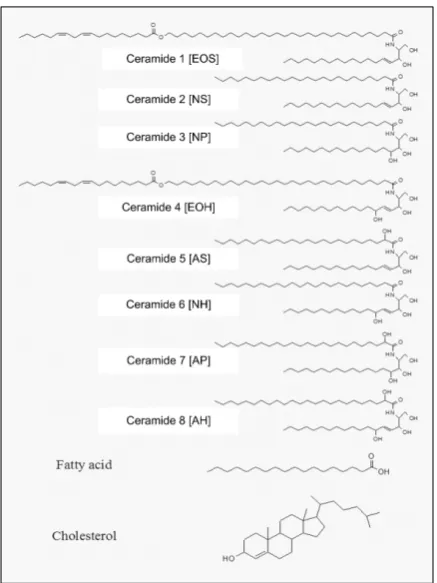
The SC intercellular lipid matrix constitutes about 15% of the dry weight of the SC and the only continuous phase of the skin barrier. The unique lamellar, bilayer organization of this lipid matrix provides the structural basis for the ex-traordinary lower permeability to water and other electrolytes through the SC.36 The major lipid species of the SC are ceramides, fatty acids, and cholesterol.37 Small amounts of cholesterol esters and cholesterol sulfates are also present in SC and both lipids play a critical role in proper structural organization of SC lipids and, therefore, in normal barrier function.38 The chemical structures of SC lipids species are illustrated in Fig. 2.
in-creased after acute disruption of barrier function, which could be an additional support for barrier recovery. In murine epidermis, mRNA expression of tumor necrosis factor (TNF)- , interleukin (IL)-1α , IL-1 and granulocyte
macrophage-colony-α β
stimulating factor (GM-CSF) are elevated after barrier disruption (Fig. 3).
[image:6.595.59.278.100.392.2]For a complete recovery of epidermal permea-bility barrier function, both the corneocytes of the SC intercellular lipids need to be replenished into the barrier-damaged skin site. While a great deal of studies have been reported on the regulation of lipid synthesis in the stratum granulosum after barrier disruption, relatively little is known about the regulation of keratinocyte differentiation after barrier disruption and its role in barrier homeostasis. Peroxisome proliferator- activated receptor (PPAR) and liver X receptor (LXR) belong to a subset of the nuclear hormone re-ceptor superfamily (class II rere-ceptors), which need retinoid X receptor (RXR) as a heterodimerizing factor for their activation.39 PPAR and LXR are ligand-activated transcriptional factors that regu-late the expression of target genes involved in many cellular functions including cell prolifera-tion, differentiation and immune/inflammation responses. Along with the fibrate hypolipidemic agents, a large number of endogenous fatty acids and fatty acid metabolites can activate PPAR. Sev
[image:6.595.99.495.495.673.2]Fig. 3. The epidermal permeability barrier homeostasis. When barrier function is perturbed, immediate secretion of preformed lamellar bodies occurs, followed by an increased synthesis of lamellar bodies and lipid precursors. Increased expression of inflammatory cytokines is observed after barrier disruption. Keratinocyte differentiation and proliferation are also increased after barrier disruption. The epidermal permeability barrier is restored through the above described homeostatic responses. Iontophoresis, sonophoresis and topical glycolic acid can also induce the down-stream homeostatic responses without changing the transepidermal water loss.
eral oxysterols, including cholesterol, can activate LXR.40 Activation of PPAR and LXR in skin, induced various cellular responses including the promotion of keratinocyte differentiation and al-leviation of epidermal hyperproliferation, as well as the reduction of epidermal inflammation.41 Re-cently, it was also shown that topical application of PPAR or LAX activator on barrier-disrupted skin improves epidermal permeability barrier recovery, inducing an increase of epidermal lipid synthesis and LB secretion.42 Since endogenous lipids produced in the keratinocyte can activate PPAR or LXR, and, in turn, regulate lipid metab-olism of the keratinocyte, these receptors are cur-rently referred to as 'liposensors' in keratinocytes, and considered major players connecting lipid metabolism and keratinocyte differentiation.
Calcium's role in barrier homeostasis
In 1985, Menon et al. reported that an epidermal calcium gradient with low calcium concentrations in the basal, proliferating layers, and a progres-sively higher concentration as one proceeds to the outer differentiated layers, i.e. the SG.43 Several years later, it was suggested that this epidermal calcium gradient plays a crucial role in skin barrier function.44 Acute barrier disruption with either acetone treatment or repeated tape strip-ping induced an immediate, marked decrease of intracellular calcium concentrations in the SG. The calcium levels in the SG then progressively nor-malized in parallel with barrier recovery over 24 hours.45Recently, Elias et al. reported the relation-ship of barrier function and the reappearance of the epidermal calcium gradient after acute barrier disruption.46 Immediate application of a vapor-permeable membrane on the barrier-disrupted skin provided an artificial permeability barrier and accelerated epidermal calcium gradient for-mation. While the exposure of barrier-disrupted skin to a cold environment retarded both the barrier recovery and calcium gradient formation, vapor-permeable membrane application with cold expo-sure resulted in significant calcium gradient for-mation after three hours, without barrier recovery. With these results, it can be postulated that barrier status dominantly regulates the formation of the epidermal calcium gradient and calcium gradient
formation or maintenance is achieved though passive, ATP-independent processes.
Many studies exploring the relationship between permeability barrier homeostasis and epidermal calcium ions have been reported. Sonophoresis and iontophoresis are both used for increasing the delivery of drugs and other bioactive materials across the SC and were shown to decrease the calcium concentrations in the upper epidermis. While the sonophoresis or iontophoresis-treated skin showed no changes in TEWL, increased secretion and synthesis of LB were observed in both skin layers.47-49 Since the secretion of LB from the SG and consequent accumulation of lamellar materials at the SC-SG junction is a hallmark for barrier homeostatic responses after barrier dis-ruption, these results showed that the regulation of LB secretion is mediated by calcium ions in the SG. Recently, it has been reported that several agonists and antagonists for G-protein-coupled receptors (GPCRs) expressed in keratinocytes, such as the - aminoγ butyric acid receptor (GABA),50 the P2X purinergic receptor,51 the NMDA type glutamate receptor,52 the 2-adrenergic receptorβ 53 and the histamine receptor,54have accelerating or delaying effects on barrier recovery after acute barrier disruption. Since the major signaling molecules for GPCRs include intracellular calcium ions, it can be concluded that the change of intra-cellular calcium ion concentration is a major signal for permeability barrier homeostasis. In addition to the GPCR activation-related calcium modulation, we have also shown that topical application of calcium chelating agents onto nor-mal skin induced the barrier homeostatic re-sponses, including loss of the epidermal calcium gradient and LB secretion.55Topical application of glycolic acid, which does not induce any changes in TEWL in normal murine skin, significantly increased LB secretion56,57 and an in vitro study using cultured keratinocytes suggests that glycolic acid could lower the calcium ion concentration, at least in part, through its chelating effects on cations, such as calcium ions.
pH AND BARRIER HOMEOSTASIS
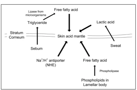
ranges from pH 4.5 to pH 5.5 in humans, is slightly acidic compared to the normal phy-siologic pH.58 SC acidity, or the "acid mantle" is currently considered to be crucial for establishing the epidermal permeability barrier, as well as for producing the epidermal antimicrobial barrier and controlling SC integrity and cohesion. Various endogenous and/or exogenous pathways are cur-rently suggested for acidifying the pH of the SC. Endogenous factors influencing SC acidification include the generation of free fatty acids from phospholipid hydrolysis catalyzed by secretory phospholipase A2 (sPLA2) and the sodium-proton antiporter-1, NHE1, which is expressed in the outer, nucleated layers of the epidermis.59 Exoge-nous mechanisms include the generation of free fatty acids by bacterial lipase, free fatty acids derived from sebum60 and eccrine gland-derived products, such as lactic acid (Fig. 4).61
In atopic eczematous lesions, the epidermal permeability barrier is perturbed and the skin pH is also significantly elevated.62 In addition to the perturbation of lipid metabolism and their mole-cular organization, increased skin pH also induces bacterial growth (e.g. Staphylococcus aureus).
Diverse functions for this acid mantle have been suggested, one of the major ones being antimi-crobial action. A few studies relating surface pH and its role in supporting the growth of normal
microflora as well as inhibiting skin pathogen growth have been previously reported.63A typical example where the impaired antimicrobial barrier is correlated with elevated skin surface pH is diaper dermatitis. Due to chronic exposure to urine and feces, diapered, neonatal skin shows a more neutral pH than uncovered skin.64 In addi-tion, pathogens that grow at neutral pH worsen diaper dermatitis,65 resulting in a vicious cycle where neutral pH-enhanced pathogen growth and inflammatory cytokine release combine to pro-duce an inflamed, colonized skin with impaired antimicrobial barriers.66
[image:8.595.55.282.96.246.2]Another important function of the acid mantle is related to the extracellular processing of LB-derived lipid precursors, which comprise the SC intercellular lipid domain. Among the various important enzymes in SC are β-glucocerebro -sidase, acid sphingomyelinase and secreted phos-pholipase A2 (sPLA2). Each of these functions optimally in an acidic environment and contri-butes to the generation of the ceramide molecules and free fatty acids required for epidermal per-meability barrier homeostasis. Acute disruption of the permeability barrier results in a slight but significant increase in skin surface pH. Along with the barrier recovery, the acid mantle reappears. Recent reports using super-acid or super-base, which could modulate skin surface pH without affecting viable epidermis, also showed that skin surface pH regulated the epidermal barrier home-ostasis.67 In addition, maintenance of intact murine SC to a neutral pH produced significant abnormalities in SC integrity and cohesiveness.68 Using neonatal hairless mice, Fluhr et al. showed that topical application of liver X receptor (LXR) activators accelerated SC acidification, as well as stimulated keratinocyte differentiation and im-proved permeability barrier homeostasis.69 Among the proposed acidification pathways, LXR acti-vators induced the increase of secretory phos-pholipase A2 (sPLA2) activity in the SC. The above described roles of the SC acid mantle in epidermal permeability barrier function suggest that both barriers are closely related and mainte-nance of skin surface pH in the normal acidic range is crucial for not only the antimicrobial barrier, but epidermal permeability barrier func-tion as well.
Skin surface pH also affects the desquamation processes. A lot of protein degrading enzymes and proteases exist in the SC and each protease has its optimal pH, respectively (Fig. 5).70,71Since each protease has its own substrate in the corneo-desmosome components, changes in pH induce abnormal desquamation and results in a scaly skin. A typical example of abnormal desquama-tion induced by alkaline pH is soap-induced xerotic skin.72
In addition, skin pH is also related to the activa-tion of the protease-activated receptor-2 (PAR-2) in skin. The protease-activated receptor (PAR) is a seven transmembrane G-protein-coupled recep-tor, distinguished by a unique mechanism of self-activation following specific proteolytic cleavage of its extracellular domains.73It was reported that PAR2 agonist peptides significantly delay epider-mal permeability barrier recovery after acute barrier disruption in murine skin,74 possibly due to the increase of intracellular calcium concentra-tion in epidermal keratinocytes. Previous studies also showed that serine protease activity was in-creased in atopic dermatitis, and it has been pos-tulated that increased PAR-2 activity could be a pathophysiologic factor for atopic dermatitis.75 Recently, we have reported the effects of PAR-2 activators on epidermal barrier functions, as well as on epidermal calcium gradients. Cockroach
allergens and house dust mite allergens, both known to be associated with the development of asthma and atopic dermatitis, could activate PAR-2 and thereby induce various cellular responses.76, 77Topically applied PAR-2 activators delayed the
barrier recovery, which might be due to the change of intracellular calcium concentration. Since the protease in cockroach and house dust mite allergens have their highest activity at neu-tral pH, maintaining an acidic pH after barrier disruption significantly prevented the inhibitory effects of both allergens on epidermal permea-bility barrier recovery.78
BARRIER DYSFUNCTIONS IN SKIN DISEASES
[image:9.595.127.459.100.270.2]balance results in an altered lipid organization and consequent abnormality in the structural or-ganization of the intercorneocyte lipid lamellae.79 Cholesterol sulfate is also known to affect other biological processes such as protease activity in the SC, which results in abnormal desquamation in the RXLI.13 In addition to the inhibitory effects on protease in the SC, cholesterol sulfate also acts as an activating factor for the eta isoform of pro-tein kinase C (PKC eta). PKC eta is a subtype of novel PKC and, in the keratinocyte, is closely as-sociated with cell cycle arrest and differentiation.80 It can, therefore, be postulated that cholesterol sul-fate is also related to the differentiation of epider-mal keratinocytes. Other genetic skin diseases, including Netherton syndrome,81characterized by defects in proteolysis, and type 2 Gaucher disease, exhibiting defects in -glucocerebrosidase, involveβ skin barrier abnormalities as a pathologic out-comes.
While various pathogenic factors are suggested for AD, impaired barrier function is currently considered an important one. Atopic dry skin displays impaired barrier function, indicated by an increased transepidermal water loss and lowered water-binding capacities. Significant de-creases in SC ceramide,82especially in ceramide 1 (CER EOS) levels is reported in AD skin.83 Bouwstra et al. suggested that the ceramide 1 in SC intercellular lipids plays a dominant role in the formation of proper molecular organization.84 In addition, Pilgram et al. reported that the abnormal lipid organization in the SC of AD patients re-sulted from the decrease of ceramide 1 and sug-gested that the impaired barrier function of AD skin was, at least in part, due to this structural change.85 Recently, we have reported a structural property of a physiologic lipid mixture containing a pseudoceramide, which showed a close simi-larity to that of SC intercellular lipids.36 Since the structural property of lipids can affect the epider-mal permeability barrier function, it might be beneficial to barrier function to use the physio-logic lipid mixture on the barrier-disrupted skin. The decrease of ceramides in AD patients is linked to an increased expression of sphingomye-lin deacylase, which converts the sphingomyesphingomye-lin to free fatty acids and sphingosylphosphorylcho-line.86In normal skin, epidermal sphingomyelin is
catalyzed by sphingomyelinase to produce cera-mide, but in AD skin, extraordinary sphigomyelin deacylase activity is presented. Recently, the ex-istence of another novel enzyme, termed as gluco-sylceramide deacylase, has been reported in AD skin lesions. In normal skin, glucosylceramide, secreted into the SC through LBs, is catalyzed by -glucocerebrosidase to produce ceramide. In AD β
skin, however, glucosylceramide is catalyzed by this novel enzyme to produce glucosylsphin-gosine.87 With this enzymatic abnormality, cera-mide level is down regulated in AD skin, and permeability barrier function is incompetent (Fig. 6). Altered ceramide metabolism in AD skin also induces a decrease in sphingosine, which is pro-duced from ceramide by ceramidase in the SC. Since sphingosine is known to have potent anti-microbial activities on Staphylococcus aureus at physiologic levels, down regulation of sphingo-sine is one of the reasons for the vulnerability of AD patients to S. aureus colonization.88
characterized by a decrease of the NMF com-ponents in skin.92 Mutations of filaggrin genes result in abnormal corneocyte structure and im-paired barrier function, leading to skin that is more vulnerable to the penetration of exogenous allergens than normal skin. Along with the abnormalities in lipid metabolisms, these findings suggest that both the bricks and the mortar have defects in AD skin, and consequent impairment of epidermal barrier function is an important pathophysiologic factor for AD.93
Another important skin disease with impaired barrier function is psoriasis. In this condition, TEWL are significantly increased, according to the severity of the lesions.94 Alterations in cera-mide content and abnormal lipid structures are also reported in psoriasis.95,96 These changes in-clude an increase in ceramide with non-hydroxyl fatty acids and sphingosine (CerNS), ceramide with 6-hydroxy-4-shingenine and omega-hydroxy fatty acid ester linked to linoleic acid (CerEOH) and decreases in ceramide with alpha-hydroxy fatty acids and sphingosine (CerAS). In addition, altered cholesterol and fatty acid levels are also observed in psoriatic lesions. This abnormality in lipid composition and components results in not only the alterations in epidermal permeability barrier function, but also alterations in corneo-cyte adhesion and desquamation. The cornified lipid envelope (CLE) structure and its compo-nents are also changed in psoriasis. While it needs to be clarified whether the abnormal per-meability barrier is primary pathophysiologic factors for these skin diseases or not, many studies have now shown that improvements in barrier function resulted in significant ameliora-tion of disease severity.
CONCLUSIONS
Epidermal permeability barrier function has the crucial role of maintaining skin homeostasis and the perturbation of barrier function has significant effects on overall skin quality. Recently, many studies have suggested that the defects in per-meability barrier function are not secondary con-sequences, but critical factors for various skin diseases. Therefore, understanding the molecular
basis of barrier function and its homeostatic responses can provide not only a more rational therapy of barrier-disrupted skin diseases, but also improve the specificity and efficacy of treat-ments for human skin with abnormal SC structure and function.
REFERENCES
1. Albanesi C, Scarponi C, Giustizieri ML, Girolomoni G. Keratinocytes in inflammatory skin diseases. Curr Drug Targets Inflamm Allergy 2005;4:329-34.
2. Hearing VJ. Biogenensis of pigment granules: a sensi-tive way to regulate melanocyte function. J Dermatol Sci 2005;37:3-14.
3. Lehmann B, Querings K, Reichrath J. Vitamin D and skin: new aspects for dermatology. Exp Dermatol 2004; 13 Suppl 4:11-5.
4. Garssen J, Vandebriel RJ, van Loveren H. Molecular aspects of UVB-induced immunosuppression. Arch Toxicol Suppl 1997;19:97-109.
5. Ghoreishi M. Heat shock proteins in the pathogenesis of inflammatory skin diseases. J Med Dent Sci 2000;47: 143-50.
6. Bowdish DM, Davidson DJ, Hancock RE. A re-evalua-tion of the role of host defense peptides in mammalian immunity. Curr Protein Prpt Sci 2005;6:35-51. 7. Braff MH, Bardan A, Nizet V, Gallo RL. Cutaneous
defense mechanisms by antimicrobial peptides. J Invest Dermatol 2005;125:9-13.
8. Elias PM. Epidermal lipids, barrier function and des-quamation. J Invest Dermatol 1983;80 Suppl:44-9. 9. Jarnik M, Simon MN, Steven AC. Cornified cell
envelope assembly: a model based on electron micro-scopic determinations of thickness and projected den-sity. J Cell Biol 1998;111:1051-60.
10. Swartzendruber DC, Wertz PW, Madison KC, Downing DT. Evidence that the corneocytes has a chemically bound lipid envelope. J Invest Dermatol 1987;88:709-13. 11. Kalinin AE, Kajava AV, Steinert PM. Epithelial barrier function: assembly and structural features of the cornified cell envelope. Bioessays 2002;24:789-800. 12. Komatsu N, Saijoh K, Sidiropoulos M, Tsai B, Levesque
MA, Elliott MB, et al. Quantification of human tissue kallikreins in the stratum corneum: dependence on age and gender. J Invest Dermatol 2005;125:1182-9. 13. Elias PM, Crumrine D, Rassner U, Hachem JP, Menon
GK, Man W, et al. Basis for abnormal desquamation and permeability barrier dysfunction in RXLI. J Invest Dermatol 2004;122:314-9.
15. Oren A, Ganz T, Liu L, Meerloo T. In human epider-mis, -defensin 2 is packaged in lamellar bodies. Expβ
Mol Pathol 2003;74:180-2.
16. Braff MH, Di Nardo A, Gallo RL. Keratinocytes stores the antimicrobial peptide cathelicidin in lamellar bodies. J Invest Dermatol 2005;124:394-400.
17. Frohm M, Agerberth B, Ahangari G, Stahle-Backdahl M, Liden S, Wigzell H, et al. The expression of the gene coding for the antimicrobial peptide LL-37 is induced in human keratinocytes during inflammatory disorders. J Biol Chem 1997;272:15258-63.
18. Nomura I, Gao B, Boguniewicz M, Darst MA, Travers JB, Leung DY. Distinct patterns of gene expression in the skin lesions of atopic dermatitis and psoriasis: a gene microarray analysis. J Allergy Clin Immunol 2003; 112:1195-202.
19. de Jongh GJ, Zeeuwen PL, Kucharekova M, Pfundt R, van der Valk PG, Blokx W, et al. High expression levels of keratinocyte antimicrobial proteins in psoriasis compared with atopic dermatitis. J Invest Dermatol 2005;125:1163-73.
20. Kim M, Jeon H, Goo J, Ahn S, Park Y, Lee S, et al. Expression of anti-microbial peptides in murine epider-mis in increased by UVB exposure. J Invest Dermatol 2006;126:A66.
21. Parker PJ, Murray-Rust J. PKC at a glance. J Cell Sci 2004;117:131-2.
22. Papp H, Czifra G, Lazar J, Gonczi M, Csernoch L, Kovacs L, et al. Protein kinase C isozymes regulate pro-liferation and high cell density-mediated differentiation in HaCaT keratinocytes. Exp Dermatol 2003;12:811-24. 23. Murata S, Uchida Y, Ichikawa S, Hirabayashi Y, Holleran WM. Regulation of glucosylceramide synthase expression in cultured human keratinocyte. J Invest Dermatol 2000;114:795.
24. Ahn BK, Jeong SK, Kim HS, Choi KJ, Seo JT, Choi EH, et al. Rottlerin, a specific inhibitor of protein kinase C-delta, impedes barrier repair responses by increasing intracellular free calcium. J Invest Dermatol 2006;126: 1348-55.
25. Odland GF, Holbrook A. The lamellar granules of epi-dermis. Curr Probl Derm 1981;9:29-49.
26. Menon GK, Ghadially R, Williams ML, Elias PM. Lamellar bodies as delivery systems of hydrolytic en-zymes: implications for normal and abnormal desqua-mation. Br J Dermatol 1992;126:337-45.
27. Ishida-Yamamoto A, Simon M, Kishibe M, Miyauchi Y, Takahashi H, Yoshida S, et al. Epidermal lamellar gran-ules transport different cargoes as distinct aggregates. J Invest Dermatol 2004;122:1137-44.
28. Ishida-Yamamoto A, Deraison C, Bonnart C, Bitoun E, Robinson R, O'Brien TJ, et al. LEKTI is localized in lamellar granules, separated from KLK5 and KLK7, and is secreted in the extracellular spaces of the superficial stratum granulosum. J Invest Dermatol 2005;124:360-6. 29. Madison KC, Howard EJ. Ceramides are transported through the Golgi apparatus in human keratinocytesin vitro. J Invest Dermatol 1996;106:1030-5.
30. Sando GN, Zhu H, Wies JM, Richman JT, Wertz PW, Madison KC. Caveolin expression and localization in human keratinocytes suggest a role in lamellar granule biogenesis. J Invest Dermatol 2003;120:531-41. 31. Elias PM, Cullander C, Mauro T, Rassner U, Komuves
L, Brown BE, et al. The secretory granular cell: The outermost granular cells as a specialized secretory cells. J Invest Dermatol Symp Proc 1998;3:87-100.
32. Guest AFG, Leyton L, Parraga M. Caveolins, caveolae, and lipid rafts in cellular transport, signalingm and disease. Biochem Cell Biol 2004;82:129-44.
33. Menon GK. Caveolins in epidermal lamellar bodies: skin is an interactive interface, not an inflexible barrier. J Invest Dermatol 2003;120:15-6.
34. Capozza F, Williams TM, Schubert W, McClain S, Bouzahzah B, Sotgia F, et al. Absence of caveolin-1 sensitized mouse skin to carcinogen-induced epidermal hyperplasia and tumor formation. Am J Pathol 2003; 162:2029-39.
35. Reolandt T, Huth M. Houben E, Crumrine D, Kutsumo A, Giddelo G, et al. Caveolin-1 incorporation into lipid rafts in the outer stratum granulosum suppresses lamellar body secretion in parallel with initiation of terminal differentiation. J Invest Dermatol 2006;126: A71.
36. Park BD, Youm JK, Jeong SK, Choi EH, Ahn SK, Lee SH. The characterization of molecular organization of multilmellar emulsions containing pseudoceramide and type III synthetic ceramide. J Invest Dermatol 2003; 121: 794-801.
37. Elias PM. Epidermal barrier function: intercellular lamellar lipid structures, origin, composition and metabolism. J Control Release 1991;15:199-262. 38. Bouwstra JA, Gooris GS, Weerheim A, Ponec M. pH,
cholesterol sulfate and fatty acids affects the stratum corneum lipid organization. J Invest Dermatol Symp Proc 1998;3:69-74.
39. Berger J, Moller DE. The mechanisms of action of PPARs. Annu Rev Med 2002;53:409-35.
40. Krey G, Braissant O, L'Horset F, Kalkhoven E, Perroud M, Parker MG, et al. Fatty acids, eicosanoids, and hypolipidemic agents identified as ligands for pero-xisome proliferators-activated receptors by coactivator-dependent receptor ligand assay. Mol Endocrinol 1997; 11:779-91.
41. Elias PM. Stratum corneum defensive functions: an integrated view. J Invest Dermatol 2005;125:183-200. 42. Man M-Q, Choi EH, Schmuth M, Crumrine D, Uchida
Y, Elias PM, et al. Basis for improved permeability barrier homeostasis induced by PPAR and LXR acti-vators: liposensors stimulate lipid synthesis, lamellar body secretion, and post-secretory lipid processing. J Invest Dermatol 2006;126:386-92.
43. Menon GK, Grayson S, Elias PM. Ionic calcium reser-voirs in mammalian epidermis: ultrastructural locali-zation by ion-capture cytochemistry. J Invest Dermatol 1985;84:508-12.
M, Feingold KR. Calcium and potassium are important regulators of barrier homeostasis in murine epidermis. J Clin Invest 1992;89:530-8.
45. Menon GK, Elias PM, Lee SH, Feingold KR. Localiza-tion of calcium in murine epidermis following disrup-tion and repair of the permeability barrier. Cell Tissue Rep 1992;270:503-12.
46. Elias PM, Ahn SK, Brown BE, Crumrine D, Feingold KR. Origin of epidermal calcium gradient: regulation of barrier status and role of active vs passive mechanisms. J Invest Dermatol 2002;119:1269-74.
47. Menon GK, Price LF, Bommannan B, Elias PM, Feingold KR. Selective obliteration of the epidermal calcium gradient leads to enhanced lamellar body secretion. J Invest Dermatol 1994;102:789-95.
48. Lee SH, Choi EH, Feingold KR, Jiang S, Ahn SK. Iontophoresis itself on hairless mouse skin induces the loss of the epidermal calcium gradient without skin barrier impairment. J Invest Dermatol 1998;111:39-43. 49. Choi EH, Kim MJ, Yeh BI, Ahn SK, Lee SH.
Ionto-phoresis and sonoIonto-phoresis stimulate epidermal cyto-kine expression at energies that do not provoke a barrier abnormality: lamellar body secretion and cyto-kine expression are linked to altered epidermal calcium levels. J Invest Dermatol 2003;121:1138-44.
50. Denda M, Inoue K, Inomata S, Denda S. -aminobutyricγ
acid (A) receptor agonists accelerate cutaneous barrier recovery and prevent epidermal hyperplasia induced by barrier disruption. J Invest Dermatol 2002;119:1041-7.
51. Denda M, Inoue K, Fuziwara S, Denda S. P2X puri-nergic receptor antagonist accelerates skin barrier repair and prevents epidermal hyperplasia induced by skin barrier disruption. J Invest Dermatol 2002;119: 1034-40.
52. Fuziwara S, Inoue K, Denda M. NMDA-type glutamate receptor is associated with cutaneous barrier homeo-stasis. J Invest Dermatol 2003;120:1023-9.
53. Denda M, Fuziwara S, Inoue K. 2-adrenergic receptorβ
antagonist accelerates skin barrier recovery and reduces epidermal hyperplasia induced by barrier disruption. J Invest Dermatol 2003;121:142-8.
54. Ashida Y, Denda M, Hirao T. Histamine H1 and H2 receptor antagonists accelerate skin barrier repair and prevent epidermal hyperplasia induced by barrier dis-ruption in a dry environment. J Invest Dermatol 2001; 116:261-5.
55. Jeong SK, Ko JY, Seo JT, Ahn SK, Lee CW, Lee SH. Stimulation of epidermal calcium gradient loss and increase in TNF- and IL-1 expressions by glycolicα α
acid in murine epidermis. Exp Dermatol 2005;4:571-9. 56. Kim TH, Choi EH, Kang YC, Lee SH, Ahn SK. The effects of topical -hydroxyacids on the normal skinα
barrier of hairless mice. Br J Dermatol 2001;144:267-73. 57. Jeong SK, Kim S, Lee EH, Choi EH, Ahn SK, Lee SH. Comparison of the effect of various chemical peeling agents on the skin barrier. Korean J Dermatol 2002;40: 1181-7.
58. Parra JL, Paye M, EEMCO Group. EEMCO guidance for the in vivo assessment of skin surface pH. Skin Pharmacol Appl Skin Physiol 2003;16:188-202. 59. Fluhr JW, Behne MJ, Brown BE, Moskowitz DG, Seldel
C, Man MQ, et al. Stratum corneym acidification in neonatal skin: secretory phospholipase A2 and the sodium/hydrogen antiproter-1 acidify neonatal rat stratum corneum. J Invest Dermatol 2004;122:320-9. 60. Bibel DJ, Miller SJ, Brown BE, Pandey BB, Elias PM,
Shinefield HR, et al. Antimicrobial activity of stratum corneum lipids from normal and essential fatty acid-deficiency mice. J Invest Dermatol 1989;92:632-8. 61. Thueson DO, Chan EK, Oechsli LM, Hahn GS. The
roles of pH and concentration in lactic acid-induced stimulation of epidermal turnover. Dermatol Surg 1998; 24:641-5.
62. Rippke F, Schreiner V, Doering T, Maibach HI. Stratum corneum pH in atopic dermatitis: impact on skin barrier function and colonization with Staphylococcus aureus. Am J Clin Dermatol 2004;5:217-23.
63. Korting HC, Hubner K, Greiner K, Hamm G, Barun-Falco O. Differences in the skin surface pH and bac-terial microflora due to long-term application of syn-thetic detergent preparations of pH 5.5 and pH 7.0. Results of a crossover trial in healthy volunteers. Acta Derm Venereol 1990;70:429-31.
64. Berg RW, Milligan MC, Sarbaugh FC. Association of skin wetness and pH with diaper dermatitis. Pediatr Dermatol 1994;11:18-20.
65. Ferrazzini G, Kaiser RR, Hirsig Cheng SK, Wehrli M, Della Casa V, Pohlig G, et al. Microbiological aspects of diaper dermatitis. Dermatology 2003;206:136-41. 66. Mauro TM. SC pH: measurement, origins, and
func-tions. In: Eilas PM, Feingold KR editors. Skin Barrier. New York; Taylor & Francis Group; 2006. p.223-30. 67. Hachem JP, Behne M, Aronchik I, Demerjian M,
Feingold KR, Elias PM, et al. Extracellular pH controls NHE1 expression in epidermis and keratinocytes: im-plications for barrier repair. J Invest Dermatol 2005;125: 790-7.
68. Fluhr JW, Kao J, Jain M, Ahn SK, Feingold KR, Elias PM. Generation of free fatty acids from phospholipids regulates stratum corneum acidification and integrity. J Invest Dermatol 2001;117:44-51.
69. Fluhr JW, Crumrine D, Man MQ, Moskowitz DG, Elias PM, Feingold KR. Topical liver X receptor activators accelerate postnatal acidification of stratum corneum and improve function in the neonate. J Invest Dermatol 2005;125:1206-14.
70. Caubet C, Jonca N, Brattsand M, Guerrin M, Bernard D, Schmidt R, et al. Degradation of corneodesmosome proteins by two serine protease of the kallikrein family, SCTE/KLK5/hK5 and SCCE/KLK7/hK7. J Invest Der-matol 2004;122:1235-44.
M. A comparative study of the effects on the skin of a classical bar soap and a syndet cleansing bar in normal use conditions and in the soap chamber test. Skin Res Technol 2001;7:98-104.
73. Macfarlane SR, Seatter MJ, Kanke T, Hunter GD, Plevin R. Proteinase-activated receptors. Phamacol Rev 2001; 53:245-82.
74. Hachem J, Uchida Y, Crumrine D, Choi E, Houben E, Brown BE, et al. Serine protease-induced alterations in permeability barrier homeostasis are mediated by pro-tease-activated receptor 2. J Invest Dermatol 2005;124: A58.
75. Steinhoff M, Neisius U, Ikoma A, Fartasch M, Heyer G, Skov PS, et al. Proteinase-activated receptor-2 mediates itch: A novel pathway for pruritus in human skin. J Neurosci 2003;23:6176-80.
76. Hong JH, Lee SI, Kim KE, Yong TA, Seo JT, Sohn MH, et al. German cockroach extract activates protease-activated receptor 2 in human airway epithelial cells. J Allergy Clin Immunol 2004;113:315-9.
77. Asokananthan N, Graham PT, Stewart DJ, Bakker AJ, Eidne KA, Thompson PJ, et al. House dust mite aller-gens induce proinflammatory cytokines from respira-tory epithelial cells: the cysteine protease allergen, Der p 1, activates protease-activated receptor (PAR)-2 and inactivates PAR-1. J Immunol 2002;169:4572-8. 78. Jeong SK, Jeon JE, Kim HJ, Ahn SK, Choi EH, Cho IS,
et al. Aeroallergens, as a protease activated receptor-2 activator, delayed epidermal permeability barrier re-covery. (under submission)
79. Bouwstra JA, Honeywell-Nguyen L, Gooris GS, Ponec M. Structure of the skin barrier and its modulation by vesicular formulations. Progress Lipid Res 2003;42:1-36.
80. Kashiwaga M, Ohba M, Chida K, Kuroki T. Protein kinase C eta (PKC eta): its involvement in keratinocyte differentiation. J Biochem (Tokyo) 2002;132:853-7. 81. Chavanas S, Bodemar T, Rochat A, Hamel-Teillac D,
Ali M, Irvine AD, et al. Mutations in SPINK5, encoding a serine protease inhibitorm cause Netherton syn-drome. Nat Genet 2000;25:141-2.
82. Imokawa G, Abe A, Jin K, Higari Y, Kawashima M, Hidano A. Decreased levels of ceremides in stratum corneum of atopic dermatitis: an etiological factor in atopic dermatitis. J Invest Dermatol 1991;96:523-6. 83. Yamamoto A, Serizawa S, Ito M, Sato Y. Stratum
corneum lipid abnormalities in atopic dermatitis. Arch Dermatol Res 1991;283:219-23.
84. Bouwstra JA, Gooris GS, Dubbelaar FER, Weerheim AM, Ijzerman AP, Ponec M. Role of ceramide 1 in the molecular organization of the stratum corneum lipids. J Lipids Res 1998;39:186-96.
85. Pilgram GSK, Vissers DCJ, van der Meulen H, Pavel S,
Lavijsen SPM, Bouwstra JA, et al. Aberrant lipid orga-nization in stratum corneum of parients with atopic dermatitis and lamellar ichthyosis. J Invest Dermatol 2001;117:710-7.
86. Hara J, Higuchi K, Okamoto R, Kawashima M, Imo-kawa G. High-expression of sphigomyelin deacylase is an important determinant of ceramide deficiency leading to barrier disruption in atopic dermatitis. J Invest Dermatol 2002;115:406-13.
87. Ishibashi M, Arikawa J, Okamoto R, Kawashima M, Takagi Y, Ohguchi K, et al. Abnormal expression of the novel epidermal enzyme, glucosylceramide deacylase, and the accumulation of its enzymatic reaction product, glucosylsphingosine, in the skin of patients with atopic dermatitis. Lab Invest 2003;83:397-408.
88. Arikawa J, Ishibashi M, Kawashima M, Takagi Y, Ichikawa Y, Imokawa G. Decreased levels of sphi-gosine, a natural antimicrobial agent, may be associated with vulnerability of the stratum corneum from patients with atopic dermatitis to colonization by
Staphylococcus aureus. J Invest Dermatol 2002;119:433-9. 89. Sugiura H, Ebise H, Tazawa T, Tanaka K, Sugiura Y, Uehara M, et al. Large-scale DNA microarray analysis of atopic skin lesions shows overexpression of an epidermal differentiation gene cluster in the alternative pathway and lack of protective gene expression in the cornified envelope. Br J Dermatol 2005;152:146-9. 90. Palmer CN, Irvine AD, Terron-Kwiatkowski A, Zhao Y,
Liao H, Lee SP, et al. Common loss-of-function variants of the epidermal barrier protein filaggrin are a major predisposing factor for atopic dermatitis. Nat Genet 2006;38:441-6.
91. Smith FJ, Irvine AD, Terron-Kwiatkowski A, Sandilands A, Campbell LE, Zhao Y, et al. Loss-of-function muta-tions in the gene encoding filaggrin cause ichthyosis vulgaris. Nat Genet 2006;38:337-42.
92. Ginger RS, Blachford S, Rowland J, Rowson M, Harding CR. Filaggrin repeat number polymorphism is associated with a dry skin phenotype. Arch Dermatol Res 2005;297:235-41.
93. Hudson T. Skin barrier function and allergic risk. Nat Genet 2006;38:399-400.
94. Tagami H, Yoshikuni K. Interrelationship between water barrier and reservoir function in pathologic stratum corneum. Arch Dermatol 1985;121:642-5. 95. Motta S, Monti M, Sesana S, Mellesi L, Ghidoni R,
Caputo R. Abnormality in water barrier function in psoriasis: role of ceramide fraction. Arch Dermatol Res 1994;130:452-6.