2921
Introduction
Ascidians have two major morphogenetic phases, embryogenesis and metamorphosis, each producing a distinct subset of chordate-specific characters (Satoh, 1994, 2001). This biphasic life cycle includes: (1) a simple swimming tadpole larva that consists of approximately 2600 cells and possesses an axial notochord, a dorsal tubular nerve cord and a muscularised tail; and (2) a sessile juvenile/adult that filter-feeds using pharyngeal gill slits (Corbo et al., 2001; Hinman and Degnan, 2001). Metamorphosis from the larval to the juvenile/adult form in ascidians requires a combination of environmental and endogenous signals (Jackson et al., 2002) and is characterised by coordinated global morphogenetic changes that include apoptosis of tail tissues, formation of a diversity of structures from endodermal and mesenchymal primordia, and cell proliferation (Cloney, 1982; Cloney, 1990; Nishida, 2002; Chambon et al., 2002). The first events of
ascidian metamorphosis, retraction of anterior palps followed by resorption of the tail, are easily observed and often occur within minutes of the larva contacting an inductive environmental signal. Analysis of the molecular and cellular processes occurring during these early events provides a means of investigating the role of exogenous environmental and intercellular cues in triggering and coordinating metamorphosis (Degnan et al., 1997).
Epidermal growth factor (EGF)-like proteins have been implicated in early metamorphosis of three ascidian species: Ciona intestinalis (Nakayama et al., 2001); Boltenia villosa (Davidson and Swalla, 2002); and Herdmania curvata (Arnold et al., 1997; Eri et al., 1999). In Herdmania, experimental analysis of Hemps protein function further implicates this pathway in the initiation of metamorphosis. The Hemps gene encodes a protein that contains four EGF-like repeats, three novel cysteine-rich repeats and a putative secretion signal Hemps, a novel epidermal growth factor (EGF)-like
protein, is expressed during larval development and early metamorphosis in the ascidian Herdmania curvata and plays a direct role in triggering metamorphosis. In order to identify downstream genes in the Hemps pathway we used a gene expression profiling approach, in which we compared post-larvae undergoing normal metamorphosis with larval metamorphosis blocked with an anti-Hemps antibody. Molecular profiling revealed that there are dynamic changes in gene expression within the first 30 minutes of normal metamorphosis with a significant portion of the genome (approximately 49%) being activated or repressed. A more detailed analysis of the expression of 15 of these differentially expressed genes through embryogenesis, larval development and metamorphosis revealed that while there is a diversity of temporal expression patterns, a number of genes are transiently expressed during larval development and metamorphosis. These and other differentially expressed genes were localised to a range of specific cell and tissue types in Herdmania larvae and post-larvae. The expression of approximately 24% of the genes that were differentially
expressed during early metamorphosis was affected in larvae treated with the anti-Hemps antibody. Knockdown of Hemps activity affected the expression of a range of genes within 30 minutes of induction, suggesting that the Hemps pathway directly regulates early response genes at metamorphosis. In most cases, it appears that the Hemps pathway contributes to the modulation of gene expression, rather than initial gene activation or repression. A total of 151 genes that displayed the greatest alterations in expression in response to anti-Hemps antibody were sequenced. These genes were implicated in a range of developmental and physiological roles, including innate immunity, signal transduction and in the regulation of gene transcription. These results suggest that there is significant gene activity during the very early stages of H. curvata metamorphosis and that the Hemps pathway plays a key role in regulating the expression of many of these genes.
Supplemental data available online
Key words: Hemps, Ascidian Herdmania curvata, Gene expression profiling
Summary
Gene expression during early ascidian metamorphosis requires
signalling by Hemps, an EGF-like protein
Rick G. Woods1,2,*, Kathrein E. Roper1,2, Marie Gauthier1,2, Lisa M. Bebell1,2, Kristin Sung1, Bernard M. Degnan2 and Martin F. Lavin1,3
1The Queensland Institute of Medical Research, PO Royal Brisbane Hospital, Herston, Brisbane 4029, Australia 2Department of Zoology and Entomology, University of Queensland, Brisbane 4072, Australia
3Central Clinical School, University of Queensland, Brisbane 4029, Australia
*Author for correspondence (e-mail: rickw@qimr.edu.au)
Accepted 11 February 2004
Development 131, 2921-2933
Published by The Company of Biologists 2004 doi:10.1242/dev.01120
2922
sequence (Arnold et al., 1997), and it is upregulated when the Herdmania larva develops competence (i.e. ability to initiate metamorphosis) and during the first few hours of metamorphosis (Eri et al., 1999). Hemps mRNA and protein are localized to the papillae and anterior epidermis of competent tadpole larvae, the region previously shown to be required for induction of metamorphosis (Degnan et al., 1997). Following contact with an inductive cue, Hemps protein is released from the anterior, spreading posteriorly into the trunk and the tunic as metamorphosis progresses. Larvae cultured in the presence of anti-Hemps antibodies do not undergo metamorphosis, although they still retract their papillae (i.e. they undergo the very first phase of the process). Conversely, incubation with recombinant Hemps protein causes competent larvae to metamorphose at high rates (Eri et al., 1999). These results point to a key role for Hemps in the regulation of Herdmania metamorphosis. While EGF signalling might play a similar role in Ciona metamorphosis (Nakayama et al., 2001), the gene encoding the putative EGF protein, Ci-meta1, does not appear to be a homologue of Hemps (Dehal et al., 2002). Indeed, a clear Hemps gene has not been detected in the Ciona genome, although there are multiple genes encoding proteins with EGF-like motifs (Dehal et al., 2002). Furthermore, recent data support an interaction between noradrenaline or adrenaline and the β1-adrenergic receptor in the nervous system in mediating metamorphosis of Ciona saviginyi (Kimura et al., 2003).
The Hemps pathway appears to activate a cascade of gene expression, starting within 3-4 hours of induction (Eri et al., 1999). This is consistent with data reported by Davidson and Swalla (Davidson and Swalla, 2001), who showed that transcription is necessary for both acquisition of competency and the completion of the initial events of metamorphosis in B. villosa. Here we characterise gene expression patterns during early Herdmania metamorphosis using a gene profiling approach (White et al., 1999) with a 4800 developmental cDNA clone set printed in a microarray format. Using the anti-Hemps antibody to block anti-Hemps, we identify genes regulated by the Hemps pathway in early metamorphosis. Genes that are activated or repressed at 30 minutes post-induction are likely to be those that are directly regulated by the Hemps signalling pathway.
Materials and methods
Extraction of total RNA and inhibition of metamorphosis Embryos or larvae were cultured in 0.2 µm filtered sea water (FSW) at 25°C and metamorphosis was induced in competent larvae (3 hours post-hatching) by subjecting the larvae to 40 mM KCl-elevated FSW (Degnan et al., 1997). Total RNA from different developmental stages was extracted using TRI-Reagent (Sigma) as described by the manufacturer. Inhibition of metamorphosis in competent larvae was carried out by the addition of anti-Hemps antibody (40 µg/ml) prior to the induction of metamorphosis as described in Eri et al. (Eri et al., 1999). Total RNA was extracted from anti-Hemps-treated and normal post-larvae 30 minutes and 4 hours after induction by 40 mM KCl-elevated FSW.
H. curvata cDNA microarray chip construction
A partially normalised H. curvata developmental cDNA library, representing gastrula, mid-tailbud, hatched larval, competent larval, 30 minutes post-induction (PI), 4 hours PI and 16 hours PI stages,
was generated using the Smart cDNA library construction kit (Clontech) following the manufacturer’s instructions. Prior to cDNA synthesis, 1 µg of total RNA from each stage was combined and ethanol precipitated. A total of 4800 random recombinant plaques were cored and the phage eluted. The insert from each of the eluted plaques was PCR-amplified using pTripleX2 sequencing forward and reverse primers (Clontech) and assessed by agarose gel electrophoresis. The PCR products were precipitated with isopropanol, washed with 70% ethanol and resuspended in 43SSC, 0.1% sarkosyl. The 4800 DNA elements were spotted (in duplicate) onto polylysine-coated glass slides using the GMS 417 Arrayer robot (Genetic Microsystems). Post-processing of the slides was accomplished according to P. O. Brown (http://cmgm.stanford.edu/pbrown/protocols/).
Probe preparation and hybridisations
Labelled cDNA was prepared from 2 µg of total RNA isolated from either normal larvae and post-larvae or anti-Hemps antibody-treated post-larvae. cDNA was transcribed using an oligo (dT)15primer using a dNTP mix with a ratio of 4:1 aminoallyl-dUTP to dTTP (Sigma). Following synthesis, aminoallyl-labelled cDNA (aa-cDNA) was purified, dried and the pellet resuspended in 100 mM sodium carbonate pH 9. Cy3 and Cy5 reactive dyes (Amersham) were added to their respective aa-cDNAs and the coupling reaction allowed to proceed for 1 hour. Unincorporated dyes were removed from the reaction by the addition of 4 M hydroxylamine. Cy3- and Cy5-labelled cDNA was then combined and purified using a PCR cleanup kit (Qiagen). Human Cot1 DNA (10 µg) and poly dA (2 µg) were added to the purified probe mix and dried prior to redissolving in 43SSC, 40% deionised formamide and 0.1% SDS. Following incubation at 95°C for 5 minutes, and then 45°C for 90 minutes, the labelled probe mix was then loaded onto the microarray chip, and hybridisation was carried out for 16 hours at 45°C. Following hybridisation, the microarray chip was washed with 0.23SSC, 0.05% SDS followed by a 0.23SSC wash and centrifuged at low speed to dry. Cy3 and Cy5 fluorescence hybridised to the cDNA elements spotted onto the microarray chip was detected with a GMS 418 Array Scanner (Genetic Microsystems) using Imagene 4.2 software (BioDiscovery, Inc.).
In order to correct for differences between microarray chips, a reference RNA mix was used. The reference consisted of pooled RNA (10 µg) isolated from the following developmental stages: egg, gastrula, mid-tailbud, hatched, competent, 30 minutes PI, 4 hours PI, 8 hours PI and 24 hours PI. The ratio between reference and test RNA was then used to determine expression levels between different microarray chips.
Data analysis
Data generated using Imagene software for both Cy3 and Cy5 channels was imported into Genespring 6 (Silicon Genetics) and normalised by applying a Lowess curve to the log-intensity versus log-ratio plot. Twenty per cent of the data was used to calculate the Lowess fit at each point. This curve was used to adjust the control value for each measurement. If the control channel was lower than 10, then 10 was used instead. Each measurement was divided by the 50th percentile of all measurements in that sample. The following filtering criteria were then applied to the normalised data: flag=0 (passed); flag=>1 (failed). The normalised data were then filtered on the standard deviation between individual spots. Any spots that had a standard deviation outside the range of 0.1 to 5 were eliminated from the analysis.
DNA sequence data from the selected clones were compared (using BLAST algorithm) to known cDNA and ESTs in the NCBI and Ciona databases. BLAST analysis of both NCBI and C. intestinalis databases with a P value <1.0e–6probability of a chance occurrence were classified significant; anything above this was considered not significant and considered unknown.
2923 Hemps and early ascidian metamorphosis
Quantitative real-time RT-PCR
Differentially expressed gene patterns identified on the arrays were confirmed using Real-time PCR. Primer pairs were designed for the selected genes using Taqman guidelines (PE Biosystems) such that a product of between 150 and 250 base pairs was generated in a PCR reaction (see Table S1 at http://dev.biologists.org/supplemental/). The annealing temperature and extension time for each of the primer pairs was optimised using Amplitaq Gold polymerase (Roche). Following optimisation, Quantitative real-time RT-PCR (QPCR) was performed in 15 µl volumes (cDNA; 13QPCR master mix (Invitrogen)); 5 pmols of each primer; 13SYBR Green (Bioscientific) using the Rotorgene Real time PCR machine (Corbett Research; Sydney, Australia). Each primer set was analysed against a standard curve determined by the expression of the ascidian Enoyl-CoA-hydratase, which was shown by array screening to be equally expressed in normal and treated larvae (see Results).
Whole-mount in-situ hybridisation
Whole-mount in-situ hybridisation was performed as previously described (Hinman and Degnan, 2001) using digoxigen-labelled probes of genes of interest approximately 300 bp in length, synthesised using T7 or T3 RNA polymerase from recombinant pBSK.
Results
Generation and characterisation of an H. curvata developmental array
We generated an H. curvata cDNA microarray chip to investigate gene expression patterns during metamorphosis and under conditions in which the activity of Hemps was neutralised with a specific antibody (Eri et al., 1999). The microarray consisted of 4800 PCR products of inserts, amplified from random clones from a developmental cDNA library that was partially normalised using a PCR-based approach, whose sizes ranged from 300-2000 bp. Sequence analysis of 700 of the 4800 clones (supplementary data) revealed 21% redundancy in the library (data not shown). Thus we estimated that out of a total of approximately 16,000 protein coding genes in this ascidian (based on Ciona estimates) (Dehal et al., 2002), approximately 24% of the transcriptome is represented on the microarray chip.
Gene expression during normal metamorphosis For each stage of metamorphosis, at least two sets of duplicate hybridisation data were obtained and imported into Genespring 6 and grouped as replicates. Post-larval RNA samples were obtained from individuals that had initiated metamorphosis within 10 minutes of being subjected to the inducer (40 mM KCl-elevated FSW). This ensured that 30-minute and 4-hour post-larval RNAs were obtained from synchronously developing individuals. To ensure that the 30-minute and 4-hour post-larval mRNA pools were not contaminated with larval transcripts, post-larvae were inspected and selected manually. At 30 minutes, post-larvae were in the process of or had just completed tail resorption. At 4 hours, post-larvae had a completely resorbed tail and were beginning to project their ampullae (Degnan et al., 1996).
Following normalisation and filtering, 3997 genes passed (16.7% failed). It was estimated that the expression of 49% of the clones (1957) on the microarray chip consistently changed by two-fold or greater in the post-larval stages relative to levels at the competent larval stage (Table 1). The expression of 2040
clones represented on the chip did not display this level of change. Of the clones that were altered in expression, approximately 90% showed a change of no more than 5-fold, with the remaining 10% between 5- and 10-fold. Differentially expressed genes were grouped into six different categories, based on their expression profile (Table 1). Based on transcript abundance, we estimated that about 14.4% of the genes (575 clones) were upregulated 2-fold or more 30 minutes PI and remained elevated at 4 hours PI (Table 1; Profile 1). An estimated 2.8% of the genes (112 clones) had a transient 2-fold or greater increase in expression at 30 minutes PI but had expression levels comparable to competent larvae at 4 hours PI, i.e. a less than 2-fold difference (Profile 2). An estimated 8.3% of the genes (334 clones) displayed no significant change in expression between competence and 30 minutes PI but increased significantly at 4 hours PI (Profile 3). The expression of an estimated 11.5% of the genes (461 clones) decreased by 2-fold or greater (relative to competence) at 30 minutes and 4 hours PI (Profile 4). An estimated 3% of the genes (123 clones) had reduced expression at 30 minutes PI but had expression levels comparable to competent larvae at 4 hours PI (Profile 5). An estimated 8% of the genes (320 clones) had insignificant changes in expression between competence and 30 minutes PI but then decreased by 2-fold or more by 4 hours PI (Profile 6). Profile 7 was comprised of the remaining 2040 clones (an estimated 45% of the genes), which did not change in expression (i.e. less than 2-fold) between competence, 30 minutes and 4 hours PI.
Perturbed gene expression in response to anti-Hemps antibody
[image:3.612.317.568.96.207.2]The ability to abrogate Hemps very early in metamorphosis, through the application of a neutralising antibody (Eri et al., 1999), provided a means to investigate the role of this event in regulating gene expression during early metamorphosis. To identify genes putatively regulated by Hemps, we determined the expression profiles of normal 30-minute-PI post-larvae with identically aged larvae that had been inhibited from undergoing metamorphosis by treatment with anti-Hemps antibody. This time point was chosen since metamorphosis is a rapid process in H. curvata larvae treated with an inductive cue, being evident within 30 minutes (Degnan et al., 1997). We also employed a longer PI time point, 4 hours, for comparison and detection of later events in the process. An estimated 24% of the clones (464) that are differentially expressed during early metamorphosis were significantly affected by this treatment (Table 1, Profiles 1–6). Of the six differentially expressed Table 1. Types of gene expression profiles observed during
early metamorphosis relative to larval expression levels
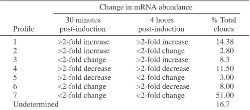
Change in mRNA abundance
30 minutes 4 hours % Total Profile post-induction post-induction clones 1 >2-fold increase >2-fold increase 14.38 2 >2-fold increase <2-fold change 2.80 3 <2-fold change >2-fold increase 8.3 4 >2-fold decrease >2-fold decrease 11.50 5 >2-fold decrease <2-fold change 3.00 6 <2-fold change >2-fold decrease 8.00 7 <2-fold change <2-fold change 51.00
2924
profiles identified in normal metamorphosis, we found that the anti-Hemps antibody treatment most significantly affected Profile 1 (upregulation of expression in both post-larval stages relative to competent larvae), in which approximately 44% of these genes (269 clones) displayed a 2-fold or greater change in treated larvae compared with normal larvae at 30 minutes, with the great majority of these (99%) being upregulated (Fig. 1A). The effect at 4 hours was much less dramatic (Fig. 1B). Two-fold or greater differences in levels of gene expression were also observed in the other profiles to a lesser extent (38% for Profile 2; 17% for Profile 3; 12% for Profile 4; 8% for Profile 5; 19.29% for Profile 6). Interestingly, 11% of the genes (204 clones) that did not change significantly (i.e. 2-fold or greater) during early metamorphosis (Profile 7) were affected by the anti-Hemps antibody treatment. In total, 16.7% of the genes assessed in this study (667 of 3997 clones) were significantly affected by the knockdown of Hemps activity. Of these, 20% were perturbed within 30 minutes of induction, suggesting that the Hemps signalling pathway is directly regulating these genes.
Confirmation of array data
To confirm that the expression profiles generated from the microarrays for individual genes were a true representation of actual changes in gene expression, we utilised quantitative real-time RT-PCR (QPCR). To get a relative abundance expression value for the genes examined, we identified a gene, Hec-coA, that was equally expressed in both the normal and anti-Hemps antibody-treated microarrays at 30 minutes and 4 hours PI; this gene coded for the enzyme enoyl-CoA hydratase (P>1.0e–14).
We confirmed that this gene was equally expressed throughout
all Herdmania developmental stages using QPCR (Fig. 2A). This compared well with another gene, Hec-Ubiq (polyubiquitin), already known to be equally expressed throughout development (Hinman and Degnan, 2001) (Fig. 2A). We employed Hec-Ubiq initially as a reference to assess Hemps expression. The results in Fig. 2B revealed that the expression of Hemps during development determined with QPCR agreed with that using northern blotting (Fig. 2C) (Eri et al., 1999). Accordingly, Hec-coA was used to normalise all other QPCR data.
We selected 15 genes with different expression profiles during normal metamorphosis for quantitation and validation using QPCR. A comparison of the data obtained at competence, 30 minutes and 4 hours PI for these genes appears in Table 2. Of the five genes upregulated at 30 minutes (Hec-pnx1, -smdp1, -meta2a, -rab, -cip2b), four showed a good correlation with the QPCR data. Only Hec-meta2a failed to show significant increase with QPCR at 30 minutes PI, but at 4 hours PI a good correlation existed between the array and
Development 131 (12) Research article
2.8 12.2 6.6 10.4 1.0 12.0 1.1 56.0
72.0 87.2 88.5 92.3 85.4 91.7 5.5 2.4 1.1 1.1 11.8 16.0
42.8
A
2.9 8.0 2.6 1.1 1.1 1.6 1.8
97.2 95.3 98.9 98.9 95.4 90.0 92.5
4.6 2.0 2.0 0.0 0.0 3.1 1.0
1 2 3 4 5 6 7
B
30 m
inut
e
P
I
[image:4.612.337.517.303.632.2]4 hour PI
Fig. 1. Distribution of microarray clones into expression profile categories during normal and anti-Hemps antibody-treated metamorphosis. Proportion of genes affected by anti-Hemps treatment at 30 minutes (A) and 4 hours (B) post-induction (PI) in each of the seven normal expression profiles. Red, greater than 2-fold increase in mRNA abundance in treated post-larvae; yellow, less than 2-fold change in mRNA abundance in treated post-larvae; blue, greater than 2-fold decrease in mRNA abundance in treated post-larvae. The figures to the right of each profile represent the
[image:4.612.54.275.426.626.2]percentage of genes in each of the categories affected by anti-Hemps antibody.
2925 Hemps and early ascidian metamorphosis
QPCR data for this gene (Table 2). Overall, for all four genes that were downregulated at either 30 minutes or 4 hours PI there was a good correlation. This correlation was also evident for six genes for which no appreciable change in gene expression occurred post-induction (Table 2). Generally there was good concordance between the data obtained by microarray analysis and those determined by QPCR.
Approximately 17% of the clones on the microarray chip were altered in their expression after treatment with anti-Hemps antibody. A subset of these genes were selected to test whether the difference in expression between treated and untreated larvae were significantly different, using both the microarray and QPCR assays. Of the 11 genes affected by anti-Hemps antibody, nine were downregulated and two were upregulated at 30 minutes PI (Fig. 3A), and in all cases QPCR data agreed with array data. A significant difference was observed between the 30-minute time point and that of untreated larvae (marked with an asterisk) for 10 of the 11 genes in at least one of the two methods used to measure expression (Fig. 3A). While there was also a reasonable correlation between the QPCR and array data at 4 hours PI, the extent of change in the clone set was less marked (Fig. 3B). Sequence-based gene classification affected by anti-Hemps antibody treatment
A subset of clones that were affected by the anti-Hemps antibody treatment were selected, sequenced and compared (using BLAST algorithm) to known cDNA and ESTs in the NCBI and Ciona intestinalis databases. Sequences that had a match to previously identified genes were then categorised into four major classes based on Lee et al. (1999). Class A coded for proteins common to many kinds of cells, class B were proteins associated with cell–cell communication, class C coded for proteins that function as transcription factors and other regulatory proteins and class D were clones of an unknown function. Furthermore, each class was then categorised into subclasses according to specific functions (see Table 3). These genes have also been classified according to the expression profile that they belong to in normal
development, and whether their expression is downregulated in treated metamorphosing larvae (D), higher in anti-Hemps antibody-treated larvae (U) or equal in expression (E), at the indicated times.
[image:5.612.319.566.75.433.2]Sixty-two Class A genes (functions that are common to most cells) were affected by the anti-Hemps antibody treatment in this study. These included homologues of genes previously shown to be involved in the metamorphosis of other ascidian species (Nakayama et al., 2001, 2002; Davidson and Swalla, 2002). Of particular interest were three putative Herdmania homologues of Ci-meta2 (Table 2): Hec-meta2 (clone 1832), –meta2a (clone 207) and –meta2b (clone 1537). Each of these genes was affected differently in larvae treated with the antibody, suggesting that they may have separate roles during Table 2. A comparison of fold change between microarray
and QPCR data through developmental stages from competence to 4 hours post-induction
30 minutes 4 hours Competence post-induction post-induction Genes Array/QPCR Array QPCR Array QPCR
Hec-pnx1 1 4.4 3.42 1.43 2.42
Hec-smdp1 1 3.29 3.44 3.17 5.3
Hec-mbl 1 0.26 0.58 1.11 0.94
Hec-meta2a 1 4.00 1.43 2.76 3.29
Hec-meta2 1 0.9 0.61 0.02 0.001
Hec-sap 1 0.56 0.66 0.46 0.59
Hec-rab 1 1.94 4 1.5 2.98
Hec-cipA 1 1.20 0.83 1.13 0.89
Hec-cip2A 1 1.27 1.58 1.68 1.162
Hec-cip2B 1 3.19 2.33 3.2 1.95
Hec-meta1 1 0.90 0.51 0.54 0.6
Hec-hsup 1 0.22 0.60 2.57 1.741
Hec-BcsX 1 1.76 1.11 0.95 0.51
Hec-lp1 1 0.1 0.1 3.0 0.84
Hec-shm 1 1.178 1.2 0.63 1.1
30 min PI: Array versus QPCR
0.1 1.0 10.
A
B
0QPCR Array
Fold change
(log)
* *
* * *
* *
* ** *
* *
* *
4 h PI: Array versus QPCR
0.1 1.0 10.0
Fold change
(log)
QPCR Array
*
* *
*
[image:5.612.48.297.106.282.2]* *
2926
De
v
elopment 131 (12)
Research ar
[image:6.612.52.741.78.566.2]ticle
Table 3. Herdmania curvata genes, identified by microarray analysis, that have an altered expression when metamorphosis is inhibited by the addition of anti-Hemps antibody
30 minutes 4 hours
Gene post-
post-Clone Identity E-Value code GE induction induction
266 gi|30146860|ref|XP_293446.2| similar to Protein CDC27Hs (Cell division cycle protein 27 homolog) (2 clones) 5.00E-05 AIII 1 U E
1832 gi|13516887|dbj|BAB40595.1| (AB041856) Ci-META2 (Hec-Meta2) 6.00E-22 AIV 1 D E
1537 gi|13516887|dbj|BAB40595.1| Ci-META2 1.00E-14 AIV 1 E D
4156 gi|5002645|emb|CAB44357.1| IF2 protein (human homolog of bacterial translation initiation factor 2) 1.00E+00 AV 1 U E
4156 gi|5002645|emb|CAB44357.1| IF2 protein (human homolog of bacterial translation initiation factor 2) 1.00E-100 AV 1 U U
3748 gi|16151619|dbj|BAB69891.1| (AB000805) mannose-binding lectin (2 clones) 1.00E-15 AVII 1 D D
2135 gi|16151619|dbj|BAB69891.1| (AB000805) mannose-binding lectin 0.0006 AVII 1 E D
1385 gi|1584024|prf||2122242A complement control protein 9.00E-19 AVII 1 E D
365 gi|16151619|dbj|BAB69891.1| (AB000805) mannose-binding lectin (2 clones) 2.00E-07 AVII 1 E D
3639 gi|21703252|gb|AAM76123.1|AF483043_1 (AF483043) complement receptor-like protein 2 9.00E-10 AVII 1 U E
4037 gi|16151619|dbj|BAB69891.1| (AB000805) mannose-binding lectin 5.00E-12 AVII 1 U E
122 gi|20822278|ref|XP_133260.1| Rab acceptor 1 (prenylated) (Hec-rab) 1.00E-11 BI 1 D E
1256 Matches to unknown of another ascidian (i.e. to Ciona scaffold) (5 clones) DIII 1 U U
1273 no match found (9 clones) N/A DIV 1 U E
3509 gi|4886910|gb|AAD32099.1|AF117616_1 (AF117616) SOUL protein 6.00E-18 AI 2 U E
933 gi|21703214|gb|AAM76104.1|AF483024_1 transposase 6.00E-15 AVII 2 D E
4130 gi|16151619|dbj|BAB69891.1| (AB000805) mannose-binding lectin (3 clones) 3.00E-16 AVII 2 D D
134 gi|391669|dbj|BAA02987.1| hikaru genki type4 product precursor 6.00E-07 BI 2 D E
1437 gi|1170275|sp|Q09101|HIG_DROME Locomotion-related protein Hikaru genki precursor 4.00E-08 BI 2 E D
1228 gi|6683496|dbj|BAA89210.1| bromodomain adjacent to zinc finger domain 1B 3.00E-33 CI 2 D E
1228 gi|6683496|dbj|BAA89210.1| bromodomain adjacent to zinc finger domain 1B 3.00E-33 CI 2 D D
197 gi|5870893|ref|NP_006832.1| solute carrier family 38, member 3; system N1 Na+ and H+-coupled glutamine transporter 3.00E-32 AI 3 U E
3768 gi|18143339|dbj|BAB79622.1| (AB057446) Ci-META3 1.00E-38 AIV 3 U E
2760 gi|21759387|sp|Q90YV5|RL13_ICTPU 60S ribosomal protein L13 6.00E-14 AV 3 D E
4289 gi|20833867|ref|XP_129647.1| (XM_129647) similar to Bifunctional aminoacyl-tRNA synthetase (2 clones) 2.00E-84 AV 3 U E
4256 gi|20833867|ref|XP_129647.1| (XM_129647) similar to Bifunctional aminoacyl-tRNA synthetase 3.00E-95 AV 3 U U
3263 gi|6225842|sp|O73888|PGD2_CHICK Glutathione-requiring prostaglandin D synthase 6.00E-36 AVII 3 E D
654 gi|28828338|gb|AAO50999.1| hypothetical protein 4.00E-06 DII 3 U E
4175 gi|21358277|ref|NP_650387.1| CG7530-PA 2.00E-17 DII 3 U E
913 gi|2498260|sp|Q28488|CRYM_MACFL Mu-crystallin 3.00E-31 AIV 4 D E
4191 gi|12381877|dbj|BAB21248.1| ribosomal protein L35 4.00E-07 AV 4 U E
216 gi|16751538|gb|AAL27683.1|AF237690_1 (AF237690) unknown (syphon associated protein) (Hec-sap) 3.00E-08 AX 4 D E
2574 gi|14010891|ref|NP_114192.1| syntenin 6.00E-52 BIII 4 D E
829 gi|4507451|ref|NP_003216.1| trefoil factor 1 8.00E-08 BIII 4 D D
2125 gi|37589760|gb|AAH59661.1| Unknown (protein for MGC:73345) 5.00E-14 DII 4 D D
3960 gi|20810547|gb|AAH29067.1| (BC029067) similar to stress-associated endoplasmic reticulum 6.00E-16 AVII 5 D E
300 gi|16151619|dbj|BAB69891.1| mannose-binding lectin (2 clones) 4.00E-09 AVII 5 D E
3960 gi|20810547|gb|AAH29067.1| (BC029067) similar to stress-associated endoplasmic reticulum 6.00E-16 AVII 5 D D
3889 gi|18491022|ref|NP_060622.2| (NM_018152) chromosome 20 open reading frame 12; bA189K21.1 (hec-sup) 9.00E-23 DII 5 D D
883 gi|4759058|ref|NP_004835.1| SH3-domain binding protein 5 (BTK-associated) 4.00E-43 AII 6 D E
894 gi|14549635|gb|AAK66965.1|AF255739_1 replication-dependent histone H2A 3.00E-48 AIII 6 D E
207 gi|13516887|dbj|BAB40595.1| Ci-META2 (Hec-meta2a) 1.00E-32 AIV 6 U E
1597 gi|6677773|ref|NP_033104.1| (NM_009078) ribosomal protein L19 3.00E-54 AV 6 D E
4195 gi|127184|sp|P07461|MLR_HALRO Myosin regulatory light chain, smooth muscle (5 clones) 8.00E-53 AX 6 E U
1531 gi|13516889|dbj|BAB40596.1| Ci-META1 (Hec-meta1) 3.00E-44 BI 6 E D
1080 gi|9966793|ref|NP_065080.1| (NM_020347) leucine zipper transcription factor-like 1 (2 clones) 4.00E-39 CI 6 D D
904 gi|20829847|ref|XP_150664.1| (XM_150664) hypothetical protein XP_150664 1.00E-09 DII 6 D E
2927
Hemps and ear
ly ascidian metamor
phosis
2108 gi|1706902|sp|P49948|FRH2_XENLA Ferritin heavy chain 2 (XL2-17) (2 clones) 6.00E-15 AI 7 E U
588 gi|12643614|sp|O55143|ATA2_MOUSE Sarcoplasmic/endoplasmic reticulum calcium ATPase 2 (Calcium pump 2)
(SERCA2) (SR Ca(2+)-ATPase 2) 1.00E-93 AI 7 E U
1208 gi|27372170|dbj|BAC53586.1| sarco-endoplasimc reticulum calcium ATPase 1.00E-60 AI 7 U D
25 gi|21314753|ref|NP_115766.2| MKI67 (FHA domain) interacting nucleolar phosphoprotein; nucleolar phosphoprotein
Nopp34; nucleolar protein interacting with the FHA domain of pKi-67 (Hec-nopp34) 3.00E-16 AII 7 D E
3250 gi|12834577|dbj|BAB22965.1| histone cell cycle regulation defective interacting protein 5~putative 2.00E-18 AIII 7 D E
3250 gi|12834577|dbj|BAB22965.1| (AK003730) data source:MGD, source key:MGI:1913290, evidence:ISS~histone cell
cycle regulation defective interacting protein 5~putative 2.00E-18 AIII 7 D D
72 gi|13359451|dbj|BAB33421.1| putative senescence-associated protein 2.00E-16 AIII 7 E U
2633 gi|34876535|ref|XP_223229.2| similar to PDZ domain actin binding protein Shroom (Hec-shm) 6.00E-27 AIV 7 D E
1296 gi|49864|emb|CAA27397.1| alpha-actin 1.00E-20 AIV 7 E U
3315 gi|2500736|sp|Q25147|S61A_HALRO PROTEIN TRANSPORT PROTEIN SEC61 ALPHA SUBUNIT 7.00E-67 AIX 7 U E
290 gi|21629316|gb|AAM68993.1|AC125735_23 ribosomal protein L38 3.00E-06 AV 7 D E
892 gi|4584080|emb|CAB40554.1| ribosomal protein L24 9.00E-24 AV 7 D E
3249 gi|22758848|gb|AAN05584.1| (AF526198) ribosomal protein L30 5.00E-50 AV 7 E D
4162 gi|15293931|gb|AAK95158.1|AF401586_1 ribosomal protein L31 4.00E-37 AV 7 E U
4305 gi|15293925|gb|AAK95155.1|AF401583_1 (AF401583) ribosomal protein L28 4.00E-15 AV 7 U E
2750 gi|12848551|dbj|BAB27994.1| (AK012052) data source:MGD, source key:MGI:1333780, evidence:ISS~putative~
ribosomal protein S19 1.00E-31 AV 7 U E
4132 gi|7335697|ref|NP_038245.1|COX2_15273 cytochrome c oxidase subunit II (2 clones) 8.00E-31 AVI 7 E U
593 gi|24233541|ref|NP_665726.1| cytochrome c oxidase, subunit Va 3.00E-27 AVI 7 U E
660 gi|25051789|ref|XP_193557.1| s-adenosylmethionine decarboxylase 1 8.00E-37 AVI 7 U U
1592 gi|16151619|dbj|BAB69891.1| (AB000805) mannose-binding lectin (4 clones) 3.00E-16 AVII 7 D D
999 gi|16151619|dbj|BAB69891.1| mannose-binding lectin 9.00E-10 AVII 7 E D
1969 gi|16151619|dbj|BAB69891.1| (AB000805) mannose-binding lectin 3.00E-16 AVII 7 E D
4176 gi|7489812|pir||T02955 probable cytochrome P450 monooxygenase - maize (fragment) 9.00E-18 AVII 7 E U
125 gi|9945306|ref|NP_064696.1| microsomal glutathione S-transferase 1 1.00E-22 AVII 7 U E
1335 gi|27503944|gb|AAH42325.1| similar to proteasome (prosome, macropain) 26S subunit, non-ATPase 2.00E-54 AVIII 7 U E
233 gi|2507247|sp|P06868|PLMN_BOVIN Plasminogen precursor 8.00E-56 AVIII 7 U E
3927 gi|11276955|pir||A59236 embryonic muscle myosin heavy chain 1.00E-116 AX 7 D D
60 gi|22507341|ref|NP_683736.1| gene trap ROSA b-geo 22 7.00E-25 AX 7 D E
4189 gi|6001952|gb|AAC32598.2| echinonectin 7.00E-22 BIII 7 E D
1251 gi|27694525|ref|XP_223330.1| similar to Transcription factor BTF3 1.00E-36 CI 7 U E
1476 Not enough information to classify (i.e. sequence < 200bp) (14 genes) N/A DI
1264 weak match (35 clones) > 1.00E-05 DII
Shown is the ascidian clone number ID, the gene identity, the predicted class (based on putative function) and the developmental profile (GE; see below) that the gene falls into during normal metamorphosis (see Table 1). Also shown is the effect of anti-Hemps antibody on expression at 30 minutes post-induction and 4 hours post-induction.
D, >2-fold decrease in mRNA abundance in larvae treated with the anti-Hemps antibody relative to untreated larvae; E, <2-fold change in mRNA abundance in larvae treated with the anti-Hemps antibody relative to untreated larvae; U, >2-fold increase in mRNA abundance in larvae treated with the anti-Hemps antibody relative to untreated larvae.
When the same gene has been identified more than once by the arrays, this is indicated in brackets. Also indicated are the gene name designations for clones further characterised in this study, indicated by (Hec-gene).
Predicted classes are subdivided into subclasses as follows.
(A) Functions that many kinds of cells use: AI, transport and binding proteins for ions and other small molecules; AII, RNA processing, polymerising, splicing and binding proteins and enzymes; AIII, cell replication, histones, cyclins and allied kinases, DNA polymerases, topoisomerases, DNA modification; AIV, cytoskeleton and membrane proteins, cellular organisers; AV, protein synthesis cofactors, tRNA synthetase, ribosomal proteins; AVI, intermediary metabolism and catabolism enzymes; AVII, intermediary metabolism and catabolism enzymes; AVIII, protein degradation and processing, proteases; AIX, transportation and binding proteins for proteins and other macromolecules; AX, proteins involved in motility including muscle components.
(B) Cell-cell communication: BI, signalling receptors including cytokine and hormone receptors, and signalling ligands; BII, intracellular signal transduction pathway molecules, including kinases and signal intermediates; BIII, extracellular matrix proteins and cell adhesion.
(C) Transcription factors and other gene regulatory proteins: CI, sequence-specific DNA-binding proteins; CII, non-DNA binding proteins that have positive or negative roles; CIII, chromatin proteins other than AIII with regulatory function.
2928
metamorphosis. In C. intestinalis it has been suggested that Ci-Meta2 may be involved in the dynamic rearrangement of cells during metamorphosis (Nakayama et al., 2001). A second gene belonging to class A, Hec-meta3 (clone 3768), an apparent homologue of Ci-meta3, which is upregulated in metamorphosing C. intestinalis juveniles (Nakayama et al., 2002), is also upregulated in Herdmania larvae treated with the anti-Hemps antibody. This protein contains domains for SPRY, which in Drosophila acts as an antagonist in fibroblast growth factor (FGF) (Hacohen et al., 1998).
A number of genes encoding proteases and factors involved in protein degradation and processing (Table 3, subclass AVII) were identified as being affected by the anti-Hemps antibody treatment. This subclass includes proteins that are implicated in innate immunity, some of which are known to be activated during larval development and metamorphosis in other ascidians (Davidson and Swalla, 2002). It has been suggested that the activation of a number of immune system-related genes during metamorphosis in B. villosa is important in the detection of endogenous signals indicative of tissue damage rather than deletion of foreign antigens (Davidson and Swalla, 2002). In this study we also report alteration in the regulation of a number of genes involved in innate immunity. Four of these genes, mannose-binding lectin (Hec-mbl), complement control protein, Hikaru genki precursor and Hikaru genki type 4 (Hec-hgp1 and Hec-hgt4, respectively) are downregulated at 30 minutes or 4 hours PI in response to blocking of metamorphosis with anti-Hemps antibody.
In addition to genes previously shown to be involved in ascidian metamorphosis, the treatment with the neutralising antibody identified other genes not previously implicated in this process. These included genes encoding structural proteins (e.g. myosins, troponins) and transcription factors (transcription initiative factor 11A gamma chain and BTF3), a gene encoding a heme-binding protein, which is homologous to a gene implicated in barnacle settlement and metamorphosis (Okazaki and Shizuri, 2000). Sixty-one of the differentially expressed genes analysed in this study (40%) did not match with sequences in GenBank or the C. intestinalis genome. The microarray analysis also indicated that Hemps has its major effect at 30 minutes PI, because at 4 hours PI approximately 50% of these genes had recovered to expression levels comparable to normal 4-hours-PI larvae (derived from data in Table 3).
Temporal and spatial expression of selected genes Fifteen genes that were expressed during early metamorphosis were classified into one of four categories based on QPCR analysis of transcript abundance through development (Fig. 4): (1) genes predominantly or transiently expressed in larvae and/or early metamorphosis (Hec-pnx, Hec-smdp1, Hec-mbl, Hec-meta2a, Hec-meta2); (2) genes predominantly expressed during metamorphosis and in the juvenile (Hec-sap, Hec-rab, Hec-cip1, Hec-cip2A, Hec-cip2B); (3) genes predominantly expressed during embryogenesis (Hsup, lp1, Hec-meta1); and (4) genes expressed at similar levels during embryogenesis and metamorphosis (Hec-bcsX, Hec-shm).
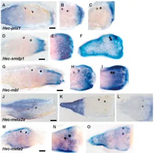
Maternal or juvenile (8-day-old) transcripts could not be detected for Hec-mbl, Hec-smdp1, Hec-meta2, Hec-meta2a and Hec-pnx1. Transcripts of these genes began accumulating after the tailbud or hatching stage, with abundance peaking between competency and 4 hours PI (Fig. 4). Whole-mount
in-situ hybridisation analysis of the spatial expression of these genes in competent larvae revealed that all these genes were expressed solely in the trunk ectoderm (Fig. 5). Hec-pnx1 was localised to the vicinity of the anterior larval palps and papillae associated tissue (PAT) cells, similar to Hemps (Eri et al., 1999) (Fig. 5A). Hec-mbl and -smdp1 transcripts were restricted to the most anterior third of the larval trunk (Fig. 5D,G), while Hec-meta2 and -meta2a were localised to the posterior two-thirds of the trunk (Fig. 5J,M). The transcripts of these genes remained localised to the same region of 30-minute post-larvae (Fig. 5B,E,H,N), with the exception of Hec-meta2a, which became localised to the resorbing tail (Fig. 5K). Hec-meta2a was also localised to the tail during tailbud formation and in newly hatched larvae (data not shown). By 4 hours PI, Hec-smdp1 and -meta2 transcripts were detected in all epidermal cells (Fig. 5F,O). Localised expression of Hec-pnx1 and -mbl did not change (Fig. 5C,G) and Hec-meta2a transcripts were not detected (Fig. 5L).
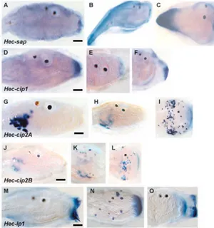
The spatial expression of five genes that were more broadly expressed across embryogenesis and metamorphosis were characterised (Fig. 6). Transcripts of three unclassified zygotic genes, Hec-cip1, Hec-cip2A and Hec-cip2B, began accumulating during late embryogenesis and increased markedly within the first 30 minutes of induction of metamorphosis (Fig. 4). These genes continued to be expressed at high levels through metamorphosis to at least the 8-day-old juvenile stage. Hec-cip1 transcripts were localised to the anterior epidermis of the larva in the vicinity of the PAT cells (Fig. 6D). At 30 minutes PI transcripts remained localised to this region and at 4 hours PI were detected in the anterior-most tunic, which corresponded to the location of the migratory PAT cells (Fig. 6E,F) (Eri et al., 1999; Davidson and Swalla, 2002). Closely related transcripts Hec-cip2A and Hec-cip2B accumulated in a similar but quantitatively unique manner. Both genes were expressed in the larval mesenchyme (Fig. 6G-L). During early metamorphosis these cells appeared to migrate from these pockets to populate internal spaces and the tunic (Fig. 6H,I,K,L).
Hec-sap, Hec-meta1 and Hec-rab were also expressed during embryogenesis but had quantitatively higher transcript levels during post-larval development and in the juvenile (Fig. 4). Hec-sap (Herdmania siphon associated protein) and Hec-meta1 transcripts were localised to the tail during embryogenesis: Hec-sap to the most posterior epidermis and Hec-meta1 to the muscle (data not shown). A dramatic change in localised Hec-sap expression accompanied the acquisition of competence, with transcripts being detected in the trunk with whole-mount in-situ hybridisation staining and most pronounced surrounding the PAT cells (Fig. 6A). Upon induction of metamorphosis Hec-sap expression was detected again in the posterior part of the resorbing tail (Fig. 6B,C), and then was expressed predominantly in the siphons of the juvenile body (data not shown).
Hec-bcsX was expressed throughout development and had a significant increase in transcript abundance during larval development and then a marked reduction between 30 minutes and 4 hours PI (Fig. 4). Hec-shm transcript abundance was lowest from hatching to 4 hours PI, increasing during later metamorphosis and in the 8-day-old juvenile. Hec-Hsup and Hec-lp1 had peaks of expression during gastrulation (Fig. 4). Whole-mount in-situ hybridisation analysis of Hec-lp1
2929 Hemps and early ascidian metamorphosis
revealed that this gene is expressed in the larval palps and anterior epidermis and a subset of tunic cells in the post-larvae (Fig. 6M-O).
Discussion
We previously demonstrated that Hemps is a key regulator of the induction of metamorphosis in H. curvata (Eri et al., 1999). By soaking larvae in an anti-Hemps antibody, tail resorption and all subsequent stages of metamorphosis were inhibited (Eri et al., 1999). In this report we have used a microarray approach to identify genes that are: (1) differentially expressed during early metamorphosis and (2) influenced by the Hemps
pathway. By investigating 30-minute and 4-hour post-larvae, we were able to focus on the immediate process of induction of metamorphosis and the role of Hemps in this process. At approximately 4 hours after induction, morphogenetic events leading to the formation of the adult body plan begin, including a marked increase in overall gene activity (Green et al., 2002). The importance of gene transcription in the early stages of metamorphosis has been demonstrated to be critical in B. villosa (Davidson and Swalla, 2001). Of particular interest in the present study are the genes that are activated or repressed at 30 minutes PI, as they are likely to be under the direct regulation of the Hemps signal and other early response signals.
hours post fertilization 0E+00 1E+04 2E+04 3E+04 4E+04 5E+04 Hec-cip1
8 d = 4402 s.d. 6322 0.0E+00 5.0E+04 1.0E+05 1.5E+05 2.0E+05 2.5E+05
0 5 10 15 20 25 30 35 40
Hec-meta2
8 d = 0 0E+00 1E+05 2E+05 3E+05 4E+05 5E+05 6E+05
8 d = 0 Hec-mbl 0.0E+00 2.5E+04 5.0E+04 7.5E+04 1.0E+05 1.2E+05
8 d = 0 Hec-smdp1 Normalised abundance 0.0E+00 5.0E+04 1.0E+05 1.5E+05 2.0E+05 2.5E+05 Hec-cip2A
8 d = 4.9E+04 s.d. 0.2E+04 0E+00 1E+05 2E+05 3E+05 4E+05 5E+05
0 5 10 15 20 25 30 35 40
Hec-cip2B
8 d = 7.5E+06 s.d. 0.6E+06 0 0.1 0.2 0.3 Hec-pnx1
8 d = 0
Hec-lp1 0E+00 1E+07 2E+07 3E+07 4E+07 5E+07
8 d = 2.6E+05 s.d. 1.1E+05 0 2000 4000 6000 8000 Hec-bcsX
8 d = 72E+04 s.d. 0.2E+04 0 1000 2000 3000 4000 5000 6000
8 d = 5431 s.d. 60 0 500 1000 1500 2000 2500
8 d = 5257 s.d. 736 Hec-rab 0 2000 4000 6000 8000 Hec-sap
8 d = 5451 s.d. 2025 0 500 1000 1500 Hec-meta1
8 d = 5539 s.d. 776 0E+00 2E+04 4E+04 6E+04 8E+04
8 d = 0 Hec-meta2a 0 200 400 600 800 1000
[image:9.612.89.532.70.510.2]0 5 10 15 20 25 30 35 40 8 d = 1467 s.d. 324 Hec-shm
*
*
*
*
*
*
*
Hec-hsup*
2930
Microarray approach to investigating transcriptional events in Herdmania metamorphosis
To investigate the importance of Hemps in the induction and regulation of early metamorphosis, we undertook a gene profiling study using a partially redundant (21%) microarray consisting of 4800 clones derived from a cDNA library of mixed developmental stages from gastrula to 16-hour post-larva. Only genes that, in a minimum of four determinations, consistently displayed greater than 2-fold changes in transcript abundance during early metamorphosis or in the anti-Hemps antibody treatment were scored. This conservative approach, which has been used in a variety of microarray studies (e.g. Butler et al., 2003; Munoz-Sanjuan et al., 2002), is likely to have missed a number of genes represented on our microarray that are differentially expressed during metamorphosis or in response to the anti-Hemps antibody treatment. QPCR analysis of a subset of the differentially expressed genes validated the microarray approach used in this study. Because of the unequal redundancy of the genes represented on the 4800 clone microarray, it is difficult to extrapolate these values to the transcriptome, even if the Herdmania genome contains ~16 000 protein-coding genes, as is the case in C. intestinalis (Dehal et al., 2002). Nonetheless, a large number of genes – probably 2000-5000 – are activated or repressed early in metamorphosis, and approximately 20% of these genes are regulated by
the Hemps pathway. These estimations are particularly striking, given that this period has significantly reduced overall transcriptional activity, and in the presence of the transcriptional inhibitor actinomyosin D tail resorption can occur (Green et al., 2002). This observation is similar to data reported by Davidson and Swalla (Davidson and Swalla, 2001), where they showed that the tail can undergo partial metamorphosis without novel transcription in B. villosa. Combined, these data suggest that early gene activation and repression during metamorphosis may not be necessary for tail resorption but are involved in downstream morphogenetic events, which include the destruction and reorganisation of the larval body plan and formation of the juvenile plan (Hinman and Degnan, 2000). The fact that treatment with anti-Hemps antibody also inhibits tail resorption suggests that this signalling pathway also influences post-transcriptional processes during early metamorphosis.
The Hemps pathway as a modulator of gene expression during early
metamorphosis
It is very likely that the Hemps protein, a key regulator of earlier events of metamorphosis (Eri et al., 1999), plays a central role in the regulation of a large number of these genes, and antibody studies, as discussed below, support this. Alternatively, a protein such as the cornichon homologue Cnib, described in B. villosa, might also play a role in this process by signalling through Hemps (Davidson and Swalla, 2001). One approach to unravelling this complexity is to identify and abrogate a key regulator in the pathway; in this case, we have targeted Hemps. We have previously shown that antibodies against Hemps abrogate normal metamorphosis and ectopic application of recombinant Hemps acts to stimulate this process (Eri et al., 1999), suggesting a key role for this peptide in induction of metamorphosis. The anterior-most cells of the trunk, which are at the base of the palps – the PAT cells – migrate through a tunnel in the juvenile tunic to the external environment around the time of competence in Herdmania (Eri et al., 1999; Green et al., 2002) and in another ascidian, B. villosa (Davidson et al., 1995; Davidson and Swalla, 2002). In Herdmania, Hemps is expressed at relatively high levels in the PAT cells and papillae (Eri et al., 1999). Upon induction of normal metamorphosis, the Hemps protein spreads to cover the anterior region of the larval trunk, suggesting that this EGF-like peptide is signalling to cells in this region (Eri et al., 1999). The anti-Hemps antibody appears to impact on metamorphosis by inhibiting this signalling event.
Microarray analysis of larvae treated with the anti-Hemps antibody indicates that genes that displayed a significant increase or decrease in transcript abundance at 30 minutes PI are probably directly regulated by the Hemps pathway. Genes affected by the anti-Hemps antibody treatment at 4 hours might be indirectly regulated by this pathway. Interestingly, most of the genes affected by the knockdown of Hemps activity are
[image:10.612.45.347.75.374.2]Development 131 (12) Research article
2931 Hemps and early ascidian metamorphosis
differentially expressed to a greater extent in the anti-Hemps antibody treatments than in normal development. For example, many of the genes that are normally upregulated in 30-minute PI post-larvae (i.e. normal expression profiles 1 and 2) are expressed at even higher levels in larvae treated with the anti-Hemps antibody. These data indicate that the knockdown of the Hemps signalling pathway does not result in maintenance of the larval gene expression profile (i.e. a large majority of the genes that are normally differentially expressed in post-larvae compared with competent larvae are not scored as equally expressed in the anti-Hemps antibody knockdown experiment). Since the knockdown of Hemps activity affects the gene expression in the 30-minute PI post-larvae to a greater extent than the 4-hour post-larvae, it appears that the Hemps pathway directly regulates early response genes at metamorphosis. Combined, these observations suggest that the Hemps signalling pathway, while crucial for the induction of metamorphosis, contributes more significantly to the modulation of early gene expression, rather than initial gene activation or repression. The extent of activation or repression of many genes during normal early metamorphosis appears to be dampened by the Hemps pathway. In the case of these genes, experimental knockdown of Hemps results in a far
greater increase or decrease in transcript abundance than observed during normal metamorphosis.
Analysis of larvae treated with the anti-Hemps antibody has led to the identification of a large number of genes that are directly or indirectly regulated at the transcriptional level by the Hemps signalling pathway. Of the genes significantly affected by the knockdown of Hemps activity by the neutralising antibody during the first 4 hours of metamorphosis (384 clones), 20% show significant difference in expression levels at 30 minute PI. These genes include homologues to genes already shown to be involved in marine metamorphosis (Nakayama et al., 2001; Okazaki and Shiruri, 2000; Davidson and Swalla, 2002). These include Hec-meta1, -meta2 and -meta3, which are homologues to genes involved in cell migration, adhesion and specialised secretory roles (Nakayama et al., 2001, 2002; Noh et al., 2003; Shao and Haltiwanger, 2003). Genes implicated in innate immunity were also identified among the genes regulated by Hemps and include Hec-mbl, Hec-hgp1 and Hec-hgp4. Homologues of the latter genes are implicated in metamorphosis in B. villosa (Davidson and Swalla, 2002). Genes affected by the anti-Hemps antibody treatment at 4 hours PI might either be regulated by the Hemps pathway or reflect differences in larval and post-larval expression profiles.
Ascidian metamorphosis: conserved and species-specific components
Ascidians have a pelagobenthic biphasic life cycle with morphologically distinct larval and adult body plans. While the biphasic life cycle is the most common form of development among metazoans (Brusca and Brusca, 2003), molecular phylogenetic analyses indicate that the ascidian life cycle might be derived from a worm-like deuterostome ancestor (Cameron et al., 2000; Swalla et al., 2000). Recently, the molecular basis of metamorphosis has been investigated in the aplousobranch C. intestinalis (Nakayama et al., 2001, 2002) and two stolidobranchs, B. villosa (Davidson and Swalla, 2002) and H. curvata (reviewed in Degnan, 2001). Progression through metamorphosis appears to be very similar in these disparate ascidians, including the ordered autolysis of larval axial structures, and proliferation and morphogenesis of larval endoderm and mesenchyme rudiments to form the adult body wall musculature, haemopoietic system and functional gut (e.g. Cloney, 1982; Hirano and Nishida, 1997, 2000). Given the shared external characteristics of metamorphosis and evolutionary history of these ascidians, it might be expected that related molecular mechanisms would underlie metamorphosis in these ascidians and that these might be different from those observed in other metazoans.
[image:11.612.49.349.75.395.2]Interestingly, 40% (61 of 151 genes) of the sequenced genes that were differentially expressed when incubated with the neutralising antibody did not match significantly with any genes in the C. intestinalis genome (Dehal et al., 2002) or Fig. 6. Localised expression of five genes that are differentially expressed during
2932
cDNA/EST database (Satou et al., 2001), or any other sequences in the GenBank database. While this observation might be related to the relatively large distances between Herdmania and Ciona, it does indicate that there are significant differences in the genetic networks operational at metamorphosis in these ascidians. This is further substantiated by the failure to identify a Hemps homologue in the Ciona database. In addition, recent data demonstrate that the noradrenaline and β1-adrenergic receptor system triggers early metamorphosis in C. savignyi (Kimura et al., 2003), pointing to different control pathways in the different ascidians. Comparison of these unknown Herdmania ESTs with the sequences of four other more closely related stolidobranch ascidians in GenBank – B. villosa (94 sequences), Halocynthia roretzi (4423), Polyandrocarpa misakiensis (501) and Botryllus schlosseri (244) failed to identify other ascidian-specific sequences.
An H. curvata homologue of the C. intestinalis metamorphosis gene Ci-meta1 is downregulated in response to anti-Hemps treatment following an inductive cue. Ci-meta1 was identified by differential screening of cDNA libraries of swimming larvae and metamorphosing C. intestinalis juveniles (Nakayama et al., 2001) and is not expressed at the larval stage but is expressed immediately after initiation of metamorphosis. Hec-meta1 encodes a 297 amino acid protein, containing two calcium-binding EGF-like domains and a secretion signal peptide. Ci-meta1 is three times larger than Hec-meta1 and contains 13 calcium-binding EGF-like repeats. While both these proteins are implicated in metamorphosis, possession of common EGF-like repeat domains does not imply that they are homologous and play the same role in metamorphosis. Indeed, whole-mount in-situ hybridisation analysis shows that Ci-meta1 expression is localised to the adhesive organ and anterior epidermis of C. intestinalis (Nakayama et al., 2001), whereas Hec-meta1 is predominantly expressed in the posterior trunk of H. curvata. Prior to metamorphosis, Hec-meta1 expression is in the epidermis of the trunk. Ci-meta1 expression is also localised to the anterior end of the larval trunk, a region in which the signal that initiates ascidian metamorphosis originates and in which Hemps protein is also localised (Degnan et al., 1997; Eri et al., 1999). In addition, both Hemps and Ci-meta1 are activated immediately at the beginning of metamorphosis and are both putative secretory proteins containing common EGF-like domains (Arnold et al., 1997; Eri et al., 1999; Nakayama et al., 2001). Thus the two genes may have similar roles in these ascidians. The identification of homologues of EGF signalling proteins implicated in B. villosa metamorphosis (Davidson and Swalla, 2001) adds further support for a central role for EGF-like proteins in ascidian metamorphosis.
We have also identified three genes that are homologues of Ci-meta2 (Nakayama et al., 2001). Two of these genes are upregulated at 30 minutes PI in normal metamorphosis and are inhibited in their response by anti-Hemps antibody. Ci-meta2 encodes a protein with a putative secretion signal and three thrombospondin repeats. This gene is upregulated in the metamorphosing juveniles and is expressed in the larval adhesive organ, neck region and dorsal trunk (Nakayama et al., 2001). The function of the protein remains unknown but is structurally related to proteins involved in cell adhesion, cell migration and those with specialised secretory roles
(Nakayama et al., 2001, 2002; Noh et al., 2003; Shao and Haltiwanger, 2003; Tomley et al., 2001).
Another gene, Hec-bcsx1, is upregulated late in metamorphosis and in the Herdmania juvenile is homologous to a barnacle metamorphosis gene (Okazaki and Shizuri, 2000). The expression of this gene is not appreciably affected by anti-Hemps antibody treatment. This gene does not change significantly in its expression until later in metamorphosis, possibly after the period the Hemps signalling pathway has greatest effect. Both array screening and QPCR revealed significant downregulation of Hec-mbl. This gene is homologous to the B. villosa mannose specific lectin (MBL) gene (Davidson and Swalla, 2002). MBL normally activates a serine protease and downstream complement signalling (Matsushita et al., 1998; Sekine et al., 2001). In addition, other genes homologous to innate immunity genes are also downregulated in response to anti-Hemps antibody. Davidson and Swalla (Davidson and Swalla, 2002) have described the differential expression of several immune system-related genes during ascidian metamorphosis. They suggested that alterations in these genes might not involve an immune response per se but that the immune system-related proteins might function in the developmental regulation of cell adhesion and migration.
In summary, we have abrogated the function of Hemps, a key regulator of metamorphosis, to identify downstream genes implicated in this process. Alterations in expression were confirmed by QPCR, and the localisation of expression of these genes was compatible with a role in metamorphosis. Using a microarray gene profiling approach, we have found that a significant portion of the genome was activated or repressed immediately upon the larva coming in contact with a cue that induces metamorphosis and that the Hemps signalling pathway affected the expression of approximately 17% of these genes, and 11% of genes that normally have no change in expression throughout metamorphosis. Based on sequence similarity of genes involved in metamorphosis in other ascidians (Nakayama et al., 2001; Davidson and Swalla, 2002) and expression patterns, it seems likely that a number of the H. curvata genes identified in this study play a conserved role in ascidian metamorphosis as well as key roles in embryogenesis. Specific disruption of these genes will allow for further dissection of the pathways involved in metamorphosis and for the assignation of functional activity to these genes.
This research was supported by an Australian Research Council grant to M.F.L. and B.M.D. We thank the staff of the University of Queensland, Heron Island Research Station for assistance in boating and animal and laboratory maintenance.
References
Arnold, J. M., Eri, R., Degnan, B. M. and Lavin, M. F. (1997). Novel gene
containing multiple epidermal growth factor-like motifs transiently expressed in the papillae of the ascidian tadpole larvae. Dev. Dyn. 210, 264-273.
Brusca, R. C. and Brusca, G. L. (2003). Invertebrates. 2nd edn. Sunderland,
MA: Sinauer Associates.
Butler, M. J., Jacobsen, T. L., Cain, D. M., Jarman, M. G., Hubank, M., Whittle, J. R., Phillips. R. and Simcox, A. (2003). Discovery of genes with
highly restricted expression patterns in the Drosophila wing disc using DNA oligonucleotide microarrays. Development 130, 659-670.
Cameron C. B., Garey, J. R. and Swalla, B. J. (2000). Evolution of the
2933 Hemps and early ascidian metamorphosis
chordate body plan, new insights from phylogenetic analyses of deuterostome phyla. Proc. Natl. Acad. Sci. USA 97, 4469-4474.
Chambon, J. P., Soule, J., Pomies, P., Fort, P., Sahuquet, A., Alexandre, D., Mangeat, P. H. and Baghdiguian, P. (2002). Tail regression in Ciona
intestinalis (Prochordate) involves a caspase-dependent apoptosis event
associated with ERK activation. Development 129, 3105-3114.
Cloney, R. A. (1982). Ascidian larvae and events of metamorphosis. Amer.
Zool. 22, 817-826.
Cloney, R. A. (1990). Urochordata-Ascidiacea. In Reproductive Biology of
Invertebrates. Vol. 4, Part B (ed. K. G. Adiyodi and R. G. Adiyodi), pp.
391-451. New Delhi: Oxford and IBH Publishing.
Corbo, J. C., DiGregorio, A. and Levine, M. (2001). The Ascidian as a
model organism in developmental and evolutionary biology. Cell 106, 535-538.
Davidson, B. and Swalla, B. J. (2001). Isolation of genes involved in ascidian
metamorphosis, epidermal growth factor and metamorphic competence.
Dev. Genes Evol. 211, 190-194.
Davidson, B. and Swalla, B. J. (2002). A molecular analysis of ascidian
metamorphosis reveals activation of an innate immune response.
Development 129, 4739-4751.
Davidson, E. H., Peterson, K. J. and Cameron, R. A. (1995). Origin of
bilaterian body plans, evolution of developmental regulatory mechanisms.
Science 270, 1319-1325.
Degnan, B. M. (2001). Settlement and metamorphosis of the ascidian
Herdmania curvata. In Biology of Ascidians (ed. C. C. Lambert, H. Yokosawa and H. Sawada), pp. 258-263. Tokyo: Springer-Verlag.
Degnan, B. M., Rhode, P. R. and Lavin, M, F. (1996). Normal development
and embryonic gene activity in the ascidian Herdmania momus. Marine.
Fresh Water Res. 47, 543-551.
Degnan, B. M., Souter, D., Degnan, S. M. and Long, S. C. (1997). Induction
of metamorphosis with potassium ions requires development of competence and an anterior centre in the ascidian Herdmania momus. Dev. Genes Evol.
206, 370-376.
Dehal, P., Satou, Y., Campell, R. K., Chapman, J., Degnan, B. M., De Tomaso, A., Davidson, B., DiGregorio, A., Gelpke, M., Goodstein, D. M. et al. (2002). The draft genome of Ciona intestinalis, insights into chordate
and vertebrate origins. Science 298, 2157-2167.
Eri, R., Arnold, J. M., Hinman, V. F., Green, K., Jones, M., Degnan, B. M. and Lavin, M. F. (1999). Hemps, a novel EGF-like protein, plays a central
role in ascidian metamorphosis. Development 126, 5809-5818.
Green, K. M., Russell, B. D., Clark, R. J., Jones, M. K., Garson, M. J., Skilleter, G. A. and Degnan, B. M. (2002). A sponge allelochemical
induces ascidian settlement but inhibits metamorphosis. Mar. Biol. 140, 355-363.
Hacohen, N., Kramer, S., Sutherland, D., Hiromi, Y. and Krasnow, M. A.
(1998). Sprouty encodes a novel anatgonist of FGF that patterns apical branching of the Drosophila airways. Cell 92, 253-263.
Hinman, V. F. and Degnan, B. M. (2000). Retinoic acid perturbs Otx gene
expression in the ascidian pharynx. Dev. Genes Evol. 210, 129-139.
Hinman, V. F. and Degnan, B. M. (2001). Homebox genes, retinoic acid and
the development and evolution of dual body plans in the ascidian Herdmania
curvata. Amer. Zool. 41, 664-675.
Hirano, T. and Nishida, H. (1997). Developmental fates of larval tissues after
metamorphosis in ascidian Halocynthia roretzi. I. Origin of mesodermal tissues of the junenile. Dev. Biol. 192, 199-210.
Hirano, T. and Nishida, H. (2000). Developmental fates of larval tissues after
metamorphosis in the ascidian, Halocynthia roretzi. II. Origin of endodermal tissues of the juvenile. Dev. Genes Evol. 210, 55-63.
Jackson, D., Leys, S. P., Hinman, V. F., Woods, R. G., Lavin, M. F. and Degnan, B. M. (2002). Ecological regulation of development, induction of
marine invertebrate metamorphosis. Int. J. Dev. Biol. 46, 679-686.
Kimura, Y., Yoshida, M. and Morisawa, M. (2003). Interaction between
noradrenaline or adrenaline and the beta 1-adrenergic receptor in the nervous system triggers early metamorphosis of larvae in the ascidian, Ciona
savignyi. Dev. Biol. 258, 129-140.
Lee, Y-H., Huang, G. M., Cameron, R. A., Graham, G., Davidson, E. H., Hood, L. and Britten, R. J. (1999). EST analysis of gene
expression in early cleavage-stage sea urchin embryo. Development 126, 3857-3867.
Matsushita, M., Endo, Y. and Fujita, T. (1998). MASP1 (MBL-associated
serine protease 1). Immunobiology 199, 340-347.
Munoz-Sanjuan, I., Bell, E., Altmann, C, R., Vonica, A. and Brivanlou, A. H. (2002). Gene profiling during neural induction in Xenopus laevis,
regulation of BMP signaling by post-transcriptional mechanisms and TAB3, a novel TAK1-binding protein. Development 129, 5529-5540.
Nakayama, A., Satou, Y. and Satoh, N. (2001). Isolation and characterization
of genes that are expressed during Ciona intestinalis metamorphosis. Dev.
Genes Evol. 211, 184-189.
Nakayama, A., Satou, Y. and Satoh, N. (2002). Further characterization of
genes expressed during Ciona intestinalis metamorphosis. Differentiation
70, 429-437.
Nishida, H. (2002). Specification of developmental fates in ascidian embryos:
molecular approach to maternal determinants and molecules. Int. Rev. Cytol.
217, 227-276.
Noh, Y. H., Matsuda, K., Hong, Y. K., Kunstfeld, R., Riccardi, L., Koch, M., Oura, H., Dadras, S. S., Streit, M. and Detmar, M. (2003). An
N-terminal 80 kDa recombinant fragment of human thrombospondin-2 inhibits vascular endothelial growth factor induced endothelial cell migration in vitro and tumor growth and angiogenesis in vivo. J. Invest. Dermatol. 121, 1536-1543.
Okazaki, Y. and Shizuri, Y. (2000). Structures of six cDNAs expressed
specifically at cypris larvae of barnacles, Balanus amphitrite. Gene 250, 127-135.
Satoh, N. (1994). Developmental Biology of Ascidians. Cambridge, UK:
Cambridge University Press.
Satoh, N. (2001). Ascidian embryos as a model system to analyze expression
and function of developmental genes. Differentiation 68, 1-12.
Satou, Y., Takatori, N., Yamada, L., Mochizuki, Y., Hamaguchi, M., Ishikawa H., Chiba S., Imai, K., Kano S., Murakami S. D. et al. (2001).
Gene expression profiles in Ciona intestinalis tailbud embryos. Development
128, 2893-2904.
Sekine, H., Kenjo, A., Azumi, K., Ohi, G., Takahashi, M., Kasukawa, R., Ichikawa, N., Nakata, M., Mizuochi, T., Matsushita, M. et al. (2001). An
ancient lectin-dependent complement system in an ascidian novel lectin isolated from the plasma of the solitary ascidian Halocynthia roretzi. J.
Immunol. 167, 4504-4510.
Shao, L. and Haltiwanger, R. S. (2003). O-fucose modifications of epidermal
growth factor-like repeats and thrombospondin type 1 repeats, unusual modifications in unusual places. Cell Mol. Life Sci. 60, 241-250.
Tomley, F. M., Billington, K. J., Bumstead, J. M., Clark, J. D. and Monaghan, P. (2001). EtMIC4, a microneme protein from Eimeria tenella
that contains tandem arrays of epidermal growth factor-like repeats and thrombospondin type-I repeats. Int. J. Parasitol. 31, 1303-1310.
White, K., Scott, A., Rifkin, T., Hurban, P. and Hogness, D. (1999).
Microarray analysis of Drosophila development during metamorphosis.