IJPSR (2014), Vol. 5, Issue 6 (Research Article)
Received on 17 December, 2013; received in revised form, 01 February, 2014; accepted, 25 April, 2014; published 01 June, 2014
ANTIHYPERGLYCEMIC AND ANTIOXIDANT POTENTIALS OF SESBANIA GRANDIFLORA
LEAVES STUDIED IN STZ INDUCED EXPERIMENTAL DIABETIC RATS
A. Sangeetha, G. Sriram Prasath and S. Subramanian*
Department of Biochemistry, University of Madras, Guindy Campus, Chennai-600 025, Tamil Nadu, India
ABSTRACT: Diabetes mellitus is a metabolic syndrome involving severe insulin dysfunction in conjugation with gross abnormalities in glucose homeostasis and lipid metabolism, which has been affecting several millions of population all over the world. Despite the introduction of hypoglycemic agents from natural as well as synthetic sources, diabetes and its secondary complications continue to be a major health problem for the medical fraternity. Sesbania grandiflora L. Pers. is an Indian medicinal plant which belongs to family Leguminosae possesses a wide array of beneficial and pharmacological properties. In the present study an attempt has been made to evaluate the antidiabetic and antioxidant nature of Sesbania grandiflora leaves. Diabetes was induced by single intraperitoneal injection of streptozotocin (45mg/Kg b.wt). Diabetic rats orally treated with leaves extract ( 300 mg/kg b.w/day) for 30 days resulted in significant (p<0.05) decrease in the levels of blood glucose, glycosylated hemoglobin, blood urea, serum uric acid, serum creatinine and diminished activities of pathophysiological enzymes such as aspartate transaminase (AST), alanine transaminase (ALT) and alkaline phosphatase (ALP). The levels of glycogen content and the activities of glycogen metabolizing enzymes were normalized in diabetic rats treated with leaves extract. The elevated oxidative stress marker and diminished antioxidant status were normalized indicating the antioxidant potential of leaves extract. The results of the present study indicate that the leaves extract possess both antidiabetic and antioxidant potent which could be attributed to the presence of pharmacologically active ingredients such as vitamins, flavonoids, saponins, tannins, diterpenes, triterpenoids, glycosides and phenols in the leaves.
INTRODUCTION: Diabetes mellitus describes a metabolic disorder of multiple etiologies characterized by chronic hyperglycemia with disturbances of carbohydrate, fat and protein metabolism resulting from defects in insulin secretion (type 1), insulin action (type 2), or both.
QUICK RESPONSE CODE
DOI:
10.13040/IJPSR.0975-8232.5(6).2266-75
Article can be accessed online on: www.ijpsr.com
DOI link: http://dx.doi.org/10.13040/IJPSR.0975-8232.5(6).2266-75
The unprecedented economic development and rapid urbanization have led to a shift in health
problems from communicable to
non-communicable diseases 1.
Nature has been a rich source of medicinal agents for thousands of years and an impressive number of modern drugs have been originally isolated from natural sources, many based on their use in traditional medicine. Higher plants, as sources of medicinal compounds, have continued to play a dominant role in the maintenance of human health since ancient times 2.
Keywords:
Sesbania grandiflora, STZ, diabetes, Antidiabetic, Antioxidant nature
Correspondence to Author:
Dr. S. Subramanian,
Assistant Professor, Department of Biochemistry, University of Madras, Guindy Campus, Chennai-600 025, Tamil Nadu, India
Herbal drugs with anti-diabetic activity are yet to be successfully formulated as modern medicines, even though they have been acclaimed for their therapeutic properties in the traditional systems of medicine 3.
Many Indian plants have been investigated for their beneficial use in different types of diabetes and reports occur in numerous scientific journals. Ayurveda and other traditional medicinal system for the treatment of diabetes describe a number of plants used as herbal drugs. Hence, they play an important role as an alternative medicine due to less side effects, availability, accessibility and affordability.
In the series of medicinal plants, Sesbania
grandiflora is one such plant that lacks scientific scrutiny. Sesbania grandiflora, L. is a short lived, fast growing soft wooded tree which belongs to family Papilonaceae4. It is commonly called as ‘sesbania’ and ‘agathi’5
. It grows up to 6-9 m height. It has been used as an important dietary nutritive source and often planted for its edible flowers and pods in Southeast Asian countries6. It is believed to have originated either in India or Southeast Asia and grows primarily in hot and
humid tropical areas of the world. The other
names include agathi, agati sesbania, August flower, Australian corkwood tree, flamingo bill, sesban, swamp pea, tiger tongue, West Indian pea, white dragon tree etc 7.
Sesbania grandiflora possess hepatoprotective 8, anticancer activity 9. For congenital bronchitis or cold in babies, leaf juice mixed with honey is often recommended 10 (Nadkarni, 1982). All parts of this unique plant are useful and have a wide spectrum of medicinal properties 11. The leaves possess analgestic, antipyretic, anti-inflammatory and antioxidant properties 12, 13, 14. In the absence of systemic studies in the literature, the present study was aimed to investigate the antidiabetic and antioxidant properties of leaves extract when administered orally in experimentally induced diabetes in rats.
MATERIALS AND METHODS:
Experimental Animals: Male albino Wistar rats (150-180 g) were purchased from TANUVAS,
Madavaram, Chennai. The rats were housed in polypropylene cages lined with husk and kept in centralized Animal house facility, University of Madras, Guindy campus, Chennai. The husk was renewed every 24 hours. The rats were fed with commercial pelleted rats chow (VRK Nutritional Solutions, Maharashtra, India) and had free access to water. The experimental rats were maintained in a controlled environment (12:12 hours light/dark cycle) and temperature (30 ± 2° C). The experiments were designed and conducted in accordance with the ethical norms approved by Ministry of Social Justices and Empowerment, Government of India and Institutional Animal Ethics Committee Guidelines for the investigation of experimental pain in conscious rats. The rats were acclimatized for one week before initiating the experiments.
Plant Material: The leaves of Sesbania grandiflora were collected from a local plantation near chengalpet. The leaves were identified and authenticated and a voucher specimen was deposited at the Centre for Advanced studies in Botany, University of Madras, Chennai. The leaves were washed for any contaminants, dried thoroughly under shade and powdered in a pulverizer and stored in an airtight container at 5°C until further use.
Preparation of Plant Extract: The powdered leaves were delipidated with petroleum ether (60 - 80°C) for overnight. It was then filtered and soxhalation was performed with 95% Ethanol. Ethanol was evaporated in a rotary evaporator at 40 – 50°C under reduced pressure. The yield was 14.7g.
Preliminary Phytochemical Screening: The ethanolic extract of Sesbania grandiflora leaves were subjected to preliminary phytochemical screening of various plant constituents15, 16.
STZ administration for the next 24 h to overcome drug induced hypoglycemia18. Neither death nor any other adverse effect was observed. After a week time, for the development and aggravation of diabetes, rats with moderate diabetes (i.e. fasting blood glucose concentration, >250 mg/dl) that exhibited hyperglycemia and glycosuria were selected for further experimentation.
Experimental Protocol: The rats were grouped into 4 groups, comprising of 6 rats in each group as follows:
Group I : Control rats
Group II : STZ induceddiabetic Rats.
Group III : Diabetic Rats treated with
Sesbania grandiflora leaves extract (300 mg/Kg bw/day) in aqueous solution orally for 30 days.
Group IV : Diabetic rats treated with gliclazide (5mg/Kg b.w /day) in aqueous solution orally for 30 days.
During the experimental period, body weight and blood glucose levels of all the rats were determined at regular intervals. At the end of the experimental
period, the rats were fasted overnight,
anaesthetized, and sacrificed by cervical
decapitation. The blood was collected with or without anticoagulant for plasma or serum separation respectively.
The liver and pancreatic tissues were dissected out and washed in ice-cold saline, which is then used for further experimental studies.
Oral Glucose Tolerance Test (OGTT): At the end of the experimental period, fasting blood samples were taken from all the groups of rats to perform oral glucose tolerance test. Four more blood samples were collected at 30, 60, 90 and 120 min intervals after an oral administration of glucose solution at a dosage of 2 g kg-1 body weight. All the blood samples were collected with EDTA for the estimation of glucose.
Biochemical parameters: Blood glucose level was
estimated by the method of glucose
oxidase/peroxidase method 19 using a commercial kit (Span Diagnostic Chemicals, India) and urea 20.
Glycosylated hemoglobin was estimated 21. Plasma was used for protein assay 22. Urine sugar was detected using urine strips. Serum was used for the determination of creatinine 23 and uric acid 24. The activities of aspartate transaminase (AST), Alanine transaminase (ALT) and Alkaline phosphatase (ALP) were assayed 25, 26.
Preparation of Tissue Homogenate: The liver and pancreatic tissues were excised, rinsed in ice- cold saline. Known amount of the tissues were homogenized in Tris–HCl buffer (100 mM, pH 7.4) at 4°C, in a Potter–Elvehjem homogenizer with a Teflon pestle at 600 rpm for 3 min. The homogenate was then centrifuged at 12,000-×g for 30 min at 4°C. The supernatant was collected as tissue homogenate, which was used to assay various parameters. The protein content in the tissue homogenate was estimated. A portion of wet liver tissue was used for the estimation of glycogen
content 27. Glycogen synthase, glycogen
phosphorylase activities were assayed in liver tissues 28, 29.
Assay of antioxidant status: The levels of lipid peroxides were determined in plasma and tissue homogenate 30, 31. The activities of enzymatic antioxidants such as SOD, catalase and GPx were assayed in the tissue homogenate of control and experimental groups of rats 32, 33, 34. The levels of non-enzymatic antioxidants such as vitamin C, vitamin E, and GSH were also determined 35, 36, 37.
Statistical Analysis: The values were expressed as mean ± S.D for six rats in each group. All data were analyzed with SPSS/16.0 student software. Hypothesis testing method included one way analysis of variance (ANOVA) followed by post hoc testing performed with least significant difference (LSD) test. A Value of P < 0.05 was considered as significant.
RESULTS: Table 1 shows the qualitative analysis of phytochemicals present in the ethanolic extract of Sesbania grandiflora leaves. From the preliminary phytochemical screening, it was found
that the Sesbania grandiflora leaves extract
TABLE 1: PHYTOCHEMICAL SCREENING OF
SESBANIA GRANDIFLORA LEAFEXTRACT
Phytoconstituents Inference
Alkaloids -
Flavonoids +
Saponins +
Tannins +
Phytosterol +
Diterpenes +
Triterpenoids -
Glycosides +
Anthraquinones -
[image:4.612.47.552.35.300.2]Phenols +
Table 2 shows the observed levels of body weight in control and experimental group of animals. The body weight of control rats was progressively increased whereas there was a significant decrease in the body weight of STZ induced diabetic rats. Diabetic rats treated with Sesbania grandiflora leaves extract as well as gliclazide for 30 days showed a significant improvement in body weight.
TABLE 2: EFFECT OF SESBANIA GRANDIFLORA EXTRACT ON CHANGES IN BODY WEIGHT OF
EXPERIMENTAL GROUPS OF RATS AFTER 30 DAYS TREATMENT
Groups Body weight (g)
Initial Final
Control 165.59 ± 2.91 229.15 ± 3.86
Diabetic 172.37 ± 2.71 149.08 ± 4.24*
Diabetic + Sesbania grandifloraextract 170.68 ± 3.06 190.11 ± 3.98@ Diabetic + gliclazide 168.59 ± 3.50 187.95 ± 4.12@
Values are given as mean ± SD for groups of six rats in each. Values are statistically significant at p < 0.05. Statistical significance was compared within the groups as follows: *compared with control, @ compared with diabetic rats.
Table 3 depicts the levels of blood glucose in certain durations after the oral administration of glucose (2g/Kg body weight) in normal and experimental groups of rats. In control rats, the
blood glucose level reached the maximum peak at 60 min after the glucose has been loaded and then was gradually reverted back to near normal levels after 120 min.
TABLE 3: EFFECT OF SESBANIA GRANDIFLORA EXTRACT ON THE BLOOD GLUCOSE LEVEL (MG/DL) IN
THE EXPERIMENTAL GROUPS OF RATS RECEIVING AN ORAL GLUCOSE LOAD
Groups Fasting 30 min 60 min 90 min 120 min
Control 86.49 ± 8.72 185.37 ± 12.49 220.13 ± 19.54 169.82 ± 14.73 112.09 ± 12.86 Diabetic 276.91 ± 20.43* 335.82 ± 22.51* 386.44 ± 26.58* 348.29 ± 25.16* 309.64 ± 22.78* Diabetic + Sesbania
grandifloraextract 159.68 ± 15.26
@ 220.52 ± 17.58@ 279.60 ± 25.38@ 215.71 ± 18.62@ 180.74 ± 16.25@
Diabetic + gliclazide 149.82 ± 12.86@ 226.54 ± 20.17@ 281.42 ± 22.59@ 190.37 ± 19.77@ 168.58 ± 15.99@ Unit: mg/dL; Values are given as mean ± SD for groups of six rats in each. Values are statistically significant at p < 0.05. Statistical significance was compared within the groups as follows: *compared with control, @ compared with diabetic rats.
Table 4 depicts the levels of blood glucose, glycosylated hemoglobin and urine sugar. STZ induced diabetic rats showed a significant elevation in the levels of blood glucose, presence of urine sugar and a simultaneous increase in glycosylated hemoglobin. Oral administration of ethanolic extract of Sesbania grandiflora leaves to the
diabetic group of rats significantly reduced the
levels of blood glucose and glycosylated
hemoglobin. Urine sugar which was present in the diabetic group of rats was found to be absent in rats treated with the extract.
TABLE 4: EFFECT OF SESBANIA GRANDIFLORA EXTRACT ON THE LEVELS OF BLOOD GLUCOSE,
GLYCOSYLATED HEMOGLOBIN, AND URINE SUGAR IN THE EXPERIMENTAL GROUPS OF RATS
Groups Glucose (mg/dl) Glycosylated hemoglobin (%) Urine sugar
Control 89.61 ± 10.72 6.51 ± 1.91 Nil
Diabetic 280.36 ± 20.15* 12.85 ± 3.46* +++
Diabetic + Sesbania grandifloraextract 164.27 ± 16.92@ 8.05 ± 1.69@ Nil Diabetic + gliclazide 155.82 ± 19.34@ 7.92 ± 1.89@ Nil
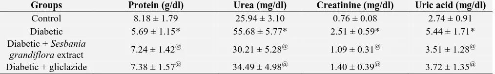
Table 5 depicts the levels of total protein, blood urea, uric acid and serum creatinine in control and experimental groups of rats. In STZ induced diabetic rats, there was a significant decrease in the total protein and increase in the levels of urea, uric acid and creatinine when compared with the control
group of rats. Administration of an ethanolic extract of Sesbania grandiflora leaves as well as the standard drug, gliclazide to the diabetic group of rats significantly decreased the levels of blood urea, uric acid, serum creatinine and increased the levels of total protein.
TABLE 5: EFFECT OF SESBANIA GRANDIFLORA EXTRACT ON THE LEVELS OF PROTEIN, UREA,
CREATININE AND URIC ACID IN PLASMA OF EXPERIMENTAL GROUPS OF RATS
Groups Protein (g/dl) Urea (mg/dl) Creatinine (mg/dl) Uric acid (mg/dl)
Control 8.18 ± 1.79 25.94 ± 3.10 0.76 ± 0.08 2.74 ± 0.91 Diabetic 5.69 ± 1.15* 55.68 ± 5.77* 2.51 ± 0.59* 5.44 ± 1.71* Diabetic + Sesbania
grandifloraextract 7.24 ± 1.42 @
30.21 ± 5.28@ 1.09 ± 0.31@ 3.51 ± 1.28@
Diabetic + gliclazide 7.38 ± 1.57@ 34.49 ± 4.98@ 1.40 ± 0.39@ 3.72 ± 1.35@ Values are given as mean ± SD for groups of six rats in each. Values are statistically significant at p < 0.05. Statistical significance was compared within the groups as follows: *compared with control, @ compared with diabetic rats.
Table 6 depicts the levels the levels of glycogen content and activities of glycogen synthase and glycogen phosphorylase in liver tissues control and experimental groups of rats. A significant decline in the glycogen level as well as in the glycogen synthase activity and a concomitant increase in the activity of glycogen phosphorylase were noted in
the liver of diabetic group of rats. Oral
administration of Sesbania grandiflora leaves
extract as well gliclazide to diabetic rats restored the level of glycogen and the activities of glycogen synthase, glycogen phosphorylase to near normalcy when compared to control group of rats.
TABLE 6: EFFECT OF SESBANIA GRANDIFLORA EXTRACT ON THE LEVELS OF LIVER GLYCOGEN
CONTENT IN THE EXPERIMENTAL GROUPS OF RATS
Groups Glycogen Glycogen synthase Glycogen phosporylase
Control 42.96 ± 5.42 794.28 ± 59.71 576.18 ± 36.22
Diabetic 25.38 ± 3.99* 539.75 ± 36.45* 899.37 ± 65.16* Diabetic + Sesbania grandiflora
extract 36.75 ± 4.18
@ 696.78 ± 33.59 @ 654.25 ± 40.17 @
Diabetic + gliclazide 37.26 ± 4.92@ 677.85 ± 40.27 @ 681.34 ± 45.91@ Units are expressed as: mg/g wet tissue for glycogen, μmoles of UDP formed/h/mg protein for glycogen synthase and μmoles of Pi liberated/h/mg protein for glycogen phosphorylase. Values are given as mean ± SD for groups of six rats in each. Values are statistically significant at p < 0.05. Statistical significance was compared within the groups as follows: *compared with control, @ compared with diabetic rats.
Table 7 depicts the levels of AST, ALT and ALP in the control and experimental group of rats. Diabetic rats showed a significant elevation in the
levels of aspartate transaminase, alanine
transaminase and alkaline phosphatase when
compared with the control group of rats.
Administration of Sesbania grandiflora leaves
extract and gliclazide to the diabetic rats resulted in a significant decrease in the levels of these markers.
TABLE 7: EFFECT OF SESBANIA GRANDIFLORA EXTRACT ON THE ACTIVITY OF AST, ALT AND ALP IN THE SERUM OF EXPERIMENTAL GROUPS OF RATS
Groups AST ALT ALP
Control 40.67 ± 8.21 20.35 ± 3.17 70.49 ± 10.91
Diabetic 109.75 ± 12.86* 68.74 ± 10.38* 202.35 ± 20.72* Diabetic + Sesbania grandifloraextract 72.38 ± 10.41@ 35.26 ± 7.14@ 98.56 ± 12.94@
[image:5.612.54.561.166.242.2]The levels of TBARS in the plasma and pancreas of control as well as experimental group of rats are presented in Table 8. STZ induced diabetic rats showed marked increase in the levels of TBARS
when compared to control rats. Treatment of
Sesbania grandiflora leaves extract to diabetic rats showed a significant decrease in the levels of TBARS.
TABLE 8: EFFECT OF SESBANIA GRANDIFLORA EXTRACT ON THE LEVEL OF TBARS IN PLASMA AND PANCREAS, OF EXPERIMENTAL GROUPS OF RATS
Groups TBARS
Plasma Pancreas
Control 3.42 ± 0.91 28.57 ± 5.64
Diabetic 7.54 ± 2.18* 72.89 ± 12.35*
Diabetic + Sesbania grandiflora extract 4.89 ± 1.52@ 40.27 ± 9.58@
Diabetic + gliclazide 5.14 ± 1.98@ 39.52 ± 10.85@
Units: mM/100 g in tissues; nM/ml in plasma. Values are given as mean ± SD for groups of six rats in each. Values are statistically significant at p < 0.05. Statistical significance was compared within the groups as follows: *compared with control, @ compared with diabetic rats.
Table 9 and 10 illustrates the activities of enzymatic and non-enzymatic antioxidants in pancreas as well as plasma of control and experimental group of rats. In STZ induced diabetic rats, there was a significant reduction in the
activities of enzymatic and non-enzymatic
antioxidants in pancreas and plasma respectively. Treatment of Sesbania grandiflora leaves extractto the diabetic rats showed improvement in the
activities of enzymatic and non-enzymatic
antioxidants.
TABLE 9: EFFECT OF SESBANIA GRANDIFLORA EXTRACT ON THE ACTIVITY OF SOD, CATALASE AND GPX, AND THE LEVEL OF GSH IN PANCREAS OF EXPERIMENTAL GROUPS OF RATS
Groups SOD Catalase GPx GSH
Control 4.55 ± 1.37 18.64 ± 2.85 6.49 ± 1.71 24.97 ± 2.54 Diabetic 1.95 ± 0.98* 8.12 ± 2.79* 3.58 ± 0.91* 10.75 ± 2.12* Diabetic + Sesbania
grandifloraextract 3.29 ± 1.45
@ 14.69 ± 2.27@ 4.86 ± 1.21@ 19.55 ± 2.37@
[image:6.612.69.544.134.221.2]Diabetic + gliclazide 3.33 ± 1.81@ 13.79 ± 3.10@ 4.51 ± 1.38@ 20.18 ± 3.42@ Activity is expressed as: 50% of inhibition of epinephrine autooxidation/min/mg of protein for SOD; µmoles of hydrogen peroxide decomposed/min/mg of protein for catalase; µmoles of glutathione oxidized/min/mg of protein for GPx; mg/100 g tissue for GSH. Values are given as mean ± SD for groups of six rats in each. Values are statistically significant at p < 0.05. Statistical significance was compared within the groups as follows: *compared with control, @ compared with diabetic rats.
TABLE 10: EFFECT OF SESBANIA GRANDIFLORA EXTRACT ON THE LEVELS OF VITAMIN C, VITAMIN E,
CERULOPLASMIN AND GSH IN PLASMA OF EXPERIMENTAL GROUPS OF RATS
Groups Vitamin C Vitamin E Ceruloplasmin GSH
Control 1.36 ± 0.12 0.65 ± 0.07 13.71 ± 1.52 28.93 ± 2.74 Diabetic 0.52 ± 0.09* 0.30 ± 0.04* 5.18 ± 0.95* 12.86 ± 2.17* Diabetic + Sesbania grandiflora extract 0.94 ± 0.11@ 0.57 ± 0.08@ 10.21 ± 1.59@ 20.72 ± 2.95@ Diabetic + gliclazide 0.89 ± 0.12@ 0.59 ± 0.04@ 9.76 ± 1.91@ 22.10 ± 3.18@ Units: mg/dl. Values are given as mean ± SD for groups of six rats in each. Values are statistically significant at p < 0.05. Statistical significance was compared within the groups as follows: *compared with control, @ compared with diabetic rats.
DISCUSSION: Plants provide an extraordinary source of natural medicines for various ailments. Moreover, secondary metabolites of plant origin serve as an invaluable chemical library for drug discovery and current medicinal chemistry in the pharmaceutical industry. The medicinal properties of plants lie in the phytoingredients that exert distinct physiological activities in the human body.
Over the past 25 years, 50% of prescription drugs have been developed from natural products and their derivatives38. In the present study, phytochemical analysis on Sesbania grandiflora leaves extract indicated the presence of flavonoids,
saponins, tannins, diterpenes, triterpenoids,
[image:6.612.52.566.527.594.2]pharmacological effects of the extract. The leaves contain essential amino acids, minerals, vitamins such as A, E, C, thiamine, riboflavin and nicotinic acid in addition to secondary phytochemicals such as pectin, triterpenoid, tannin, glycosides and saponin 39, 40.
The leaves extract was reported to be non-toxic. It has been reported that the oral administration of leaves extract even at the maximum single dose of 2000 mg/kg body weight was found to be safe, since no mortality 41.
STZ acts as a cytotoxin for beta-cells of the islet of langerhans, causes diabetes by inducing β-cell necrosis 42. Diabetic rats exhibit gradual weight loss as compared with the normal group. This process is due to muscle wasting and depletion of protein in tissues. A decrease in body weight was observed in diabetic group indicating the increased proteolysis. Diabetic rats treated with Sesbania grandiflora leaves extract for 30 days showed a significant improvement in body weight as compared to diabetic animals, which shows the beneficial effects of the extract in controlling the muscular wasting.
Diabetes mellitus is characterized by decreased glucose tolerance due to low secretion of insulin or its action. Oral glucose tolerance test (OGTT) is a test of immense value in favor of using fasting plasma glucose concentration to facilitate the diagnosis of diabetes mellitus. OGTT revealed that the blood glucose levels in control rats reach peak at 60 minutes after the oral glucose load and gradually return backs to normal levels after 120 minutes. In diabetic rats, the peak increases in blood glucose concentration was observed after 60 minutes and remained high over the next 60 minutes. However, oral treatment with leaves extract showed definite lower peak blood glucose values, 60 minutes after glucose load also gives lower values almost at the end of 120 minutes indicating the improved glucose tolerance in diabetic rats treated with Sesbania grandiflora
leaves extract.
Diabetes mellitus is characterized by persistent hyperglycemia which results from reduced glucose utilization by various tissues. STZ induction causes specific damage in cells and thus exerts a
pronounced increase in blood glucose
concentration. It is well established that gliclazide is used as an antihyperglycemic drug, which stimulate the insulin secretion from pancreas and it is often used as a standard drug in STZ induced diabetic models to compare the antidiabetic
property of various plant extracts. Oral
administration of with Sesbania grandiflora leaves extract to STZ induced diabetic rats resulted in significant reduction in blood glucose level indicating the hypoglycemic nature of the leaves extract.
Persistent hyperglycemia results in glycation of hemoglobin that leads to the formation of
glycosylated hemoglobin43. Glycosylated
hemoglobin is an easily measurable biochemical marker that strongly correlates with the level of ambient glycemia during a 2- to 3-month period and is a more accurate and reliable measure than fasting blood glucose level 44. The concentration of glycosylated hemoglobin strongly predicts the risk of eye, kidney and neural disease in diabetes mellitus and is regarded as a key target for the diagnosis and prognosis of diabetes-related complications 45.
Oral treatment with Sesbania grandiflora leaves extract significantly decreased the levels of glycosylated hemoglobin, suggesting that it may prevent oxidative damage caused by the glycation reaction in diabetic conditions. These results on the levels of glucose and glycosylated hemoglobin indicate the beneficial effects of in the maintenance of glucose homeostasis. Urine sugar which was present in diabetic rats was found to be absent in the rats treated with the extract indicating the improved glycemic control.
The accelerated proteolysis of uncontrolled diabetes occurs as a result of deranged glucagon mediated regulation of cyclic AMP formation in insulin deficiency 47. This readily accounts for the observed decrease in the total protein content in
diabetes mellitus. The serum creatinine
concentration is the variable used not only to assess impairment of kidney function but also to detect the toxic effects of certain compounds derived from medicinal plants on kidney, in order to determine its efficacy in the treatment of diabetic rats. Serum uric acid is significantly associated with the risk of diabetes. Serum uric acid has been shown to be associated with oxidative stress and production of tumour necrosis factor 48.
In addition, a recent study in rats showed that fructose-induced hyperuricemia plays a pathogenic role in metabolic syndrome 49 (Nakagawa, 2006). Thus, lowering uric acid may be a novel treatment target for preventing diabetes. The levels of urea, serum creatinine and uric acid were restored to near normalcy by treatment with Sesbania grandiflora
leaves extract as well as gliclazide in STZ induced diabetic rats.
Liver plays a unique role in controlling carbohydrate metabolism by maintaining glucose concentrations in a normal range over both short and long periods of time. Liver produces glucose by breaking down glycogen (glycogenolysis) and by de novo synthesis of glucose (gluconeogenesis) from non-carbohydrate precursors such as lactate, amino acids and glycerol. Glycogen is the primary intracellular storable form of glucose and its availability in various tissues is a direct manifestation of insulin action as insulin facilitates intracellular glycogen deposition by stimulating the activity of glycogen synthase and inhibiting glycogen phosphorylase 50.
Glycogen synthase is a crucial and rate-limiting enzyme which catalyzes the transfer of glucose
from UDP-glucose to glycogen. Glycogen
phosphorylase is a rate-limiting enzyme of glycogenolysis and is regulated by phosphorylation and by allosteric binding of AMP, ATP, glucose-6-phosphate and glucose51. In diabetes, the glycogen
levels, glycogen synthase activity and
responsiveness to insulin signaling are diminished
and glycogen phosphorylase activity is
significantly increased. Oral administration of leaves extract to diabetic rats restored the glycogen content and the activities of glycogen metabolizing enzymes demonstrating the role of leaves extract in the regulation of glycogen metabolism. Earlier, we
have reported similar findings with Murraya
koenigii leaves extract 52.
AST and AST are the intracellular enzymes that have escaped into the blood stream and serve as a clinical index of tissue injury chiefly hepatocyte as well as renal injury. ALP acts as a marker of biliary function and cholestasis. It is assumed that elevation in the levels of serum ALT, AST and ALP are considered as predictors of diabetes 53. The increased activities of ALT, AST and ALP in the serum of diabetic rats may be primarily due to the leakage of these enzymes from liver as well as kidney into the blood stream54. Oral administration of leaves extract to diabetic group of rats showed a notable decline in the activity of these enzymes to their basal levels, indicating its non-toxic as well as tissue protective nature.
Oxidative stress is associated with the molecular mechanism of the decreased insulin biosynthesis and secretion, which is the main etiology of glucose toxicity. Because pancreatic islet cells
show extremely weak manifestation of
antioxidative machinery 55, 56, it is thought that the pancreas may be more susceptible to oxidative stress than other tissues and organs. Several conditions are known to disturb the balance between ROS production and cellular defense mechanisms.
The elevated cytotoxic and highly reactive oxidative stress markers such as lipid peroxides causes oxidative damage to proteins and DNA and the reduced cellular nonenzymatic and enzymatic antioxidant levels in diabetic conditions further increases the severity of tissue dysfunction resulting in decreased insulin synthesis, secretion, and finally resulting in β cell death.
Furthermore, the levels of activities of enzymatic antioxidants such as SOD, CAT, GPx, and GST were significantly improved in extract treated diabetic rats. Also, the plasma levels of nonenzymatic antioxidants such as vitamin C, vitamin E, reduced glutathione, and ceruloplasmin are found to be increased. The observed improvement in the antioxidant status reflects the antioxidant property of the leaves extract.
CONCLUSION: The results of the present study shows that Sesbania grandiflora leaves extract possess antidiabetic and antioxidant nature. Phytochemical screening indicated the presence of pharmacologically active ingredients in the leaves. The improved glycemic control is evident from the results of OGTT. The improvement in body weight gain indicates the beneficial effect of the leaves in controlling muscle wasting. The leaves extract significantly normalizes the biochemical alterations that occurred during diabetic mellitus.
The normalization in the activities of
pathophysiological enzymes indicates the non-toxic nature of the leaves extract. The improved enzymatic and non-enzymatic antioxidant status indicates the antioxidant property of the leaves extract. In conclusion, the observed antidiabetic and antioxidant property could be due to the presence of biologically active ingredients in the leaves. Thus, the study provides a scientific rationale for the use of Sesbania grandiflora leaves in the traditional system of medicine. Extraction, isolation and identification of active ingredients from the leaves may provide valuable lead molecules with wide range of medicinal values.
REFERENCES
1. American Diabetes Association. Diagnosis and classification of diabetes mellitus. Diabetes Care. 2012 Jan; 35 Suppl 1:S64-71.
2. Farombi EO. African indigenous plants with chemotherapeutic potentials and biotechnological approach to the production of bioactive prophylactic agents. Afr. J. Biotech. 2003; 2: 662-671
3. Wadkar KA, Magdum CS, Patil SS and Naikwade NS. Antidiabetic potential and Indian medicinal plants. J
Herbal Med and Toxicol. 2008; 2:45-50.
4. Suresh Kashyap and Sanjay Mishra . Phytopharmacology of Indian Medicinal Plant Sesbania Grandiflora. The
Journal of Phytopharmacology. 2012; 1(2): 2320- 480.
5. Gutteridge RC. The perennial sesbania species. In: Gutteridge, R.C and Shelton, H.M (Eds): Forage Tree Legumes in Tropical Agriculture. 1994; 49-64.
6. Ramesh T, Mahesh R and Begum VH. Effect of Susbania
grandiflora on lung antioxidant defense system in cigarette
smoke exposed rats. International Journal of Biological
Chemistry. 2007; 1(3): 141-148.
7. Kar A and Borthakur SK. Wild vegetables of Karbi -Anglong district, Assam. Natural Product Radiance. 2008; 7: 448-460.
8. Kasture VS, Deshmukh VK and Chopde CT. Anxiolytic and anticonvulsive activity of Sesbania grandiflora leaves in experimental animals. Phytother Res 2002; 16: 455-460. 9. Doddola S. Evaluation of Sesbania grandiflora for antiurolithiatic and antioxidant properties. Natural
Medicines. 2010; 62 (3): 300-07.
10. Nadkarni KM. Indian Materia Medica, Vol 1. Popular Prakashan: Mumbai. 1982; 52-54.
11. Govindan S and Shanmugasundaram ERB. Evaluation of the nutritive value of agathi (Sesbania grandiflora) leaf protein concentrates. The Ind J Nutr Dietet. 1987; 24:370-375.
12. Shrivastav N and Janin SK. Plants bearing antifertility properties. Hamdard Med 1993; 36: 91-98
13. Tamboli SA. Anti-inflammatory activity of Sesbania
grandiflora. Ind Drug 1996; 33: 504-506.
14. Tamboli SA. Analgesic and antipyretic activity of
Sesbania grandiflora. Ind Drug 2000; 37: 95-98.
15. Harborne JB. Phytochemical methods. Chapman and Hall Int., New York, Third Edition 1998.
16. Kokate CK: Pharmacognosy. Nirali Prakasham, Mumbai, India, Sixteenth Edition 2001.
17. Rakieten N, Rakieten ML and Nadkarni MV. Studies on the diabetogenic action of streptozotocin (NSC-37917). Cancer Chemotherapy Reports. 1963; 29: 91-98.
18. Fischer LJ and Rickert DE. Pancreatic islet cell toxicity CRC. Critical Reviews in Toxicology. 1975; 3: 231-263. 19. Trinder P. Determination of blood glucose using an
oxidaseperoxidase system with a non-carcinogenic chromogen. J Clin Pathol. 1969; 22:158-161.
20. Natelson S, Scott ML and Begga E. A rapid method for the estimation of urea in biological fluids by means of the reaction between diacetyl and urea. Am J Clin Pathol. 1951; 21: 275-281.
21. Nayak SS and Pattabiraman TN. A new colorimetric method for the estimation of glycosylated hemoglobin. Clin Chem Acta. 1981; 109: 267-274.
22. Lowry OH, Rosebrough NJ, Farr AL and Randall RJ. Protein measurement with the Folin phenol reagent. J Biol Chem. 1951; 193: 265-275.
23. Brod J and Sirota JH. The renal clearance of endogenous “creatinine” in man. J Clin Invest. 1948; 27: 645-654. 24. Caraway WI. Uric acid. In: Standard methods of clinical
chemistry. (Ed.) Seligson D. Vol.4, Academic Press, New York. 1963; pp.239-247.
25. King J. The hydrolases-acid and alkaline phosphatases. In: Practical clinical enzymology. (Ed.) Van D. Nostrand Co, London. 1965; pp.199-208.
26. King J. The transaminases: alanine and aspartate transaminases. In: Practical Clinical Enzymology (ed.) D.Van, Nostrand Co., London. 1965; pp. 363–395 27. Morales MA, Jabbagy AJ and Terenizi HR. Mutations
affecting accumulation of glycogen. Neurospora News. 1973; 20: 24–25.
28. Leloir LF and Goldemberg SH. Glycogen synthetase from rat liver: (Glucose)n + (UDPG)→(Glucose)n+1 +UDP, In: Colowick SP, Kalpan NO (Eds.), Methods in Enzymology. Academic Press, New York, 1962; pp. 145–147.
in the perfused isolated rat heart. J Biol Chem. 1963; 238: 1592-1597.
30. Yagi K. Simple fluorimetric assay for lipid peroxides in blood plasma. Biochem Med. 1976; 15: 212-215.
31. Ohkawa H, Ohishi N and Yagi K. Assay for lipid peroxides in animal tissues by thiobarbituric acid reaction. Anal Biochem. 1979; 95: 351-358.
32. Misra HP and Fridovich I. The role of superoxide anion in the auto oxidation of epinephrine and a simple assay of superoxide dismutase. J Biol Chem. 1972; 247: 3170-3175.
33. Takahara S, Hamilton BH, Nell JV, Kobra TY, Ogura Y and Nishimura ET. Hypocatalasemia, a new genetic carrier state. J Clin Invest. 1960; 29: 610 619.
34. Rotruck JT, Pope AL, Gasther HE, Hafeman DG and Hoekstra WG. Selenium biochemical role as a component of glutathione peroxidase. Science.1973; 179: 588-590. 35. Omaye ST, Turnbull JD and Sauberlich HE. Selected
methods for the determination of ascorbic acid in animal cells, tissues and fluids. Methods Enzymol. 1979; 62: 3-11. 36. Desai ID. Vitamin E analysis methods for animal tissues.
Methods in Enzymol. 1984; 105: 138-147.
37. Sedlak J and Lindsay RH. Estimation of total, protein bound and non-protein sulfhydryl groups in tissue with Ellmans reagent. Anal Biochem. 1968; 25: 293-305. 38. Chang CL, Chen YC, Chen HM, Yang NS and Yang WC.
Natural cures for type 1 diabetes: a review of phytochemicals, biological actions, and clinical potential. Curr Med Chem. 2013; 20(7):899-907.
39. Ching LS and Mohamed S. Alpha-tocopherol content in 62 edible tropical plants. J Agric Food Chem 2001; 49: 3101-3105.
40. Das P, Raghuramulu N and Rao KC. Determination of in vitro availability of iron from common foods. J Hum Ecol 2005; 18: 13-20.
41. Doddola S, Pasupulati H, Koganti B and Prasad KV. Evaluation of Sesbania grandiflora for antiurolithiatic and antioxidant properties. J Nat Med. 2008; 62: 300-307 42. Szkudelski T. The mechanism of alloxan and
streptozotocin action in β cells of the rat pancreas. Physiol Res. 2001; 50:537-546.
43. Yabe-Nishimura C. Aldose reductase in glucose toxicity: a potential target for the prevention of diabetic complications. Pharmacol Rev.1998; 50:21-33.
44. Goldstein DE, Little RR, Lorenz RA, Malone JI, Nathan DM and Peterson CM. American Diabetes Association. Tests of glycemia in diabetes. Diabetes Care. 2004; 27:S91-S93.
45. Howlett J and Ashwell M (2008). Glycemic response and health: summary of a workshop. Am J Clin Nutr. 2008; 87:212S-216S.
46. Rannels DE, Marker DE and Morgan HE. Biochemical actions of hormones. In: G. Litwack (ed), Academic Press, New York. 1997; Vol. 1: pp135-195.
47. Dighe RR, Rojas FJ, Birnbaumer L and Garber AJ. Glucagon stimulable adenylyl cyclase in rat liver. The impact of streptozotocin-induced diabetes mellitus. Journal of Clinical Investigation. 1984; 73:1013-1023.
48. Butler R, Morris AD, Belch JJ and Hill A et.,al. Allopurino normalizes endothelial dysfunction in type 2 diabetics with mild hypertension. Hypertension. 2000; 35:746-751.
49. Nakagawa T, Hu H, Zharikov S and Tuttle KR et.,al. A causal role for uric acid in fructose-induced metabolic syndrome. American Journal of Physiology - Renal Physiology. 2006; 290:F625– F631.
50. Pederson BA, Schroeder JM, Parker GE, Smith MW, DePaoli-Roach AA and Roach PJ (2005). Glucose metabolism in mice lacking muscle glycogen synthase. Diabetes. 2005; 54(12):3466-3473.
51. Greenberg CC, Jurczak MJ, Danos AM and Brady MJ. Glycogen branches out: new perspectives on the role of glycogen metabolism in the integration of metabolic pathways. Am J Physiol Endocrinol Metab. 2006; 291(1): E1-8.
52. Arulselvan P and Subramanian S. Effect of Murraya
koenigii leaf extract on carbohydrate metabolism studied
in streptozotocin induced diabetic rats. International Journal of Biological chemistry. 2007; 1 (1): 21-28. 53. Hanley AJ, Williams K, Festa A, Wagenknecht LE,
D'Agostino RB Jr, Kempf J, Zinman B and Haffner SM. Insulin resistance atherosclerosis study. Elevations in markers of liver injury and risk of type 2 diabetes: the insulin resistance atherosclerosis study. Diabetes. 2004; 53:2623-2632.
54. El-Demerdash FM, Yousef MI and El-Naga NI. Biochemical study on the hypoglycemic effects of onion and garlic in alloxan-induced diabetic rats. Food Chem. Toxicol. 2005; 43:57–63.
55. Tiedge M, Lortz S, Drinkgern J and Lenzen S. Relation between antioxidant enzyme gene expression and antioxidative defense status of insulin-producing cells. Diabetes 1997; 46: 1733-1742.
56. Robertson RP. Oxidative stress and impaired insulin secretion in type 2 diabetes. Curr Opin Pharmacol 2006; 6: 615-619.
All © 2013 are reserved by International Journal of Pharmaceutical Sciences and Research. This Journal licensed under a Creative Commons Attribution-NonCommercial-ShareAlike 3.0 Unported License.
This article can be downloaded to ANDROID OS based mobile. Scan QR Code using Code/Bar Scanner from your mobile. (Scanners are available on Google Playstore)
How to cite this article: