INTRODUCTION
Similar to the stratified squamous epidermis, the epithelium of foregut-derived tissues, including the esophagus, arises from a layer of simple columnar cells that stratify and differentiate. In the epidermis, these processes are associated with multiple signaling pathways, including those driven by bone morphogenetic protein (BMP) and Wnt (Botchkarev, 2003; Botchkarev and Sharov, 2004). However, it remains unknown whether these pathways are also active in the embryonic foregut during development and in the steady state.
The canonical BMP signaling pathway is activated by a secreted ligand (e.g. Bmp4, Bmp7) interacting with a complex of type I and type II transmembrane receptors. This initiates signal transduction by phosphorylation of Smads 1/5/8 (R-Smads), which interact with a co-Smad (Smad4) to regulate downstream target genes (Miyazono et al., 2009). BMP signaling is modulated by extracellular antagonists, including noggin (Nog), chordin (Chrd) and follistatin (Fst), that interfere with productive binding to receptors (Massague et al., 2005). In the epidermis, conditional deletion of Bmpr1a leads to increased epithelial proliferation (Kobielak et al., 2003; Qiao et al., 2006; Yuhki et al., 2004). Deletion of Smad4in the epithelium also results in basal cell hyperproliferation and leads to squamous cell cancer (Qiao et al., 2006).
The mouse esophagus separates from the foregut tube between embryonic day (E) 9.5 and E11.5. This separation process is regulated by multiple signaling pathways, including Wnt, BMP and Shh, which may act upstream of Bmp4 (Litingtung et al., 1998;
Morrisey and Hogan, 2010; Roberts et al., 1998). Analysis of Nog -null mutants has shown that BMP signaling regulates the initial dorsal-ventral patterning of the single foregut tube and the separation into dorsal esophagus and ventral trachea (Li et al., 2007; Que et al., 2006). Here, we show that Nog also regulates the transition from simple columnar to stratified squamous epithelium in the developing esophagus. Combining gain- and loss-of-function studies, we further show that BMP signaling plays multiple roles in the development of the esophagus and forestomach, the epithelium of which appears histologically as a continuation of the esophagus.
MATERIALS AND METHODS Mouse strains
Heterozygous BRE-lacZ (Blank et al., 2008), NoglacZ/+(Brunet et al., 1998) and Bmp7lacZ/+(Godin et al., 1998) reporter mice were maintained on the C57Bl/6 ⫻ 129/SvEv background. To conditionally activate BMP signaling in foregut endoderm, Shh-GFP-Cre(Shh-Cre) mice (Harfe et al., 2004) were crossed with Rosa26CAG-loxpstoploxp-caBmpr1a/+(Bmpr1aCA/+) mice. In this line, a constitutively active Bmpr1a (caBmpr1a) was inserted into the Rosa26locus preceded by a CMV early enhancer/chicken -actin (CAG) promoter and a floxed neo cassette (see Fig. S2A in the supplementary material). The caBmpr1a construct has a single amino acid substitution (Q to D) within the glycine-serine (GS) domain, giving the protein strong kinase activity even in the absence of ligand or type II receptor (Zou et al., 1997). To conditionally delete Bmpr1a, Shh-Cre;Bmpr1aflox/flox or Shh-Cre;Bmpr1aflox/null compound mutants were generated (Yuhki et al., 2004).
In situ hybridization
In situ hybridization was performed as previously described (Que et al., 2006; Que et al., 2007). Digoxigenin (DIG)-labeled antisense cRNA probes against mouse Bmpr1a, Bmp7, Nog, Fst, Fstl1, Chrd, Grem1 andCer1
were synthesized using T3, T7 or SP6 RNA polymerases. Grem1was a kind gift from Dr Clifford Tabin (Harvard University).
Immunohistochemistry, immunostaining and X-gal staining Tissues were fixed in 4% paraformaldehyde for 3-4 hours at 4°C and embedded in paraffin for sectioning. Cryosections were also used for immunostaining with phospho (p) Smad1/5/8 antibody (1:1000; kind gift Development 137, 4171-4176 (2010) doi:10.1242/dev.056077
© 2010. Published by The Company of Biologists Ltd
1Department of Cell Biology, Duke University Medical Center, Durham, NC 27710, USA. 2Department of Molecular Medicine, Maine Medical Center Research Institute, 81 Research Drive, Scarborough, ME 04074, USA.
*Present address: Department of Biomedical Genetics, University of Rochester, Rochester, NY 14642, USA
†Author for correspondence (jianwen_que@urmc.rochester.edu)
Accepted 6 October 2010 SUMMARY
The stratification and differentiation of the epidermis are known to involve the precise control of multiple signaling pathways. By contrast, little is known about the development of the mouse esophagus and forestomach, which are composed of a stratified squamous epithelium. Based on prior work in the skin, we hypothesized that bone morphogenetic protein (BMP) signaling is a central player. To test this hypothesis, we first used a BMP reporter mouse line harboring a BRE-lacZ allele, along with in situ hybridization to localize transcripts for BMP signaling components, including various antagonists. We then exploited a Shh-Cre allele that drives recombination in the embryonic foregut epithelium to generate gain- or loss-of-function models for the Bmpr1a (Alk3) receptor. In gain-of-function (Shh-Cre;Rosa26CAG-loxpstoploxp-caBmprIa) embryos, high levels of ectopic BMP signaling stall the transition from simple columnar to multilayered undifferentiated epithelium in the esophagus and forestomach. In loss-of-function
experiments, conditional deletion of the BMP receptor in Shh-Cre;Bmpr1aflox/floxembryos allows the formation of a multilayered squamous epithelium but this fails to differentiate, as shown by the absence of expression of the suprabasal markers loricrin and involucrin. Together, these findings suggest multiple roles for BMP signaling in the developing esophagus and forestomach.
KEY WORDS: BMP signaling, Esophagus, Forestomach, Stratification, Differentiation, Mouse
BMP signaling in the development of the mouse esophagus
and forestomach
Pavel Rodriguez1, Susana Da Silva1, Leif Oxburgh2, Fan Wang1, Brigid L. M. Hogan1and Jianwen Que1,*,†
D
E
V
E
LO
P
M
E
N
from Dr Edward Laufer, Columbia University). The following primary antibodies were used on paraffin sections: mouse anti-p63 (1:200; Santa Cruz Biotechnology); mouse anti-Krt14 (1:200; Thermo Scientific); rat anti-Krt8 (1:100; DSHB); rat anti-phospho-histone H3 (1:1000, Upstate Biotechnology); rabbit anti-loricrin (1:100) and anti-involucrin (1:10,000) (kind gifts from Dr Terry Lechler, Duke University); and rabbit anti-Krt5 (1:100; Covance). Whole embryos, isolated esophagi and stomachs were stained with X-gal and processed as previously described (Que et al., 2006).
RESULTS AND DISCUSSION
Dynamic BMP signaling in the developing mouse esophagus and forestomach
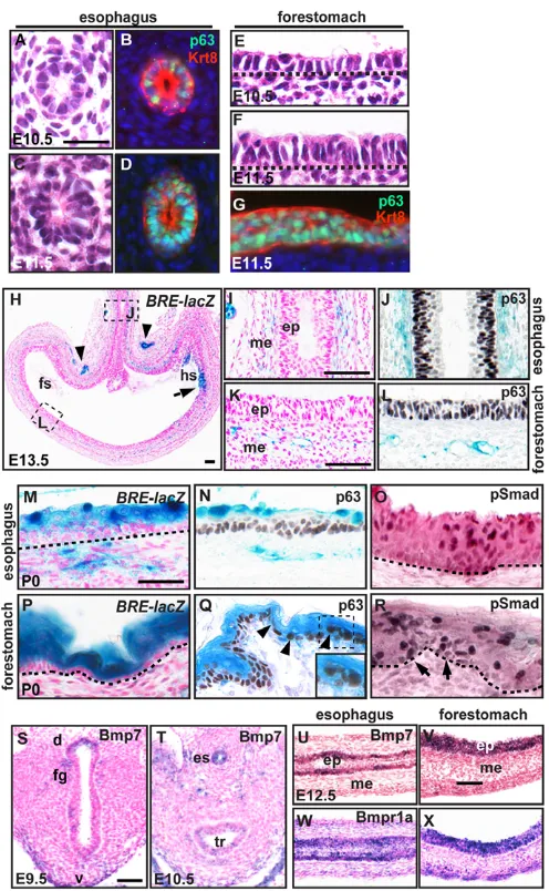
The epithelium of the embryonic esophagus and forestomach starts to stratify at early E11.5. At E10.5, the esophagus is lined by keratin 8 (Krt8)-positive simple columnar epithelial cells, a subset of which express p63 (Trp63 – Mouse Genome Informatics) (Fig. 1A,B), a transcription factor that is required for epithelial stratification (Daniely et al., 2004). By E11.5, the epithelium is 1-2 cells thick (Yu et al., 1-2005), with all cells expressing p63 and Krt8, but not Krt5 and Krt14, which only starts to be expressed from E14.5 (Fig. 1C,D; data not shown). Similarly, the epithelium in the forestomach starts to stratify between E10.5 and E11.5 (Fig. 1E-G).
To investigate canonical BMP pathway activation in the developing esophagus and forestomach, we used the BRE-lacZ transgenic reporter line, which expresses -galactosidase (-gal) under the control of BMP response elements (BREs) from Id1 (Blank et al., 2008). At E9.5, -gal-positive cells were observed in the epithelium and mesenchyme of the ventral foregut (see Fig. S1A in the supplementary material), which generates the trachea at E11.5 (see Fig. S1B in the supplementary material). Although the simple columnar epithelium of the hindstomach and mesenchyme showed active BMP signaling at E13.5 (Fig. 1H), the epithelium of the esophagus and forestomach remained negative (Fig. 1H-L). By E14.5, -gal activity was seen in a limited number of suprabasal cells in both the esophagus and forestomach, and this increased by E15.5 (see Fig. S1C-H in the supplementary material). At postnatal day (P) 0, all suprabasal cells exhibited -gal activity (Fig. 1M-R) and ~75% of these cells were also positive for pSmad1/5/8. Interestingly, ~5% of basal cells were positive for both activities (Fig. 1O,R).
Expression of BMP ligands and inhibitors in the developing esophagus and forestomach
In situ hybridization showed that between E9.5 and E15.5, Bmpr1a is ubiquitously expressed, whereas Bmp7 transcripts are enriched in the esophageal and forestomach epithelium (Fig. 1S-X; data not shown). The expression of Bmp7 in the epithelium of the esophagus and forestomach was confirmed by analysis of the Bmp7-lacZ‘knock-in’ allele (see Fig. S1I,J in the supplementary material). At E15.5, low levels of Bmp6 were detected in the differentiating suprabasal cells (see Fig. S1K,L in the supplementary material).
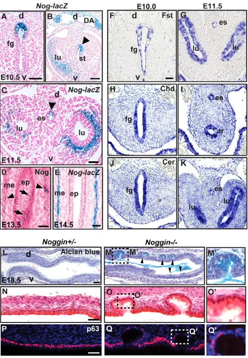
Although the epithelial cells express Bmp7and Bmpr1afrom at least E10.5 to E13.5, BRE-lacZ-positive cells were only observed from E14.5. We hypothesized that the absence of BMP signaling in the epithelium is due to extracellular BMP inhibitors that block ligand-receptor interaction. Nogis expressed in the dorsal E9.5 foregut (Li et al., 2007; Que et al., 2006). This expression pattern was maintained as the separation proceeds at ~E10.5-11.5, and Nog-lacZexpression was restricted to a subpopulation of epithelial
[image:2.612.314.562.58.460.2]cells in the esophagus and in the dorsal forestomach (Fig. 2A-C). Although at E13.5 Nog-lacZ expression was very low in the esophageal epithelium, Nogtranscripts could still be detected by in Fig. 1. BMP signaling in mouse foregut development.
(A-G)Transverse sections of E10.5-11.5 esophagus (A-D) and longitudinal sections of E10.5-11.5 forestomach (E-G) stained with Hematoxylin and Eosin (H&E) (A,C,E,F) or antibodies against p63 and Krt8 (B,D,G). (H-L)Longitudinal sections of E13.5 BRE-lacZesophagus and forestomach. X-gal staining is present in the blood vessels (arrowheads in H) and hindstomach epithelium (arrow in H),
esophageal (I,J) and forestomach (K,L) mesenchyme. (M-R)At P0, BRE-lacZis active mostly in the suprabasal layers of the esophagus and forestomach, whereas p63 is only found in the basal layer of the epithelium. BRE-lacZreporter activity correlates with pSmad1/5/8 nuclear localization (O,R). The inset in Q shows that p63-positive basal cells are also positive for X-gal staining (arrowheads), and this correlates with pSmad staining in a subset of basal cells (arrows in R). (S-V)In situ hybridization shows that Bmp7is present in the dorsal epithelium of the E9.5 (S, transverse section) and E10.5 (T) foregut, E12.5 esophageal (U) and forestomach (V) epithelium. (W,X)Bmpr1ais ubiquitously expressed at E12.5 in the esophagus and forestomach. Nuclei in H,I,K,M,P,S-X are counterstained with Nuclear Fast Red. Dotted lines indicate the basal membrane. ep, epithelium; me, mesenchyme; fs, forestomach; hs, hindstomach; fg, foregut; d, dorsal; v, ventral; es, esophagus; tr, trachea. Scale bars: 25m in A-G; 50m in H-X.
D
E
V
E
LO
P
M
E
N
situ hybridization (Fig. 2D). However, at E14.5, Nog-lacZwas only detected in the inner circular muscle layer of the esophagus (Fig. 2E). We also examined transcripts of other putative BMP inhibitors including Fst, follistatin-like 1 (Fstl1), Chrd, gremlin 1 (Grem1) and cerberus (Cer1) (reviewed by Umulis et al., 2009). Between E10.5 and E11.5, Fst, Chrd, Cer1 and Fstl1were expressed in the epithelium, whereas Grem1 was found only in the mesenchyme (Fig. 2F-K; data not shown). After E12.5, only Cer1 expression was maintained at a high level in the epithelium, whereas Chrd, Grem1 and Fstl1 were expressed in the mesenchyme (see Fig. S1M-R in the supplementary material; data not shown). Interestingly, Fstwas expressed in the epithelium and mesenchyme
of the E12.5 forestomach at a moderate level, which contrasts with the very low levels found in the esophagus (see Fig. S1O,P in the supplementary material).
Ectopic activation of BMP signaling in the epithelium
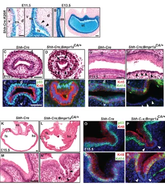
Previously, we reported esophageal atresia and tracheoesophageal fistula (EA/TEF) in nearly 60% of Nog-null mutants (Que et al., 2006). In the remaining 40% of mutants, the foregut separates normally. However, in the E18.5 dorsal esophagus we found multiple abnormal gland-like pits lined with simple columnar epithelium (Fig. 2L-Q⬘; n12). The epithelial cells lining the pits were positive for Alcian Blue staining, which is indicative of mucus production (Fig. 2M,M⬘), and the number of p63-positive cells was reduced compared with the wild type (Fig. 2P-Q⬘). No expression of the tracheal transcription factor Nkx2.1 or the Clara cell marker Scgb1a1 was detected (data not shown), excluding the possibility that the cells have acquired a respiratory identity. It is noteworthy that there were no abnormalities in the forestomach of Nog-null mutants at any stage. Here, other BMP inhibitors in the forestomach, such as Fst, might play a compensatory role in the event of Nog deletion (see Fig. S1P in the supplementary material). Our results suggest that the formation of a multilayered epithelium from a simple columnar epithelium between E10.5 and E14.5 is inhibited by the loss of Nog and by the resultant increase in canonical BMP signaling. To further explore this idea, we used a combination of a new conditional Rosa26allele that incorporates constitutively active Bmpr1a (Bmpr1aCA) (see Fig. S2A in the supplementary material) and a Shh-Creallele that is preferentially expressed in the ventral esophagus and forestomach between E10.5 and E11.5 (Harris-Johnson et al., 2009) (Fig. 3A). By E13.5, most of the epithelial cells in these tissues of Shh-Cre;R26Rembryos were labeled by X-gal staining, suggesting that the recombination later extends dorsally (Fig. 3B). All of the Shh-Cre;Bmpr1aCA/+ mutants died before P5, probably owing to respiratory distress (J.Q. and B.L.M.H., unpublished). The activation of BMP signaling was confirmed by pSmad1/5/8 staining, which showed nuclear signals in most epithelial cells of the E15.5 mutant esophagus and forestomach, as compared with the control (see Fig. S2B-E in the supplementary material).
Examination of the E15.5 Shh-Cre;Bmpr1aCA/+mutant revealed clusters of simple columnar epithelial cells along the esophagus, preferentially in the ventral region (Fig. 3C-J; n9). These simple epithelial cells maintained a high level of Krt8, whereas the number of p63-positive cells was apparently reduced (Fig. 3F). Moreover, the simple columnar epithelium was negative for Krt5 and Krt14, two cytokeratins that are characteristic of basal cells of stratified/multilayered epithelia (Fig. 3J). In the mutant forestomach, scattered regions of simple columnar (Krt8-positive, Krt5/14-negative, p63 low) epithelium were also observed at E15.5 and P0 (Fig. 3K-R and see Fig. S2F,G in the supplementary material; n24). At E15.5, phospho-histone H3 staining revealed no significant differences in cell proliferation between the simple columnar and stratified epithelium in the mutants and control (data not shown).
[image:3.612.52.297.59.412.2]The simple columnar epithelium was present only on the ventral side of the esophagus of Shh-Cre;Bmpr1aCA/+mutants, which correlated with the ventral activation of Shh-Crebefore E12.5 (Fig. 3A). At E13.5, even though Shh-Crewas expressed on the dorsal side of the esophagus (Fig. 3B), the resultant ectopic BMP signaling was incapable of reversing the stratified epithelium back to a monolayer (Fig. 3D,H), suggesting that there is a limited time Fig. 2. Expression of BMP inhibitors in the developing esophagus
and forestomach.(A-C)Transverse sections of X-gal-stained E10.5 and E11.5 Nog-lacZmouse embryos show that Nogis expressed in the epithelium of the dorsal esophagus and forestomach (arrowhead). (D)In situ hybridization shows that the Nogtranscript is present in the epithelium (arrow) and mesenchyme (arrowhead) of the E13.5 esophagus. (E)At E14.5, Nog-lacZis active only in the muscle of the esophagus. (F-K)In situ hybridization of transverse sections of E10.0-11.5 embryonic foregut shows that Fst(F,G), Chrd(H,I) andCer1(J,K) are transcribed in the foregut epithelium. (L-Q⬘) Longitudinal sections of the E18.5 Nog+/–and Nog–/–esophagus stained with Alcian Blue (L-M⬘), H&E (N-O⬘) and antibody to p63 (P-Q⬘). The Nog–/–esophagus shows multiple gland-like pits that are present dorsally and contain simple columnar epithelium, which stain positive for Alcian Blue (M, arrowheads). In these pits, the number of p63-positive cells is reduced. lu, lung; st, stomach. Scale bars: 50m.
D
E
V
E
LO
P
M
E
N
period during which the fate of the epithelium can be influenced by ectopic signals. Similar observations have been reported in the chick gizzard, where ectopic Bmp2 can only switch the luminal to glandular epithelium within a certain time window (Yasugi and Mizuno, 2008).
Conditional deletion of Bmpr1ain the
forestomach epithelium
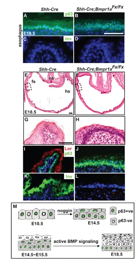
After E14.5 the BMP pathway is active in the differentiating suprabasal cells of the esophagus and forestomach (see Fig. S1C-H in the supplementary material). To address whether BMP signaling is necessary for the differentiation of the epithelium, we deleted Bmpr1a by generating Shh-Cre;Bmpr1aflox/flox and Shh-Cre;Bmpr1aflox/null mutants. Since similar phenotypes were observed in the Shh-Cre;Bmpr1aflox/floxand Shh-Cre;Bmpr1aflox/null mutants, subsequent analyses employed the former. Most mutants died in utero or at birth, presumably owing to heart defects, but a few did survive to P9 (n3). The loss of Bmpr1a transcripts in the mutant esophagus and forestomach at E15.5 was confirmed by in situ hybridization (see Fig. S2H-K in the supplementary material). In the esophageal epithelium of the E18.5 and surviving P9 mutants, expression of p63 persisted in the upper layers (Fig. 4A,B; data not shown), suggesting that these cells are not fully differentiated. There was no involucrin or loricrin immunostaining in the mutant epithelium, but even the wild-type epithelium expressed low levels of these proteins at this stage (Fig. 4C,D; data not shown). By contrast, the loss of Bmpr1a in the forestomach led
to a more obvious defect in the differentiation of suprabasal cells (Fig. 4E-L). The affected regions remained p63 positive, but lacked involucrin and loricrin (Fig. 4I-L). In addition, some of these p63-positive cells remained proliferative, as indicated by phospho-histone H3 staining (see Fig. S2L,M in the supplementary material). These results suggest that BMP signaling is required for the differentiation of the suprabasal cells in the esophagus and forestomach. Interestingly, other studies have shown that the inhibition of BMP signaling by overexpression of Nogunder a Krt5 promoter in the epidermis also results in alterations in eyelid epithelial differentiation, and the levels of involucrin and loricrin are reduced (Sharov et al., 2003). Conversely, the addition of Bmp4 to cultured human oral epithelium increases the expression of involucrin (Kim et al., 2006). These studies suggest that BMP signaling regulates the differentiation of suprabasal cells in stratified epithelium.
[image:4.612.52.387.54.426.2]In conclusion, our results suggest that BMP signaling has two distinct functions in the development of the mouse esophagus and forestomach (Fig. 4M). Initially, between E10.5 and E14.5, the inhibition of BMP signaling by Nog is necessary for epithelial stratification to occur. The deletion of Nog or expression of constitutively active BMP receptor maintains a simple columnar epithelium. Then, starting at ~E14.5, BMP signaling is required for the differentiation of the suprabasal cells. In the absence of Bmpr1a, the epithelial stratification does occur but the suprabasal cells do not fully differentiate. In this study, we have only investigated the role of BMP signaling in the embryonic and early Fig. 3. Activation of BMP signaling in the Shh-Cre;Bmpr1aCA/+mutant inhibits stratification of the esophageal and forestomach epithelium.(A,B)Longitudinal sections of X-gal-stained E11.5 and E13.5 Shh-Cre;R26Rmouse esophagus and stomach. At E11.5, Cre activity is in the ventral esophageal epithelium (A), with non-uniform expression in the forestomach epithelium (arrowheads). By E13.5, the recombination pattern is more uniform throughout the esophagus and forestomach (B). (C,D)Transverse sections of E15.5 control and Shh-Cre;Bmpr1aCA/+mutant esophagus stained with H&E. Note that the mutant esophagus contains epithelium that is non-stratified ventrally (arrowheads), but stratified dorsally (arrows). (E,F)The ventral columnar epithelium in the
Shh-Cre;Bmpr1aCA/+mutant has few p63-positive cells, whereas most are positive for Krt8. (G,H)Longitudinal sections of E15.5 mutant esophagus show non-stratified epithelium ventrally (arrowheads) and stratified epithelium dorsally. (I,J)The ventral columnar epithelium is negative for Krt5 and Krt14 (arrowheads), whereas the stratified dorsal epithelium is positive for both. (K-N)H&E staining shows that simple columnar epithelium is present (arrowheads) in the Shh-Cre;Bmpr1aCA/+ mutant but not in the control. M and N show the boxed regions in K and L, respectively, at high magnification. (O-R)The corresponding simple columnar epithelium present in the mutant (arrowheads) has few or no cells that are positive for p63, Krt5 and Krt14. Scale bars: 50m.
D
E
V
E
LO
P
M
E
N
postnatal development of the esophagus and forestomach. In humans, the pathological condition Barrett’s esophagus and the related esophagitis have been associated with multiple pathways, including abnormal BMP and Shh signaling (Chang et al., 2007;
Milano et al., 2007; Wang et al., 2010). In the future, it will be important to determine the role of the BMP signaling pathway in the steady-state maintenance of the normal esophageal epithelium and in various injury and repair models.
Acknowledgements
We thank members of the B.L.M.H. laboratory and Sarah Fausett, Chandra Davenport, Drs John Klingensmith and Terry Lechler of Duke University Medical Center for critical reading and helpful suggestions. We are also grateful to Drs Dan Vasiliauskas, Susan Morton, Tom Jessell and Edward Laufer for the phospho-Smad1/5/8 antibody. This work was supported by NIH K99/R00 (project number 1K99DK082650-01A2 to J.Q.) and a Howard Hughes Medical Institute Medical Research Training Fellowship (to P.R.). Deposited in PMC for release after 6 months.
Competing interests statement
The authors declare no competing financial interests.
Supplementary material
Supplementary material for this article is available at
http://dev.biologists.org/lookup/suppl/doi:10.1242/dev.056077/-/DC1
References
Blank, U., Seto, M. L., Adams, D. C., Wojchowski, D. M., Karolak, M. J. and Oxburgh, L.(2008). An in vivo reporter of BMP signaling in organogenesis reveals targets in the developing kidney. BMC Dev. Biol. 8, 86.
Botchkarev, V. A.(2003). Bone morphogenetic proteins and their antagonists in skin and hair follicle biology. J. Invest. Dermatol. 120, 36-47.
Botchkarev, V. A. and Sharov, A. A.(2004). BMP signaling in the control of skin development and hair follicle growth. Differentiation72, 512-526.
Brunet, L. J., McMahon, J. A., McMahon, A. P. and Harland, R. M.(1998). Noggin, cartilage morphogenesis, and joint formation in the mammalian skeleton. Science280, 1455-1457.
Chang, C.-L., Lao-Sirieix, P., Save, V., De La Cueva Mendez, G., Laskey, R. and Fitzgerald, R. C.(2007). Retinoic acid-induced glandular differentiation of the oesophagus. Gut56, 906-917.
Daniely, Y., Liao, G., Dixon, D., Linnoila, R. I., Lori, A., Randell, S. H., Oren, M. and Jetten, A. M.(2004). Critical role of p63 in the development of a normal esophageal and tracheobronchial epithelium. Am. J. Physiol. Cell Physiol.
287, C171-C181.
Godin, R. E., Takaesu, N. T., Robertson, E. J. and Dudley, A. T.(1998). Regulation of BMP7 expression during kidney development. Development125, 3473-3482.
Harfe, B. D., Scherz, P. J., Nissim, S., Tian, H., McMahon, A. P. and Tabin, C. J.
(2004). Evidence for an expansion-based temporal Shh gradient in specifying vertebrate digit identities. Cell118, 517-528.
Harris-Johnson, K. S., Domyan, E. T., Vezina, C. M. and Sun, X.(2009). beta-Catenin promotes respiratory progenitor identity in mouse foregut. Proc. Natl. Acad. Sci. USA106, 16287-16292.
Kim, S. G., Chae, C. H., Cho, B. O., Kim, H. N., Kim, H. J., Kim, I. S. and Choi, J. Y.(2006). Apoptosis of oral epithelial cells in oral lichen planus caused by upregulation of BMP-4. J. Oral Pathol. Med. 35, 37-45.
Kobielak, K., Pasolli, H. A., Alonso, L., Polak, L. and Fuchs, E.(2003). Defining BMP functions in the hair follicle by conditional ablation of BMP receptor IA. J. Cell Biol. 163, 609-623.
Li, Y., Litingtung, Y., Ten Dijke, P. and Chiang, C.(2007). Aberrant Bmp signaling and notochord delamination in the pathogenesis of esophageal atresia. Dev. Dyn. 236, 746-754.
Litingtung, Y., Lei, L., Westphal, H. and Chiang, C.(1998). Sonic hedgehog is essential to foregut development. Nat. Genet. 20, 58-61.
Massague, J., Seoane, J. and Wotton, D.(2005). Smad transcription factors. Genes Dev. 19, 2783-2810.
Milano, F., van Baal, J. W., Buttar, N. S., Rygiel, A. M., de Kort, F., DeMars, C. J., Rosmolen, W. D., Bergman, J. J., van Marle, J., Wang, K. K. et al.(2007). Bone morphogenetic protein 4 expressed in esophagitis induces a columnar phenotype in esophageal squamous cells. Gastroenterology132, 2412-2421.
Miyazono, K., Kamiya, Y. and Morikawa, M.(2009). Bone morphogenetic protein receptors and signal transduction. J. Biochem. 147, 35-51.
Morrisey, E. E. and Hogan, B. L.(2010). Preparing for the first breath: genetic and cellular mechanisms in lung development. Dev. Cell18, 8-23.
Qiao, W., Li, A. G., Owens, P., Xu, X., Wang, X. J. and Deng, C. X.(2006). Hair follicle defects and squamous cell carcinoma formation in Smad4 conditional knockout mouse skin. Oncogene25, 207-217.
Que, J., Choi, M., Ziel, J. W., Klingensmith, J. and Hogan, B. L.(2006). Morphogenesis of the trachea and esophagus: current players and new roles for noggin and Bmps. Differentiation74, 422-437.
[image:5.612.51.283.53.490.2]Que, J., Okubo, T., Goldenring, J. R., Nam, K. T., Kurotani, R., Morrisey, E. E., Taranova, O., Pevny, L. H. and Hogan, B. L.(2007). Multiple dose-dependent
Fig. 4. Deletion of Bmpr1ainhibits differentiation of the esophageal and forestomach epithelium.(A-D)Longitudinal sections of the E18.5 Shh-Crecontrol and Shh-Cre;Bmpr1aflox/flox mutant mouse esophagus show a multilayered stratified epithelium with p63-positive cells in the upper layers of the mutant. Note that involucrin (Inv) is absent from both control and mutant esophagus. (E-H)Longitudinal sections of E18.5 Shh-Cre;Bmpr1aflox/floxmutant and control forestomach stained with H&E. G and H are high magnifications of the boxed regions in E and F, respectively. (I-L)Immunohistochemistry shows that the mutant forestomach maintains a multilayered p63-positive epithelium that fails to express the suprabasal markers loricrin (Lor) and involucrin, whereas the control restricts p63 staining to the basal layer and expresses loricrin and involucrin in the suprabasal layers. (M)Model proposing that between E10.5 and E14.5 the inhibition of BMP signaling, as mediated by an inhibitor such as noggin, is necessary to allow the stratification of simple columnar to multilayered
epithelium. Then, after E14.5-15.5, active BMP signaling is required for differentiation of the suprabasal cells. Scale bars: 50m.
D
E
V
E
LO
P
M
E
N
roles for Sox2 in the patterning and differentiation of anterior foregut endoderm. Development134, 2521-2531.
Roberts, D. J., Smith, D. M., Goff, D. J. and Tabin, C. J.(1998). Epithelial-mesenchymal signaling during the regionalization of the chick gut. Development
125, 2791-2801.
Sharov, A. A., Weiner, L., Sharova, T. Y., Siebenhaar, F., Atoyan, R., Reginato, A. M., McNamara, C. A., Funa, K., Gilchrest, B. A., Brissette, J. L. et al.
(2003). Noggin overexpression inhibits eyelid opening by altering epidermal apoptosis and differentiation. EMBO J. 22, 2992-3003.
Umulis, D., O’Connor, M. B. and Blair, S. S.(2009). The extracellular regulation of bone morphogenetic protein signaling. Development136, 3715-3728.
Wang, D. H., Clemons, N. J., Miyashita, T., Dupuy, A. J., Zhang, W., Szczepny, A., Corcoran-Schwartz, I. M., Wilburn, D. L., Montgomery, E. A., Wang, J. S. et al.(2010). Aberrant epithelial-mesenchymal Hedgehog
signaling characterizes Barrett’s metaplasia. Gastroenterology138, 1810-1822.
Yasugi, S. and Mizuno, T.(2008). Molecular analysis of endoderm regionalization. Dev. Growth Differ. 50 Suppl. 1, S79-S96.
Yu, W. Y., Slack, J. M. and Tosh, D.(2005). Conversion of columnar to stratified squamous epithelium in the developing mouse oesophagus. Dev. Biol. 284, 157-170.
Yuhki, M., Yamada, M., Kawano, M., Iwasato, T., Itohara, S., Yoshida, H., Ogawa, M. and Mishina, Y.(2004). BMPR1A signaling is necessary for hair follicle cycling and hair shaft differentiation in mice. Development131, 1825-1833.
Zou, H., Wieser, R., Massague, J. and Niswander, L.(1997). Distinct roles of type I bone morphogenetic protein receptors in the formation and differentiation of cartilage. Genes Dev. 11, 2191-2203.