Evaluation of gellan gum fluid gels as modified release oral liquids
Mohammed H Mahdi, Barbara R Conway and Alan M Smith*
Department of Pharmacy, University of Huddersfield, Queensgate, Huddersfield, HD1 3DH, UK
*Correspondence: Dr. Alan M Smith
Tel:- +44-1484-472-350
Fax:- +44-1484-472-350
a.m.smith@hud.ac.uk
For Submission to:
Abstract
Oral liquids are often preferred for drug administration to patients for whom swallowing is difficult,
however formulating modified release versions can be challenging. A potential route to achieve
modified release in oral liquids is by using fluid (sheared) gels formed by introducing a shear field
during gelation in gel-forming biopolymers. These fluid gels can act as pourable viscoelastic fluids
but retain true gel micro/nano structure. Here, we have demonstrated that fluid gels have potential
as paediatric oral liquids preventing release of ibuprofen in simulated gastric fluid. Subsequent
release at pH 7.4 was affected by the duration of exposure and magnitude of acid pH with a linear
relationship between onset of release and the preceding acidic exposure duration. Delayed release
was a result of increasing gel stiffness, a consequence of the acidity of the initial release media and
exposure time. A much faster release rate was measured when exposure time in acid was 10 min
compared with 60 min. This study highlights the potential to design fluid gels that are tuned to have
a specified stiffness at a particular pH and exposure time. This could enable the preparation oral
1. Introduction 1
There is an ever increasing demand for the development of age appropriate dosage forms, especially 2
for paediatric patients and older adults who have difficulties in swallowing. This is most apparent in 3
modified release formulations where the functional excipients responsible for controlling drug 4
release can become ineffective due to manipulation prior to administration to children. Even over 5
the counter antipyretic formulations have an increased risk of side effects in children. Worryingly, 6
there are very few oral modified-release drug delivery platforms suitable for administration to 7
paediatrics. Generally, for children and patients who find swallowing is difficult, syrup-based oral 8
liquids are the preferred dosage form however formulating these dosage forms to have modified 9
release properties can be challenging. Recently researchers have looked to develop such dosage 10
forms using enteric coated micro-particles (Dalmoro et al., 2010) and ion exchange resins (Cuña et 11
al., 2000) however these systems are often costly, suffer from poor mouth feel and are only suitable 12
for use with specific drugs. There is therefore a real need for alternative formulations. A potential 13
route to achieve modified release in oral liquids is by using polysaccharide solutions which undergo 14
a sol gel transition on exposure to stomach acid. Indeed several authors have evaluated the oral 15
sustained delivery of drugs such as theophylline, ambroxol, paracetamol and cimetidine in various 16
in situ gelling polysaccharides which have included xyloglucan (Miyazaki et al., 2003; Itoh et al., 17
2008; Itoh et al., 2010), pectin (Itoh et al., 2008; Kubo et al., 2004; Miyazaki et al., 2005; Kubo et 18
al., 2005), and sodium alginate (Itoh et al., 2010; Kubo et al., 2003). Although these systems have 19
shown some promise as vehicles there are issues associated with their use such as leaching of water 20
soluble drugs and lengthy gastric retention due to large bulk gel formation in situ (Kubo et al., 21
2003). These issues could potentially be overcome by using fluid gels. 22
Fluid gels (also referred to as sheared gels) can be defined as suspensions of gel particles prepared 23
by introducing a shear field while gelation is occurring in biopolymer solutions. These fluid gels 24
linked gel microstructure within the particles. The formation of these gelled particles has been 26
previously described by a nucleation and growth mechanism, with the applied shear field limiting 27
the molecular ordering to within individual gel particles by physically ensuring that the original 28
formed gel nucleation sites remain separate from one another (Norton et al., 1999). Along with the 29
bulk viscosity, the size and strength of these micron-sized, gelled particles can also be controlled by 30
varying the concentration of polymer and shearing rate used during production (Gabriele et al 2009; 31
Fernández Farrés et al., 2014). This creates an attractive opportunity to incorporate drugs into an 32
acid-resistant fluid gel which could potentially delay release in the stomach. 33
Gellan gum is a biopolymer particularly suited for producing fluid gels for such applications. It is a 34
microbial exopolysaccharide produced by Sphingomonas elodea (Doner et al.,1997; Dai et al. 35
2008) and consists of repeating tetrasaccharide units of glucose, glucuronic acid, glucose and 36
rhamnose residues (Chandrasekaran et al., 1988). Gellan gum is an EU approved food additive 37
(E418) that has been investigated by several groups for applications in pharmaceuticals (Deasy and 38
Quigley, 1991; Carlfors et al., 1998) and as a biomaterial (Smith et al., 2007; Oliveira et al., 2010; 39
Jahromi et al., 2011). At temperatures above 85 °C the gellan gum exists as a random coil, which 40
forms helical structures upon cooling resulting in a “weak gel” formed by tenuous association of 41
ridged ordered structures (Norton et al., 1984) rather than by stronger associations of junction zones 42
present in normal polysaccharide gels (Rees et., al 1982). However, on addition of ions such as 43
hydrogen, sodium, potassium and calcium true, self-supporting gels are formed. This occurs via a 44
mechanism of aggregation of gellan double helices either by suppression of the negatively charged 45
groups on the polymer with monovalent ions or by direct site binding of the helices with divalent 46
cations (Grasdalen and Smidsrod, 1987; Sworn et al., 1995; Morris et al., 2012). The mechanical 47
properties and gelation temperature can be controlled by salt concentration and species (Ogawa, 48
1996). The ability of gellan gum to form acid-insoluble gels renders it a particularly attractive 49
candidate for developing oral bioresponsive drug delivery systems. Indeed, these have been 50
(Smith et al., 2010) and as floating in situ gelling systems (Rajinikanth and Mishra, 2008). 52
Furthermore oral sustained delivery using gellan solutions (which formed acid gels in the stomach) 53
has also been explored and bioavailability from the gels formed in situ was similar to that of a 54
commercially available suspension (Kubo et al., 2003). Unlike tablet or capsule formulations, there 55
is no standard technique for measuring the dissolution properties of oral liquids. Biopharmaceutical 56
measurements of such formulations are usually performed using modified USP dissolution 57
apparatus which can lead to high variability. This is a particularly important issue when designing 58
medicines for children as extrapolating adult biopharmaceutical measurements is difficult due to the 59
difference in gastrointestinal physiology in paediatric patients (Batchelor et al., 2013). Moreover, 60
large variations in physiology within paediatric populations are also evident from birth through to 61
adolescence (Bowles et al., 2010) which further complicates the design suitable biopharmaceutical 62
methodologies. 63
In the present study gellan gum fluid gels loaded with ibuprofen, (a BCS Class II drug that is 64
currently available as modified release tablets) were investigated as a modified release oral liquid. 65
Fluid gel formulations were investigated over a range of pH and acid exposure times to evaluate 66
how variations in gastric physiology may impact the mechanical properties of these physiologically 67
responsive fluid gels and the consequential release behaviour. 68
69
2. Material and Methods 70
2.1. Materials
71
Low acyl gellan gum (KelcogelTM) was kindly donated by CP Kelco (USA). Ibuprofen powder 72
(Ibuprofen 38) was obtained from BASF. All other materials were obtained from Sigma–Aldrich, 73
Poole, UK. 74
2.2. Preparation of fluid gels
Fluid gels were prepared by adding low acyl gellan gum at concentrations from 0.1 to 1% w/w to 76
deionised water at 85 °C while stirring. Once fully dissolved, the solutions were allowed to cool to 77
~60 °C then a paediatric dose of ibuprofen (20 mg/ml) was added and the pH was adjusted to 7.4 78
using 1 M NaOH. Solutions were then cooled further at 2 °C min-1 whilst being sheared using 79
Bohlin Gemini Nano HR rheometer at a shear rate of 500 s-1. To evaluate the potential to vary the 80
particle size during formulation, fluid gels were prepared with changes to the processing conditions. 81
To investigate the effect of cooling rate, 0.75% w/w gellan gum fluid gels were prepared as 82
described above at a fixed shear rate of 500 s-1 with cooling rates of 0.5 °C min-1, 2 °C min-1 and 10 83
°C min-1. Similarly, to investigate the effect of shear rate, 0.75% w/w gellan gum fluid gels were 84
prepared at a fixed cooling rate of2 °C min-1 using shear rates of 100 s-1,500 s-1 and1000 s-1. 85
2.3. Preparation of control formulations
86
2.3.1 Viscosity test controls
87
To ensure the fluid gel formulations had a suitable viscosity profile a marketed paediatric ibuprofen 88
suspension was used as a standard comparison and referred to as C1. 89
2.3.2 Dissolution test controls
90
To ensure ibuprofen could be fully dissolved in the dissolution media (PBS pH 7.4) at the 91
formulated dose following 20 min exposure to acid at pH 1.2 (and any delayed release was not an 92
effect of the pKa of the ibuprofen), control solutions were prepared by adding drug (20 mg/ml) to 93
deionized water at ~60 °C which were cooled to room temperature and the pH was adjusted to 7.4 94
using 1 M NaOH (referred to as C2). 95
To ensure the same grade of ibuprofen was used in all dissolution experiments formulations based 96
upon standard ibuprofen suspensions were prepared as a control by adding 0.3% w/w xanthan gum 97
and 0.2% w/w sorbitol to deionized water / glycerol 50:50 at 85°C while stirring (to prevent any 98
fully dissolved, the solution was allowed to cool to ~60 °C then a paediatric dose of ibuprofen (20 100
mg/ml) was added. The suspension was then cooled to room temperature and referred to as C3. 101
2.4. Viscosity measurements
102
Viscosity of all samples was determined taken at 25 °C using the Bohlin Gemini Nano HR 103
rheometer using the 55 mm parallel plate geometry across shear rates ranging from 1 s-1 - 1000 s-1. 104
2.5. Microscopy
105
Fluid gel samples were imaged using an optical microscope (Keyence VHX digital microscope RZ 106
x 250- x2500 real zoom lens in high dynamic range). Samples were prepared for imaging by 107
dispersing the fluid gel samples in 10 ml of 50 mM CaCl2. The suspension was then centrifuged at
108
13000 rpm and the pellet was then examined under the microscope. CaCl2 was used as the diluent
109
during the processing of the sample prevent aggregation of the gel particles during the 110
centrifugation step. 111
2.6. Dissolution studies
112
A modified USP I apparatus (baskets at a stirring rate of 100 rpm) was used to study in vitro drug 113
release. Each formulation (5 ml) was placed into dialysis tubing (12500 MWCO) then submerged 114
(within the baskets) in small volume vessels containing 200 ml dissolution media at pH values of 115
1.2, 2, 3, 4, 5, and 7.4 for 20 min. The media were subsequently changed to pH 7.4 phosphate 116
buffered saline (sodium chloride 137 mM, potassium chloride 2.7 mM, disodium hydrogen 117
phosphate 10 mM and potassium dihydrogen phosphate 2.0 mM). All buffers used were prepared at 118
the same ionic strength and pH 7.4 was used to represent the highest pH the formulations may 119
encounter during intestinal transit (terminal ilium). To understand how release in simulated 120
intestinal conditions was affected by residence time in acidic media, samples were also exposed to 121
pH 1.2 and pH 2 environments for time periods increasing from 5 min to 120 min before changing 122
prepared at concentrations ranging from 10-1000 g/ml and measured using UV spectrophotometer 124
at a wavelength of 254 nm to generate calibration curves which were plotted for all pH values. The 125
concentration of ibuprofen released from the sample was determined from the corresponding 126
calibration curves. All experiments were carried out in triplicate. 127
2.7. Rheological Measurements
128
The following rheological measurements were performed to investigate how gel stiffness changes 129
during the in vitro dissolution tests and therefore enable correlation of stiffness (G‟) to drug release. 130
To understand how elastic modus (G′) was affected by residence time in acidic media, 5 ml of the 131
formulation was placed into a dialysis tube (12500 MWCO)then submerged in 200 ml 0.1 M HCl 132
at pH 1.2 for time periods increasing from 5 to 120 min before loading the sample on the rheometer. 133
To study the impact the change of dissolution media (to PBS pH 7.4) has on the stiffness of the gel 134
following exposure to acid, another set of samples was also exposed to pH 1.2 for 10 and 60 min 135
(batch A and B respectively). The medium was then changed to pH 7.4 for a period of time from 30 136
to 600 min for batch A and 60 to 1200 min for batch B, prior to loading on the rheometer. 137
Rheological measurements were carried out using a Bohlin Gemini Nano HR rheometer. Oscillation 138
mode was used to determine viscoelasticity of the gel. Mechanical spectra were obtained by taking 139
measurements of the elastic (storage) modulus (G′), viscous (loss) modulus (G′′) and complex 140
dynamic viscosity (*). The measurements were recorded at 10 rad/s angular frequency and 0.5% 141
strain using a 55 mm parallel-plate geometry with a 0.5 mm gap. The strain amplitude chosen was 142
within the linear viscoelastic region of the samples. All measurements were taken at 37 °C. 143
2.8. Statistical analysis
144
Statistical significance (P < 0.05) between test groups was determined by one-way analysis of 145
3. Results 147
3.1.Rheological Measurements
148
Using a rheometer, the formation of fluid gels can be characterised during manufacture alongside 149
real-time measurements of the characteristic changes in viscosity that occur during formation. 150
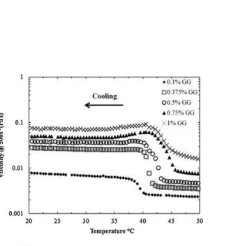
Figure 1 shows the relative viscosity vs temperature of a 0.1%, 0.375%, 0.5% 0.75% and 1% w/w 151
gellan gum fluid gel during manufacture. As the temperature is decreased there is an increase in 152
viscosity that occurs at the onset of gelation of the gellan, a maximal viscosity is then reached 153
which is the temperature beyond which no further particles are formed (Tmax), followed by a plateau
154
in viscosity as the formed particles are smoothed. The results indicate that the viscosity of fluid gel 155
is concentration dependant; onset of gelation increases from ~40 °C for 0.1 % gellan gum to ~45 °C 156
for 1% gellan gum. Furthermore, the final viscosity (at 500 s-1 and 20 °C) of the fluid gels increases 157
with increasing concentration from ~0.01 Pas for 0.1% w/w gellan gum up to ~0.1 Pas for 1% w/w 158
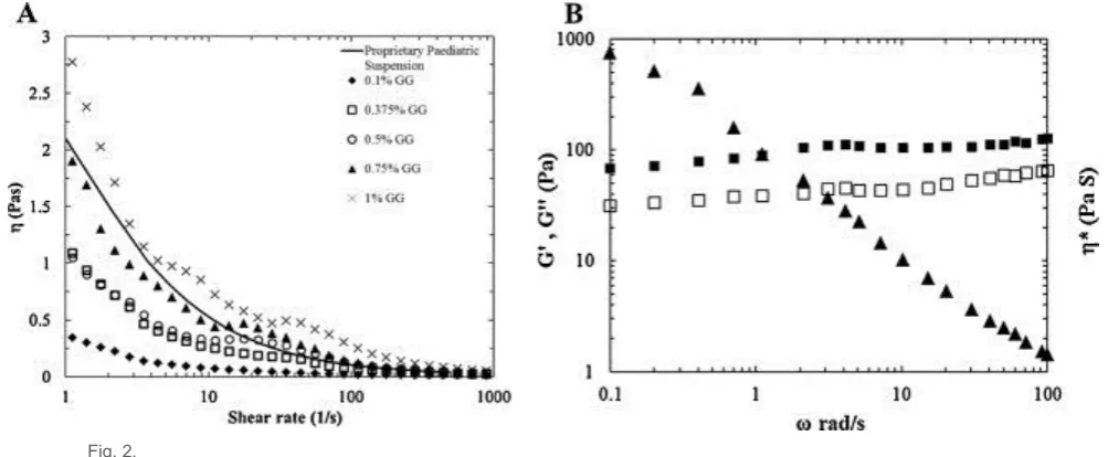
gellan gum. To evaluate the potential of gellan gum fluid gels as a modified oral liquid, samples 159
were tested and compared with a proprietary ibuprofen suspension. The viscosity profiles of gellan 160
gum fluid gel formulations (0.1-1 % w/w) are shown in figure 2A and have a shear thinning 161
viscosity profile. The 0.75% fluid gel sample exhibited a viscosity profile that was most similar to 162
that of a standard ibuprofen paediatric suspension. In addition the yield stress was sufficient to 163
allow inversion of the fluid gel sample without any flow however following mild shaking of the 164
sample it is easily poured on a dispensing spoon as illustrated in figure 3 (Supplementary Video 1) .
165
This formulation was therefore used in further investigations. Dynamic small deformation 166
oscillatory measurements of G‟ and G‟‟ (Fig 2B) highlight the viscoelasticity of the 0.75% w/w 167
fluid gel with G‟ slightly greater than G‟‟ across a range of frequencies; this is typical „weak gel‟ 168
rheological behaviour. Figure 3 shows the effect of cooling rate on the viscosity during formation of 169
cooling rate of 2 °C/min (Fig 4B). The viscosity of the fluid gels during formation increased with 171
increasing cooling rates and viscosity decreased when shear rate was increased. 172
173
3.2 Effect of gellan gum concentration
174
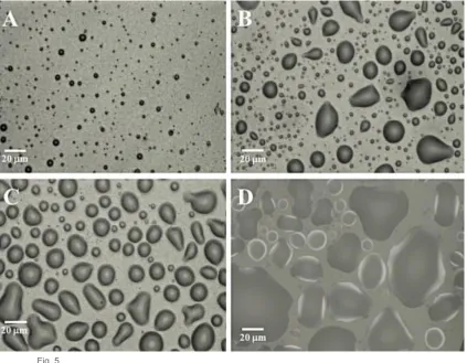
Microscopy images in figure 5 reveal particle sizes of fluid gel are highly dependent on 175
concentration. At 0.1% gellan the particles were in the region of 1-5 m and were generally 176
spherical in shape (Fig. 5A). As the concentrations increased to 0.5% the particles had a larger, 177
binomial size distribution with a population of micron sized particles (similar to 0.1% w/w) and a 178
population and a population in the region of 10-20 m (Fig. 5B). At 0.75% w/w the particles appear 179
less polydisperse than at 0.5% and more spherical with the majority of the population in the region 180
of 20 m (Fig. 5C). When the concentration is increased further to 1% the particles were much 181
larger and irregular in shape (Fig. 5D). 182
183
3.3 Effect of cooling rate and shear rate 184
Figure 6 shows the effect of increasing cooling rates on the particle size of 0.75% w/w at fixed 185
shear rate of 500 s-1 (Fig 6A-C) and the effect of increasing shear rates on the particle size of same 186
concentration of gellan at fixed cooling rate of 2 °C/min (Fig 6D-F). These micrographs indicate 187
that a smaller particle size can be obtained by decreasing cooling rate and increasing the shear rate 188
when forming the fluid gels. 189
190
3.3 Dissolution behaviour
191
To investigate the effects of exposure to low pH for the fluid gels a 5 ml sample of each was 192
dispensed into 0.1M HCl at pH 1.2. A proprietary ibuprofen suspension (C1) was also used for 193
comparison. The proprietary formulation formed a cloudy dispersion in the acid which is attributed 194
to the poor solubility of ibuprofen at low pH. The gellan fluid gel on the other hand formed an acid 195
pieces for over 6 hours. This was supported by dissolution experiments which showed no ibuprofen 197
was released at pH 1.2 (results not shown) and in Figure 7 where ibuprofen crystals can be seen to 198
remain entrapped within the fluid gel particles. 199
Figure 8 illustrates the in vitro release of ibuprofen from different gellan gum fluid gel 200
concentrations ranging from (0.0 % (ibuprofen alone) to 0.75% w/w) determined at pH 1.2 then the 201
release media was changed after 20 minutes to PBS pH 7.4 . The results show that there was a small 202
quantity of ibuprofen released in acidic media for the gels containing lower concentrations of gellan 203
and control formulations (C2 and C3). At 0.75% w/w however, there was no release in acid medium 204
and subsequent release was retarded in PBS for 30 min. 205
To account for the wide variation in stomach pH found in paediatric patients, release characteristics 206
were determined in vitro at different pH values (1.2, 2, 3, 4, 5 and 7.4) then the release medium was 207
changed after 20 minutes to PBS pH 7.4. Figure 9 highlights that the release of ibuprofen from the 208
gellan gum fluid gel was strongly affected by pH of the dissolution media. There was no significant 209
difference (p > 0.05) in release between samples initially immersed in pH 7.4, pH 5 and pH 4. At 210
pH 3 however, subsequent release of ibuprofen was retarded. The retardation of release became 211
progressively more pronounced as the pH was dropped further, to the point where exposure to pH 212
1.2 for just 20 min delayed the onset of drug release for a further 60 min when transferred to pH 213
7.4. The duration of exposure to acidic pH was shown to dramatically affect the lag time to onset of 214
release following transfer to pH 7.4. Figure 10 illustrates the linear relationship between onset of 215
release in pH 7.4 and the preceding exposure time at pH 1.2 and pH 2. The onset of release in pH 216
7.4 was shown to be dramatically affected by the acidity of the initial dissolution medium taking 217
almost 3 hours after exposure to pH 1.2 (for 2 hours) compared with 30 minutes to onset of release 218
following exposure to pH 2 (for 2 hours). This lag time was shown to be dependent on gel stiffness. 219
Figure 11 shows onset of release time rises exponentially with increase in G‟, which in turn is 220
dependent on exposure time to pH 1.2 as highlighted in figure 12. Interestingly, when the fluid gel 221
to increase, albeit at a slower rate, until a plateau was reached (following 90 minutes in pH 7.4) 223
where G‟ is approximately 1200 Pa. When the gel was exposed to pH 1.2 for 60 minutes the 224
stiffness was almost an order of magnitude greater than after 10 minutes exposure. However 225
following transfer to PBS pH 7.4 the stiffness gradually decreased over a period of 180 minutes to 226
the plateau where G‟ is approximately 1200 Pa. The relationship between gel stiffness and release 227
in pH 7.4 is highlighted in figure 13. Following 60 minutes exposure to 0.1 M HCl the gel stiffness 228
was 8000 Pa which gradually decreased on transfer to PBS. No released drug was detected until the 229
stiffness of the gel had reduced to ~2000 Pa (which took 2 hours), following which zero order 230
release 0.15 mg/min was apparent (Fig. 13A). When the sample was exposed to pH 1.2 for 10 231
minutes, the gel stiffness was only ~600 Pa and gradually increased to ~1300 Pa on transfer to PBS 232
pH 7.4. In this system the zero order drug release occurred within 40 minutes and at an increased 233
rate of 0.44 mg/min (Fig. 13B). After this time, the gel disintegrated and was no longer included in 234
this study. These results highlight that increased gel stiffness can reduce the release rate. 235
236
4. Discussion 237
The use of fluid gels as a platform technology for pharmaceutical formulations has great potential 238
due to the tuneable mechanical properties and their ease of manufacture. It has been previously 239
shown that fluid gels can be prepared with many different biopolymers including gelatin (de 240
Carvalho and Djabourov, 1997), agarose (Norton et al., 1998), -carrageenan (Garrec and Norton, 241
2012; Gabriele et al., 2009) and gellan gum (Sworn et al., 1995). Most of these investigations have 242
been focused towards applications in foods to improve stability and improve texture. Here we have 243
investigated the potential of gellan gum fluid gels as a modified release oral drug delivery system. 244
The preparation of fluid gels is a simple process, producing gelled particles that are dispersed in an 245
un-gelled medium. Production using a rheometer allows the cooling rate and the shear rate to be 246
accurately controlled and the characteristic change in viscosity monitored (the process however, is 247
formed containing ibuprofen, the onset of ordering increased with increasing gellan concentration 249
(Fig 1) which can be explained by the consequential increase in concentration of the counterions to 250
the charged group of the polymer promoting aggregation (Morris et al 2012). Interestingly, this 251
onset of ordering occurs at a slightly lower temperature that has been previously reported for gellan 252
gum fluid gels without a drug load (Sworn et al., 1995). This is thought to be due to the competitive 253
inhibition by the negatively charged ibuprofen binding some of the Na+ ions (introduced during pH 254
adjustment with NaOH) reducing the overall ionic strength of the bulk, consequently reducing the 255
viscosity and gelation temperature. Once manufactured, the bulk fluid gels containing ibuprofen 256
showed shear thinning behaviour similar to that of a proprietary paediatric oral ibuprofen 257
suspension with the 0.75% w/w fluid gel having the closest match (Fig 2). However, at very low 258
shear rates the viscosity was sufficient for the preparation to be inverted without any steady state 259
flow as illustrated in figure 3 (Supplementary Video 1). This is due to the weak gel properties of the 260
ibuprofen gellan fluid gel (Fig 2B) which are thought to be a result of particle-particle interactions 261
(Garrec et al., 2013). 262
Oral liquid formulations with relatively high values of zero shear viscosity that rapidly shear thin to 263
enable dispensing would be greatly beneficial by suspending the drug more efficiently during 264
product storage while not impacting on the ease of administration. Furthermore, producing oral 265
liquid formulations with modified release properties would provide an alternative dosage form for 266
paediatric patients in particular. The physical properties of gellan gum fluid gels can be tuned by 267
simply changing the concentration of the polymer or by the rate of cooling and/or shear rate during 268
fluid gel formation (Fig 4). This has previously been demonstrated in food based applications with 269
agarose and carrageenan fluid gels (Norton et al 1998; Gabriele et al 2009). This allows the particle 270
size to be controlled as shown in figures 5 and 6. Gellan gum has previously shown promise as a 271
sustained release oral liquid which gels in situ (Miyazaki et al., 1999). In this study we present a 272
gellan gum oral liquid which was formulated to have a physically cross-linked microstructure prior 273
ungelled gellan effectively immobilising the pre-gelled particles. This system was shown to prevent 275
the dispersion of ibuprofen in the gastric fluid as occurred with a proprietary oral liquid and the 276
drug remained associated with the gellan gum for over 6 hours at pH 1.2. A problem often 277
associated with hydrogel drug delivery systems is drug leaching through the pores of the gel. In this 278
system however, the poor solubility of ibuprofen resulted in precipitation within the gel when 279
exposed to 0.1M HCl pH 1.2, illustrated by the opaque nature of the gel with the precipitated drug 280
particles remaining entrapped within gel particles as illustrated in figure 7. 281
To develop a modified release oral liquid designed particularly for children it is vitally important to 282
take into account paediatric gastrointestinal physiology when designing in vitro biopharmaceutical 283
tests. Variables such as stomach acid volume, gastric pH and small intestinal transit time, which are 284
important for drug release, are well documented (Bowles et al., 2010). For example, in paediatric 285
patients the age at which gastric acid secretion reaches adult values is often quoted as 6 months, 286
however in reality the pH remains variable and the time that intragastric pH is maintained below pH 287
2 increases as a function of age. Nagita et al. (1996) reported that gastric acidity rapidly increased 288
from infancy to 3 years of age and then slowly increased and attained adult levels (< pH 2 for 65% 289
of a 24 h period) by adolescence (age 14). In vitro release data shown in figure 8 reveal that even at 290
concentrations as low as 0.1%, gellan fluid gels have the ability to retard the release of ibuprofen 291
following 20 minutes exposure to 0.1 M HCl pH 1.2 compared with the control formulations (C2 292
and C3). There is however, still some ibuprofen (approximately 5%) released while exposed to pH 293
1.2. Increasing gellan concentration further slows release and at 0.75%, no ibuprofen was measured 294
during acid exposure. Moreover, when the medium was changed to pH 7.4, there was a lag time of a 295
further 30 minutes before onset of release. 296
The effects of varying acidic pH on the subsequent release of ibuprofen from the 0.75% gellan gum 297
fluid gel following transfer to pH 7.4 was also evaluated. It was found that the release of ibuprofen 298
9). There was no significant difference in release between samples that were initially exposed to pH 300
4, pH 5, and pH 7.4. However, as the pH was decreased below the pKa of the carboxyl group of the
301
gellan gum (~3.4), an acid gel was formed, preventing the dissolution of the gel, thus retarding 302
ibuprofen release. This lag time became progressively more pronounced as the pH was dropped 303
further and the acid gel strengthened; exposure to pH 1.2 for just 20 min prevented the onset of 304
release for a further 60 min following transfer to pH 7.4. Moreover, there was a linear relationship 305
between onset of release in pH 7.4 and the preceding exposure time at pH 1.2 for up to 120 min 306
(Fig. 10). A linear relationship was also found following exposure to pH 2 although the effect was 307
substantially less pronounced. This was thought to be due to fewer H+ ions present at pH 2 308
compared with pH 1.2. This will result in formation of a weaker acid gel with an associated increase 309
in hydration and dissolution of the ibuprofen when transferred to pH 7.4. Indeed, the stiffness of the 310
gel had an exponential relationship with onset of release in pH 7.4 media (Fig. 11). Furthermore the 311
stiffness of the gellan was dependent on the duration of exposure to acidic pH which has also 312
recently been reported by Bradbeer et al. (2014). Interestingly, regardless of the duration of acid 313
exposure, the stiffness of gellan fluid gels eventually plateaued at approximately 1200 Pa when 314
transferred to pH 7.4 (Fig. 12). Subsequently, the gel stiffness as a function of exposure time 315
relates to in vitro release. When the gel was exposed to pH 1.2 for 60 min, G‟ was approximately 316
8000 Pa and release was retarded, probably due to the time required for ion exchange to occur 317
between the H+ cross-linked gel and the phosphate buffer. This exchange gradually reduces the gel 318
strength until drug release is enabled. Moreover, the diffusion of the phosphate buffer into the gel 319
also increases the solubility of the ibuprofen by increasing the pH within the gel. This is thought to 320
have facilitated drug diffusion into the surrounding release medium increasing release rate as 321
highlighted, with a much faster release rate of 0.44 mg/min when the exposure time in acid was 322
only 10 min compared with 0.15 mg/min following 60 min exposure (Fig. 13). This dependence on 323
acidic residence time and the strength of acidic pH may be problematic in determining reproducible 324
to overcome this issue would need to be addressed if such a carrier was to be used in clinical 326
practice. However, by understanding the three way relationship between acid exposure time, gel 327
stiffness and onset of release, there is potential for controlling release behaviour by tuning the fluid 328
gels to have a specified stiffness at a particular pH and duration of exposure. Furthermore, 329
producing formulations using this relatively simple method is particularly attractive and by careful 330
design of processing parameters, the microgel particles‟ size, shape, viscoelasticity and behaviour in 331
physiological fluids can be manipulated to suit the application. This could open the door to multiple 332
applications of fluid gel systems in pharmaceutical technology in addition to use as modified release 333
oral liquids. 334
335
5. Conclusion 336
In this study we have demonstrated that fluid gels have the potential to be formulated with a similar 337
viscosity profile to that of a marketed paediatric oral liquid with a yield stress sufficient that the 338
sample can be inverted without any immediate flow but shear thins sufficiently by shaking, to be 339
poured onto a dispensing spoon. Furthermore, we have shown that it is possible to modify the 340
release of ibuprofen from gellan gum fluid gels, providing a simple and effective technology in 341
formulating modified release oral liquids. The release behaviour of ibuprofen from gellan gum fluid 342
gels in a simulated intestinal pH environment was dependent on the stiffness of the gel following 343
exposure to simulated gastric pH media. The stiffness, and hence drug release, could be controlled 344
with exposure time and acidity of the simulated gastric pH environment. This work highlights the 345
potential application of gellan gum fluid gels as modified release oral liquids while at the same 346
time, illustrates the importance of understanding how subtle differences in patient physiology could 347
impact on drug release from such formulations. A realization of this is very important especially 348
Acknowledgements 350
The authors would like to thank the University of Huddersfield for funding the PhD studies of 351
Mohammed Mahdi and Professor Ian Norton and Dr Fotis Spyropoulos of the University of 352
Birmingham for their helpful discussions. 353
354
References 355
Babu, R.J., Sathigari, S., Kumar, M.T., Pandit, J.K., 2010. Formulation of controlled release gellan 356
gum macro beads of amoxicillin. Current Drug Delivery7, 36-43 357
Batchelor, H.K., Kendall, R., Desset-Brethes, S., Alex, R., Ernest, T. B., 2013. Application of in 358
vitro biopharmaceutical methods in development of immediate release oral dosage forms intended 359
for paediatric patients. European Journal of Pharmaceutics and Biopharmaceutics 85, 833-842 360
Bowles, A., Keane, J., Ernest, T., Clapham, D., Tuleu, C., 2010. Specific aspects of gastrointestinal 361
transit in children for drug delivery design. International Journal of Pharmaceutics 395, 37-43 362
Bradbeer, J. F., Hancocks, R., Spyropoulos, F., Norton, I. T., 2014. Self-structuring foods based on 363
acid-sensitive low and high acyl mixed gellan systems to impact on satiety. Food Hydrocolloids 35, 364
522-530 365
Carlfors, J., Edsman, K., Petersson, R., Jörnving, K., 1998. Rheological evaluation of Gelrite® in 366
situ gels for ophthalmic use. European Journal of Pharmaceutical Science 6, 113–119 367
Chandrasekaran, R., Puigjaner, L.C., Joyce, K.L., Arnott, S., 1988. Cation interactions in gellan: an-368
Cuña, M., Vila Jato, J.L., Torres, D., 2000. Controlled-release liquid suspensions based on ion-370
exchange particles entrapped within acrylic microcapsules. International Journal Pharmaceutics 371
199, 151-158 372
Dai, L., Liu, X.X., Liu, Y.L., Tong, Z., 2008. Concentration dependence of critical exponents for 373
gelation in gellan gum aqueous solutions upon cooling. European Polymer Journal 44, 4012-4019. 374
Dalmoro, A., Lamberti, G., Titomanlio, G., Barba, A.A., d‟Amore M., 2010. Enteric micro-375
particles for targeted oral drug delivery. AAPS PharmsciTech 11, 1500-1507 376
Deasy, P.B., Quigley, K.J., 1991. Rheological evaluation of deacetylated gellan gum (Gelrite®) for 377
pharmaceutical use. International Journal of Pharmaceutics 73, 117-123 378
De Carvalho, W., Djabourov, M., 1997. Physical gelation under shear for gelatin gels. Rheologica 379
Acta 36, 591-609 380
Doner, L.W., 1997. Rapid purification of commercial gellan gum to highly soluble and gellable 381
monovalent cation salts. Carbohydrate Polymers 32, 245-247. 382
Gabriele, A., Spyropoulos, F., Norton, I.T., 2009. Kinetic study of fluid gel formation and 383
viscoelastic response with kappa-carrageenan. Food Hydrocolloids23, 2054-2061 384
Garrec, D.A., Guthrie, B., Norton, I.T., 2013. Kappa carrageenan fluid gel material properties. Part 385
1: Rheology. Food Hydrocolloids 33, 151-159 386
Garrec, D.A., Norton, I.T., 2012. Understanding fluid gel formation and properties. Journal Food 387
Engineering 112, 175-182 388
Itoh, K., Tsuruya, R., Shimoyama, T., Watanabe, H., Miyazaki, S., Emanuele, A.D., Attwood, D., 390
2010. In situ gelling xyloglucan/alginate liquid formulation for oral sustained drug delivery to 391
dysphagic patients. Drug Development Industrial Pharmacy 36, 449-455 392
Itoh, K., Yahaba, M., Takahash,i A., Tsuruya, R., Miyazaki, S., Dairaku, M., Togashi, M., Mikami, 393
R., Attwood, D., 2008. In situ gelling xyloglucan/pectin formulations for oral sustained drug 394
delivery. International Journal Pharmaceutics 356, 95-101 395
Jahromi, S.H., Grover, L.M., Paxton, J.Z., Smith, A.M., 2011. Degradation of polysaccharide 396
hydrogels seeded with bone marrow stromal cells. Journal of the Mechanical Behavior of 397
Biomedical Materials 4, 1157-1166 398
Kubo, W., Itoh, K., Miyazaki, S., Attwood, D., 2005. Oral sustained delivery of theophylline and 399
cimetidine from in situ gelling pectin formulations in rabbits. Drug Development and Industrial 400
Pharmacy 31, 819-825 401
Kubo, W., Konno, Y., Miyazaki, S., Attwood, D., 2004. In situ gelling pectin formulations for oral 402
sustained delivery of paracetamol. Drug Development Industrial Pharmacy 30, 593-599 403
Kubo, W., Miyazaki, S., Attwood, D., 2003. Oral sustained delivery of paracetamol from in situ-404
gelling gellan and sodium alginate formulations. International Journal of Pharmaceutics 258, 55-64 405
Miyazaki, S., Aoyama, H., Kawasaki, N., Kubo, W., Attwood, D., 1999. In situ-gelling gellan 406
formulations as vehicles for oral drug delivery. Journal Control Release 60, 287-295 407
Miyazaki, S., Endo, K., Kawasaki, N., Kubo, W., Watanabe, H., Attwood, D., 2003. Oral sustained 408
delivery of paracetamol from in situ gelling xyloglucan formulations. Drug Development Industrial 409
Miyazaki, S., Kubo, W., Itoh, K., Konno, Y., Fujiwara, M., Dairaku, M., Togashi, M., Mikami, R., 411
Attwood, D., 2005. The effect of taste masking agents on in situ gelling pectin formulations for oral 412
sustained delivery of paracetamol and ambroxol. International Journal of Pharmaceutics 297, 38-49 413
Morris, E.R., Nishinari, K., Rinaudo, M., 2012. Gelation of gellan – A review. Food hydrocolloids 414
28, 373-414. 415
Nagita A., Amemoto K., Yoden A., Aoki S., Sakaguchi M., Ashida K., Mino M., 1996. Diurnal 416
variation in intragastric pH in children with and without peptic ulcers. Pediatric Research 40, 528-417
532 418
Norton, I.T., Foster, T., Brown, R., 1998. The science and technology of fluid gels. In: (Williams, 419
P.A. & G.O. Phillips, Eds.), Gums and Stabilisers for the Food Industry 9, 259-269 420
Norton, I.T., Goodall, D.M., Frangou, S.A., Morris, E.R., Rees D.A., 1984. Mechanism and 421
dynamics of conformational ordering in xanthan polysaccharides. Journal of Molecular Biology 422
175, 371–394 423
Norton, I.T., Jarvis, D.A., Foster, T.J., 1999. A molecular model for the formation and properties of 424
fluid gels. International Journal of Biological Macromolecules26, 255-261 425
Ogawa, E., 1996. Conformational transition of polysaccharide sodium-gellan gum in aqueous 426
solutions. Macromolecules, 29, 5178-5182. 427
Oliveira, J.T., Martins, L., Picciochi, R., Malafaya, P.B., Sousa, R.A., Neves, N.M., Mano, J.F., 428
Reis, R.L., 2010. Gellan gum: a new biomaterial for cartilage tissue engineering applications. 429
Journal of Biomedical Materials Research, 93, 852-863 430
Rajinikanth, P.S., Mishra, B., 2008. Floating in situ gelling system for stomach site-specific 431
Rees, D.A., Morris, E.R., Thom, D., Madden, J., 1982. Shapes and interactions of carbohydrate 433
chains. In The Polysaccharides, Vol. 1, 195-290 (G.O. Aspinall, ed.), Academic press New York 434
Smith, A.M., Ingham, A., Grover, L.M., Perrie, Y., 2010. Polymer film formulations for the 435
preparation of enteric pharmaceutical capsules. Journal of Pharmacy and Pharmacology 62, 167-436
172 437
Smith, A.M., Shelton, R.M., Perrie, Y., Harris, J.J., 2007. An initial evaluation of gellan gum as a 438
material for tissue engineering applications. Journal of Biomaterials Applications 22, 241-254. 439
Sworn, G., Sanderson, G.R., Gibson, W., 1995. Gellan gum fluid gels. Food Hydrocolloids 9, 265-440
271 441
442
Figure Captions 443
Figure 1 Viscosity of gellan gum during fluid gel formation (cooling at 2°C /min at a shear 444
rate of 500 s-1) for 0.1% (filled diamonds) 0.375 % (open squares) 0.5% (open circles) 0.75% 445
(filled triangles) and 1% (black crosses) w/v gellan gum loaded with 20 mg/ml ibuprofen. 446
447
Figure 2 A) Viscosity vs. shear rate at 25°C for 0.1% (filled diamonds) 0.375 % (open squares) 448
0.5% (open circles) 0.75% (filled triangles) and 1% (black crosses) w/v gellan gum loaded 449
with 20 mg/ml ibuprofen. Black line indicates a proprietary ibuprofen paediatric suspension. 450
B) Mechanical spectrum (0.5% strain; 37 °C) of a 0.75% Gellan Gum fluid gel loaded with 20 451
mg/ml ibuprofen showing variation of G’ (filled squares), G’’ (open squares) and * (filled 452
Figure 3 Images illustrating the shear thinning behaviour of an ibuprofen loaded fluid gel 454
sample with the ability to invert without any flow. 455
456
Figure 4 Viscosity of 0.75% w/v gellan gum loaded with 20 mg/ml ibuprofen during fluid gel 457
formation using A) different cooling rates; 10 °C/min (open circles), 2 °C/min (filled 458
diamonds), 0.5 °C/min (open triangles) at a shear rate of 500 s-1 and B) different shear rates 459
cooling at 2 °C/min; 1000 s-1 (open diamonds), 500 s-1 (filled diamonds), 100 s-1 (black 460
crosses). 461
462
Figure 5 Light microscopy images of gellan gum fluid gels prepared at different 463
concentrations loaded with 20mg/ml ibuprofen A) 0.1% w/v B) 0.5 % w/v C) 0.75 % w/v D) 1 464
% w/v. 465
466
Figure 6 Light microscopy images of 0.75% w/v gellan gum loaded with 20 mg/ml ibuprofen 467
prepared at a shear rate of 500 s-1 using different cooling rates (A-C) A) 0.5 °C/min B) 468
2°C/min C) 10 °C/min and different shear rates cooling at 2 °C/min (D-F); D)100 s-1 E) 500 s-1 469
F) 1000 s-1 470
471
Figure 7 Light microscopy images of gellan gum fluid showing crystallised ibuprofen 472
entrapped within gel particles 473
Figure 8 Cumulative % release of ibuprofen from fluid gels prepared at different 475
concentrations of gellan gum compared with a standard ibuprofen suspension. Dotted line 476
indicates the point the media was changed from 0.1 M HCl at pH 1.2 to PBS at pH 7.4. Values 477
are represented as mean ± SD (n=3) 478
479
Figure 9 Cumulative % release of ibuprofen from 0.75% w/v gellan gum fluid gel loaded with 480
20 mg/ml ibuprofen exposed to different acidic pH values for a period of 20 minutes. Dotted 481
line indicates the point the media was changed to PBS at pH 7.4. Values are represented as 482
mean ± SD (n=3) 483
484
Figure 10 Relationship between onset of release at pH 7.4 and preceding exposure time in 485
simulated gastric fluid at pH 1.2 (filled diamonds) and pH 2 (open diamonds). 486
487
Figure 11 Exponential relationship between the onset of release in SIF pH 7.4 as a function of 488
gel stiffness (G’). 489
490
Figure 12 Effect of time exposed to pH 1.2 on gel stiffness (G’) and subsequent stiffness on 491
transfer to pH 7.4. The red line (filled diamonds) indicates the stiffness of the gel when 492
exposed to pH 1.2 (0.5% strain; 37 °C at 10rad s-1). The green dashed line (open triangles) 493
represents the stiffness of the gel in PBS at pH 7.4 following 10 min exposure to pH 1.2. The 494
blue dashed line (filled squares) represents the stiffness of the gel in PBS at pH 7.4 following 495
60 min exposure to pH 1.2. 496
Figure 13 Cumulative % release (primary vertical axis) and gel stiffness (G’) (secondary 498
vertical axis) versus time following A) 60 min exposure to pH 1.2 and B) 10 min exposure to 499
pH 1.2. 500
501
502
503
[image:24.595.60.402.170.523.2]504
Fig. 1. 505
Viscosity of gellan gum during fluid gel formation (cooling at 2 °C/min at a shear rate of 500 s−1) for 0.1% (filled diamonds), 506
0.375% (open squares), 0.5% (open circles), 0.75% (filled triangles) and 1% (black crosses) w/v gellan gum loaded with 507
20 mg/ml ibuprofen. 508
510
Fig. 2. 511
(A) Viscosity vs. shear rate at 25 °C for 0.1% (filled diamonds), 0.375% (open squares), 0.5% (open circles), 0.75% (filled 512
triangles) and 1% (black crosses) w/v gellan gum loaded with 20 mg/ml ibuprofen. Black line indicates a proprietary ibuprofen 513
paediatric suspension. (B) Mechanical spectrum (0.5% strain; 37 °C) of a 0.75% gellan gum fluid gel loaded with 20 mg/ml 514
ibuprofen showing variation of G′ (filled squares), G″ (open squares) and η* (filled triangles) with angular frequency. 515
516
[image:25.595.117.582.354.489.2]517
Fig. 3. 518
Images illustrating the shear thinning behaviour of an ibuprofen loaded fluid gel sample with the ability to invert without any 519
flow. 520
521
[image:25.595.310.550.547.739.2]522
Viscosity of 0.75% w/v gellan gum loaded with 20 mg/ml ibuprofen during fluid gel formation using (A) different cooling rates; 524
10 °C/min (open circles), 2 °C/min (filled diamonds), 0.5 °C/min (open triangles) at a shear rate of 500 s−1 and (B) different 525
shear rates cooling at 2 °C/min; 1000 s−1 (open diamonds), 500 s−1 (filled diamonds), 100 s−1 (black crosses). 526
527
[image:26.595.59.483.153.482.2]528
Fig. 5. 529
Light microscopy images of gellan gum fluid gels prepared at different concentrations loaded with 20 mg/ml ibuprofen (A) 530
0.1% w/v, (B) 0.5% w/v, (C) 0.75% w/v and (D) 1% w/v. 531
533
Fig. 6. 534
Light microscopy images of 0.75% w/v gellan gum loaded with 20 mg/ml ibuprofen prepared at a shear rate of 500 s−1 using 535
different cooling rates (A–C). (A) 0.5 °C/min, (B) 2 °C/min, (C) 10 °C/min and different shear rates cooling at 2 °C/min (D–F), 536
(D) 100 s−1, (E) 500 s−1 and (F) 1000 s−1. 537
538
539
Fig. 7. 540
Light microscopy images of gellan gum fluid showing crystallised ibuprofen entrapped within gel particles. 541
[image:27.595.58.340.409.638.2]543
Fig. 8. 544
Cumulative % release of ibuprofen from fluid gels prepared at different concentrations of gellan gum compared with a 545
standard ibuprofen suspension. Dotted line indicates the point the media was changed from 0.1 M HCl at pH 1.2 to PBS at pH 546
7.4. Values are represented as mean ± SD (n = 3). 547
548
549
Fig. 9. 550
Cumulative % release of ibuprofen from 0.75% w/v gellan gum fluid gel loaded with 20 mg/ml ibuprofen exposed to different 551
acidic pH values for a period of 20 min. Dotted line indicates the point the media was changed to PBS at pH 7.4. Values are 552
represented as mean ± SD (n = 3). 553
[image:28.595.56.357.408.652.2]555
Fig. 10. 556
Relationship between onset of release at pH 7.4 and preceding exposure time in simulated gastric fluid at pH 1.2 (filled 557
diamonds) and pH 2 (open diamonds). 558
559
560
Fig. 11. 561
Exponential relationship between the onset of release in SIF pH 7.4 as a function of gel stiffness (G′). 562
[image:29.595.55.341.370.574.2]564
Fig. 12. 565
Effect of time exposed to pH 1.2 on gel stiffness (G′) and subsequent stiffness on transfer to pH 7.4. The red line (filled 566
diamonds) indicates the stiffness of the gel when exposed to pH 1.2 (0.5% strain; 37 °C at 10 rad s−1). The green dashed line 567
(open triangles) represents the stiffness of the gel in PBS at pH 7.4 following 10 min exposure to pH 1.2. The blue dashed line 568
(filled squares) represents the stiffness of the gel in PBS at pH 7.4 following 60 min exposure to pH 1.2. (For interpretation of 569
the references to colour in this figure legend, the reader is referred to the web version of this article.) 570
571
572
Fig. 13. 573
Cumulative % release (primary vertical axis) and gel stiffness (G′) (secondary vertical axis) vs. time following (A) 60 min 574
exposure to pH 1.2 and (B) 10 min exposure to pH 1.2. 575
[image:30.595.59.553.401.585.2]