Proteasome function is required for platelet
production
Dallas S. Shi, … , Dean Y. Li, Andrew S. Weyrich
J Clin Invest.
2014;124(9):3757-3766. https://doi.org/10.1172/JCI75247.
The proteasome inhibiter bortezomib has been successfully used to treat patients with
relapsed multiple myeloma; however, many of these patients become thrombocytopenic,
and it is not clear how the proteasome influences platelet production. Here we determined
that pharmacologic inhibition of proteasome activity blocks proplatelet formation in human
and mouse megakaryocytes. We also found that megakaryocytes isolated from mice
deficient for PSMC1, an essential subunit of the 26S proteasome, fail to produce
proplatelets. Consistent with decreased proplatelet formation, mice lacking PSMC1 in
platelets (
Psmc1
fl/flPf4-Cre
mice) exhibited severe thrombocytopenia and died shortly after
birth. The failure to produce proplatelets in proteasome-inhibited megakaryocytes was due
to upregulation and hyperactivation of the small GTPase, RhoA, rather than NF-
k
B, as has
been previously suggested. Inhibition of RhoA or its downstream target, Rho-associated
protein kinase (ROCK), restored megakaryocyte proplatelet formation in the setting of
proteasome inhibition in vitro. Similarly, fasudil, a ROCK inhibitor used clinically to treat
cerebral vasospasm, restored platelet counts in adult mice that were made
thrombocytopenic by tamoxifen-induced suppression of proteasome activity in
megakaryocytes and platelets (
Psmc1
fl/flPdgf-Cre-ER
mice). These results indicate that
proteasome function is critical for thrombopoiesis, and suggest inhibition of RhoA signaling
as a potential strategy to treat thrombocytopenia in bortezomib-treated multiple myeloma
patients.
Research Article
Hematology
Find the latest version:
http://jci.me/75247/pdf
Introduction
Thrombocytopenia (low platelet count) is observed in numerous diseases and can be life threatening due to bleeding complica-tions. Bortezomib, a reversible chemotherapeutic inhibitor used to treat patients with relapsed multiple myeloma, often induces thrombocytopenia within a few days of therapy initiation (1–3). Bortezomib-induced thrombocytopenia is dose-limiting, and if severe, bortezomib is withheld (2, 3). Although the mechanisms by which bortezomib induces thrombocytopenia is not clear, its primary mode of action is inhibition of the proteasome. The clini-cal observation that platelet counts rise above pretherapy levels upon cessation of bortezomib treatment suggests that bortezomib affects thrombopoiesis (1, 3).
Like other cells, megakaryocytes and anucleate platelets pos-sess proteasome activity (4, 5). While the specific functions of the proteasome in platelet precursors (e.g., megakaryocytes) is rela-tively unknown (6), there is evidence that bortezomib alters the function of platelets (7–11). It has also been hypothesized, but not proven, that bortezomib inhibits megakaryocyte development via nuclear factor κB (NF-κB) (12). The aim of the present work was
to precisely define the roles of the proteasome in thrombopoi-esis and, in doing so, determine whether bortezomib-induced thrombocytopenia can be reversed. Using a combination of phar-macologic and genetic tools, we showed that inhibition of protea-some activity in megakaryocytes blocks proplatelet formation. In addition, conditional deletion of proteasome activity in mouse megakaryocytes led to severe thrombocytopenia and postnatal death. Decreased thrombopoiesis in proteasome-inhibited mice was caused by accumulation and increased activity of RhoA, and inhibitors of the RhoA signaling pathway restored platelet produc-tion. These findings demonstrated that the megakaryocyte pro-teasome controls the final stages of platelet production and also provided a potential option for restoring platelet counts in throm-bocytopenic patients treated with bortezomib.
Results
Pharmacologic inhibition of the proteasome blocks platelet production.
Due to its thrombocytopenic side effects, bortezomib is typically administered as a bolus twice weekly for 2 weeks (days 1, 4, 8, and 11), followed by a 10-day rest period (3). To ascertain the immedi-ate effects of this inhibitor on plimmedi-atelets, mice were administered a clinically relevant dose (2 mg/kg body weight) of bortezomib, and platelet counts and proteasome activity were measured. Con-sistent with its well-known effect on platelet counts in patients, bortezomib induced a mild thrombocytopenia within 24 hours of
The proteasome inhibiter bortezomib has been successfully used to treat patients with relapsed multiple myeloma; however, many of these patients become thrombocytopenic, and it is not clear how the proteasome influences platelet production. Here we determined that pharmacologic inhibition of proteasome activity blocks proplatelet formation in human and mouse megakaryocytes. We also found that megakaryocytes isolated from mice deficient for PSMC1, an essential subunit of the 26S proteasome, fail to produce proplatelets. Consistent with decreased proplatelet formation, mice lacking PSMC1 in platelets (Psmc1fl/flPf4-Cre mice) exhibited severe thrombocytopenia and died shortly after birth. The failure to produce proplatelets
in proteasome-inhibited megakaryocytes was due to upregulation and hyperactivation of the small GTPase, RhoA, rather
than NF-κB, as has been previously suggested. Inhibition of RhoA or its downstream target, Rho-associated protein
kinase (ROCK), restored megakaryocyte proplatelet formation in the setting of proteasome inhibition in vitro. Similarly, fasudil, a ROCK inhibitor used clinically to treat cerebral vasospasm, restored platelet counts in adult mice that were made
thrombocytopenic by tamoxifen-induced suppression of proteasome activity in megakaryocytes and platelets (Psmc1fl/fl
Pdgf-Cre-ER mice). These results indicate that proteasome function is critical for thrombopoiesis, and suggest inhibition of RhoA signaling as a potential strategy to treat thrombocytopenia in bortezomib-treated multiple myeloma patients.
Proteasome function is required for platelet production
Dallas S. Shi,1,2,3 Matthew C.P. Smith,1,3,4 Robert A. Campbell,1 Patrick W. Zimmerman,1 Zechariah B. Franks,1 Bjorn F. Kraemer,1Kellie R. Machlus,5,6 Jing Ling,1 Patrick Kamba,1 Hansjörg Schwertz,1,7 Jesse W. Rowley,1,3 Rodney R. Miles,8 Zhi-Jian Liu,6,9
Martha Sola-Visner,6,9 Joseph E. Italiano Jr.,5,6,10 Hilary Christensen,11 Walter H.A. Kahr,11,12 Dean Y. Li,1,2,3,4,13 and Andrew S. Weyrich1,3
1The Molecular Medicine Program, 2Department of Human Genetics, 3Department of Internal Medicine, and 4Department of Oncological Sciences, University of Utah, Salt Lake City, Utah, USA. 5Division of Hematology, Department of Medicine, Brigham and Women’s Hospital, Boston, Massachusetts, USA. 6Harvard Medical School, Boston, Massachusetts, USA. 7Department of Immunology
and Transfusion Medicine, Ernst-Moritz-Arndt University of Greifswald, Greifswald, Germany. 8Department of Pathology, University of Utah, Salt Lake City, Utah, USA. 9Division of Newborn Medicine
and 10Vascular Biology Program, Department of Surgery, Children’s Hospital, Boston, Massachusetts, USA. 11Division of Haematology/Oncology, Program in Cell Biology, The Hospital for Sick Children,
Toronto, Ontario, Canada. 12Departments of Paediatrics and Biochemistry, University of Toronto, Toronto, Ontario, Canada. 13Key Laboratory for Human Disease Gene Study of Sichuan Province,
Institute of Laboratory Medicine, Sichuan Academy of Medical Sciences and Sichuan Provincial People’s Hospital, Chengdu, Sichuan, People’s Republic of China.
Authorship note: Dallas S. Shi and Matthew C.P. Smith contributed equally to this work. Conflict of interest: The authors have declared that no conflict of interest exists. Submitted: January 17, 2014; Accepted: June 5, 2014.
proplatelet formation in murine megakaryocytes. Bortezomib sig-nificantly decreased proplatelet formation in fetal liver–derived megakaryocytes (Figure 2A). Similar responses were observed in human megakaryocytes, and removal of bortezomib from the incubation media restored proplatelet formation (Figure 2B and data not shown). To confirm that this effect was specific to pro-teasome inhibition, megakaryocytes were treated with MG132 or lactacystin. Both proteasome inhibitors phenocopied the effects of bortezomib (Supplemental Figure 4 and data not shown). The inability to form proplatelets was accompanied by a notable increase in cell spreading on immobilized fibrinogen (Figure 2B), which indicates that the proteasome regulates key cytoskeletal proteins in megakaryocytes.
Phenotypic consequences of proteasome inhibition are independent
of NF-κB and integrin αIIb. Bortezomib’s antitumor activity in
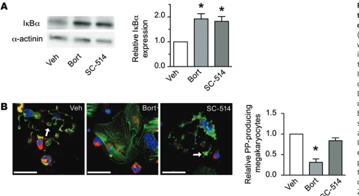
mul-tiple myeloma has been attributed to inhibition of NF-κB in plasma cells (14). Therefore, others have speculated that proteasome inhibi-tors may induce thrombocytopenia via the NF-κB signaling path-way (12). To test this hypothesis, we first treated megakaryocytes with bortezomib and examined the expression of nuclear factor of κ light polypeptide gene enhancer in B cells inhibitor, α (IκBα), which sequesters NF-κB in the cytoplasm until it is phosphorylated, ubiqui-tinated, and degraded (15). As expected, bortezomib increased the expression of IκBα in megakaryocytes (Figure 3A), which demon-strated that inhibition of the proteasome blocks the NF-κB signaling pathway. We also found that inhibition of IκB kinase with SC-514, which induces IκBα phosphorylation, increased IκBα protein lev-els in megakaryocytes (Figure 3A). However, unlike bortezomib, SC-514 did not halt proplatelet production (Figure 3B).
In addition to regulating NF-κB activity, Mitchell and col-leagues previously demonstrated that the proteasome is capable of degrading pro–integrin αIIb (5). Based on this published work, and
our present finding that inhibition of the proteasome decreased treatment (Figure 1A). The thrombocytopenia was transient and
temporally correlated with inhibition of proteasome activity in cir-culating platelets (Figure 1A).
To assess whether bortezomib-induced thrombocytopenia was due to accelerated clearance, we injected Dylight 488 conjugated to GPIBβ into the bloodstream to track the lifespan of platelets in vivo in the presence of bortezomib or its vehicle. As expected, the num-ber of labeled platelets decreased over 96 hours as platelets were cleared from the circulation (13). However, the number of labeled platelets was similar between bortezomib and vehicle treatment groups at every time point tested (Figure 1B). This suggested that bortezomib did not induce platelet activation in the bloodstream, which would facilitate platelet clearance. Consistent with this notion, we found that bortezomib did not directly induce activa-tion of integrin αIIbβ3 (Supplemental Figure 1; supplemental
materi-al available online with this article; doi:10.1172/JCI75247DS1), nor did it alter agonist-induced activation of integrin αIIbβ3 or surface
expression of P-selectin in mouse platelets (Supplemental Figures 1 and 2). Similarly, bortezomib did not influence PAC-1 binding to human platelets in the presence or absence of agonist stimulation (Supplemental Figure 3 and data not shown).
Since acute administration of bortezomib did not shorten the lifespan of circulating platelets, we hypothesized that the bortezo-mib-induced thrombocytopenia was due to a decrease in platelet production. To test this, we depleted platelets with an antibody against GPIBα and then treated mice with bortezomib or its vehi-cle to determine whether bortezomib prevented platelet counts from rebounding. Platelet counts rebounded at a slower pace with bortezomib treatment than with the vehicle control (Figure 1C).
[image:3.585.39.363.56.313.2]The data in Figure 1 suggested that bortezomib-induced thrombocytopenia was due to a defect in the formation of platelets from megakaryocytes. To examine this further, we determined whether inhibition of the proteasome with bortezomib blocked
tory particle that is critical for ubiquitin-mediated protein degrada-tion by the 26S proteasome complex (20–22). It is conserved at the protein level in human and mouse megakaryocytes (Supplemental Figure 8). mRNA for Psmc1 was also expressed in both species, although human megakaryocytes had lower levels of the transcript compared with mouse megakaryocytes (Supplemental Table 1).
Psmc1fl/fl mice were crossed with platelet factor 4 Cre
recombi-nase (Pf4-Cre) mice to disrupt proteasome activity in megakaryo-cytes and platelets. Psmc1fl/fl Pf4-Cre mice had significantly reduced
protein for PSMC1 in megakaryocytes, but not other tissues (Supple-mental Figure 9). Ubiquinated proteins also accumulated in mega-karyocytes from Psmc1fl/fl Pf4-Cre mice (Supplemental Figure 10).
Despite a marked reduction in PSMC1 protein, the number of megakaryocytes from Psmc1fl/fl Pf4-Cre mice in bone marrow or
spleen was not reduced compared with Psmc1fl/wt mice
(Supplemen-tal Figure 11). Unlike their littermate controls, however, Psmc1fl/fl
Pf4-Cre mice had severe thrombocytopenia at postnatal day 1 (P1),
and the majority of Psmc1fl/fl Pf4-Cre mice died before weaning
(Fig-ure 5, A and B). The reduction in platelet counts was more severe than in c-Mpl knockout pups at the same age (Supplemental Figure 12). In addition to reduced numbers of platelets, Psmc1fl/fl Pf4-Cre
mice had lower hematocrits than Psmc1fl/wt mice, and bleeding was
seen in the abdomen and limbs (Figure 5, C and D). Pathological signs of hemorrhage were also present in the bladder and testes of all animals and occasionally observed in the brain, lymph nodes, and intestines (Figure 5E and data not shown).
Ultrastructure examination of megakaryocytes from Psmc1fl/fl
Pf4-Cre mice revealed less cytoplasm compared with
megakaryo-cytes from Psmc1fl/wt mice (Figure 6A). In addition, Psmc1fl/fl
Pf4-Cre megakaryocytes lacked demarcation membranes, which were
readily visible in Psmc1fl/wt megakaryocytes (Figure 6A). Similar
to mouse megakaryocytes treated with bortezomib (Figure 2A), megakaryocytes from Psmc1fl/fl Pf4-Cre mice failed to produce
pro-platelets (Figure 6B).
Inhibition of RhoA-dependent signaling prevents thrombocytopenia induced by genetic disruption of proteasome activity. As predicted from
the pharmacological data, megakaryocytes from Psmc1fl/fl Pf4-Cre
mice expressed higher levels of total RhoA protein and RhoA-GTP the formation of proplatelets when megakaryocytes adhere to
fibrinogen, we sought to determine whether bortezomib regulated the activity of integrin αIIbβ3 in megakaryocytes. Bortezomib did
not alter the expression of mature integrin αIIb protein, nor did it
increase binding of soluble fibrinogen or PAC-1 to human mega-karyocytes (Supplemental Figure 5 and data not shown). Bortezo-mib also had no effect on adherence of human megakaryocytes to fibrinogen (Supplemental Figure 6). Together, these data indicate that bortezomib does not directly block proplatelet formation through NF-κB– or integrin αIIbβ3–dependent mechanisms.
Phenotypic consequences of proteasome inhibition require RhoA.
The changes in actin polymerization observed in megakaryocytes treated with proteasome inhibitors were reminiscent of cytoskel-etal changes in endothelial cells that rely on the small GTPase RhoA (16). Indeed, we found that bortezomib increased total RhoA protein expression (Figure 4A). Bortezomib also increased RhoA-GTP activity and phosphorylation of myosin light chain (MLC) kinase, which is downstream of RhoA (Figure 4, A and B).
RhoA-dependent signaling has been linked to the production of proplatelets (17, 18). Therefore, we treated human megakaryo-cytes with Y27632, a selective inhibitor of the Rho-associated protein kinase p160ROCK, or with C3 transferase, a direct RhoA inhibitor. Y27632 and C3 transferase rescued proplatelet forma-tion in bortezomib-treated cells (Figure 4C and Supplemental Fig-ure 7). This response was likely due to inhibition of downstream RhoA effectors, because Y27632 decreased phosphorylation of MLC kinase in the presence of bortezomib (Figure 4B). In agree-ment with the rescue of proplatelet formation observed in bort-ezomib-treated human megakaryocytes, mouse megakaryocytes treated with bortezomib plus Y27632 or with bortezomib plus fasudil, a more clinically relevant p160ROCK inhibitor, formed proplatelets (Figure 4D). These results in mouse megakaryocytes were similar to a recent report by Murai et al. (19).
Genetic deletion of the proteasome results in severe thrombocyto-penia and death. To further dissect the role of the proteasome in
thrombopoiesis, we focused on protease (prosome, macropain) 26S subunit, ATPase 1 (Psmc1; gene ID 19179) in mouse megakary-ocytes and platelets. Psmc1 is an essential subunit of the 19S
[image:4.585.43.304.54.255.2]Our findings provide definitive proof that the megakaryocyte proteasome is required for the final stages of platelet production. Evidence for this is 2-fold: first, pharmacologic inhibition of pro-teasome activity in late-stage human or mouse megakaryocytes significantly blunted proplatelet formation; and second, platelet production was significantly reduced in Psmc1fl/fl Pf4-Cre mice, in
which genetic deletion of Psmc1 does not occur until megakaryo-cytes express platelet factor 4, which activates the Cre recombi-nase (23). In addition to regulating thrombopoiesis, others have shown that the proteasome is important for the proliferation of megakaryocyte precursors (24) and the degradation of cyclin B and pro–integrin αIIb in megakaryocytes (5, 25).
Like their parent megakaryocytes, anucleate platelets also possess proteasome activity (4, 10), and several groups have demonstrated that pharmacologic inhibition of the proteasome regulates platelet function (8–11, 26). Under the conditions of our experiments, bortezomib did not affect indices of platelet activa-tion in mouse or human platelets that included activaactiva-tion of integ-rin αIIbβ3 and translocation of P-selectin to the surface of platelets.
However, similar to Gupta and coworkers (8), we observed that bortezomib reduced the aggregation of human platelets when low concentrations of thrombin were used as the agonist (Supplemen-tal Figure 14). Although more work is needed, results generated by multiple independent groups strongly indicate that protein degra-dation systems regulate platelet function (6).
Other groups have shown that pharmacologic inhibition regu-lates the function of platelets ex vivo (8–11, 26), but bortezomib did not accelerate the clearance of labeled platelets under the conditions of our present studies. Our results contrasted those of Nayak and colleagues (7), who showed that pharmacologic inhibi-tion of the proteasome reduced the half-life of platelets in mice. One potential explanation for these discordant findings is that the bolus dose of bortezomib used in our studies only produced a mild thrombocytopenia and did not completely abolish platelet protea-some activity (Figure 1A). Although Nayak’s group did not measure cellular proteasome activity (7), it is possible that they achieved more efficient pharmacologic inhibition of the proteasome in plate-lets and other vascular cells. Different routes of drug
administra-Figure 3. Proteasome-dependent forma-tion of proplatelets in human megakaryo-cytes occurs independently of NF-κB. (A) Human megakaryocytes were treated with vehicle, bortezomib, or the NF-κB inhibitor SC-514. Shown are a represen-tative Western blot for IκBα as well as IκBα expression levels, as measured by densitometry, relative to vehicle control. Data are mean ± SEM (n = 3). (B) Morphol-ogy of megakaryocytes treated with vehicle, bortezomib, or SC-514. Megakaryocytes were stained with WGA (red), phalloidin (green), and DAPI (blue). Arrows denote proplatelets. Images are representative of 3 independent experiments. Also shown is the number of proplatelet-producing megakaryocytes rela-tive to vehicle control. Data are mean ± SEM of 3 independent experiments. Scale bars: 25 μm. *P < 0.05 vs. vehicle.
(Figure 7A). Fasudil also rescued proplatelet formation in bone mar-row–derived megakaryocytes from Psmc1fl/fl Pf4-Cre mice (Figure 7B).
Next we generated inducible conditional knockouts by cross-ing Psmc1fl/fl mice with platelet-derived growth factor–Cre estrogen
receptor (Pdgf-Cre-ER) mice, which allowed for time-restricted dele-tion of Psmc1 in megakaryocytes and platelets after administradele-tion of the competitive estrogen receptor ligand tamoxifen. Although Pdgf is expressed by other cells besides megakaryocytes, Pdgf-Cre-ER mice were used because Pf4-Cre-ER mouse lines are not currently avail-able. Like Psmc1fl/fl Pf4-Cre mice (Figure 5, A and B), administration
of tamoxifen to Psmc1fl/fl Pdgf-Cre-ER mice at P1 resulted in
throm-bocytopenia and early postnatal mortality (Supplemental Figure 13, A and B). When tamoxifen was administered to adult Psmc1fl/fl
Pdgf-Cre-ER mice, platelet counts were reduced by approximately 50%
after 6 days compared with Psmc1fl/wt mice (Figure 8A). In the
pres-ence of fasudil, however, tamoxifen did not significantly decrease platelet counts in Psmc1fl/fl Pdgf-Cre-ER mice (Figure 8A). Consistent
with these rescue experiments, staining of megakaryocytes in crude bone marrow showed that the in vivo fasudil treatment rescued pro-platelet formation (Figure 8B). These results are consistent with our in vitro findings that fasudil maintained proplatelet formation in bortezomib-treated megakaryocytes (Figure 4D).
Discussion
In this study, we found that pharmacologic or genetic disruption of proteasome activity in megakaryocytes inhibits proplatelet forma-tion. Pharmacologic inhibition was reversible in megakaryocytes treated in vitro with bortezomib, and thrombocytopenia was tran-sient when bortezomib was administered as a bolus in vivo. When inhibition of proteasome activity was sustained, as was the case with genetic deletion of Psmc1 in megakaryocytes and platelets, mega-karyocytes did not form proplatelets, and Psmc1fl/fl Pf4-Cre mice had
[image:5.585.36.401.55.255.2]development or blood/lymphatic vessel separation, which could lead to excessive bleeding (28). In this regard, several groups have shown that platelet C-type lectin-like receptor 2 (CLEC-2) receptors regulate lymphatic vascular development, and, like Psmc1fl/fl Pf4-Cre mice (29–
33), platelet-specific knockout of CLEC-2 results in postnatal lethality (32). It should also be noted that a very low threshold of platelet func-tion sufficiently maintains vascular funcfunc-tion (27, 33, 34), raising the possibility that Psmc1fl/fl Pf4-Cre mice produce dysfunctional platelets
that are incapable of maintaining vascular integrity. Indeed, recent studies have demonstrated that immune-type receptors in platelets are critical for the prevention of inflammation-induced hemorrhage (35). Thus, it is entirely possible that in addition to being reduced in number, platelets from Psmc1fl/fl Pf4-Cre mice express an abnormal
repertoire of proteins resulting in platelet dysfunction.
Studies in megakaryocytes revealed that genetic or pharma-cologic interruption of proteasome activity led to accumulation of IκBα and RhoA. Although both proteins were upregulated, we found that the final stages of proplatelet formation required RhoA signaling rather than inhibition of NF-κB, as previously suggested (12). The inability to sprout proplatelets resembled studies in neu-tion and types/concentraneu-tions of proteasome inhibitors between
the studies may also explain the divergent results. Further studies are needed to resolve the in vivo pharmacology of proteasome inhi-bition and its relation to thrombocytopenia. However, our present studies clearly showed that platelet counts rebounded at a slower pace in mice subjected to platelet depletion in the presence of bort-ezomib. These data, in combination with the severe thrombocyto-penia we observed in Psmc1fl/fl Pf4-Cre mice, demonstrated that the
proteasome directly modulates platelet production.
We found that thrombocytopenia was more severe in Psmc1fl/fl
Pf4-Cre mice compared with c-Mpl knockout mice (Supplemental
Figure 12), which have normal life expectancies (27). Consistent with a marked reduction in platelet counts, Psmc1fl/fl Pf4-Cre mice had low
hematocrits and hemorrhaging in the abdominal region. Occasional hemorrhaging was also observed in the brain, lymph nodes, and intestines. This suggests that severe thrombocytopenia is the primary driver of postnatal death in Psmc1fl/fl Pf4-Cre mice. Hemorrhaging
in Psmc1fl/fl Pf4-Cre mice may occur because platelet numbers are
[image:6.585.43.439.58.473.2]simply too low to prevent bleeding. Alternatively, insufficient plate-let counts in Psmc1fl/fl Pf4-Cre mice may result in abnormal vascular
rons, in which acute inhibition of the proteasome blocks activity-dependent growth of new dendritic spines. It is not known what proteins are degraded by the proteasome in order to stimulate new spine growth; however, inactivation of RhoA leads to neurite outgrowth (36, 37). This suggests that, similar to megakaryocyte proplatelet formation, the proteasome may control neuronal out-growth by degrading RhoA. Moreover, RhoA signaling has been shown to maintain normal megakaryocyte development, which is critical for platelet production (18).
Malfunction of the proteasome in human diseases may lead to aberrant platelet production or abnormal platelet generation. Disruption of proteasome activity could occur at multiple check-points, since human megakaryocytes expressed the full reper-toire of proteasome components at the mRNA level (Supplemen-tal Table 1). Identifying the complete portfolio of target proteins
[image:7.585.36.371.52.529.2]degraded by the proteasome in megakaryocytes will shed addi-tional light on the mechanisms that control thrombopoiesis and the phenotype of platelets as they enter the circulation. Under-standing the functions of the proteasome in platelets, which is active and capable of degrading proteins (7–9), also requires fur-ther investigation. From an immediate perspective, our present findings demonstrated that bortezomib directly inhibits protea-some activity in megakaryocytes and thereby decreases platelet production. Our findings also established fasudil as a potential treatment for preventing and/or reversing bortezomib-induced thrombocytopenia in multiple myeloma patients. Additionally, inhibitors of the RhoA signaling pathway may have efficacy in the treatment of other thrombocytopenic disorders caused by abnormal platelet production, especially if the disease is driven by proteasome-dependent mechanisms.
Figure 5. Genetic ablation of proteasome activ-ity in megakaryocytes causes severe throm-bocytopenia and postnatal death. (A) Platelet counts at P1 in Psmc1fl/fl Pf4-Cre and Psmc1fl/wt
mice, expressed relative to Psmc1fl/fl mice. Bars
show mean ± SEM of 6 independent experi-ments. *P < 0.05 vs. Psmc1fl/fl. (B) Mortality rates
in Psmc1fl/fl, Psmc1fl/wt, and Psmc1fl/fl Pf4-Cre
mice at P1 and P21. Shown are ratios of expected versus observed genotypes, determined by χ2 analysis, at P1 and P21
(n = 88). *P < 0.05 vs. P1, determined by χ2 distri-bution table. (C) Hematocrits in Psmc1fl/fl Pf4-Cre
relative to Psmc1fl/wt mice at P1. Data are mean
± SEM of 6 independent experiments. *P < 0.05 vs. Psmc1fl/wt. (D) Left: Images of Psmc1fl/wt and
Psmc1fl/fl Pf4-Cre mice at P1. Evidence of bleeding
was observed in the abdominal region (arrow). Middle and right: Limbs of Psmc1fl/wt and
Psmc1fl/fl Pf4-Cre mice. Hemorrhaging was
observed in the limb of the Psmc1fl/fl Pf4-Cre
mouse (arrow). (E) Whereas P1 histological sec-tions of Psmc1fl/wt mice demonstrated normal
histology, bleeding was observed in the bladder and testis of a Psmc1fl/fl Pf4-Cre mouse. Boxed
Methods
Differentiation of human and mouse megakaryocytes
Cord blood from normal full-term deliveries was obtained, and CD34+
hematopoietic progenitors were isolated and differentiated into mega-karyocytes as previously described (38, 39). Mature megamega-karyocytes were placed on immobilized human fibrinogen–coated surfaces in the presence of specific inhibitors or their vehicle, and the number of megakaryocytes that possessed proplatelets was counted by an inde-pendent blinded observer. On average, 12% ± 3% of vehicle-treated megakaryocytes had proplatelet extensions.
Mouse megakaryocytes were isolated from fetal liver as previ-ously described (40). Mouse bone marrow–derived megakaryocytes were obtained using modifications of a published report (41). For the bone marrow megakaryocytes, C57BL/6 mice (8–10 weeks of age) were euthanized, and cells were obtained from the bone marrow of femur and tibia by flushing the bone marrow. Cells were homog-enized by pipetting followed by passage through a 100-μm filter. The cell population was resuspended in 10% fetal bovine serum–supple-mented DMEM with 2 mM l-glutamine, penicillin/streptomycin, and fibroblast condition media containing thrombopoietin. The cells were cultured for 5 days (37°C and 5% CO2), and mature megakaryocytes were layered over a bovine serum albumin (BSA) gradient as described previously (42). Fetal liver and bone marrow–derived megakaryo-cytes were subsequently resuspended in culture media as described above, then placed on immobilized BSA or fibrinogen in the presence or absence of inhibitors, and megakaryocytes with proplatelets were counted. On average, 34% ± 1% of vehicle-treated fetal liver–derived megakaryocytes produced proplatelets. Proplatelet formation in vehi-cle-treated bone marrow–derived megakaryocytes was 50% ± 1%.
Inhibitors used in these in vitro studies (all diluted in DMSO) included bortezomib (100 nM; Selleck Chem), Y27632 (10 μM; Sig-ma-Aldrich), fasudil (10 μM; Selleck Chem), C3 transferase (10 μM; Cytoskeleton Inc.), SC-514 (0.5 μM; Calbiochem), MG132 (10 μM; Sig-ma-Aldrich), and lactacystin (10 μM; Sigma-Aldrich). Inhibitors were administered at different times, as indicated in the figure legends.
Next-generation RNA-Seq
Fetal liver–derived megakaryocytes for RNA-Seq were provided by J. Thon (Harvard Medical School, Boston, Massachusetts, USA). RNA from human CD34-differentiated or fetal liver–derived proplate-let-producing megakaryocytes was isolated and prepped for deep sequencing as previously described (43–45). In brief, RNA was pre-pared for sequencing according to Illumina’s (DNA vision) TruSeq kit V2 for poly-A RNA. Libraries were sequenced 36 (human) and 50 (mouse) base pairs on an Illumina sequencer. Reads were aligned using Novoalign (Novocraft Technologies) software followed by pro-cessing, including RPKM assignment, using the USeq analysis pack-age (46). The processed RNA-Seq data and aligned reads were depos-ited in GEO (accession no. GSE58202; ref. 47).
Protein expression analyses and assessment of RhoA activity
Cell lysates were placed in laemmli buffer, proteins were separated by SDS-page, and IκBα and phospho-MLC (Cell Signaling) were analyzed by Western blotting. To measure RhoA activity, platelets were placed in Mg2+ lysis buffer supplemented with protease (Roche Applied Science)
and phosphatase inhibitors (Sigma-Aldrich). A small portion of the lysate was retained as total cell lysate, and the rest was incubated with the assay reagent. GTP-bound forms were eluted from the assay reagent using Laemmli sample buffer. Total RhoA and RhoA-GTP bound pro-tein were analyzed by Western blotting using a pulldown kit (Millipore).
Mouse in vivo studies
In vivo measurements of platelets. In Figure 1A, platelet counts were
determined at various time points with a Hemavet 950 (Drew Sci-entific). For platelet clearance, C57BL/6 mice were injected intra-venously (i.v.) on day 0 with anti-GPIBβ Dylight 488 (0.1 μg/g body weight; Emfret Analytics) and intraperitoneally (i.p.) with bortezo-mib (2 mg/kg body weight) or its vehicle (10% DMSO in 0.9% saline). Blood samples (30 μl) were taken daily, diluted in Hanks balanced salt solution, and stained with a phycoerythrin-conjugated anti-mouse CD41 antibody (BD Biosciences), and clearance of Dylight 488–posi-tive platelets was measured as previously described (48).
[image:8.585.45.287.57.353.2]For estimation of platelet production, C57BL/6 mice were injected i.v. on day 0 with anti-GPIBα antibodies (3 μg/g body weight; Emfret Analytics) to deplete circulating platelets. On day 1,
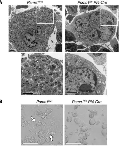
Figure 6. Platelet territories and proplatelets fail to form in PSMC1-deficient megakaryocytes. (A) Whereas Psmc1fl/wt mouse
megakaryo-cytes showed a large cytoplasmic region compared with the nucleus, those from a Psmc1fl/fl Pf4-Cre mouse had less cytoplasm compared
with the multilobed nucleus. Boxed regions are shown at higher magnification below, in which the demarcation membrane exhibited in the Psmc1fl/wt megakaryocytes (arrowheads) was not observed in the
Psmc1fl/fl Pf4-Cre megakaryocyte. (B) Transmission images of
mega-karyocytes derived from Psmc1fl/wt and Psmc1fl/fl Pf4-Cre mice at P1.
Proplatelet formation (arrows) was absent in Psmc1fl/fl Pf4-Cre mice.
Pdgf-Cre-ER pups on P1, and then mortality was monitored from
P2 to P21. For studies in adult Psmc1fl/fl Pdgf-Cre-ER mice,
tamoxi-fen was administered 8 weeks after birth. In these mice, fasudil (5 mg/kg) was injected i.p. to Psmc1fl/fl Pdgf-Cre-ER mice 4 and 48
hours after tamoxifen administration, and blood was retrieved from tail veins on day 6 after tamoxifen to determine circulating plate-let counts. In a subset of mice (n = 2 per treatment group), ex vivo assessment of proplatelet formation was performed. For these stud-ies, mice were euthanized via CO2 asphyxiation followed by cervi-cal dislocation. Femurs were isolated, and bone marrow cords were flushed with HEPES-tyrodes buffer with 100 U/ml penicillin/strep-tomycin. Crude bone marrow cords were sliced into multiple sec-tions, and buffer was replaced with HEPES-tyrodes buffer with 5% mouse serum and 100 U/ml penicillin/streptomycin. Bone marrow sections were then incubated at 37°C for 2 hours and stained with platelet counts were assessed to confirm depletion; shortly after,
mice were treated i.p. with bortezomib or its vehicle as above. Blood samples (2 μl) were taken daily for the remainder of the experiment, and platelet counts were measured by flow cytometry as previously described (13).
Knockout of the proteasome in mouse megakaryocytes and platelets.
Megakaryocyte and platelet ablation of proteasome activity was achieved by crossing Psmc1fl/fl mice (provided by J. Mayer, Baylor
College of Medicine, Houston, Texas, USA) with Pf4-Cre or
Pdgf-Cre-ER mice (Jackson Labs), generating Psmc1fl/wt and Psmc1fl/fl
Pf4-Cre mice or Psmc1fl/wt and Psmc1fl/fl Pdgf-Cre-ER mice. Psmc1fl/fl mice
[image:9.585.108.473.54.237.2]were also used as controls in select studies. Knockdown was con-firmed in megakaryocytes using an antibody against PSMC1 (Novus Biologicals), and platelet counts were assessed as recently described (49). Tamoxifen (0.25 mg/kg) was administered i.p. to Psmc1fl/fl
Figure 7. Genetic deletion of Psmc1 in megakaryocytes is associated with increased RhoA protein and activity. (A) Representative Western blot of total RhoA and RhoA-GTP in megakaryocytes derived from Psmc1fl/wt and Psmc1fl/fl Pf4-Cre mice at P1. Also shown is densitometry quantification
relative to Psmc1fl/wt control; for RhoA-GTP, megakaryocytes were isolated from 10 P1 mice, lysed, and then the lysates were pooled together for
each pulldown experiment (see Methods). Data are mean ± SEM of 3 (total RhoA) and 2 (RhoA-GTP) experiments. *P < 0.05 vs. Psmc1fl/wt.
(B) Bone marrow–derived megakaryocytes from Psmc1fl/wt and Psmc1fl/fl Pf4-Cre mice were treated with vehicle or fasudil, and the number of
proplatelet-producing megakaryocytes was quantified and expressed relative to Psmc1fl/wt controls. Data are mean ± SEM of 3 independent
experi-ments. *P < 0.05 vs. vehicle-treated Psmc1fl/wt.
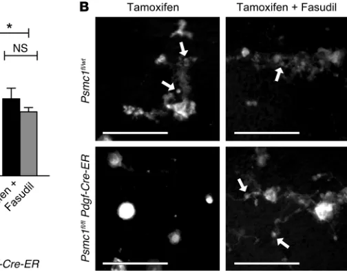
Figure 8. Inhibition of RhoA signaling rescues platelet counts in adult mice in which proteasome activity is conditionally deleted. (A) Tamoxifen was administered to adult Psmc1fl/wt and Psmc1fl/fl
Pdgf-Cre-ER mice, followed by treatment with fasudil or saline control (4 and 48 hours after tamoxifen). Shown are platelet counts at day 6 after tamoxifen admin-istration relative to Psmc1fl/wt controls treated with
tamoxifen alone. Data are mean ± SEM of 9 experi-ments performed on independent mice. *P < 0.05 as indicated. No significant difference was observed between groups treated with tamoxifen plus fasudil. (B) Representative images of Dylight 488–positive megakaryocytes present in crude bone marrow iso-lated from Psmc1fl/wt and Psmc1fl/fl Pdgf-Cre-ER mice
[image:9.585.289.541.542.739.2]All data graphed relative to controls (Figures 2–5, 7, and 8 and Sup-plemental Figures 4, 7, 10, and 13) were generated by comparing the average of the data set in each treated group with that of the control group (as the denominator).
Study approval
The human studies were approved by the University of Utah’s Institu-tional Review Board (IRB no. 392). All participating subjects provided informed consent. Cord blood from normal full-term deliveries was obtained after informed consent by the mothers (IRB no. 11919). The mouse studies were approved by the University of Utah’s Institutional Animal Care and Review Board (IACUC nos. 12-10002 and 12-11017) or by the Children’s Hospital in Boston (IACUC no. A3431-01).
Further information can be found in the supplemental material and citations therein (51, 52).
Acknowledgments
We thank Diana Lim for preparation of the figures, critical comments, and consultation regarding responsible and effective display of the images. We also thank Guy Zimmerman for helpful discussions regarding the roles of the proteasome in platelets and megakaryo-cytes. This work was funded by NIH grants HL066277 and HL112311 (to A.S. Weyrich), HL112311, HL084516, and AR064788 (to D.Y. Li), GM103806 (to J.W. Rowley), and HL68130 (to J.E. Italiano Jr.); by the American Heart Association (13POST13930019 to K.R. Machlus; 11POST7290019 to R.A. Campbell; 0625098Y and 09BG1A 2250381 to H. Schwertz); and by the Canadian Institutes of Health Research (MOP-259952 to W.H.A. Kahr). In addition, H. Schwertz was funded by a Lichtenberg-Professorship from the Volkswagen Foundation.
Address correspondence to: Dean Li or Andrew S. Weyrich, Eccles Institute of Human Genetics, 15 North 2030 East, Bldg. 533, Rm. 4150, Salt Lake City, Utah 84112, USA. Phone: 801.585.0950; E-mail: [email protected] (D. Li), andy.weyrich@u2m2. utah.edu (A.S. Weyrich).
anti-GPIBβ Dylight 488. Megakaryocytes were imaged on a Nikon Eclipse TS100 fluorescence microscope and quantified by counting 10 fields (×20) per well.
Measurement of blood hematocrit
Hematocrit was measured in Psmc1fl/wt and Psmc1fl/fl Pf4-Cre mice at P3.
After anesthesia (ketamine-xylazine; 0.2 mg/g body weight), blood was acquired via the retro-orbital venous plexus. Blood was collected into heparinized capillary tubes and spun at 50,000 g for 10 minutes to obtain a hematocrit for each mouse.
Histopathology
Organs from P1 mice were collected and fixed in neutral buffered for-malin. Tissues were embedded in paraffin, sectioned at 10 μm, and stained with hematoxylin and eosin (H&E). Slides were then assessed by a hematopathologist.
Electron microscopy
Femurs from mice were collected, and bone marrow was flushed into glutaraldehyde 2.5% in PBS. Fixed samples were kept at 4°C, shipped to the Hospital for Sick Children in Toronto, and further processed and imaged as previously described (50).
Statistics
For multiple-group comparisons, data were subjected to 1-way analysis of variance (ANOVA), and Tukey’s post-hoc test was used to assess statistical significance among groups. 2-way ANOVA with Newman-Keuls post-hoc test was used to assess statistical signifi-cance for the data in Figure 8B. 2-tailed Student’s t test was used when comparisons were made between 2 groups. Differences in mortality were assessed by χ2 test, and observed outcomes were
graphed relative to calculated expected outcomes (Figure 5B and Supplemental Figure 13B). When possible, quantifications were done by a blinded observer. P values less than 0.05 were considered statistically significant.
1. Adams J, et al. Proteasome inhibitors: a novel class of potent and effective antitumor agents. Cancer Res. 1999;59(11):2615–2622.
2. Richardson PG, et al. A phase 2 study of bortezo-mib in relapsed, refractory myeloma. N Engl J Med. 2003;348(26):2609–2617.
3. Field-Smith A, Morgan GJ, Davies FE. Bort-ezomib (Velcadetrade mark) in the treatment of multiple myeloma. Ther Clin Risk Manag. 2006;2(3):271–279.
4. Ostrowska H, Ostrowska JK, Worowski K, Radziwon P. Human platelet 20S proteasome: inhibition of its chymotrypsin-like activity iden-tification of the proteasome activator PA28. A preliminary report. Platelets. 2003;14(3):151–157. 5. Mitchell WB, Li J, French DL, Coller BS. αIIbβ3
biogenesis is controlled by engagement of αIIb in the calnexin cycle via the N15-linked glycan. Blood. 2006;107(7):2713–2719.
6. Kraemer BF, Weyrich AS, Lindemann S. Protein degradation systems in platelets. Thromb Hae-most. 2013;110(5):920–924.
7. Nayak MK, Kulkarni PP, Dash D. Regulatory role of proteasome in determination of platelet life
span. J Biol Chem. 2013;288(10):6826–6834. 8. Gupta N, Li W, Willard B, Silverstein RL, McIntyre
TM. Proteasome proteolysis supports stimulated platelet function and thrombosis. Arterioscler Thromb Vasc Biol. 2014;34(1):160–168. 9. Kumari S, Dash D. Regulation of β-catenin
stabilization in human platelets. Biochimie. 2013;95(6):1252–1257.
10. Nayak MK, Kumar K, Dash D. Regulation of proteasome activity in activated human platelets. Cell Calcium. 2011;49(4):226–232.
11. Avcu F, Ural AU, Cetin T, Nevruz O. Effects of bortezomib on platelet aggregation and ATP release in human platelets, in vitro. Thromb Res. 2008;121(4):567–571.
12. Lonial S, et al. Risk factors and kinetics of throm-bocytopenia associated with bortezomib for relapsed, refractory multiple myeloma. Blood. 2005;106(12):3777–3784.
13. Mason KD, et al. Programmed anuclear cell death delimits platelet life span. Cell. 2007;128(6):1173–1186.
14. McConkey DJ. Bortezomib paradigm shift in myeloma. Blood. 2009;114(5):931–932.
15. Spinelli SL, et al. Platelets and megakaryocytes contain functional nuclear factor-kappaB. Arte-rioscler Thromb Vasc Biol. 2010;30(3):591–598. 16. Whitehead KJ, et al. The cerebral cavernous
malformation signaling pathway promotes vascular integrity via Rho GTPases. Nat Med. 2009;15(2):177–184.
17. Gobbi G, et al. Proplatelet generation in the mouse requires PKCepsilon-dependent RhoA inhibition. Blood. 2013;122(7):1305–1311. 18. Suzuki A, et al. RhoA is essential for maintaining
normal megakaryocyte ploidy and platelet gen-eration. PLoS One. 2013;8(7):e69315. 19. Murai K, et al. Bortezomib induces
throm-bocytopenia by the inhibition of proplatelet formation of megakaryocytes [published online ahead of print April 21, 2014]. Eur J Haematol. doi:10.1111/ejh.12342.
20. Rubin DM, Glickman MH, Larsen CN, Dhru-vakumar S, Finley D. Active site mutants in the six regulatory particle ATPases reveal multiple roles for ATP in the proteasome. EMBO J. 1998;17(17):4909–4919.
Gold-berg AL, Finley D. The axial channel of the prote-asome core particle is gated by the Rpt2 ATPase controls both substrate entry product release. Mol Cell. 2001;7(6):1143–1152.
22. Smith DM, Fraga H, Reis C, Kafri G, Goldberg AL. ATP binds to proteasomal ATPases in pairs with distinct functional effects, implying an ordered reaction cycle. Cell. 2011;144(4):526–538. 23. Tiedt R, Schomber T, Hao-Shen H, Skoda RC.
Pf4-Cre transgenic mice allow the generation of lineage-restricted gene knockouts for studying megakaryocyte platelet function in vivo. Blood. 2007;109(4):1503–1506.
24. Galimberti S, et al. PS-341 (Bortezomib) inhibits proliferation induces apoptosis of megakaryo-blastic MO7-e cells. Leuk Res. 2008;32(1):103–112. 25. Zhang Y, Wang Z, Liu DX, Pagano M, Ravid K.
Ubiquitin-dependent degradation of cyclin B is accelerated in polyploid megakaryocytes. J Biol Chem. 1998;273(3):1387–1392.
26. Necchi V, et al. Ubiquitin/proteasome-rich particulate cytoplasmic structures (PaCSs) in the platelets and megakaryocytes of ANKRD26-related thrombo-cytopenia. Thromb Haemost. 2012;109(2):263–271.
27. Murone M, Carpenter DA, de Sauvage FJ. Hema-topoietic deficiencies in c-mpl and TPO knock-out mice. Stem Cells. 1998;16(1):1–6.
28. Bertozzi CC, Hess PR, Kahn ML. Platelets: covert regulators of lymphatic development. Arterioscler Thromb Vasc Biol. 2010;30(12):2368–2371. 29. Benezech C, et al. CLEC-2 is required for
devel-opment maintenance of lymph nodes. Blood. 2014;123(20):3200–3207.
30. Herzog BH, et al. Podoplanin maintains high endo-thelial venule integrity by interacting with platelet CLEC-2. Nature. 2013;502(7469):105–109.
31. Osada M, et al. Platelet activation receptor CLEC-2 regulates blood/lymphatic vessel sepa-ration by inhibiting prolifesepa-ration, migsepa-ration, tube formation of lymphatic endothelial cells. J Biol Chem. 2012;287(26):22241–22252.
32. Finney BA, et al. CLEC-2 and Syk in the mega-karyocytic/platelet lineage are essential for development. Blood. 2012;119(7):1747–1756. 33. Bertozzi CC, et al. Platelets regulate lymphatic
vascular development through CLEC-2-SLP-76 signaling. Blood. 2010;116(4):661–670. 34. Levin J, et al. Pathophysiology of
thrombocyto-penia and anemia in mice lacking transcription factor NF-E2. Blood. 1999;94(9):3037–3047. 35. Boulaftali Y, et al. Platelet ITAM signaling is
criti-cal for vascular integrity in inflammation. J Clin Invest. 2013;123(2):908–916.
36. Hamilton AM, et al. Activity-dependent growth of new dendritic spines is regulated by the pro-teasome. Neuron. 2012;74(6):1023–1030. 37. Jeon CY, et al. Control of neurite outgrowth by RhoA
inactivation. J Neurochem. 2012;120(5):684–698. 38. Foulks JM, et al. PAF-acetylhydrolase expressed
during megakaryocyte differentiation inactivates PAF-like lipids. Blood. 2009;113(26):6699–6706. 39. Denis MM, et al. Escaping the nuclear confines:
signal-dependent pre-mRNA splicing in anucle-ate planucle-atelets. Cell. 2005;122(3):379–391. 40. Thon JN, Italiano JE. Visualization and
manipula-tion of the platelet and megakaryocyte cytoskel-eton. Methods Mol Biol. 2012;788:109–125. 41. Mazharian A, Watson SP, Severin S. Critical
role for ERK1/2 in bone marrow fetal liver-derived primary megakaryocyte differentiation, motility, proplatelet formation. Exp Hematol. 2009;37(10):1238–1249.
42. Thon JN, et al. Cytoskeletal mechanics of
pro-platelet maturation and pro-platelet release. J Cell Biol. 2010;191(4):861–874.
43. Cecchetti L, Tolley ND, Michetti N, Bury L, Weyrich AS, Gresele P. Megakaryocytes differentially sort mRNAs for matrix metalloproteinases and their inhibitors into platelets: a mechanism for regulating synthetic events. Blood. 2011;118(7):1903–1911. 44. Kahr WH, et al. Mutations in NBEAL2, encoding
a BEACH protein, cause gray platelet syndrome. Nat Genet. 2011;43(8):738–740.
45. Rowley JW, et al. Genome-wide RNA-seq analysis of human and mouse platelet transcriptomes. Blood. 2011;118(14):e101–e111.
46. Nix DA, Courdy SJ, Boucher KM. Empirical methods for controlling false positives and estimating confidence in ChIP-Seq peaks. BMC Bioinformatics. 2008;9.
47. Edgar R, Domrachev M, Lash AE. Gene Expres-sion Omnibus: NCBI gene expresExpres-sion and hybridization array data repository. Nucleic Acids Res. 2002;30(1):207–210.
48. Josefsson EC, White MJ, Dowling MR, Kile BT. Platelet life span and apoptosis. Methods Mol Biol. 2012;788:59–71.
49. Liu ZJ, et al. Expansion of the neonatal platelet mass is achieved via an extension of platelet life span. Blood. 2014;123(22):3381–3389. 50. Kahr WH, et al. Abnormal megakaryocyte
devel-opment and platelet function in Nbeal2(–/–) mice. Blood. 2013;122(19):3349–3358. 51. Holly SP, et al. Chemoproteomic discovery of
AADACL1 as a regulator of human platelet activa-tion. Chem Biol. 2013;20(9):1125–1134. 52. Ramskold D, Wang ET, Burge CB, Sandberg R.