Evaluation of the
BRCA
1/2 mutation as a prognostic
marker in primary peritoneal serous cancer
Naoto Furukawa1*, Sumire Ohno1, Takahiko Kasai2, Sachiko Morioka1, Fuminori Ito1, Yasuhito Tanase1, Shoji Haruta1, Seiji Kanayama1, Ryuji Kawaguchi1, Shozo Yoshida1, Hiroshi Kobayashi1
1Department of Obstetrics and Gynecology, Nara Medical University, Kashihara, Japan 2Diagnostic Pathology, Nara Medical University, Kashihara, Japan
Email: *furunao0813@gmail.com
Received 2 October 2013; revised 30 October 2013; accepted 6 November 2013
Copyright © 2013 Naoto Furukawa et al. This is an open access article distributed under the Creative Commons Attribution License,
which permits unrestricted use, distribution, and reproduction in any medium, provided the original work is properly cited.
ABSTRACT
Introduction: The present study was a retrospective investigation of the relation between immunohisto-
chemical BRCA1/2 status and prognosis in patients
with primary peritoneal serous cancer (PPSC). Mate- rials and Methods: We retrospectively evaluated 14 consecutive patients diagnosed with PPSC other than hereditary breast and ovarian cancer between 2005 and 2010. All patients had serum CA125 levels >40 U/mL prior to starting first-line chemotherapy with paclitaxel and carboplatin. Paclitaxel was adminis- tered as a 3-hour intravenous infusion at a dose of
175 mg/m2 on day 1, and carboplatin was delivered at
an area under the curve of 5 based on the Calvert method. Patients received six cycles of first-line che- motherapy, except patients whose disease was deter- mined to be progressive during the chemotherapy
regimen. BRCA1/2 and p53 protein expression was
determined by immunohistochemistry of patient tis- sue samples. The Cox proportional hazards model was used to evaluate univariate and independent mul- tivariate associations with the effect of clinical pa- rameters, such as age at diagnosis; tumor histology; tumor grade; and rate of change in CA125, and
BRCA1/2, p53 status on overall survival. Probability
values of less than 0.05 were considered to indicate statistical significance. Results: Two cases (14%) had
the BRCA1 mutation, and none had the BRCA2 mu-
tation. Eleven cases (79%) were positive for p53. In
the univariate analysis, factors significantly associ- ated with overall survival were (pre-chemotherapy CA125-pre-2nd chemotherapy CA125)/pre-chemo-
therapy CA125 (p = 0.0034) and (pre-chemotherapy
CA125-pre-3rd chemotherapy CA125)/pre-chemo-
therapy CA125 (p = 0.0245). BRCA1 and p53 status
were not predictors of overall survival. Multivariate analysis performed with overall survival as an end- point revealed that none of the factors examined was significant. Median survival rate of patients without a
BRCA1 mutation was 23.5 months (2 - 82 months),
and all died. By contrast, one patient with a BRCA1
mutation remains alive at 85 months, and the other
patient died at 64 months. Conclusion: BRCA1 might
be a predictor of overall survival in patients with PPSC receiving chemotherapy.
Keywords:BRCA1; Primary Peritoneal Serous Cancer;
Prognosis
1. INTRODUCTION
Ovarian cancer is increasing annually in Japan [1], and the total number of ovarian cancer cases in 2006 was 7913 [2]. Standard therapy for ovarian cancer comprises primary surgical cytoreduction followed by platinum- based chemotherapy, but 25% to 90% of cases of ovarian cancer are diagnosed during progression [3]. Paclitaxel and carboplatin as first line chemotherapy are adminis- tered to patients with ovarian cancer, and overall re- sponse rates of 59% [4] and 68% [5] are reported in stud- ies in which paclitaxel (175 - 185 mg/m2) and car-
boplatin (area under the curve of 5 - 6) are administered every 3 weeks. Despite a favorable initial response rate to treatment, early recurrences and platinum resistance are frequently encountered. Several reports have dis- cussed the diverse pathogenesis of ovarian cancers. Low- grade serous carcinomas are associated with mutations in KRAS/BRAF and evolve slowly [6]. High-grade serous carcinomas are, in contrast, associated with mutations in
p53, BRCA1, or BRCA2, and evolve rapidly [6,7]. BRCA1 and BRCA2 act as tumor suppressors involved in repairing double-strand DNA breaks by homologous recombination. BRCA1/2 germline mutations occur in 11% to 15.3% and BRCA1/2 somatic mutations occur in 19% of women with unselected ovarian cancers [8]. Pa-tients with BRCA1 and BRCA2 mutations have increased sensitivity to platinum chemotherapy and a better prog- nosis [9]. Primary peritoneal serous carcinoma (PPSC) is histologically and clinically similar to stage III-IV ovar- ian high-grade serous carcinoma, and is rare. The fre- quency of BRCA1/2 germline mutations in PPSC is re- ported to be 15.8% [10]. Another group reported that germline-BRCA1 mutations are found in 26% of PPSC patients [11]. Few reports, however, describe the fre-quency of somatic mutations of BRCA1/2 in patients with PPSC, or the relation between BRCA1/2 status with chemosensitivity and prognosis in patients with PPSC. BRCA immunohistochemistry identifies BRCA mutations with a sensitivity of 80% and a specificity of 93%, sug- gesting that BRCA immunohistochemistry is a promising screening method for BRCA mutation detection [12].
In the present study, we retrospectively investigated the relation between immunohistochemical BRCA1/2 status and prognosis in patients with PPSC.
2. MATERIALS AND METHODS
2.1. Patient Selection
The present study was conducted in accordance with the principles of the Declaration of Helsinki. We retrospec- tively evaluated 14 consecutive patients diagnosed with PPSC (all stage IIIC) other than hereditary breast and ovarian cancer between 2005 and 2010. Genetic testing was not available during this period, however, so heredi- tary breast and ovarian cancer were diagnosed by asking patients detailed questions regarding family history. All patients underwent a laparotomy or laparoscopy to obtain a small tissue sample for examination under a micro- scope, but none of them underwent debulking surgery. Diagnostic criteria include normal sized ovaries; extra- ovarian site involvement greater than surface involve- ment of the ovary; an ovarian component of less than 5 × 5 mm within the ovary and otherwise confined to the surface of the ovary; and histologic characteristics pre- dominantly of the serous type [13]. All patients had se- rum CA125 levels >40 U/mL prior to starting first-line chemotherapy with paclitaxel and carboplatin. Paclitaxel was administered as a 3-hour intravenous infusion at a dose of 175 mg/m2 on day 1, and carboplatin was deliv-
ered at an area under the curve of 5 based on the Calvert method. Patients received 6 cycles of first-line chemo- therapy, except patients whose disease was determined to be progressing during the chemotherapy regimen. The
retrieved clinical data included patient age at diagnosis, tumor histology, tumor grade, CA125, and overall sur- vival from the medical records. Rates of change in CA125 were calculated as follows: (pre-chemotherapy CA125—pre-2nd chemotherapy CA125)/pre-chemother- apy CA125; and (pre-chemotherapy CA125—pre-3rd chemotherapy CA125)/pre-chemotherapy CA125. Pre- chemotherapy CA125 represents the CA125 level before administering the first dose of chemotherapy, Pre-2nd
chemotherapy CA125 represents the CA125 level before administering the second dose of chemotherapy; and Pre- 3rd chemotherapy CA125 represents the CA125 level before administering the third dose of chemotherapy.
2.2. Immunohistochemistry
BRCA1/2 and p53 protein expression was determined by immunohistochemistry of patient tissue samples. Immu- nohistochemistry staining was performed according to standard techniques. Staining was carried out with a mouse monoclonal IgG antibody, anti-BRCA1 (OP92- 100UGCN, Merck Chemicals Ltd, Nottingham, UK), and anti-BRCA2 (MAB2476, R & D Systems, Minne- sota, US). Two independent pathologists blinded to the clinical data and each other’s opinion scored the expres- sion based on the staining intensity. Scoring was as fol- lows: 0, 0%; 1, 1 to <10%; 2, 10 to <50%; 3, 50 to <90%; 4, if >90% of the cells were positive. The tumor was considered BRCA1/2 mutation-positive when less than 10% of the tumor cells had a positive reaction. The tu- mor was considered p53-positive when 50% or more of the tumor cells had a positive reaction.
2.3. Statistical Analysis
All statistical analyses were conducted using SPSS soft- ware Version 17 (SPSS Inc., Chicago, IL, USA).The Cox proportional hazards model was used to evaluate uni- variate and independent multivariate associations with the effect of clinical parameters, and BRCA1/2and p53 status on overall survival. Probability values of less than 0.05 were considered to indicate statistical significance.
3. RESULTS
Table 1. Patient characteristics.
Median age (range) 58 (37 - 79)
Grade 2 6
Histology (serous)
Grade 3 8
[image:3.595.309.538.112.219.2]Median pre-treatment CA125 (range) U/ml 906 (344 - 13998)
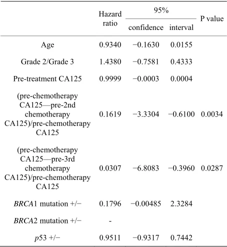
Table 2. Univariate analysis of risk factors associated with overall survival.
95% Hazard
ratio confidence interval P value
Age 0.9340 −0.1630 0.0155
Grade 2/Grade 3 1.4380 −0.7581 0.4333
Pre-treatment CA125 0.9999 −0.0003 0.0004
(pre-chemotherapy CA125—pre-2nd
chemotherapy CA125)/pre-chemotherapy
CA125
0.1619 −3.3304 −0.6100 0.0034
(pre-chemotherapy CA125—pre-3rd
chemotherapy CA125)/pre-chemotherapy
CA125
0.0307 −6.8083 −0.3960 0.0287
BRCA1 mutation +/− 0.1796 −0.00485 2.3284
BRCA2 mutation +/−
-p53 +/− 0.9511 −0.9317 0.7442
Multivariate analysis performed with overall survival as an endpoint revealed that neither was a significant factor
(Table 3). Median survival rate was 23.5 months (2 - 82
months) in patients with no BRCA1 mutation, and all had died. By contrast, one patient with a BRCA1 mutation remains alive (85 months), and the other patient died at 64 months. Median survival rate was 23 months (2 - 85 months) in patients that were p53-positive, and 27 months (22 - 36 months) in patients that were p53-nega- tive.
4. DISCUSSION
PPSC is reported to arise from the extra-ovarian perito- neum [14]. A recent study, however, demonstrated an association with tubal intraepithelial carcinoma in 47% of patients with PPSC [15], suggesting the potential role of the distal fallopian tube as an organ of serous car- cinogenesis. Symptoms include abdominal distension and pain with ascites caused by peritonitis carcinomatosa. Most patients do not consult a gynecologist until the dis- ease is in an advanced stage, and patients with PPSC have a worse prognosis than those with ovarian cancer.
Table 3. Multivariate analysis of risk factors associated with overall survival.
95% Hazard
ratio confidence interval
(pre-chemotherapy CA125—pre-2nd chemotherapy CA125)/pre-chemotherapy CA125
0.5597 −3.8685 3.5259
(pre-chemotherapy CA125—pre-3rd chemotherapy CA125)/pre-chemotherapy CA125
0.0913 −10.4273 4.8152
Median survival is 12 to 18 months [16]. PPSC is an ovarian cancer subtype, and therefore the chemotherapy regimen is the same as that for ovarian cancer. Patients with ovarian cancer with a BRCA mutation are consid- ered to be platinum-sensitive, and thus have better prog- nosis than those with no BRCA mutation. Overall sur- vival is 52.7 months in patients with low/intermediate BRCA1 mRNA expression, which is significantly better than the overall survival of 18.2 months in patients with high BRCA expression [9]. In the present study, median survival was 22 months in patients with positive BRCA1 expression and 64 months in the 2 patients with negative BRCA1 expression. Based on the present study, BRCA1 may be also a predictor of the prognosis of PPSC, al- though the sample size was small. By contrast, no BRCA2 mutation was detected in the present study. Some reports documented an overall survival advantage con- ferred by BRCA2 mutations compared with either having no BRCA mutation or having a BRCA1 mutation in ovar- ian cancer [17,18]. In these reports, the frequency of the BRCA2 mutation was half that of the BRCA1 mutation. In the present study, it is therefore not surprising that no cases of BRCA2 mutation were detected because the BRCA1 mutation was detected in only two cases. No relation between p53 status and overall survival was de- tected in the present study. Over half the PPSC patients were p53-positive, and BRCA1 mutation carriers had a higher overall incidence of p53 mutations than those with wild-type BRCA1 [11]. The p53signature, which is defined as a morphologically normal, strongly p53-im- munopositive segment of tubal secretory (non-ciliated) cells spanning at least 12 consecutive nuclei [19], has been found in patients with BRCA germline mutations, with an incidence ranging from 11% to 71% of cases, and also in normal controls (19% - 50%) [20-22]. To our knowledge, however, no reports have described a relation between p53 status and overall survival in patients with PPSC.
[image:3.595.58.285.207.454.2]therapy with advanced ovarian cancer [23,24], suggest- ing that the CA125 regression rate may be predictor of overall survival in patients with PPSC without surgery. In the present study, however, the CA125 regression rate was detected as a factor in univariate analysis but not in multivariate analysis. Possible explanations for CA125 regression rate not being detected as a predictor of over- all survival in the present study might be the more ag- gressive behaviour of PPSC compared with ovarian can- cer and the small sample size. Limitations of the present study include the small sample size, the fact that it is a retrospective study, hereditary breast and ovarian cancer might be included because genetic testing was not avail- able.
5. CONCLUSION
In conclusion, the BRCA1 mutation was detected in 14% in patients with PPSC and these patients had a better prognosis than patients not having the BRCA1 mutation. BRCA1 might be a predictor of overall survival in pa- tients with PPSC receiving chemotherapy. There is a need for new biomarkers, however, because most pa- tients with PPSC have a poor prognosis and have no BRCA1 mutation.
REFERENCES
[1] Ushijima, K. (2009) Current status of gynecologic cancer in Japan. Journal of Gynecologic Oncology, 20, 67-71. http://dx.doi.org/10.3802/jgo.2009.20.2.67
[2] Matsuda, T., Marugame, T., Kamo, K., et al. (2012)
Cancer incidence and incidence rates in Japan in 2006: based on data from 15 population-based cancer registries in the monitoring of cancer incidence in Japan (MCIJ) project. Japanese Journal of Clinical Oncology, 42, 139- 147.http://dx.doi.org/10.1093/jjco/hyr184
[3] Metser, U., Jones, C., Jacks, L.M., Bernardini M.Q. and Ferguson, S. (2011) Identification and quantification of peritoneal metastases in patients with ovarian cancer with multidetector computed tomography: Correlation with surgery and surgical outcome. International Journal of Gynecological Cancer, 21, 1391-1398.
http://dx.doi.org/10.1097/IGC.0b013e31822925c0 [4] Vasey, P.A., Jayson, G.C., Gordon, A., et al. (2004)
Phase III randomized trial of docetaxel-carboplatin versus paclitaxel-carboplatin as first-line chemotherapy for ovarian carcinoma. Journal of the National Cancer Insti-tute, 96, 1682-1691.
http://dx.doi.org/10.1093/jnci/djh323
[5] du Bois, A., Luck, H.J., Meier, W., et al. (2003) A ran-
domized clinical trial of cisplatin/paclitaxel versus car- boplatin/paclitaxel as first-line treatment of ovarian can- cer. Journal of the National Cancer Institute, 95, 1320- 1329.
http://dx.doi.org/10.1093/jnci/djg036
[6] Shih Ie, M. and Kurman, R.J. (2004) Ovarian tumori-
genesis: A proposed model based on morphological and molecular genetic analysis. American Journal of Pathol- ogy, 164, 1511-1518.
http://dx.doi.org/10.1016/S0002-9440(10)63708-X [7] Koul, A., Malander, S., Loman, N., et al. (2000) BRCA1
and BRCA2 mutations in ovarian cancer: Covariation
with specific cytogenetic features. International Journal of Gynecological Cancer, 10, 289-295.
http://dx.doi.org/10.1046/j.1525-1438.2000.010004289.x [8] Hennessy, B.T., Timms, K.M., Carey, M.S., et al. (2010)
Somatic mutations in BRCA1 and BRCA2 could expand
the number of patients that benefit from poly (ADP ribose) polymerase inhibitors in ovarian cancer. Journal of Clini- cal Oncology, 28, 3570-3576.
http://dx.doi.org/10.1200/JCO.2009.27.2997
[9] Quinn, J.E., James, C.R., Stewart, G.E., et al. (2007) BRCA1 mRNA expression levels predict for overall sur-
vival in ovarian cancer after chemotherapy. Clinical Cancer Research, 13, 7413-7420.
http://dx.doi.org/10.1158/1078-0432.CCR-07-1083 [10] Alsop, K., Fereday, S., Meldrum, C., et al. (2012) BRCA
mutation frequency and patterns of treatment response in
BRCA mutation-positive women with ovarian cancer: A
report from the Australian Ovarian Cancer Study Group.
Journal of Clinical Oncology, 30, 2654-2663. http://dx.doi.org/10.1200/JCO.2011.39.8545
[11] Schorge, J.O., Muto, M.G., Lee, S.J., et al. (2000) BRCA1-related papillary serous carcinoma of the perito-
neum has a unique molecular pathogenesis. Cancer Re- search, 60, 1361-1364.
[12] Skytte, A.B., Waldstrom, M., Rasmussen, A.A., Cruger, D., Woodward, E.R., Kolvraa, S. (2011) Identification of
BRCA1-deficient ovarian cancers. Acta Obstetricia et Gynecologica Scandinavica, 90, 593-599.
http://dx.doi.org/10.1111/j.1600-0412.2011.01121.x [13] Chu, C.S., Menzin, A.W., Leonard, D.G., Rubin, S.C. and
Wheeler, J.E. (1999) Primary peritoneal carcinoma: A re- view of the literature. Obstetrical & Gynecological Sur- vey, 54, 323-335.
http://dx.doi.org/10.1097/00006254-199905000-00023 [14] Eltabbakh, G.H., Piver, M.S., Natarajan, N. and Mettlin,
C.J. (1998) Epidemiologic differences between women with extraovarian primary peritoneal carcinoma and women with epithelial ovarian cancer. Obstetrical & Gy- necological, 91, 254-259.
http://dx.doi.org/10.1016/S0029-7844(97)00650-9 [15] Folkins, A.K., Jarboe, E.A., Roh, M.H. and Crum, C.P.
(2009) Precursors to pelvic serous carcinoma and their clinical implications. Gynecologic Oncology, 113, 391- 396.http://dx.doi.org/10.1016/j.ygyno.2009.01.013 [16] Ramos, J., Gunturu, K.S., Krishnamoorthy, S., Kenney, B.
and Saif, M.W. (2011) Primary peritoneal carcinoma in complete remission: A case report. Anticancer Research, 31, 4397-4400.
[17] Pal, T., Permuth-Wey, J., Kapoor, R., Cantor, A. and Sutphen R. (2007) Improved survival in BRCA2 carriers with ovarian cancer. Familial Cancer, 6, 113-119. http://dx.doi.org/10.1007/s10689-006-9112-x
proved survival for BRCA2-associated serous ovarian
cancer compared with both BRCA-negative and BRCA1- associated serous ovarian cancer. Cancer, 118, 3703- 3709.http://dx.doi.org/10.1002/cncr.26655
[19] Lee, Y., Miron, A., Drapkin, R., et al. (2007) A candidate
precursor to serous carcinoma that originates in the distal fallopian tube. The Journal of Pathology, 211, 26-35. http://dx.doi.org/10.1002/path.2091
[20] Shaw, P.A., Rouzbahman, M., Pizer, E.S., Pintilie, M. and Begley, H. (2009) Candidate serous cancer precur- sors in fallopian tube epithelium of BRCA1/2 mutation
carriers. Modern Pathology, 22, 1133-1138.
http://dx.doi.org/10.1038/modpathol.2009.89
[21] Folkins, A.K., Jarboe, E.A., Saleemuddin, A., et al. (2008)
A candidate precursor to pelvic serous cancer (p53 sig- nature) and its prevalence in ovaries and fallopian tubes from women with BRCA mutations. Gynecologic On- cology, 109, 168-173.
http://dx.doi.org/10.1016/j.ygyno.2008.01.012
[22] Mehra, K.K., Chang, M.C., Folkins, A.K., et al. (2011)
The impact of tissue block sampling on the detection of p53 signatures in fallopian tubes from women with BRCA
1 or 2 mutations (BRCA+) and controls. Modern Pathol-ogy, 24, 152-156.
http://dx.doi.org/10.1038/modpathol.2010.171
[23] Vasudev, N.S., Trigonis, I., Cairns, D.A., et al. (2011)
The prognostic and predictive value of CA-125 regres- sion during neoadjuvant chemotherapy for advanced ovarian or primary peritoneal carcinoma. Archives of Gynecology and Obstetrics, 284, 221-227.
http://dx.doi.org/10.1007/s00404-010-1655-2
[24] Furukawa, N., Sasaki, Y., Shigemitsu, A., et al. (2013)
CA-125 cut-off value as a predictor for complete interval debulking surgery after neoadjuvant chemotherapy in pa- tients with advanced ovarian cancer. Journal of Gyneco- logic Oncology, 24, 141-145.