http://dx.doi.org/10.4236/fns.2016.75036
How to cite this paper: Watanabe, K., Horinishi, N., Matsumoto, K. and Sogabe, Y. (2016) Identification and Enumeration Method of Both Eukaryotic and Prokaryotic Microorganisms in Food Sample. Food and Nutrition Sciences, 7, 345-354. http://dx.doi.org/10.4236/fns.2016.75036
Identification and Enumeration Method of
Both Eukaryotic and Prokaryotic
Microorganisms in Food Sample
Katsuji Watanabe
1*, Naoto Horinishi
1, Kunimasa Matsumoto
1, Yuji Sogabe
21Department of Life, Environment and Materials Science, Graduate School of Engineering, Fukuoka Institute of Technology, Fukuoka, Japan
2Global Application Development Center, Shimadzu Co., Kyoto, Japan
Received 23 January 2016; accepted 25 April 2016; published 28 April 2016
Copyright © 2016 by authors and Scientific Research Publishing Inc.
This work is licensed under the Creative Commons Attribution International License (CC BY). http://creativecommons.org/licenses/by/4.0/
Abstract
The method to analyze both eukaryotic and prokaryotic microorganisms without preliminary crobial information of sample seemed to be useful not only for research and investigation of mi-croorganisms but also for industry using mimi-croorganisms. In the present manuscript, preparation of a new DNA primers, new reference database for 18S rDNA for our newly developed method [1]- [3], and analyses of eukaryotic and prokaryotic microorganisms in fermentation products were presented. In komekouji, Aspergillus spp., was enumerated to be 46.5 × 10⁶ MPN g−1, and
Penicil-lium spp., was enumerated to be 1.5 × 10⁶ MPN g−1. In dry yeast, Saccharomyces group, were enu-merated to be 8600 × 106 MPN g−1. In komekouji-miso, no eukaryotic microorganism was detected, while the other Bacillus spp., was numerically dominant (21.5 × 10⁶ MPN g−1) as prokaryotic mi-croorganisms, followed by B. subtilis group (4.65 × 10⁶ MPN g−1), and the other Firmicutes (3.7 × 10⁶ MPN g−1). The komekouji-miso included lower number of Actinobacteria (0.15 × 10⁶ MPN g−1),
Burkhokderia sp. (1.5 × 10⁶ MPN g−1), and the other α,β,γ-proteobacteria (0.12 × 10⁶ MPN g−1). In sake-kasu, both prokaryote and eukaryote were not detected by the method. Present results indi-cated that using both universal primers for eukaryotic and prokaryotic microorganisms, each groups of prokaryotic and eukaryotic microorganisms were enumerated without any preliminary information nor setting up standard curve, which were required for real time PCR.
Keywords
Eukaryotic Microorganisms, Prokaryotic Microorganisms, Multiple Enzyme Restriction Fragment Length Polymorphism Analysis, the Most Probable Number Method, Microchip Electrophoresis
1. Introduction
By PCR-based analysis methods such as real time quantitative PCR (qPCR) or multiplex PCR, eukaryotic and
prokaryotic microorganisms [4]-[7] could be detected or quantified by the same procedure by changing used
DNA primers. By qPCR using universal primer for the both microorganisms, total numbers of eukaryote and prokaryote in sample could be enumerated, however we could not know what kinds of microorganisms were
mainly included without analyzing amplified DNA [4]. By the multiplex PCR or qPCR using several selective
primers, the exact numbers of each specific microorganisms could be detected or enumerated, however we could not know what kinds of microorganisms were mainly included, because selective primer targeted the specific
microorganisms and afford no-information as to microorganisms which were not covered by the used primers [4]
[8]-[10]. Although denaturing gradient gel electrophoresis (DGGE) [11]-[14] provided taxonomical information of each microbial group and had been used for exploration of microbial diversity. We could not know relative abundance of each microbial group exactly, because of the PCR bias, which altered the ratio of each microbial
groups in the amplified DNA from that of original DNA in sample [15]-[18]. Moreover we could not compare a
number of eukaryote to that of prokaryote obtained by different PCR primers, respectively [11]-[14], because the amplification rate and PCR bias were varied with the used DNA primers.
The method to compare the both eukaryote and prokaryote directly without preliminary microbial information of sample seemed to be useful not only for research and investigation of microorganisms but also for industry using microorganisms.
Until now, we had developed a new affiliation system based on multiple enzyme restriction fragment
poly-morphism analysis (MERFLP), by which bacteria was systematically affiliated [1], and the method to provide
numbers of each taxonomically different bacterial groups in the former papers [2][3]. Although the same pri-mers for 16S rDNA (41f/1066r) had been used until now, the other DNA primer could additionally be used for the analysis. In the present manuscript, preparation of a new DNA primers, PCR condition, and new reference DNA database for 18S rDNA, and microbial analysis of fermentation products by the both primers for the 16S rDNA and the 18S rDNA were presented.
2. Materials and Methods
2.1. Samples and Isolation of Microorganisms
Precision of the affiliation and enumeration of microorganisms in sample was found to be depend on a microbial diversity in the sample. Kome-kouji (K), which included kouji proliferated on steamed rice and used as seed for fermentation, was selected as a representative sample including higher ratio of Aspergillus oryzae. Dry yeast (P)
was selected as a representative sample including higher ratio of Saccaromyces sp. Komekouji-miso (M), which
was a traditional fermentation product of soybean by addition of kome-kouji, was selected as a representative sample including lower ratio of eukaryotic microorganisms and higher ratio of prokaryotic microorganisms. Sake-kasu (S), which was a residual material after filtering rice wine from moromi where rice was fermented using kouji and yeast, was selected as a representative sample including lower ratio of both eukaryotic and pro-karyotic microorganisms.
For MPN, serial 10-fold dilutions (10−3 to 10−9) prepared from sample (1 g fresh wt.) were inoculated to test vials (3 replicates) including potato dextrose medium (potato extract 4 g, dextrose 20 g∙L−1). Microbial DNAs were extracted after 3 days of the incubation. Reference strains of Penicillium roqueforti (NBRC5459), Gibe-rellazeae (NBRC7189), Aspergillus oryzae (NBRC30113) and Saccharomyces uvarum (NBRC10968T) were purchased from NBRC (NITE Chiba Japan).
Microorganisms were newly isolated from komekouji (K; 3 isolates), sake-kasu (K; 5 isolates), dry yeast (P; 3 isolates), and komekouji-miso (M; 5 isolates) using an agar plate containing potato dextrose medium. As euka-ryotic microorganisms were not isolated from sake-kasu (K) and komekouji-miso (M), prokaeuka-ryotic microorgan-isms were isolated from the both samples.
2.2. PCR Amplification of 18S rDNA and 16S rDNA and Their Restriction Digestion
rDNA analyses.
DNA primers for 18S rDNA (forward primer EF41; 5’GGAAKGGRTGYAYTTAYYAG3’, reverse primer EF31; 3’GYTTDSACCRRTAAATCTCCT5’) were newly designed based on those of the reported DNA
pri-mers (EF4/EF3), and PCR condition was also little modified from those of the reported PCR condition [19][20]
because some of isolated fungi were not amplified by the EF4/EF3. After denaturation at 94˚C for 1 min, 18S
rDNA was amplified with the following parameters; 40 cycles of denaturation at 94˚C for 1 min, primer
anneal-ing at 51˚C for 1 min, and primer extension of 72˚C for 3 min. After amplification cycles, the final elongation step lasted for 10min at 72˚C.
Amplification of 16S rDNA was according to the former study [21][22] using the 41f forward primer (V2),
and the 1066r reverse primer (V6) [23]. After restriction digestion of the PCR product (10 μl) by each restriction enzyme, HaeIII or HhaI or Rsa I or AluI (10 units, Takara Bio Co. Ltd. Shiga Japan) in buffer solution (10xLow salt buffer, Takara Bio Co. Ltd.) and 5 folds dilution by de-ionized water, restriction fragment lengths were measured by microchip electrophoresis system (MCE-202 MultiNA; Shimadzu Co., Ltd. Kyoto Japan).
2.3. Reference MERFL Database for EF41/EF31 and 41f/1066r Used for the Phylogenetic
Estimation
The database for 18S rDNA was newly edited using the method of Watanabe and Okuda [1] as described
pre-viously [21]. For EF41/EF31 primers, 2154 post-amplification sequence files, which were consisted from 897
eukaryotic genera, including uncultured and unidentified microorganisms, were mainly re-edited using small subunit rRNA files in RDP II release 9_61 [24] under 5 - bases mismatches in the both in primer annealing sites. By checking the database it was re-confirmed that the primers selectively amplified fungi and yeast with no cross reactivity to bacteria, algae, plants and nematodes as reported for the original EF4/EF3 primer [19]. Com-pared to the database for 41f/1066r primers, which had also been edited from small subunit rRNA files in RDP
II release 9_61 [24], total number of MERFLs for EF41/EF31 primers (2154 MERFLs) were much lower
(30,844 MERFLs), which was caused from lower number of 18S rDNA registered in RDP II release 9_61 [24],
and lower ratio of the registered 18S rDNA with whole length of this gene in spite that MERFL database for
EF41/EF31 primers required almost whole length of the 18S rDNA (1555 bp DNA for S. cerevisiae (SayCe101),
1558 bp DNA for A. oryza (Aspe. Oryza), and 1557 bp DNA for P. roqueforti (GQ458035).
For the phylogenetic analysis fragment smaller than 100 bp were eliminated from the both theoretical MERFL
database and measured MERFL as described previously [21][22].
2.4. Selection of MERFL Originated from Homogeneous rDNA Sequence from Mixed
MERFLs and Phylogenetic Estimation
As the reference MERFL databases were for the homogeneous 16S rDNA and 18S rDNA, the measured MERFL originated from the homogeneous 16S rDNA and 18S rDNA had to be selected among the mixed MERFLs for phylogenetic estimation. The major RFs (H) having the highest relative mole concentration (ratio of fluorescent intensity to fragment size) were selected among the mixed RFs as described previously [2]. The 2nd major RFs (M) were selected among the remained FRs after subtraction of the major RFs. The 3rd major RFs (L) were selected among the remained FRs after subtraction of the 2nd major RFs.
For similarity search, the theoretical MERFL (B) having the lowest pairwise distance (DABME) to the measured
MERFL (A), which was an average of all the pairwise distance for used restriction enzymes (DABs) calculated
according to Nei and Li [25], were searched in the reference database as described previously [21]. The equation (1 − DABME) × 100) was used as similarity (%).
In similarity search, identical reference MERFL (100%) was searched by using all the MERFL preferentially. In case when the 100% identical reference MERFL was not found, combinations of fewer RFs was used for the
next searches (Table 1). In case when the 100% identical reference MERFL (100%) was not found using 2 RFs,
the reference MERFL with the highest similarity was indicated in Table 1[2][3].
2.5. Estimation of Each Microbiological Numbers by MPN
Most probable numbers of each groups (A - M) were estimated by 3 tube, 3 decimal dilution experiment [26]
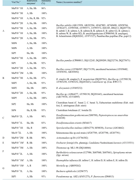
Table 1.Affiliation of microorganisms by MERFLPa of 16S rDNA(A-H) and 18S rDNA(I-M).
Vial No.b Restriction enzymesc
Similarity
(%) Name (Accession number) d
A
MuP10−31H A, Ha, Hh 86%
Bacillus subtilis (AB115958, AB192294, AF447803, AF549498, AF058766,
AY962473, AY995568, AY995571, AY995572, D26185, D84213, DQ207730, B. subtili 3, B. subtrrn A, B. subtrrn B, B. subtrrn E, B. subtrr G2, B. subtrrn J, B. subtrrn W, B. subtrr H2), B. amyloliquefaciens (CP000560, B. amylique), B. licheniformis (DQ020262, AY971527), Paenibacillus popilliae (Pae. popil 2) MuP10−33H A, Hh 100%
MuP10−41H A, Ha, R, Hh 92%
MuP10−42H A, Ha, Hh 100%
MuP10−43H A, Ha, Hh 100%
MuP10−51H A, Ha, Hh 100%
MuP10−52H A, Ha, Hh 95%
MiP4 A, Ha, Hh 100%
MiP6 A, Hh 100%
SiP5 Ha, Hh 100%
B
MuP10−63H A, Ha, Hh 100%
Bacillus pumilus (CP000813, DQ112245, DQ209209, DQ232736, DQ275671)
SiP1 A, Ha, Hh 92%
MuP10−32H A, Ha, Hh 92% Bacillus cereus (AY986507, DQ173159), uncultured bacterium (AY858402,
AY858392, AB185692) MuP10−52M A, Hh 100%
MuP10−53H A, Ha, Hh 87% B. simplex (B. simplex3), B. megaterium (DQ207561), Bacillus sp. (AY590138,
AY965250, AY965252, DQ201643), Caulobacter sp. (Cau. RWC17) MiP3 A, Ha, Hh 93%
MiP2 Ha, Hh 100% B. alveyuensis (AY605232)
MuP10−52H A, Ha, Hh 95% Bacillus sp. (AJ864277, AY590138, DQ201643), uncultured bacterium
(AB179530, AY534095) MiP5 A, Ha, Hh 100%
C
SiP2 Ha, Hh 100% Clostridium barati (C. barati 2, C. barati 3), Eubacterium multiforme (Eub. mul-for), E. nitritogenes (Eub. nitrit)
SiP4 Ha, R, Hh 93% Clostridium botulinum (C. botulin D)
MuP10−32L A, Hh 90% Desulfotomaculum geothermicum (X80789), Peptostreptococcus anaerobius (L04168)
MuP10−51L Ha, Hh 83% Desulfotomaculum orientis (M34417)
MuP10−61H Ha, R 100% Sporolactobacillus inulinus (AB362770, M58838), S.terrae (AJ634662)
Mu10−42L A, Hh 100% Selenomonas-like sp.oral strain (AF287789, AF287790, AF287791)
D MuP10−42M R, Hh 90% Actinomadura sp.E6 (AY766302)
E MuP10−31Me R, Hh 100% Oxobacter fennigii (Ox. phennig), Candidatus Neohrichiami kurensis (AY135531)
F
MuP10−33M A, Hh 100% Thiomonas sp. ML1-98 (DQ146066)
MuP10−62L A, Hh 100% Burkholderia solanacearum (U27986, X607040, X607041), Spiroplasma mirum
(Spp. mirum)
G
MuP10−31Me A, Hh 100% Haemophilus influenza (H. inflrrn C, H. inflrrn D, H. inflrrn E, H. inflrrn D)
MuP10−31Me A, R 100% Moritella sp. (AB059262)
MuP10−33L A, Ha 100% Buchnera aphidicola (AJ296757)
Continued
H
MuP10−32M A, Hh 93%
unidentified and uncultured bacterium (AB179495, AF317770, AF371874, AY177783, DQ129379)
MuP10−41M Ha, Hh 93%
MuP10−43M A, Hh 93%
MuP10−52L R, Hh 90%
MuP10−62M Ha, Hh 93%
I
RefESU R, Hh, A 100%
Saccharomyces cerevisiae (SayCe 101), S. dairenensis (SayDair8),
Zygosaccharomyces microellipsoides (Zygs. micro), Z. mrakii (Zygs. mraki),
Z. mellis (Zygs. melli), Torulospora delbrueckii (TorDe113), T. globosa
(Trls. globo), T. pretoriensis (Trls. preto), Kluyveromyces delphensis (KluDeip4),
K. polysporus (KluPoly8), Kazachstania vitcola (X99526), Candida colliculosa
(Can. colli) PiE1 Ha, R, Hh, A 100%
PiE2 Ha, R, Hh, A 100%
PiE3 Ha, R, Hh, A 100%
PuE10−81H Ha, R, Hh, A 100%
PuE10−82H Ha, R, Hh, A 100%
PuE10−83H Ha, R, Hh 100%
PuE10−92H Ha, R, Hh, A 100%
J RefEGZ Ha, R 92% Gibberella zeae (AB250414), G. intermedia (JN236216)
K
RefEPC Ha, R, Hh, A 100%
P. chrysogenum (AFF411201, AF548086), P. expansum (DQ912698),
P. camemberti (GQ458020)
KiE2 Ha, Hh, A 93%
KuE10−61H , Hh, R, A 97%
L
RefEAO Ha, R, Hh, A 100%
Aspergillus clavatus (Asp. Clav9), A.awamori (Aspe. awamo), A. fumigatus
(Aspe. fumig, Asp. fumi 2, Asp. fumi 9), A. niger (Aspe. niger), A. parasiticus
(Aspe. psiti), A. restrictus (AspRes10), A. terreus (Asp. Ter15), A.sparsusr
(AspSpar2), P. tardum (AF245256), P. purpurogenum (AF245261, AF245264),
P. glabrum (AF245270), P. decumbens (EU136028), Eremascus albus
(Erms. albus), Elaphomyces maculatus (EloMacul), Eurotium herbariorum
(EutHerba) KiE3 Ha, Hh, A 100%
KiE4 Ha, Hh, A 100%
KuE10−51H Ha, Hh, A 100%
KuE10−52H Ha, Hh, R, A 95%
KuE10−53H HaHh, A 100%
KuE10−62H Ha, Hh, A 97%
KuE10−63H Ha, Hh, A 97%
aGrouping was based on affiliation by MERFL; Bacillus subtilis group (Group A), the other Bacillus spp. (Group B), the other Firmicutes (Group C),
α-Proteobacteria (Group D), β-Proteobacteria (Group E), γ-Proteobacteria (Group F), δ-Proteobacteria (Group G), unidentified and uncultured bac-terial group (Group H), Hemiascomycetes (Group I), Sordariomycetes (Group J), Eurotiomycetes (Penicillium spp.; Group K), and Eurotiomycetes (Aspergillus spp.; Group L). bThe 1st letter in vial indicates origins of microorganisms; “Ref” stands for reference strains (SU; Saccharomyces uva-rum (NBRC10968T), GZ; Gibberella zeae (NBRC7189), PC; Penicillium roqueforti (NBRC5459), AO; Aspergillus oryzae (NBRC30113)), “M” stands for the sample from komekouji-miso, “S” stands for the sample from sake-kasu, “P” stands for the sample from dry yeast, and “K” stands for the sample from komekouji The next letter in the samples means origins of DNAs; “i” means DNA from microbial isolate, and “u” means DNA from culture medium. The next letter indicates used DNA primers; “P” stand for 41f/1066r primers for 16S DNA, and “E” stand for EF4/EF3 primers for 18S rDNA. Vial number in the isolate represents strain number. Exponential of vial number in un isolated DNA (u) represents the decimal dilution of the vial. The 2nd number of vial number (1-3) represents number in 3 replicates for the each decimal dilution. H of last letter represents MERFL ori-ginating from the major 16S rDNA, M represents from the 2nd major 16S rDNA, and L represents from the 3rd major 16S rDNA. cRestriction en-zymes used for similarity search; “Ha”, “R”, “Hh”, and “A” stand for Hae III, RsaI, Hha I, andAluI. For the measured MERFLP which had no com-pletely identical theoretical MERFLP, the theoretical MERFLP having the highest similarity using all the RFLPs was presented with the similarity as described in the materials and method. dSpecies name (accession number) of the theoretical MERFL having the highest similarity with the measured MERFL. eAdditional name (accession number) of the theoretical MERFL using the different restriction enzymes.
3. Results and Discussions
3.1. Affiliation of Isolated and Non-Isolated Microorganisms by MERFLP
Affiliations of 27 MERFLs for 18S rDNA of reference strains (4 strains), isolates (13 strains), and each MPN vials (10 genomes), and those of 36 MERFLs for 16S rDNA of isolates (10 isolates), and each MPN vials (26
genomes) were summarized in Table 1. MERFLs for 16S rDNA were divided into 8 groups for the MPN
calculation as the followings; B.subtilis group (Group A, 10 MERFLPs), Bacillus spp. (Group B, 9 MERFLs),
Table 2. Most probable numbers of each groups (A - L) and 5% confidence limits obtained using FDA’s Bacterial Analytical Manual [26]. In sake-kasu (S), both prokaryote and eukaryote were not detected by the method (<0.3 × 10⁵ MPN g−1). T*; Total numbers of microorganisms.
Groups
komekouji-miso dry yeast komekouji,
Three dilutions Score
×105 MPN g−1
matter
5% limits Low/High
Three
dilutions Score
×105 MPN g−1 matter
5% limits Low/High
Three dilutions Score
×105 MPN g−1 matter
5% limits Low/High
A 10−410−510−6 3-2-0 4.65 0.9/21 nd <0. 3 - nd <0.3 -
B 10−510−610−7 3-1-0 21.5 4.5/90 nd <0. 3 - nd <0.3 -
C 10−510−610−7 1-1-0 3.7 0.65/10 nd <0. 3 - nd <0.3 -
D 10−410−510−6 0-1-0 0.15 0.01/0.6 nd <0. 3 - nd <0.3 -
E + F + G 10−310−410−5 3-0-0 0.12 0.02/0.5 nd <0. 3 - nd <0.3 -
E 10−510−610−7 0-1-0 1.5 0.08/5.5 nd <0. 3 - nd <0.3 -
H 10−510−610−7 1-1-0 3.7 0.65/10 nd <0. 3 - nd <0.3 -
I nd <0.3 - 10−710−810−9 3-1-0 8600 360000 1800/ nd <0.3- -
K nd <0.3 - nd <0. 3 - 10−510−610−7 0-1-0 1.5 0.075/5.5
L nd <0.3- - nd <0. 3 - 10−510−610−7 3-2-0 46.5 9/210
T* 10−510−610−7 3-3-0 120 21/500 10−710−810−9 3-1-0 8600 1800/ 360000 10
−510−610−7 3-3-0 120 21/500
MERFLs), β-Proteobacteria (Group F, 2 MERFLs), γ-Proteobacteria (Group G, 2 MERFLs), and uncultured and
unnamed bacterium (Group H, 5 MERFLs) (Table 1). MERFLs for 18S rDNA were divided into 5 groups as the
followings; Hemiascomycetes (Group I, 8 MERFLs), Sordariomycetes (Group J, one MEPFL), Eurotiomycetes (Penicillium spp.; Group K, 3 MERFLs), and Eurotiomycetes (Aspergillus spp.; Group L, 8 MERFLs) (Table
1).
Most of MERFLs of isolates were placed in the same OUT as the MPN vials of the original samples as the
following (Table 1); in Group A, two isolates (MiP4, MiP6) and the 7 vials (MuP10−31H, MuP10−33H,
MuP10−41H, MuP10−42H, MuP10−43H, MuP10−51H, MuP10−52H) were placed in the same OUT (Table 1); in
Group B, two isolates (MiP3,MiP5) and two vials (MuP10−53H,MuP10−52H) were placed in the same OUT
(Table 1); in Group I, two isolates (PiE1, PiE2) and four vials (PuE10−81H, PuE10−82H, PuE10−83H,
PuE10−92H) were placed in the same OUT (Table 1); in Group K, one isolate (KiE2) and one vial (KuE10−61H)
were placed in the same OUT (Table 1); in Group L, two isolates (KiE3, KiE4) and five vials (KuE10−51H,
KuE10−52H, KuE10−53H, KuE10−62H, KuE10−63H) were placed in the same OUT (Table 1). These results
in-dicated that microorganism included in food sample were properly affiliated using the MPN vials without isola-tion. As total MERFLs of most of the MPN vials in komekouji (K) and dry yeast (P) were the same as those of isolates and reference strains, the both samples were suggested to include almost pure single microorganism.
Bacillus spp. (Group A and B) were amplified preferentially to the other gram positive bacterial group (Group C and D) and gram negative bacterial group (Group E, F and G) by 41f/1066r primers as the followings; all the
Bacillus spp. (Group A and B) were the major MERFLs, indicated as “H”, in the MPN vials, while 20% of the other gram positive bacterial group (Group C and D) was the major MERFLs, and no gram negative bacterial
group (Group E, F and G) were the major MERFLs (Table 1), which were coincident with the former
manu-scripts [2][3].
For 18S rDNA, difference between the theoretical MERFL and the measured MERFL, which caused to de-crease precision of pyrogenetic estimation as described in the former papers [21][22], was inevitably caused by used restriction enzyme as the followings; contiguous fragments of 354 bp and 356 bp in theoretical Alu I RF of
of Hha I RFs of G.zaea (Group J) were misread as one fragment in the respective measured RFs; contiguous
fragments of 177 bp and 174 bp and those of 155 bp and 151 bp in theoretical Hae III RFs of Saccharomyces
spp. (Group K) were misread as one fragment in the respective measured RFs, the fragments of 99 bp in
theo-retical Rsa I RF of Saccharomyces spp. (Group K) were sometimes measured over 100 bp fragment due to the
measuring error. As the relative mole ratios of these contiguous fragments were almost twice as those of the other fragments in the same RF, they were manually divided into 2 fragments in this study. Fragments from 100 bp to 103 bp of the measured Rsa I RF of Saccharomyces spp. (Group K) were manually eliminated in this study. Development of a program, which achieved these data processing automatically, may be useful to reduce a time for analysis.
Until now, following 5 restriction enzymes, HaeIII, HhI, Rsa I, ScrF1, and Alu I, have been used because they had more restriction sites in 16S rDNA than the others, in order to affiliate bacterial isolates precisely by using a
fewer restriction enzymes [21] [22]. However such the restriction enzyme increased the opportunity of
over-lapped fragment, when they were used for longer DNA or sample having huge microbial diversity without isola-tion. Another combination of restriction enzymes, which had fewer restriction sites in 16S rDNA or 18S rDNA, was found suitable for such the analyses (unpublished results). Analysis using the other combination of restric-tion enzymes will be presented in the following manuscript.
3.2. Enumeration of Each Prokaryotic and Eukaryotic Microbial Groups by MPN
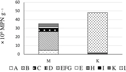
In komekouji (K), Group L, including Aspergillus spp., was enumerated to be 46.5 × 10⁶ MPM g−1, and Group K, including Penicillium spp., was enumerated to be 1.5 × 10⁶ MPM g−1, and no other eukaryotic nor prokaryo-tic microorganisms were detected (Table 2,Figure 1). As the sample (K) included almost pure Aspergillus spp., the sample might be analyzed without cultivation as same as lactic acid bacterial groups in yoghurt [27]. In dry yeast (P), Group I, including Saccharomyces group, was enumerated to be 8600 × 106 MPM g−1, which was higher than that estimated by dilution plate count (220 × 106 CFU g−1; unpublished result), and no other euka-ryotic nor prokaeuka-ryotic microorganisms was detected (Table 2). As the sample (P) included almost pure Saccha-romyces group, the sample might be analyzed without cultivation as same as lactic acid bacterial groups in yog-hurt [27].
In komekouji-miso (M), eukaryotic microorganism was not detected. As prokaryotic microorganisms, Group B (the other Bacillus spp.) was numerically dominant, which was enumerated to be 21.5 × 10⁶ MPM g−1, fol-lowed by Group A (B. subtilis group.; 4.65 × 10⁶ MPM g−1), and Group C (the other Firmicutes; 3.7 × 10⁶ MPM
g−1) (Table 2, Figure 1). Number of Group A was similar level as those reports, in which B. subtilis originated
from soybean was present at 104 ~ 106 CFU g−1 during miso fermentation, and decreased to under 104 ~ 105
CFU g−1[28][29]. Because of the difference of used incubation medium, total bacterial number in komekouji-
[image:7.595.210.415.523.640.2]miso (K) in this study was higher than that of the former study using MRS medium for lactic acid bacteria [27], and lactic acid bacteria was not detected in this study (Table 2). No Aspergillus spp. added with komekoji, nor
Figure 1. Numbers of microbial groups estimated by MPN and MERFLP in ko-mekouji-miso (M), and komekouji (K). Number of the other Bacillus spp. (Group A; ), B. subtilis (Group B; ), the other Firmicutes (Group C; ), Actino-bacteria (Group D; ), α,β,γ-Proteobacteria (Group E, F, G; ), Burkholderia sp. (Group E; ), unidentified and uncultured bacteria (Group H; ), Saccharo-myces sp. (Group I; ), Penicillium spp. (Group K; ), and Aspergillus spp.
(Group L; ) were presented.
0 10 20 30 40 50 60
M K
A B C D EFG E H I K L
×
10
6M
P
N
g
yeast, which progressed alcohol fermentation, were also detected in komekouji-miso (<0.3 × 10⁵ MPN g−1)
(Table 2). The sample M also included lower number of Group D (Actinobacteria; 0.15 × 10⁶ MPM g−1), Group
E (Burkhokderia sp.; 1.5 × 10⁶ MPM g−1), and Group E, F, G (the other α, β, γ proteobacteria; 0.12 × 10⁶ MPM
g−1) (Table 2, Figure 1). In sake-kasu (S), both prokaryote and eukaryote were not detected by the method,
while bacteria affiliated to be Goup A (SiP5), Group B (SiP1), Group C (SiP2, SiP4), and Group G (SiP3) were isolated from the lowest dilution agar plate. As skekasu was residue filtered rice wine from moromi after finish of alcohol fermentation and sterilization by heating at 60 to 65℃, eukaryotic microorganisms, such as
Aspergil-lus spp. and Saccharomyces spp., which progressed alcohol fermentation, nor prokaryotic bacteria were not
founded in MPN vials, but spore forming bacteria such as Bacillus subtilis (group A; SiP5), B. pumilus (group B; SiP1), Clostridium barati (group C; SiP2), and C. botulinum (group C; SiP4) were isolated in lowest dilution agar plate as heat tolerant bacteria (Table 1).
As PCR amplification were not tested under 10−5 dilution vials and lower detection limit was high (<0.3 × 10⁵
MPN g−1) in the present experiment except for prokaryotic microorganisms in komekouji-miso (K) (Table 1,
Table 2), the lower detection limit could be lowered by using more effective DNA purification method.
4. Conclusions
By DGGE [8]-[11], which used directly extracted DNA, exact ratio of each microbial groups was not obtained
due to PCR bias and it was no use to compare the numbers and composition of prokaryotic microorganisms to
those of eukaryotic microorganisms because of a difference in the PCR amplification rates [15]-[18]. As
num-bers of each microbial groups were estimated by MPN not by an amount of the amplified DNA in this method as described in the former manuscripts [1][2], the difference in the primers did not affect the numbers and compo-sition of numerically dominant microorganisms but their effect was limited in numerically minor microorgan-isms. Present results indicated that using both universal primers for eukaryotic and prokaryotic microorganisms, each group of prokaryotic and eukaryotic microorganisms were simultaneously enumerated without any prelim-inary information nor setting up standard curve, which were required for real time PCR. The method seemed useful not only to evaluate whole microbial compositional change during traditional fermentation process, and manure composting process, but also the variety of other purpose such as to ensure food safety by evaluating unintended or unexpected contamination or proliferation of opportunistic pathogens or foodborne pathogens or non-pathogenic spoilage microorganisms during food distribution system.
In this study, universal PCR primers for eukaryotic and prokaryotic microorganisms were used to evaluate what kinds of microorganisms was mainly included in the sample. Another combination of universal primers and selective primers will afforded more detailed information of each microbial group, and functional or toxico-logical information of each microbial group will similarly be obtained by changing DNA primers.
Acknowledgements
We thank Mr. N. Osako, and Mr. R. Kizuka, the former undergraduate students of Fukuoka Institute of Tech-nology, for analysis of samples.
References
[1] Watanabe, K. and Okuda, M. (2003) Method and System for Searching for Relationships between Base Sequences in Genes. Japanese Patent 3431135; (2006) US Patent 7,006,924.
[2] Watanabe, K., Horinishi, N. and Matumoto, K. (2015a) Antibiotic-Resistant Bacterial Group in Field Soil Evaluated by a Newly Developed Method Based on Restriction Fragment Length Polymorphism Analysis. Advances in Microbiolo-gy, 5, 807-816. http://dx.doi.org/10.4236/aim.2015.512085
[3] Watanabe, K., Horinishi, N., Matumoto, K., Tanaka, A. and Yakushido, K. (2015) Bacterial Groups Concerned with Maturing Process in Manure Production Analyzed by a Method Based on Restriction Fragment Length Polymorphism Analysis. Advances in Microbiology, 5, 832-841. http://dx.doi.org/10.4236/aim.2015.513088
[4] Smith, C.J. and Osborn, A.M. (2009) Advantages and Limitations of Quantitative PCR(Q-PCR)-Based Approaches in Microbial Ecology. FEMS Microbiology Ecology, 67,6-20. http://dx.doi.org/10.1111/j.1574-6941.2008.00629.x [5] Park, E., Chang, H., Kim, K., Nam, Y., Roh, S.W. and Bae, J. (2009) Application of Quantitative Real-Time PCR for
[6] Lakaniemi, A.M., Hulatt, C.J., Wakeman, K.D., Thomas, D.N. and Puhakka, J.A. (2012) Eukaryotic and Prokaryotic Microbial Communities during Microalgal Biomass Production. Bioresource Technology, 124,387-393.
http://dx.doi.org/10.1016/j.biortech.2012.08.048
[7] Teranishi, H. and Ouchi, K. (2014) Detection of Bacteria, Fungi, and Viruses by a Real-Time PCR Assay Using Uni-versal Primers and Probes from Blood in Patients with Febrile Neutropenia. Kawasaki Medical Journal, 40,1-11.
[8] Mills, D.A., Johannsen, E.A. and Cocolin, L. (2002) Yeast Diversity and Persistence in Botrytis-Affected Wine Fer-mentations. Applied and Environmental Microbiology, 68,4884-4893.
http://dx.doi.org/10.1128/AEM.68.10.4884-4893.2002
[9] Hierro, N., Esteve-Zarzoso, B., González, Á., Mas, A. and Guillamón, J.M. (2006) Real-Time Quantitative PCR (QPCR) and Reverse Transcription-QPCR for Detection and Enumeration of Total Yeasts in Wine. Applied and Envi-ronmental Microbiology, 72,7148-7155. http://dx.doi.org/10.1128/AEM.00388-06
[10] Guo, R., Chen, Z., Chen, N. and Chen, M.D. (2010) Quantitative Real-Time PCR Analysis of Intestinal Regular Fungal Species in Fecal Samples From Patients With Chronic Hepatitis B Virus Infection. Science, 41,591-596.
http://dx.doi.org/10.1309/lmmc0wvzxd13pujg
[11] Lottmann, J., O’Callaghan, M., Baird, D. and Walter, C. (2010) Bacterial and Fungal Communities in the Rhizosphere of Field-Grown Genetically Modified Pine Trees (Pinus radiata D.). Environmental Biosafety Research, 9, 25-40. http://dx.doi.org/10.1051/ebr/2010007
[12] Duartea, S., Pascoala, C., Alvesb, A., Correiab, A. and Cássioa, F. (2010) Assessing the Dynamic of Microbial Com-munities during Leaf Decomposition in a Low-Order Stream by Microscopic and Molecular Techniques. Microbiolog-ical Research, 165, 351-362. http://dx.doi.org/10.1016/j.micres.2009.06.002
[13] Suzuki, C., Takenaka, M., Oka, N., Nagaoka, K. and Karasawa, T. (2012) A DGGE Analysis Shows That Crop Rota-tion Systems Influence the Bacterial and Fungal Communities in Soils. Soil Science and Plant Nutrition, 58, 288-296. http://dx.doi.org/10.1080/00380768.2012.694119
[14] Xu, Y., Xie, Y.B., Zhang, X.R., Chen, C., Xiang, H. and Xie, Q. (2013) Monitoring of the Bacterial and Fungal Biodi-versity and Dynamics during Massa Medicata Fermentata Fermentation. Applied and Environmental Microbiology, 97, 9647-9655.
[15] Suzuki, M.T. and Giovannoni, S.J. (1996) Bias Caused by Template Annealing in the Amplification of Mixtures of 16S rRNA Genes by PCR. Applied and Environmental Microbiology, 62, 625-630.
[16] Ishii, K. and Fukui, M. (2001) Optimization of Annealing Temperature to Reduce Bias Caused by a Primer Mismatch in Multitemplate PCR. Applied and Environmental Microbiology, 67, 3753-3755.
http://dx.doi.org/10.1128/AEM.67.8.3753-3755.2001
[17] Neilson, J.W., Jordan, F.L. and Maier, R.M. (2013) Analysis of Artifacts Suggests DGGE Should Not Be Used for Quantitative Diversity Analysis. Journal of Microbiological Methods, 92, 256-263.
http://dx.doi.org/10.1016/j.mimet.2012.12.021
[18] Direito, S.O.L., Zaura, E., Little, M., Ehrenfreund, P. and Röling, W.F.M. (2014) Systematic Evaluation of Bias in Mi-crobial Community Profiles Induced by Whole Genome Amplification. Environmental Microbiology, 16, 643-657. http://dx.doi.org/10.1111/1462-2920.12365
[19] Smit, E., Leeflang, P., Glandorf, B., Elsas, J.D.V. and Wernars, K. (1999) Analysis of Fungal Diversity in the Wheat Rhizosphere by Sequencing of Cloned PCR-Amplified Genes Encoding 18S rRNA and Temperature Gradient Gel Electrophoresis. Applied and Environmental Microbiology, 65, 2614-2621.
[20] Gao, Z., Li, B., Zheng, C. and Wang, G. (2008) Molecular Detection of Fungal Communities in the Hawaiian Marine Sponges Suberites zeteki and Mycale armata. Applied and Environmental Microbiology, 74,6091-6101.
http://dx.doi.org/10.1128/AEM.01315-08
[21] Watanabe, K., Okuda, M. and Koga, N. (2008) A Newly Developed System Based on Multiple Enzyme Restriction Fragment Length Polymorphism—An Application to Proteolytic Bacterial Flora Analysis. Soil Science and Plant Nu-trition, 54, 204-215. http://dx.doi.org/10.1111/j.1747-0765.2007.00230.x
[22] Watanabe, K. and Koga, N. (2009) Use of a Microchip Electrophoresis System for Estimation of Bacterial Phylogeny and Analysis of NO3− Reducing Bacterial Flora in Field Soils. Bioscience Biotechnology and Biochemistry, 73,
479-488. http://dx.doi.org/10.1271/bbb.70712
[23] Weidner, S., Arnold, W. and Puhler, A. (1996) Diversity of Uncultured Microorganisms Associated with the Seagrass Halophilastipulacea Estimated by Restriction Fragment Length Polymorphism Analysis of PCR-Amplified 16S rRNA Genes. Applied and Environmental Microbiology, 62, 766-771.
[25] Nei, M. and Li, W.H. (1979) Mathematical Model for Studying Genetic Variation in Terms of Restriction Endonuc-leases. Proceedings of the National Academy of Sciences of the United States of America, 76, 5269-5273.
http://dx.doi.org/10.1073/pnas.76.10.5269
[26] Blodgett, R. (2010) FDA, Bacterial Analytical Manual, Appendix 2 Most Probable Number from Serial Dilutions. http://www.fda.gov/Food/FoodScienceResearch/LaboratoryMethods/ucm109656.htm
[27] Matsumoto, K., Shimada, K., Horinishi, N. and Watanabe, K. (2016) Evaluation of the Method Based on Restriction Fragment Length Polymorphism Analysis as Simple Analysis Method of Lactic Acid Bacteria in Foods. Food and Nu-trition Sciences, 7, 163-172. http://dx.doi.org/10.4236/fns.2016.73018
[28] Onda, T., Yanagida, F., Tsuji, M., Shinohara, T. and Yokotsuka, K. (2003) Time Series Analysis of Aerobic Bacterial Flora during Miso Fermentation. Letters in Applied Microbiology, 37, 162-168.
http://dx.doi.org/10.1046/j.1472-765X.2003.01371.x
[29] Onda, T., Yanagida, F., Uchimura, T., Tsuji, M., Shinohara, T., Ogino, S. and Yokotsuka, K. (2003) Analysis of Lactic Acid Bacterial Flora during Miso Fermentation. Food Science and Technology Research, 9, 17-24.

