Address for correspondence
Hatice Özdemir
E-mail: dentist_hatice@hotmail.com
Funding sources
None declared
Conflict of interest
None declared
Acknowledgements
The authors thank the companies Zhermack and 3M ESPE for their product support, and Memiş Özdemir for his statistical assistance.
Received on July 25, 2018 Reviewed on November 24, 2018 Accepted on December 17, 2018 Published online on March 19, 2019
Abstract
Background. The dimensional accuracy of impression materials has been evaluated for a long time, but thus far, digital radiography has not been used for this purpose. The dimensional accuracy of impression materials is very important for the final adaptation of dental prostheses.
Objectives. The objective of this study was to evaluate the effects of different disinfectant solutions and storage times on the dimensional stability of different impression materials by means of digital radiogra-phy.
Material and methods. Polyether (PE), hydrocolloid (IH), condensation silicone (CS), and addition silicone (AS) materials were used for preparing impressions, taken from an acrylic master model with 2 vertical and 2 horizontal reference points. Water (W), sodium hypochlorite (SH) and a disinfectant solu-tion without aldehyde (Z) were applied on the impressions. Half of the impressions were poured over immediately and half of them – 1 day after. Digital radiography was used to determine the dimensional accuracy of the impression materials. The data was analyzed with a variance analysis and Tukey’s multiple comparison test.
Results. While PE showed the smallest dimensional changes, IH showed the greatest in all lines. Applying SH and pouring 1 day after caused the greatest dimensional changes in all impression materials.
Conclusions. Different disinfectant solutions and storage times had a different effect on the impressions, but the dimensional changes were clinically acceptable.
Key words: dimensional accuracy, storage time, impression material, digital radiography, disinfection so-lution
Słowa kluczowe: wierność odtwarzania kształtu, czas przechowywania, materiał wyciskowy, radiogra-fia cyfrowa, roztwór odkażający
Cite as
Özdemir H, Azlağ Pekince K. Evaluation of the effect of storage time and disinfectant solutions on the dimensional accuracy of impression materials with digital radiography . Dent Med Probl. 2019;56(1):67–74. doi:10.17219/dmp/101649
DOI
10.17219/dmp/101649
Copyright
© 2019 by Wroclaw Medical University This is an article distributed under the terms of the Creative Commons Attribution Non-Commercial License (http://creativecommons.org/licenses/by-nc-nd/4.0/)
Evaluation of the effect of storage time and disinfectant solutions
on the dimensional accuracy of impression materials with digital radiography
Ocena wpływu czasu przechowywania oraz roztworów odkażających
na wierność odtwarzania kształtu przez materiały wyciskowe
z użyciem radiografii cyfrowej
Hatice Özdemir
1,A–F, Kader Azlağ Pekince
2,A,E1 Department of Prosthodontics, Faculty of Dentistry, Atatürk University, Erzurum, Turkey 2 Department of Oral and Maxillofacial Radiology, Faculty of Dentistry, Karabük University, Turkey A – research concept and design; B – collection and/or assembly of data; C – data analysis and interpretation; D – writing the article; E – critical revision of the article; F – final approval of the article
Introduction
Dental impression materials are used to register the form and relation of the teeth and the surrounding oral tissues. The accuracy of the impression is related to the dimensional stability of the impression material, and this is affected by many factors, such as the impression tech nique, impression tray, selection of a proper impression material, and properties of the material.1–3 An accurate impression is the most important stage for the construc tion of a dental prosthesis.4 Polysulfide polymers, conden sation silicones and the addition type of silicones, syn thetic elastomers containing polyether, and hydrocolloids are widely used in dentistry to obtain the copy of oral tis sues.5 Impression materials are exposed to some factors, which may result in dimensional changes.6 The dimen sional stability and accuracy of impression materials are significant for the adaptation of the final prosthetic resto ration. The dimensional accuracy of impressions is mostly influenced by the procedure of disinfection and the time period after which the model is poured over with dental stone. In terms of the latter, the best results are obtained by pouring the dental stone immediately after removing the impression from the mouth. For practical reasons, the impressions may nevertheless sometimes not be poured over immediately. Some producers claim that recently produced impression materials can be kept in appropriate conditions for longer periods of time without loss of clini cal accuracy. Glutaraldehyde and sodium hypochlorite are frequently used for disinfecting dental impressions.7 So dium hypochlorite, which is an effective disinfectant re commended by the American Dental Association (ADA), is used in a 1:10 dilution for a 10minute immersion to disinfect irreversible hydrocolloid impressions.8 The use of glutaraldehyde is not preferred due to its harmful and irritant effect. Therefore, dentists may prefer aldehyde free disinfectant solutions.
Various dimensional measuring techniques are used in clinical and academic dentistry for dental stone models and scanned models. Manual measurements are performed with a Vernier caliper or needlepoint calipers, and they are the standard method to evaluate the accuracy of den tal models. Manual measurements have some advantages, such as the ease of application, low cost and immediate ap plicability. Alternatively, 3dimensional computer dental models, scanned with an optical or laser beam, are con sidered appropriate in clinical applications. In this study, the dimensional changes of different impression materials were evaluated with digital radiography, which differs from the methods described in the current literature. Digital ra diography is considered to be more economical than the 3dimensional computer application and more reliable than the manual methods.8,9
The aim of this study was to evaluate the effect of dif ferent disinfectant solutions and storage times on the dimensional stability of different impression materials.
The hypothesis of the study was that different storage times and disinfectant solutions would affect the dimen sional stability of impression materials in different ways, depending on the type of impression material.
Material and methods
In this study, a denture base from heatpolymerized acrylic, prepared on a stone model of the edentulous max illa, was used as a master model. Holes were drilled in the places corresponding to the right and left canines and tuber maxillary ridges on the acrylic base with a steel round drill of a diameter of 0.2 mm, and 4 different reference points were determined. Then, metal balls of a diameter of 1 mm were placed in the reference points of the master model and fixed with dental wax. Two vertical (line 1 and line 2) and 2 horizontal (line 3 and line 4) reference lines were identi fied on both sides between the 4 different reference points. The reference lines were determined as follows:
– line 1 – the distance between the steel balls in the region of the left canine and left tuber maxilla;
– line 2 – the distance between the steel balls in the re gion of the right canine and right tuber maxilla;
– line 3 – the distance between the steel balls in the region of the right and left canines;
– line 4 – the distance between the steel balls in the region of the right and left tuber maxillae.
Therefore, it was aimed to evaluate the dimensional changes that might occur in the impression materials both horizontally and vertically by measuring the verti cal and horizontal reference lines. The master model was fixed inside the lower part of a dental prosthesis flask with dental white stone. Thus, it was ensured that the model remained stable during impression procedures.
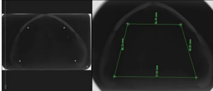
The master model was placed in a digital radiography
unit (PHOTxII; Takara Belmont USA, Inc., Somerset,
USA) and its image was taken with occlusal radiography. The reference measurements, determined by the reference points, were carefully performed digitally by 2 researchers (Fig. 1). These measurements were used as references and compared to those of the subsequent stone models. The dimensional changes that occurred due to the impression materials were evaluated by subtracting the 1st reference measurement value from those of the stone models.
Four different impression materials were used to pro duce the stone models (Table 1). A total of 240 impres sions were taken from the master model.
Apart from polyethers (PE), all impressions of the mas ter model were taken using a standard metal tray (Jesco form®; Aesculap AG, Tuttlingen, Germany). The irrevers ible hydrocolloid (IH) impression material was prepared by mixing powder and water in the amount suggested by the producer in an automatic mixing machine (HurrimixTM; Zhermack SpA, Badia Polesine, Italy). Room temperature water was consistently used for the mixtures so that the hardening time of the impression material could not be af fected. The condensation (Ctype) silicones (CS) were pre pared by mixing a catalyzer and the base at the rates sug gested by the producer with clean bare hands. Gloves were not used, because the powder in latex gloves may affect the hardening reaction of the impression material. The double mixing technique was used for the siliconebased impres sion materials (CS and addition silicone – AS). It was also applied for the light bodies of the siliconebased impression materials. A special tray was used for the PE impression material. A tray adhesive (EXAMIXTM; GC America, Inc., Alsip, USA) was applied in a thin layer to the inside and edges of the special tray. The PE impression material was mixed in an automatic mixing machine (PentamixTM, 3M ESPE, Seefeld, Germany), placed in the special impression tray, and an impression was taken from the master model.
The impressions were separated into 6 equal groups, and 3 different disinfectant solutions were applied with 2 different storage times. The disinfectants were: water (W), 1% sodium hypochlorite (SH) and an aldehydefree disinfectant solution – Zeta 7 spray (Z) (Zhermack SpA).
The disinfection procedures were as follows: W – wash ing with water for 45 s; SH – applying 1% SH and keeping it for 10 min; and Z– applying the ready impression disin fectant solution and keeping it for 3 min. While half of the impressions were immediately poured over, the other half
were poured over 1 day after. The IH impressions were kept in a closed plastic box. Other impressions were kept at normal room temperature (23°C), humidity 55 ±5%.
Type IV hard stone (Moldano®; Heraeus Kulzer GmbH,
Hanau, Germany) was used to obtain the stone models from the impressions. The hard stone mixture was pre pared by mixing powder (100 g) and water (30 mL) in the amount suggested by the producer. The required precau tions were taken to prevent air bubbles in the stone during mixing and pouring it into the impression.
The resulting stone models were immediately removed from the impressions as soon as the stone was hardened.
As with the master model, metal balls of a diameter of 1 mm were placed in the reference points of the stone mo dels and fixed with dental wax (Fig. 2), and then occlu sal radiographs were taken. The measurements between the steel balls were performed on the radiographic images. All measurements were repeated 5 times and their aver ages were calculated. The digital measurements on the stone models were carefully performed by 2 researchers.
The mean, standard deviation (SD) and percentage
values of the data of the line 1, line 2, line 3, and line 4 reference measurements were calculated. An analysis of variance (ANOVA) was conducted to evaluate the ef fect of the different impression material types, storage times and disinfectant solutions on 4 different reference measurements. Tukey’s multiple comparison test was performed to compare the averages. A pvalue ≤0.05 was considered statistically significant.
Results
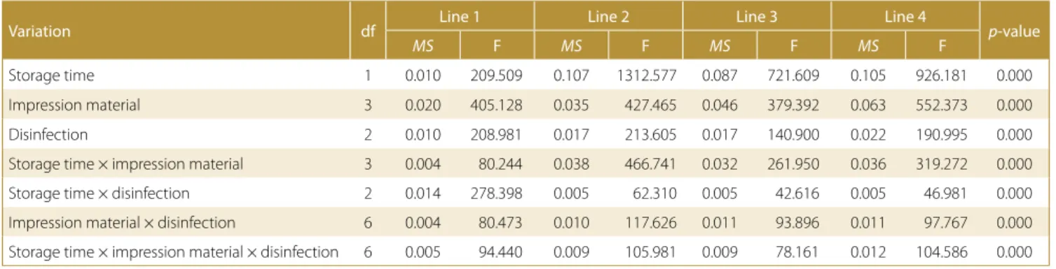
The results of ANOVA regarding the effect of the im pression material type, storage time and disinfectant on the dimensional changes in the impression materials are presented in Table 2. According to the Table, both the fac tors and the interactions between them were found to be statistically significant in all measurements performed in 2 vertical and 2 horizontal directions (p < 0.001).
The mean and SD values obtained with Tukey’s mul
tiple comparison test for line 1 are presented in Table 3. While the smallest dimensional change was observed in PE, the greatest dimensional change was observed in IH, and the differences between the impression mate rials were found statistically significant in the poured immediately groups. The effect of different disinfectant solutions was also observed, but the differences were Fig. 2. The radiographic image of the metal balls and reference lines on the
stone model
Table 1. Impression materials used in the present study
Impression material Product Manufacturer Lot number Code
Polyether Impregum™ Penta™ Soft 3M ESPE, Neuss, Germany 624182 PE Irreversible hydrocolloid Hydrogum 5 Zhermack SpA, Badia Polesine, Italy 212976 IH Addition silicone Elite P&P Zhermack SpA, Badia Polesine Italy 234875 AS Condensation silicone Zetaflow Zhermack SpA, Badia Polesine Italy 242997 CS
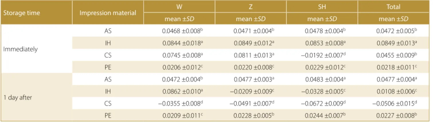
not statistically significant in the pouredimmediately groups. Moreover, all impression materials poured over immediately showed dimensional expansion. Consider ing the effect of different storage times, there were no statistically significant differences between the poured immediately and poured1dayafter groups, except for the case when SH was applied. While the smallest dimensional change was observed in PE, the greatest dimensional change was observed in IH, and the dif ferences between the impression materials were found statistically significant in the poured1dayafter groups, except for AS and CS. Water and Z had the same effect on the dimensional accuracy of all impression mate rials, but SH showed a statistically significant effect in all poured1dayafter groups. While the AS and CS groups after applying SH showed dimensional contraction, all other groups showed dimensional expansion. The per centages of the dimensional changes observed in the impression materials were also found to be statistically significantly different from each other. However, the di mensional changes observed in the impression materials were clinically acceptable (−0.047–0.187%) (Fig. 3).
The mean and SD values obtained with Tukey’s mul
tiple comparison test for line 2 are presented in Table 4. While the smallest dimensional change was observed in PE, the greatest dimensional change was observed in IH. There were no statistically significant differences between AS and CS in the pouredimmediately groups. The effect
of different disinfectant solutions was also varied, but the differences were not statistically significant, except for CS, with SH applied in the pouredimmediately group. Furthermore, all impression materials that were poured over immediately showed dimensional expansion, except for the CS group where SH was applied. Considering the effect of different storage times, statistically significant differences were observed between the pouredimmedia tely and poured1dayafter groups. While the smallest dimensional change was observed in CS, the greatest di mensional change was observed in AS, and the differences between the impression materials were found statistically significant in the poured1dayafter groups. This might be caused by expansion and contraction occurring at the same time. Addition silicone and PE showed dimensional expansion, and CS showed dimensional contraction after applying different disinfection solutions. The IH group Table 3. Multiple comparison test results with mean ± standard deviation (SD) for line 1
Storage time Impression material W Z SH Total
mean ±SD mean ±SD mean ±SD mean ±SD
Immediately
AS 0.0338 ±0.007c 0.0343 ±0.002c 0.0352 ±0.002c 0.0344 ±0.004c
IH 0.0727 ±0.007a 0.0741 ±0.004a 0.0756 ±0.003a 0.0741 ±0.005a
CS 0.0522 ±0.010b 0.0569 ±0.007b 0.0627 ±0.014b 0.0573 ±0.011b
PE 0.0149 ±0.014d 0.0157 ±0.006d 0.0163 ±0.007d 0.0156 ±0.009d
1 day after
AS 0.0342 ±0.006c 0.0349 ±0.003c 0.0358 ±0.004a 0.0350 ±0.004b
IH 0.0733 ±0.004a 0.0752 ±0.004a −0.0170 ±0.008c 0.0438 ±0.004a
CS 0.0564 ±0.006b 0.0645 ±0.008b −0.0190 ±0.005c 0.0340 ±0.004b
PE 0.0151 ±0.005d 0.0169 ±0.006d 0.0176 ±0.007b 0.0165 ±0.006c
W – water; Z – Zeta 7 spray (aldehyde-free disinfectant solution); SH – sodium hypochlorite. There are statistically significant differences between the values indicated with different letters (p < 0.05).
Table 2. Results of the analysis of variance (ANOVA)
Variation df Line 1 Line 2 Line 3 Line 4 p-value
MS F MS F MS F MS F
Storage time 1 0.010 209.509 0.107 1312.577 0.087 721.609 0.105 926.181 0.000 Impression material 3 0.020 405.128 0.035 427.465 0.046 379.392 0.063 552.373 0.000 Disinfection 2 0.010 208.981 0.017 213.605 0.017 140.900 0.022 190.995 0.000 Storage time × impression material 3 0.004 80.244 0.038 466.741 0.032 261.950 0.036 319.272 0.000 Storage time × disinfection 2 0.014 278.398 0.005 62.310 0.005 42.616 0.005 46.981 0.000 Impression material × disinfection 6 0.004 80.473 0.010 117.626 0.011 93.896 0.011 97.767 0.000 Storage time × impression material × disinfection 6 0.005 94.440 0.009 105.981 0.009 78.161 0.012 104.586 0.000 df – degrees of freedom; MS – mean square; F – F-test of ANOVA.
showed dimensional expansion when W was applied, and after applying Z and SH, showed dimensional contraction. The percentages of the dimensional changes observed in the impression materials were also found to be statisti cally significantly different from each other. However, the dimensional changes observed in the impression mate rials were clinically acceptable (−0.087–0.211%) (Fig. 4).
The mean and SD values obtained with Tukey’s multiple comparison test for line 3 are presented in Table 5. While the smallest dimensional change was observed in PE and CS, the greatest dimensional change was observed in IH. There were no statistically significant differences between PE and CS in the pouredimmediately groups. The effect of different disinfectant solutions was varied and the dif ferences were statistically significant, except for IH in the pouredimmediately groups. Moreover, all impression materials that were poured over immediately showed
dimensional expansion, except for the CS group where SH was applied. Considering the effect of different storage times, statistically significant differences were observed between the pouredimmediately and poured1day after groups. While the smallest dimensional change was observed in CS, the greatest dimensional change was ob served in AS, and the differences between the impres sion materials were found statistically significant in the poured1dayafter groups. This might be caused by ex pansion and contraction occurring at the same time. Ad dition silicone and PE showed dimensional expansion, and CS showed dimensional contraction after applying different disinfection solutions. The IH group showed di mensional expansion when W was applied, and after ap plying Z and SH, showed dimensional contraction. The percentages of the dimensional changes observed in the impression mate rials were also found to be statistically significantly different from each other. However, the di mensional changes observed in the impression materials were clinically acceptable (−0.135–0.223%) (Fig. 5).
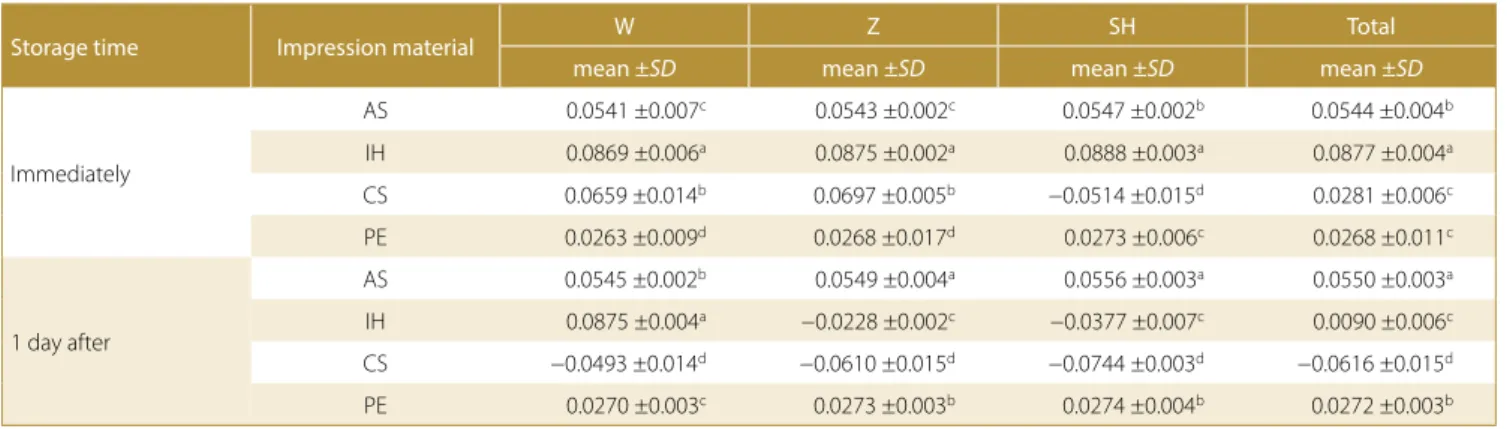
The mean and SD values obtained with Tukey’s multiple comparison test for line 4 are presented in Table 6. While the smallest dimensional change was observed in PE and CS, the greatest dimensional change was observed in IH. There were no statistically significant differences between PE and CS in the pouredimmediately groups. The effect of different disinfectant solutions was also varied and the differences were statistically significant, except for IH Table 4. Multiple comparison test results with mean ± standard deviation (SD) for line 2
Storage time Impression material W Z SH Total
mean ±SD mean ±SD mean ±SD mean ±SD
Immediately
AS 0.0468 ±0.008b 0.0471 ±0.004b 0.0478 ±0.004b 0.0472 ±0.005b
IH 0.0844 ±0.018a 0.0849 ±0.012a 0.0853 ±0.008a 0.0849 ±0.013a
CS 0.0745 ±0.008a 0.0811 ±0.013a −0.0192 ±0.007d 0.0455 ±0.009b
PE 0.0206 ±0.012c 0.0220 ±0.008c 0.0229 ±0.012c 0.0218 ±0.011c
1 day after
AS 0.0472 ±0.004b 0.0477 ±0.003a 0.0483 ±0.004a 0.0477 ±0.004a
IH 0.0862 ±0.010a −0.0209 ±0.009c −0.0328 ±0.005c 0.0108 ±0.006c
CS −0.0355 ±0.008d −0.0491 ±0.007d −0.0672 ±0.009d −0.0506 ±0.015d
PE 0.0209 ±0.011c 0.0228 ±0.005b 0.0244 ±0.007b 0.0227 ±0.008b
There are statistically significant differences between the values indicated with different letters (p < 0.05).
Table 5. Multiple comparison test results with mean ± standard deviation (SD) for line 3
Storage time Impression material W Z SH Total
mean ±SD mean ±SD mean ±SD mean ±SD
Immediately
AS 0.0401 ±0.010c 0.0406 ±0.008b 0.0417 ±0.006b 0.0408 ±0.008b
IH 0.0801 ±0.015a 0.0808 ±0.015a 0.0818 ±0.004a 0.0809 ±0.012a
CS 0.0642 ±0.009b 0.0708 ±0.034a −0.0495 ±0.012d 0.0285 ±0.006c
PE 0.0199 ±0.009d 0.0237 ±0.010b 0.0286 ±0.009c 0.0241 ±0.009c
1 day after
AS 0.0405 ±0.012b 0.0412 ±0.003a 0.0424 ±0.008a 0.0414 ±0.008a
IH 0.0811 ±0.004a −0.0174 ±0.012c −0.0269 ±0.006c 0.0123 ±0.005c
CS −0.0405 ±0.003d −0.0591 ±0.007d −0.0738 ±0.004d −0.0578 ±0.015d
PE 0.0214 ±0.004c 0.0264 ±0.003b 0.0297 ±0.004b 0.0258 ±0.005b
There are statistically significant differences between the values indicated with different letters (p < 0.05).
in the pouredimmediately groups. Also, all impression ma terials that were poured over immediately showed dimen sional expansion, except for the CS group where SH was applied. Considering the effect of different storage times, statistically significant differences were observed between the pouredimmediately and poured1dayafter groups. While the smallest dimensional change was observed in CS, the greatest dimensional change was observed in AS, and the differences between the impression materials were found statistically significant in the poured1dayafter groups. Addition silicone and PE showed dimensional expansion, and CS showed dimensional contraction after applying different disinfection solutions. The IH group showed dimensional expansion when W was applied, and after applying Z and SH, showed dimensional contraction. The percentages of the dimensional changes observed in the impression materials were also found to be statisti cally different from each other. However, the dimensional changes observed in the impression materials were clini cally acceptable (−0.090–0.156%) (Fig. 6).
Discussion
The results obtained in this study support our hypothe sis. In the study, the dimensional changes observed in the impression materials were found to be different from each other in relation to the type of impression material, stor age time and type of disinfectant solution. However, the dimensional changes occurring in the impression materi als were at a clinically acceptable level according to the ADA specification No. 19 (<0.5%).9
While reviewing the literature, it can be observed that the dimensional changes in impression materials have been evaluated manually or with laser scanning and computed tomography.8,9 It has been reported that there are some drawbacks in such methods; specifically, the manual methods are not sensitive enough and the other methods imply increased cost.9 In this study, digital den tal radio graphy, which had never been used until this time in the evaluation of dimensional changes, was uti lized. It is considered that it is technically and financially more economical than computed tomography, and more sensitive than the manual methods. However, it does not enable monitoring 3dimensional tooth movements, as in computed tomography. Zilberman et al., in their study in which they evaluated the dimensional stability of im pression mate rials with the conventional measurement method and the 3dimensional computed model method, reported that the conventional method proved to be bet ter.10 In the present study, all reference measurements on the stone model were evaluated with single radiography, using occlusal radiography procedures. Moreover, 4 dif ferent reference measurements – in total 2 vertical (line 1 and line 2) and 2 horizontal (line 3 and line 4) – were per formed on the master model to evaluate the dimensional changes occurring in the impression materials in mul tiple ways. After obtaining the radiographs of the stone models, the measurements were digitally performed on a special system of the radiography software program. The measurements were repeated 5 times in the digital environment, in an attempt to increase the sensitivity of the results.
Fig. 5. Line 3 mean differences between dental impression materials
Table 6. Multiple comparison test results with mean ± standard deviation (SD) for line 4
Storage time Impression material W Z SH Total
mean ±SD mean ±SD mean ±SD mean ±SD
Immediately
AS 0.0541 ±0.007c 0.0543 ±0.002c 0.0547 ±0.002b 0.0544 ±0.004b
IH 0.0869 ±0.006a 0.0875 ±0.002a 0.0888 ±0.003a 0.0877 ±0.004a
CS 0.0659 ±0.014b 0.0697 ±0.005b −0.0514 ±0.015d 0.0281 ±0.006c
PE 0.0263 ±0.009d 0.0268 ±0.017d 0.0273 ±0.006c 0.0268 ±0.011c
1 day after
AS 0.0545 ±0.002b 0.0549 ±0.004a 0.0556 ±0.003a 0.0550 ±0.003a
IH 0.0875 ±0.004a −0.0228 ±0.002c −0.0377 ±0.007c 0.0090 ±0.006c
CS −0.0493 ±0.014d −0.0610 ±0.015d −0.0744 ±0.003d −0.0616 ±0.015d
PE 0.0270 ±0.003c 0.0273 ±0.003b 0.0274 ±0.004b 0.0272 ±0.003b
There are statistically significant differences between the values indicated with different letters (p < 0.05).
If irreversible hydrocolloid impression materials are to be kept without being poured over immediately, they lose water in their structure and shrink, or if there is water in the environment, they tend to absorb it, and then they are exposed to dimensional changes by expanding.11,12 There fore, impressions taken with irreversible hydrocolloid im pression materials should not be kept in the mouth for more than 10 min and should be poured over within 1 h at the latest. Otherwise, shrinkage or expansion may be ob served in the impression.13 Alkurt et al., in their study in which they evaluated the effect of storage time on the di mensional change of different impression materials with computed tomography, reported that irreversible hydro colloids exhibited the greatest dimensional change and this change was at a clinically acceptable level.14 In our study, the IH group was determined to be the group ex hibiting the greatest dimensional change. Furthermore, the researchers stated that the use of computed tomo graphy was noneconomic and required professional help. In the present study, PE was determined to be the im pression material exhibiting the smallest dimensional change, depending on the applied storage time and disin fectant solutions. The differences between other impres sion materials were found to be statistically significant. On the other hand, Alkurt et al. reported in their study that polyether impression material was dimensionally the most stable impression material, taking into account different storage times.14 Polyether impression mate rials are significantly sensitive impression materials that do not create byproducts when they harden. However, due to their hydrophilic structure, they may be exposed to dimensional changes if they are kept in a humid and aqueous medium. In this study, the impressions taken were dried after applying the disinfectant solutions and kept in an environment of 23°C with 55 ±5% humidity. Kamble et al., in their study in which they examined the effect of different disinfectant procedures on the dimen sional change of elastomeric impression materials, stated that polyether impression material was exposed to more dimensional change compared to addition silicone; how ever, this change was at a clinically acceptable level.15
Silicone impression materials are separated into 2 groups as addition and condensation silicones according to whether they create a byproduct during the hardening reactions or not. While byproducts are not formed in addition sili cones, volatile byproducts are formed in condensation sili cones. In this regard, addition silicones are dimensionally more stable.5 As it was shown in the results of the present study, AS was exposed to less dimensional change com pared to CS, and the dimensional stability of both materials was found to be significantly higher compared to IH. Since mixing in latex gloves during the preparation of CS exhibits an inhibitory effect on the polymerization reaction, it was mixed with clean bare hands.
Since the present study did not imitate intraoral con ditions, new studies can be conducted under invivo
conditions using the same techniques. Thus, the reliabi lity of digital radiography in the determination of dimen sional changes can be tested. Moreover, the reliability of digital radiography can be evaluated by comparing it with the previously used methods.
Conclusions
The type of impression material causes some dimension al changes. While PE is dimensionally the most stable im pression material, the changes observed in IH are greater. Different disinfectant solutions applied to impression materials cause dimensional changes. Notably, the appli cation of SH caused the greatest dimensional change in all the impression materials.Significant differences were observed between the pouredimmedia tely and poured after1day groups. More dimensional changes were iden tified in the impressions stored for 1 day. The dimensional changes were clinically acceptable according to the ADA specification No. 19 for all lines (<0.5%).
ORCID iDs
Hatice Özdemir https://orcid.org/0000-0001-8512-0471 Kader Azlağ Pekince https://orcid.org/0000-0001-9110-9586
References
1. Markovic D, Puskar T, Hadzistevic M, Potran M, Blazic L, Hodolic J. The dimensional stability of elastomeric dental impression mate-rials. Contemp Mater. 2012;III-1:105–110.
2. Stober T, Johnson GH, Schmitter M. Accuracy of the newly formu-lated vinyl siloxanether elastomeric impression materials. J Prosthet Dent. 2010;103(4):228–239.
3. Hamalian TA, Nasr E, Chidiac JJ. Impression materials in fixed prostho-dontics: Influence of choice on clinical procedure. J Prostho dont. 2011;20(2):153–160.
4. Piwowarczyk A, Ottl P, Büchler A, Lauer HC, Hoffmann A. In vitro study of dimensional accuracy of selected materials for monophase elastic impression making. Int J Prosthodont. 2002;15(2):168–174. 5. Johnson GH. Impression materials. In: Craig RG, Powers JM, eds.
Restor-ative Dental Materials. 11th ed. St. Louis, MO: Mosby; 2001:348–368.
6. Khinnavar PK, Dhanya Kumar BH, Nandeeshwar DB. An in vitro study to evaluate the effect on dimensional changes of elastomers during cold sterilization. J Indian Prosthodont Soc. 2015;15(2):131– 137.
7. McCabe JF, Storer R. Elastomeric impression materials. The mea-surement of some properties relevant to clinical practice. Br Dent J. 1980;149:73–79.
8. Memarian M, Fazeli MR, Jamalifar H, Azimnejad A. Disinfection effi-ciency of irreversible hydrocolloid impressions using different con-centrations of sodium hypochlorite: A pilot study. J Contemp Dent Pract. 2007;8(4):27–34.
9. Revised American Dental Association Specification no. 19 for Non-aqueous, Elastomeric Dental Impressions. J Am Dent Assoc. 1977;94(4):733–741.
10. Zilberman O, Huggare JA, Parikakis KA. Evaluation of the validity of tooth size and arch width measurements using conventional and three-dimensional virtual orthodontic models. Angle Orthod. 2003;73(3):301–306.
11. Nicholls JI. The measurement of distortion: Theoretical consider-ations. J Prosthet Dent. 1977;37(5):578–586.
stabili-ty of irreversible hydrocolloid impression material. Am J Orthod. 1979;75(4):438–446.
13. Anseth KS, Bowman CN, Brannon-Peppas L. Mechanical properties of hydrogels and their experimental determination. Biomaterials. 1996;17(17):1647–1657.
14. Alkurt M, Duymuş ZY, Dedeoglu N. Investigation of the effects of storage time on the dimensional accuracy of impression mate-rials using cone beam computed tomography. J Adv Prosthodont. 2016;8(5):380–387.
15. Kamble SS, Khandeparker RV, Somasundaram P, Raghav S, Babaji RP, Varghese TJ. Comparative evaluation of dimensional accuracy of elastomeric ımpression materials when treated with autoclave, microwave, and chemical disinfection. J Int Oral Health. 2015;7:22–24.