Synaptic Plasticity and Morphogenesis in the Developing Postnatal Cerebral
Cortex
Jacqueline de Marchena Powell
A dissertation submitted to the faculty of the University of North Carolina at Chapel
Hill in partial fulfillment of the requirements for the degree of Doctor of Philosophy in
the
Curriculum in Neurobiology.
Chapel Hill
2010
© 2010
ABSTRACT
Jacqueline de Marchena Powell
Synaptic Plasticity and Morphogenesis in the Developing Postnatal Cerebral Cortex
(Under the direction of Benjamin Philpot, Ph.D. and Franck Polleux, Ph.D.)
Learning how neurons interact, to create functional circuits, is crucial for
understanding the basis of cognition and for shedding insight into the underpinnings
of neurological disorders. This work will describe how changes in (1) postsynaptic
N-methyl D-aspartate receptor (NMDAR) subunit composition and (2) dendritic spine
morphology, influence synaptic transmission and synaptic plasticity in the
developing cerebral cortex. Synaptic NMDARs undergo a dramatic
activity-dependent change in subunit composition, going from being primarily
NR2B-containing, to being increasingly NR2A-containing. Interestingly, this change in
synaptic subunit composition correlates with developmental changes in the
properties of synaptic plasticity. Using pharmacology, I have elucidated how NR2A-
and NR2B-type NMDARs contribute to synaptic plasticity at distinct developmental
time points. Using this approach, I demonstrate that the degree of NMDAR activation
required for the induction of long term potentiation (LTP) increases with age. This
work also focuses on the role of slit-robo GTPase activating protein 2 (srGAP2) on
shaping the morphology of postsynaptic specializations, called dendritic spines.
While the significance of dendritic spines is highly contentious, it has been shown
This work demonstrates that srGAP2 is expressed at the synapse and that it has the
ability to induce an elongation of dendritic spine shape, through the synergistic
action of both its FBAR and RhoGAP domains. From a physiological perspective,
srGAP2 also influences synaptic transmission, by altering the shape and the
complement of receptors at the postsynaptic membrane. As a whole, this doctoral
work highlights the importance of changes in postsynaptic receptor composition and
In dedication to:
Ashton, for giving me the courage to find my way
Mom, for inspiring me to dream Dad, for showing me to love learning
Venessa, for keeping it real
ACKNOWLEDGEMENTS
Paul Manis, Benjamin Philpot, Franck Polleux, Serena Dudek, Mark Zylka,
Rebekah Corlew, Lindsey Wilfley, Vera Valakh, Maile Henson, Adam Roberts,
Paul Middlebrooks, Koji Yashiro, Janet Berrios, Micheal Wallace,
Dante Bortone, Randal Hand, Sabrice Guerrier, Marie Rougie,
Julien Crochet, Lisa Plummer, Jaeda Coutinho-Budd, Meghan Morgan-Smith,
Scott Hutton, Lorelei Taylor, Stacy Herzer, Eric Chen,
Bruce Herzer, Yukako Yokota, Julie Williams, Brandon Williams,
Venessa Figueroa, Michelle de Marchena, Albert de Marchena Jr., Erika de Marchena,
George & Ivonne de Marchena, Heidi, Hannah, and Heather,
Melanie & Eduardo de Marchena,
Ashley de Marchena, Matthew Shapiro, Eduardo de Marchena Jr., Tiffany de Marchena,
Earl Bryson Powell, Lauren Powell, Franny & Bryce Powell, Ned & Diane Powell,
Mary Powell, Brandon Powell, Becky Powell,
Octavio & Mercedes de Marchena, Clara & Salvador Jaramillo, Zorida Lopez,
Kirk Peppas & MAC swimming, Dr. Graham, Dr. Stone, Dr. Vaida, Mr. Terry,
Mom & Dad
TABLE OF CONTENTS
LIST OF FIGURES . . . x
LIST OF ABBREVIATIONS . . . . . . xii
Chapters
I. Introduction . . . . . . . 1
Cortical connectivity and synaptic plasticity . . . .. . . 3
Composition and characteristics of
NMDARs in the developing cortex. . . . . . 5
Subcellular expression of NMDAR subtypes . . . 9
NMDA recetor subunit composition and synaptic
plasticity. . . .10
Using pharmacology to assess subunit-specific
functions. . . .12
Synaptic morphology and development of
dendritic spines . . . .14
Functional significance of dendritic spines . . . .18
Actin cytoskeleton and dendritic morphology . . . 22
BAR-domain containing proteins and
membrane dynamics. . . .. . . .25
Bibliography . . . 32
II. NMDA receptor antagonists reveal age-dependent
differences in the properties of visual cortical plasticity. . . .40
Summary . . . .41
Results . . . .45
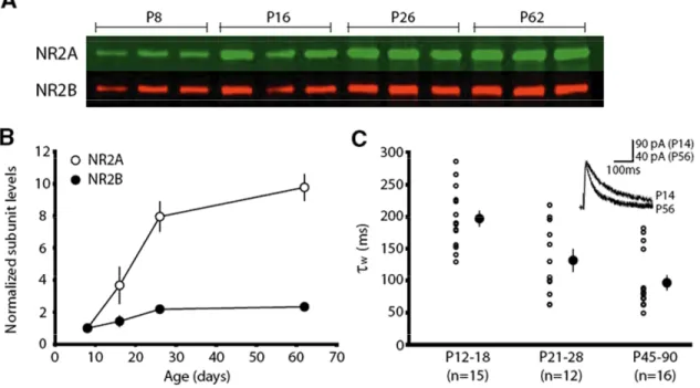
Developmental changes in NMDAR subunit Composition and function in mouse visual cortex . . . .45
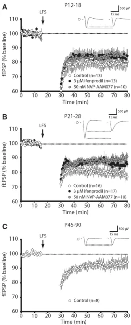
LFS-LTD is developmentally sensitive to NMDAR antagonism. . . . .47
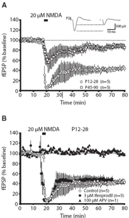
NR2B-type NMDARs not required for expression of chemically-induced LTD. . . .49
NVP-AAM077completely blocks LTP in adult, but not juvenile, animals. . . 51
The developmental effect of NVP-AAM077 is not the result of NR2A-type specificity. . . .52
Developmental differences in the requirements for LTP can explain why NVP-AAM077 has a more profound effect on adult LTP . . . 57
Discussion . . . 62
Experimental Procedures. .. . . 67
Biochemical Fractions . . . 67
Immunoblot Analysis. . . 68
Pharmacological Agents . . . .68
Statistics . . . 68
Acknowledgements. . . 69
Bibliography. . . .. . . 70
III. srGAP2 regulates dendritic spine morphogenesis and synaptic transmission through the synergistic action of its F-BAR and RhoGAP domains. .. . . 77
Summary . . . .78
Introduction . . . .79
Results . . . .82
Supplemental Data. . . 92
IV. Discussion. . . .103
Developmental changes in LTP. . . .103
srGAP2 at the synapse. . . 105
Does srGAP2 playa role in synaptogenesis?. . . .105
Is srGAP2 recruited to the synapse in an activity dependent manner? . . . 105
While Chapter Three provides evidence that genetic loss of srGAP2 upregulates the prevalence of silent synapses, this needs to be directly tested. . . 106
Does genetic loss of srGAP2 influence endocytosis?. . . 106
If stGAP2 acts to prevent AMPA receptor internalization, does it negativelt regulate LTD?. . . 107
Is the function of srGAP2 cell-autonomous?. . . 107
Does srGAP2play an important role in modulating presynaptic release onto L5 pyramidal neurons?. . . 107
Does srGAP2 modulate dendritic spine morphology by modulating the activity of Rac1? . . . 108
LIST OF FIGURES
Figure1.1 Neural anatomy and connectivity . . . 4
1.2 Excitatory glutamatergic and inhibitory GABAergic synapses. . . .6
1.3 NMDAR subunit composition. . . .8
1.4 Dendritic spine development. . . 17
1.5 Physiological significance of dendritic spines. . . 20
1.6 The actin cytoskeleton and small G-proteins play critical roles at the synapse . . . .24
1.7 BAR containing proteins sculpt cellular morphology. . . 31
2.1 Developmental expression of NR2A and NR2B in the mouse visual cortex. . . .46
2.2 NVP-AAM077 and ifenprodil have developmentally- specific effects on the expression of LFS-LTD. . . 48
2.3 Chemically induced LTD is insensitive to ifenprodil application . . . 50
2.4 The ability of NVP-AAM077 to block LTP in the visual cortex is age-dependent . . . .51
2.5 Unlike ifenprodil,, NVP-AAM077 does not modulate NMDAR current kinetics in a developmentally predictable manner . . . .54
2.6 NVP-AAM077 does not modulate NMDAR current amplitude in a developmentally predictable manner. . . .56
2.7 Developmental increase in the sensitivity of LTP to NMDAR antagonists. . . .59
2.8 Juvenile and adult animals have the same postsynaptic field potential responses to 100Hz stimulation. . . 61
morphologies . . . 85
3.3 Genetically modified Layer5 pyramidal neurons can be fluorescently targeted for electrophysiological
recordings . . . .
. . .
. 873.4 Expressing the F-BAR domain of srGAP2 reduces
mEPSC frequency and amplitude . . . .
. . . .
. 883.5 srGAP2 KO L5 pyramidal neurons exhibit a
decreased mEPSC frequency . . . . .
. . .
. 893.6 Spine density is increased in the apical
dendrites of srGAP2 KO animals . . .
. . .
903.7 LTP in CA1 of the hippocampus may be
augmented in srGAP2 KO animals . . .
. . .
. 913.8 Genetic loss of srGAP2 does not affect
dendritic branching patterns in DIV 21 cultures . . . 92
3.9 srGAP2 KO neurons are shorter and wider
than WT neurons, making them less filopodial-like
at DIV14. . . .
. . .
. 933.10 The F-BAR domain of srGAP2 is enriched in
dendritic spines . . . .. . . .94
3.11 The F-BAR domain of srGAP2 is specifically enriched along the dendritic spine shaft, relative
LIST OF ABBREVIATIONS
AMPA - α amino-3-hydroxy-5-methyl-4-isoxazolepropionic acid receptor
APV - ((2R)-amino-5-phosphonovaleric acid; (2R)-amino-5-phosphonopentanoate)
CNQX - 6-Cyano-7-nitroquinoxaline-2,3-dione disodium salt hydrate
DIV – Days in vitro
FBAR - Fer Cip4 Homolgy (FCH) Domain and Bin/Amphiphysin/Rvs-homology (BAR) Domain
LTD – Long Term Depression
LTP – Long Term Potentiation
mEPSC – mini excitatory postsynaptic current
NMDA - N-Methyl D-Aspartate
NMDAR – N-Methyl D-Aspartate Receptor
NR1 – NMDAR 1 Subunit
NR2 – NMDAR 2 Subunit
NR2A - NMDAR 2A Subunit
NR2B - NMDAR 2B Subunit
NVP-AAM077 - (R)-[(S)-1-(4-bromo-phenyl)-ethylamino]-(2,3-dioxo-1,2,3,4 tetrahydroquinoxalin-5-yl)-methyl]-phosphonic acid
P - Postnatal day
RhoGAP – Rho Family GTPase Activating Protein
SH3 – SRC Homolgy 3 Domain
srGAP2 – Slit Robo GTPAse Activating Protein 2
CHAPTER ONE
INTRODUCTION
The brain is anatomically organized in a topographic manner, with distinct
segregation of sensory and motor areas. This regionalization allows sensory input from
each modality to converge upon a defined subpopulation of neurons, which become
specialized in processing discrete types of information. While brain topography is defined
by birth, neural connections within these sensory processing areas are refined in an
activity-dependent manner during early postnatal development. The focus of this thesis is to better
understand the mechanisms that sculpt neural circuitry in the developing cortex. A broad
approach, focusing on both synaptic physiology and morphology, has been taken in
addressing this multifaceted question.
This body of research first focuses on how the composition of postsynaptic N-Methyl
D-Aspartate Receptors (NMDARs) affects the properties of neurotransmission and synaptic
plasticity over the course of postnatal development. Because NMDAR activation is required
for most types of synaptic plasticity, they are profoundly important for sculpting neuronal
circuitry during early postnatal development. While it is well known that NMDARs are critical
regulators of synaptic plasticity, relatively little is known about how the distinct properties of
different types of NMDARs affect mechanisms of long-term potentiation (LTP) or long-term
depression (LTD). NMDAR subunit composition confers distinct properties onto the receptor
by tethering the receptor to different intracellular effector molecules, changing the receptor’s
gating properties, and profoundly influencing the receptor’s current kinetics. Interestingly,
activity-dependent manner. Yet, exactly how this affects synaptic plasticity in the developing
cortex remains elusive.
While electrophysiology has been used extensively to understand how synaptic
function is modulated by the complement of neurotransmitter receptors that are present on
the postsynaptic membrane, far less is known about how the morphology of postsynaptic
specializations, called dendritic spines, shape neurotransmission. During early postnatal
development, dendritic spines are very thin and highly dynamic. But, over the first three
postnatal weeks, spines become increasingly stable and assume a more mature “stubby”
morphology, with a thin neck and a mushroom-like head. To better understand how the
shape of dendritic spines influences synaptic transmission and synaptic plasticity, I focused
on a membrane deforming protein called slit-robo GTPase Activating Protein 2 (srGAP2),
which is expressed at the synapse. Interestingly, this protein has been shown to directly
bind and deform lipid membranes through its FBAR domain. However it can also hydrolyze
and inactivate the small G Protein, Rac1, a known regulator of the actin cytoskeleton.
Therefore, srGAP2 can synergistically influence membrane dynamics through direct
interactions with the cell membrane and indirect interactions through the actin cytoskeleton.
This thesis will first review what is currently known about NMDAR subtypes, their
relative expression over the course of postnatal development, and what is currently known
about their contribution to synaptic plasticity. This will then be followed by a description of
how properties of synaptic plasticity, in the primary visual cortex, have been shown to
change over the course of development. The overlap between changing NMDAR subunit
composition and concomitant changes in synaptic plasticity will be addressed.
This thesis will also address how structural changes in neuronal morphology play a
pivotal role in shaping neural circuitry. How dendritic spine morphology is hypothesized to
affect neurotransmission, will be reviewed. Finally, the function of FBAR and RhoGAP
Cortical connectivity and synaptic plasticity
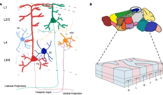
The cerebral cortex is dorsally located within the brain and is subdivided into six
layers, which receive input from different brain regions. Each layer then sends information
to different brain areas (Fig. 1A). This thesis will focus on the plasticity of synaptic
connections made by Layer (L)4 granule cell neurons onto L2/3 pyramidal cell neurons of
the developing cerebral cortex. The molecular mechanisms shaping the morphology of
dendritic spines on L5 pyramidal neurons will also be discussed.
While the broad topographical organization of the brain is defined prenatally,
synaptic plasticity plays an important role in shaping the functional circuitry of the developing
postnatal cortex. One cortical region that has particularly well-defined mechanisms of
synaptic plasticity is the primary visual cortex. In the binocular region of the primary visual
cortex, input from the right and left eye drive activity in defined cortical columns (Fig. 1B).
However, during an early stage of development known as the “critical period” both eyes
compete for the ability to drive the activity of these cortical columns. This is an
activity-dependent process that begins at three weeks and ends at five weeks of life in mice
(Gordon and Stryker 1996). During this period, if one eye is occluded so that no sensory
driven activity can be elicited, the open eye will begin to drive the activity of columns that
were previously activated by the occluded eye (Wiesel and Hubel 1965). The relative
change in the ability of each eye to drive the activity of cortical columns is called ocular
dominance plasticity. This type of plasticity is restricted to the critical period and is very
prominent in highly visual species like cats and humans. In mice, there is not a clear
anatomical segregation of cortical neurons into discrete columns (Antonini et al. 1999) but
the binocular region of the visual cortex still exhibits robust ocular dominance plasticity
Changing the strength of synaptic connections, in an activity-dependent manner,
defines the basis of synaptic plasticity. At its foundation, the concept of synaptic plasticity
rests on the premise that repeated stimulation of a presynaptic cell onto a postsynaptic cell
will result in a stronger synaptic connection. Conversely, cells that are weakly connected
will undergo synaptic weakening (Malenka and Bear 2004). Both excitatory synpases,
which use glutamate as a neurotransmitter, and inhibitory synapses that use GABA as a Figure 1: The anatomy of the brain is highly organized, with complex
connectivity that can be modified in an activity-dependent fashion. (A) The cortex
consists of six layers of neurons that receive information from different afferent populations. Sensory information from the thalamus, synapses onto layer (L) 4 spiny stellate neurons (SSC). L4 neurons then synapse onto L2/3 pyramidal neurons (Pyr), which go onto make robust intracortical synaptic connections onto both pyramidal neurons and interneurons, such as basket (BC) and chandelier cells (Ch). However, the axons of L2/3 neurons project collosally to contalateral cortex, while the axons of L5 pyramidal neurons project ventrally, to deeper brain
structures. (B) Within the demarcated primary visual cortex, specific columns of
neurotransmitter (Fig. 2), can undergo synaptic strengthening and weakening (Hensch et al.
1998; Scannevin and Huganir 2000; Maffei et al. 2006). However, in this thesis I will focus
entirely on the modulation of glutamatergic synapses.
Glutamatergic synapses release glutamate onto a complement of postsynaptic
receptors, including AMPA and NMDA receptors. Both depolarize the postsynaptic neuron
but AMPA receptors produce large, rapid currents in response to glutamate release, while
NMDA receptors have slower current kinetics (Hollmann and Heinemann 1994). NMDA
receptors and AMPA receptors that contain the GluR2 subunit, are permeable to calcium
(Sprengel and Seeburg 1993). This has important implications for the molecular
mechanisms that mediate the expression of synaptic plasticity (Blitzer 2005). A main focus
of this thesis is to elucidate how different types of NMDARs contribute to the expression of
synaptic strengthening, known as LTP, and synaptic weakening, known as LTD.
Composition
and characteristics of NMDARs in the developing cortex
NMDARs allow current influx when presynaptic release of glutamate is coupled to
postsynaptic depolarization. Thus, they have been coined “coincident receptors” due to
their requirement for temporally paired pre- and post-synaptic activity. This unique property
allows NMDARs to play an important role in synaptic plasticity, learning, and memory.
NMDARs contain two NR1 subunits and two ancillary subunits (NR2A-D, NR3A-B) that
confer unique glutamate-binding and kinetic properties onto the receptor (McBain and Mayer
1994; Flint et al. 1997; Laube et al. 1998). Within the cortex, NR2A and NR2B are the most
widely expressed. Thus, the majority of research focusing on the role of NMDAR subunits in
Figure 2: Excitatory glutamatergic and inhibitory GABAergic synapses create the functional connectivity of the cortex. Glutamatergic synaptic contacts are characterized by postsynaptic densities, consisting primarily of PSD-95, that anchor AMPA and NMDA-type glutamate receptors at the synapse. These glutamatergic synapses are subcellularly confined to small dendritic protrusions, called dendritic spines. Glutamate depolarizes neurons by allowing for sodium and calcium influx. Conversely, GABAergic synapses do not have a postsynaptic density and are not exclusively localized at dendritic spines. Furthermore, in the mature cortex, GABA typically hyperpolarizes neurons by promoting the influx of chloride. Thus, excitatory
and inhibitory synaptic contacts differ in (1) the neurotransmitter that is released from
The exact composition of NMDARs in the developing cortex is difficult to ascertain,
with NR1/NR2B dimers, NR1/NR2A dimers, and NR1/NR2B/NR2A triheteromers being
difficult to discriminate (Kohr 2006). However, it is well established that the general
composition of synaptic NMDARs goes from being primarily NR2B-containing, to
increasingly NR2A-containing over the course of development (Monyer et al. 1994;
Watanabe et al. 1994)(Fig 2). This developmental upregulation of the NR2A subunit is
activity-dependent (Quinlan et al. 1999) and is controlled at the transcriptional level
(Hoffmann et al. 2000; Yashiro and Philpot 2008). While the significance of this transition is
unclear, NR2A and NR2B subunits confer important differences onto the channel properties
and intracellular binding partners of NMDARs. NR1/NR2A dimeric receptors have a higher
open probability and faster deactivation kinetics than NR1/NR2B receptors (Monyer et al.
1994; Vicini et al. 1998). Functionally, this means that NR1/NR2A dimers have fast current
kinetics, while NR1/NR2B dimers have slower currents that result in a greater amount of
charge transfer (Erreger et al. 2005). Interestingly, calcium-imaging studies have shown that
NR1/NR2B dimers allow the influx of a greater amount of calcium ions per unit of charge
than NR1/NR2A dimers (Sobczyk et al. 2005). Because calcium mediates a host of second
messenger pathways, this difference in calcium permeability may have important
implications for how synaptic plasticity is regulated by these subunits. Not surprisingly,
triheteromeric NR1/NR2A/NR2B NMDARs have characteristics that are intermediate to pure
NR2A or NR2B dimers (Vicini et al. 1998). One study using serial immunoprecipitation,
reported that in adult hippocampus, 2/3 of NMDARs were diheteromeric, while only 1/3 of
NMDARs were triheteromeric (Al-Hallaq et al. 2007). However, the relative amount of
NMDAR dimers to NMDAR triheteromers is difficult to ascertain due to developmental and
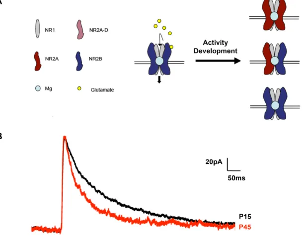
Figure 3: NMDAR subunit composition, at glutamatergic synapses, in the developing
cortex. (A) NMDARs consist of two obligatory NR1 subunits and two ancillary
subunits that can be either NR2A-D or NR3A-B. These secondary subunits confer unique glutamate binding affinities and current kinetics onto the receptor. NDMARs are permeable to sodium and calcium when glutamate is bound to the receptor and when the postsynaptic membrane potential is sufficiently depolarized to release the channel’s magnesium block. Because the receptor only allows depolarization to occur when presynaptic release and postsynaptic depolarization are temporally synchronized, the NMDAR has been coined a “coincident receptor” of both pre- and post-synaptic activity. Interestingly, over the first three postnatal weeks, NMDARs undergo a profound activity-dependent shift from being primarily NR2B containing, to
largely NR2A containing. (B) This developmental change in NMDAR subunit
Subcellular expression of NMDAR subtypes
While it is clear that different NMDAR subunits confer distinct properties onto
NMDARs, it is unclear whether different types of NMDARs preferentially populate distinct
subcellular regions (synaptic versus extrasynaptic) or whether the composition of NMDARs
changes in an input-specific manner. Gaining a better understanding of the where NMDAR
subunits are expressed and how their expression changes in response to activity, will shed
light onto the physiological importance of different NMDAR subunits.
NR2A-containing NMDARs may preferentially populate the central region of the
synapse. This is supported by a study showing that NMDAR mediated mEPSCs have faster
current kinetics than evoked NMDAR currents (Dalby and Mody 2003). This suggests that
NR2A-containing NMDARs, with relatively fast current kinetics, populate the central region
of the synapse that is sensitive to single vesicles of neurotransmitter release. However,
upon action potential-evoked release, more peripherally located NR2B-containing receptors
that have slower current kinetics are activated, changing the current decay kinetics. This
interpretation hinges on the premise that univesicular release is subsaturating at the
synaptic cleft, and that only evoked release can stimulate all the NMDARs at the synapse.
However, this concept is highly contested and may differ between developmental stages
and brain regions (Clements 1996). Yet, the idea that NR2A-containing NMDARs are
present in the central portion of the synpase is further supported by the finding that there are
no NMDAR components to mEPSCs recorded in NR2A knockout mice (Townsend et al.
2003). There are also a number of studies that demonstrate the presence of extrasynaptic
NR2B-containing NMDARs (Stocca and Vicini 1998; Tovar and Westbrook 1999).
Over the course of development, NMDAR subunit composition changes from being
largely NR2B-containing, to increasingly NR2A-containing. While it is known that
unknown whether this transition occurs in a synapse-specific manner. However, there are
several lines of evidence suggesting that NMDAR composition is regulated on the synaptic
level. First, inter- and intra-cortical synapses have different NR2A/NR2B ratios—
demonstrating that synapses receiving inputs from different populations of afferents have
different complements of NMDARs (Kumar and Huguenard 2003). Also, on the single-cell
level, NR2B is differentially expressed on apical and basal dendrites (Kawakami et al. 2003).
Additionally, synaptic plasticity appears to change the composition of NMDARs at the
synapse. In line with this, the induction of LTP in the CA1 region of the hippocampus causes
an input specific increase in the NR2A/NR2B ratio. Therefore, synapse-specific differences
in NMDAR subunit composition may explain why different synapses exhibit different degrees
of synaptic plasticity (Matsuzaki et al. 2004).
NMDA receptor subunit composition and synaptic plasticity
NMDAR subunit composition may affect plasticity by modulating NMDAR current
kinetics and by influencing intracellular binding to distinct signaling molecules. There are a
number of reasons to speculate that the NR2B subunit promotes LTP. (1) The NR2B
subunit allows for more charge transfer and has greater calcium permeability than the NR2A
subunit (Monyer et al. 1994; Vicini et al. 1998; Erreger et al. 2005). (2) NR2B preferentially
interacts with CamKII, a molecule known to be important for the induction of LTP (Strack
and Colbran 1998; Lisman et al. 2002). (3) Overexpressing the NR2B c-terminus, which
limits the intracellular interaction of the NR2B subunit to its endogenous binding partners like
CamKII, decreases the magnitude of LTP (Barria and Malinow 2005). (4) Exogenous
expression of NR2A, which reduces the relative amount of NR2B at the synapse, attenuates
LTP in hippocampus (Barria and Malinow 2005). (5) Genetic loss of NR2A does not affect
lines of evidence provide strong support for the role of NR2B in LTP. However, while
NR2B-containing NMDARs are relatively downregulated at the synapse during development, LTP
persists at maturity (Kirkwood et al. 1997; Jiang et al. 2007). This suggests that the relative
amount of NR2A and NR2B at the synapse may not dictate the polarity of plasticity, as much
as modulate the degree of synaptic stimulation required to induce plasticity in the postnatal
cortex (Yashiro and Philpot 2008). Because increasing expression of NR2A would limit
charge transfer, reduce calcium permeability, and attenuate the amount of CamKII proximal
to the synapse, its expression may increase the amount of synaptic stimulation required for
LTP.
The idea that NR2A upregulation modulates the amount of stimulation required to
induce synaptic plasticity, is supported by studies that manipulate the expression of NR2A in
the developing cortex. Using dark rearing to prevent NR2A upregulation in the visual cortex,
it has been shown that the frequency of stimulation required to induce LTP and LTD is
reduced. This effectively increases the range of frequencies that can be used to evoke LTP
in these animals. Therefore, it has been speculated that the developmental change in the
NR2A/NR2B ratio causes a shift in the LTP/LTD induction threshold. This would effectively
create a lower threshold for LTP induction at early developmental timepoints, when cortical
circuitry is being established. Studies done on NR2A knockout animals also provide strong
evidence that NR2A upregulation is required for increasing the threshold for LTP, over the
course of development.
Also, dark rearing does not reduce the threshold for LTP induction in these knockouts,
suggesting that NR2A is required for activity-dependent changes in the synaptic plasticity
threshold (Philpot et al. 2007). Furthermore, the LTP threshold in NR2A knockout mice is
greatly diminished, with 1Hz stimulation being sufficient to drive LTP (Philpot et al. 2007).
While the NR2B subunit of the NMDAR has been shown to be important for LTP,
NR2A upregulation increases the stringency for LTP induction, by diluting the amount of
NR2B at the synapse, it does not prevent the expression of LTP all together. Yet, a direct
association between NR2A and known LTD signaling pathways, like the PP1/PP2B
(calcineurin pathway) (Yashiro and Philpot 2008), has yet to be shown. Because of this, it
does not appear that each subunit is coupled to LTP or LTD in a binary fashion. Rather, it
seems that the developmental transition in NMDAR subunit composition regulates plasticity
over the course of postnatal development by changing the amount of stimulation required to
induce LTP, in an activity-dependent fashion.
Using pharmacology to assess subunit-specific functions
One approach to better understand how current through distinct subtypes of
NMDARs contribute to synaptic plasticity, is to use pharmacological antagonists that
selectively block particular subunits. While much can be gained from these types of studies,
it should be re-emphasized that NMDARs in the cortex heterogeneous—and this can
strongly influence their susceptibility to NMDAR antagonists. As stated earlier, it has been
estimated that 2/3 of cortical NMDARs are dimeric NR1/NR2A or NR1/NR2B-containing
receptors, while 1/3 of cortical NMDARs are triheteromeric NR1/NR2A/NR2B- containing
NMDARs (Al-Hallaq et al. 2007).
Ifenprodil is a well-characterized antagonist for NR2B-containing NMDARs. It binds
the amino terminus of the NR2B subunit in a non-competitive fashion (Gallagher et al. 1996;
Perin-Dureau et al. 2002; Mosley et al. 2009). Performing whole-cell recordings, in the
presence of ifenprodil, it has been demonstrated that more current is carried by
NR2B-containing NMDARs at early developmental timepoints when compared to later
developmental timepoints. This is completely congruent with its biochemically defined
expression pattern (Monyer et al. 1994; Stocca and Vicini 1998; de Marchena et al. 2008).
and NR2A subunits (Kew et al. 1996; Tovar and Westbrook 1999). This makes it impossible
to assess the contribution of trihetomeric NMDARs, which may represent as much as 1/3 of
the total NMDAR population (Al-Hallaq et al. 2007). It has also been reported that the
efficacy of ifenprodil is strongly influenced by the concentration of glutamate at the synaptic
cleft. Ifenprodil has actually been shown to potentiate NMDAR currents at low glutamate
concentrations (Kew et al. 1996). However, the broad applicability of this phenomenon to
cortical synapses is unclear.
More recently, an antagonist was developed to specifically block NR2A-containing
NMDARs. This antagonist, NVP-AAM077, was initially reported to be 100 times more
selective for NR2A, over NR2B-containing receptors in a cell line that was exogenously
expressing human NMDARs (Auberson et al. 2002). However, subsequent reports found
that NVP-AAM077 was only 6-12 times more specific for NR2A, when rodent NMDARs were
expressed (Feng et al. 2004). Furthermore, it was found that NVP-AAM077 blocks a
significant amount of NMDAR current in brain preparations obtained from NR2A knockout
animals (Neyton and Paoletti 2006; de Marchena et al. 2008).
Yet, because the drug was initially reported to have such strong specificity for
NR2A-containing NMDARs, many investigators began use NVP-AAM077 alongside ifenprodil to
probe for NR2 subunit-specific contributions to synaptic plasticity. Interestingly, two reports
emerged demonstrating that plasticity in the hippocampus and in the perirhinal cortex was
strongly dependent on subunit-specific activation (Liu et al. 2004; Massey et al. 2004). In
these studies, LTP was blocked by the NR2A-specific antagonist NVP-AAM077 and LTD
was blocked by the NR2B-specific antagonist ifenprodil. These findings suggested that LTP
was dependent on NR2A-containing NMDARs and LTD was dependent on NR2B-containing
NMDARs. However, three separate laboratories were unable to replicate these findings,
reporting that LTD in hippocampus was completely insensitve to ifenprodil application. LTP
the highest possible specificity for NR2A-containg NMDARs (Berberich et al. 2005; de
Marchena et al. 2008).
However, if properly interpreted, pharmacology can be used to better understand
how different types of NMDARs contribute to synaptic plasticity. The work described in
Chapter Two of this thesis employs pharmacology to carefully assess how the properties of
synaptic plasticity change over the course of development. In this work, we found that
NVP-AAM077 only attenuates LTP at late developmental time points. Using a subsaturating
concentration of APV, a pan-NMDAR antagonist, we mirrored the differential effect of
NVP-AAM077 on LTP. We propose that this is the result of a developmental increase in the
amount of NMDAR activation needed to elicit LTP (de Marchena et al. 2008).
Synaptic morphology and the development of dendritic spines
While this introduction has focused on the role of postsynaptic NMDARs, the
morphology of synapses is also very important in shaping neural circuitry. While the
postsynaptic element of synapses cluster neurotransmitter receptors, its general shape
affects its response to neurotransmitter and its ability to drive dendritic depolarization.
Dynamic changes in the shape of the synapse and the width of the synaptic cleft are also
thought to underlie elements of synaptic plasticity (Engert and Bonhoeffer 1999; Yuste and
Bonhoeffer 2001; Hofer et al. 2009).
90% of glutamatergic synapses are located on dendritic protrusions, known as
dendritic spines (Nimchinsky et al. 2002). These spines have a characteristic morphology
that undergo dynamic changes during development (Dailey and Smith 1996; Ziv and Smith
1996; Matus 2000; Yuste and Bonhoeffer 2004). The spine contains an electron dense
region, known as the postsynaptic density, where the majority of glutamate receptors and
accompanying signaling molecules are clustered (Gray 1959; Parnavelas et al. 1977). This
region. These inhibitory synapses do not form dendritic spines or contain a postsynaptic
density (DeFelipe and Farinas 1992). However, the biological purpose of dendritic spines
and the reason why they are specific to glutamatergic synapses, remains debated.
Dendritic spines begin to form during the first week of postnatal life and increase in
density until the fourth postnatal week (Nimchinsky et al. 2002). At this point, there is a
gradual reduction in the number of dendritic spines. Therefore, there is an initial
overproduction of dendritic spines, which is followed by elimination. Early in development
thin, long filopodial-like protrusions emerge from dendrites. The terminal filopodial-like
protrusions that extend from the ends of developing dendrites, guide the formation of
branches on the dendritic tree (Luo 2002). However, in CA1 of the hippocampus, collateral
filopodia have been shown to extend from the middle of dendritic processes and make
putative synaptic contacts (Fiala et al. 1998). These axo-dendritic contacts then go on to
form dendritic spines (Fiala et al. 1998).
Dendritic spines are conventionally subdivided into three categories: stubby, thin,
and mushroom-like (Peters and Kaiserman-Abramof 1970). Developmentally, dendritic
spines transition from being stubby, to thin, and then finally to mushroom-shaped (Yuste and
Bonhoeffer 2004). However, dynamic filopodia play an important role in initiating synaptic
connections that have the potential to form dendritic spines. Because developing
neocortical neurons have been reported to form as many as 50,000 new filopodial
protrusions per day (Portera-Cailliau et al. 2003), these extensions are uniquely able to
probe the extracellular environment for appropriate synaptic connections. This active
process allows developing neurons to create functional synaptic circuitry. However, while
several models have been proposed, the exact mechanism that induces the formation of
dendritic spines is still an area of active debate (Yuste and Bonhoeffer 2004).
One conceptualization, coined the Sotello Model, posits that dendritic spines form in
the evidence for this model comes from the cerebellum, looking at synaptic connections
between granule and Purkinje cells. In mutants where granule cells are missing, resulting in
a loss of presynaptic innervation onto Purkinje cells, dendritic spine formation is unperturbed
(Mariani et al. 1977). Purkinje neurons in these knockouts even have normal postsynaptic
specializations without pre-synaptic apposition. Similar findings have been reported using
X-irradiation to ablate granule cells in the cerebellum (Altman and Anderson 1972). It also
appears that dendritic spines, at least in the cerebellum, emerge before the presynaptic
specializations can be visualized in the proximity of the spine. These spines are referred to
as “naked spines” and their presence suggests that spines can form independently of
presynaptic specializations. However, the rules that govern spinogenesis may differ across
different neuronal sub-types, as naked spines are rarely observed in the neocortex (Arellano
et al. 2007).
Another theory on spine formation is that filopodia are spine precursors that directly
seek out presynaptic afferents (Ziv and Smith 1996; Yuste and Bonhoeffer 2004). Once a
functional synaptic contact is made, dendritic spines are thought to mature from these
elongated protrusions. However, two lines of evidence suggest that this is an unlikely
scenario. First, filopodial protrusions are made by many neurons that do not go on to form
dendritic spines (Mason 1983; Wong et al. 1992; Linke et al. 1994). Therefore, while
dynamic filopodial protrusions may play a role in dendritic spine formation, they must serve
other functions that are unrelated. Secondly, the idea that filopodia are precursors to
dendritic spines is contrary to the ontogeny of dendritic spine development in the neocortex
and cerebellum, which progress from being stubby, to being longer and mushroom-like
(Harris et al. 1992; Yuste and Bonhoeffer 2004).
The most likely mechanism guiding spinogenesis in the neocortex is the Miller/
Peters hypothesis, where axonal contacts induce the formation of dendritic spines through
range of developmental time points, in the CA1 region of the hippocampus, provides
convincing evidence that synaptic connections precede the formation of dendritic spines
(Fiala et al. 1998). These early synaptic contacts do not exhibit any type of postsynaptic
protrusions and are referred to as shaft synapses. These contacts go on to form stubby
spines, before ultimately making mature mushroom-shaped spines. However, the extensive
number of filopodia that are generated during the early postnatal period probably act to
provide cellular surface area for the formation of synapses (Fiala et al. 1998). Therefore,
while filopodia appear to play an important role in allowing dendrites to make contacts with
axons, spine formation itself seems to be coincide with the presence of a presynaptic
contact.
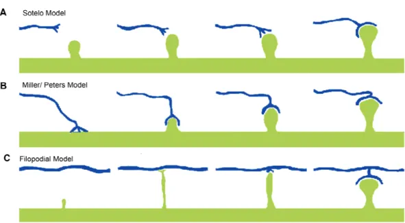
Figure 4: Genesis and maturation of dendritic spines in the postnatal cortex (Yuste
et al. 2004). (A) The Sotelo Model of dendritic spine formation, suggests that
dendritic spines form in a cell autonomous fashion, independent of cues from
presynaptic axons. (B) The Miller/Peters Model claims that the axon induces
Functional significance of dendritic spines
Although dendritic spines are prominent features of glutamatergic synapses, how
they influence neural circuitry is just beginning to be understood. Spines are highly dynamic
structures that can change in shape, appear de novo, or disappear altogether. Although
changes in spine morphology and turnover are more robust during early postnatal
development, the fact that this phenomena persists into adulthood suggests that
morphological changes in spines continue to play an important role in sculpting neural
communication throughout life (Grutzendler et al. 2002; Trachtenberg et al. 2002).
Live imaging experiments on fluorescently labeled neurons in vivo reveal a
developmental decrease in spine dynamics (Grutzendler et al. 2002; Trachtenberg et al.
2002). During early postnatal development there is rapid addition and retraction of filopodial
and spine-like processes. Interestingly, as development proceeds, the overall density of
dendritic spines decreases and the spines that remain are increasingly persistent
(Grutzendler et al. 2002). This suggests that there is an early phase of dynamic connectivity
that is followed by a process of refinement. This progressive elimination of dendritic spines
reflects the pruning of synaptic connections that occurs through input competition, a process
known to play a central role in neural development (Lendvai et al. 2000).
Spines have been shown to change their dynamics in an activity dependent fashion.
Globally preventing neural activity, by perfusing tetrodotoxin (TTX), causes a robust
increase in spine density (Bravin et al. 1999). A similar increase in spine density was found
subsequent to monocular deprivation in the binocular region of the primary visual cortex
(Hofer et al. 2009). This may reflect a homeostatic mechanism that acts in response to
decreased levels of synaptic activity. Interestingly, the change in spine density that follows
monocular deprivation persists and repeated deprivation fails to produce any additional
changes in spine density. Therefore, it appears that the spines underlying an alternative
recruited to respond to a change in the sensory environment. Whisker trimming also affects
the protrusive motility of dendritic spines in the barrel cortex (Lendvai et al. 2000). It should
be noted that this change in spine motility is restricted to the critical period, with no effect
observed for sensory deprivation occurring before or after this developmental window. High
frequency stimulation protocols that are typically used to induce LTP in organotypic slices
have also been shown to increase spine density in CA1 of the hippocampus (Engert and
Bonhoeffer 1999). Therefore, it appears that directly manipulating activity with drugs,
stimulation protocols, and natural sensory-evoked activity can all influence spine dynamics.
The shape of dendritic spines also plays an important role in modulating postsynaptic
characteristics. Elongated spine necks are thought to create a diffusional bottleneck that
impedes the flow of calcium and important intracellular signaling molecules from the
proximity of the synapse (Yuste et al. 2000). Spine specific increases in calcium
concentration have been visualized, with calcium sensitive dyes, following synaptic
stimulation (Denk et al. 1996). Diffusional exchange has also been measured using
fluorescence recovery after photobleaching (FRAP) experiments—confirming the anatomical
assumption that spines can act as semiautonomous microdomains (Svoboda et al. 1997).
However it is also hypothesized that spine shape is capable of not only influencing its
diffusional characteristics, but also its electrical properties (Nimchinsky et al. 2004; Tsay and
Yuste 2004; Spruston 2008). Specifically, the length of the spine neck may act to limit how
depolarization, at the spine head, translates into depolarization of the dendritic shaft. This is
important because voltage sensitive calcium channels may restrict calcium entry to this
small microdomain, if depolarization is largely restricted to the spine head. This physical
restriction of calcium diffusion could underlie input-specific mechanisms governing plasticity.
The ability of spines to restrict depolarization is also important because it influences how
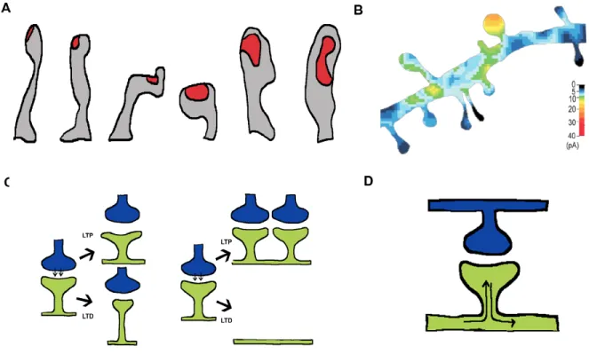
Figure 5: The physiological significance of dendritic spines (A) There is a strong correlation between dendritic spine shape and the size of the postsynaptic density (PSD), illustrated in red. The three spines on the left have an elongated, thin, filopodial-like shape and they have relatively small PSDs. However, the three spines on the right have a stubby, bulbous morphology and concomitantly larger PSDs
(Arellano et al. 2007). (B) Stubby spines are also more responsive to uncaged
glutamate. Thus, it appears that the large PSDs found in stubby spines correspond with the presence of large postsynaptic regions of glutamatergic receptors (Matsuzaki
et al. 2004). (C) Dendritic spines have also been speculated to play an important role
in structural plasticity. The shape of the dendritic spine can be modified to increase or decrease the density of postsynaptic receptors (as illustrated on the left) in response to either long-term potentiation (LTP) or long-term depression (LTD). The gross number of dendritic spines along the shaft can also change in response to synaptic
plasticity (as illustrated on the right). (D) Dendritic spines have also been speculated to
restrict depolarization is dependent on the resistivity of the spine neck, which is highly
coupled to spine length and width.
While spine neck resistivity has been estimated to be as high as 150 MΩ, this value
is not predicted to be high enough to significantly modulate synaptic currents (Svoboda et al.
1997). However, several lines of evidence suggest that spine head volume and spine neck
length can both affect somatic depolarization. Using glutamate uncaging to map the
glutamate sensitivity of individual dendritic spines, a direct correlation was found between
spine head volume and the degree of depolarization that was measured at the soma
(Matsuzaki et al. 2001). Therefore, larger, more mushroom-shaped spines solicited a
greater depolarization at the soma than longer more filopodial-like spines. Although differing
densities of AMPA receptors at differently shaped spines could account for this finding,
subsequent studies have found a direct correlation between spine neck length and degree
of depolarization at the soma (Araya et al. 2006). Therefore, while biophysical evidence
suggests that spines cannot act as electrically isolated units, experimental evidence
provides functional evidence that they do.
In summary, dendritic spines are highly dynamic structures that act to probe the
neural environment and modulate synaptic connectivity. While Ramon y Cajal was the first
to describe their structure and postulate their function in the nervous system,a renewed
interest in their dynamics came when it was discovered that spines were rich in actin
(Bonhoeffer and Yuste 2002). The presence of actin in dendritic spines was considered
strong evidence for their motility, before live imaging experiments were able to demonstrate
this definitively (Fischer et al. 1998). Now molecular mechanisms that influence the actin
cytoskeleton, in order to shape spine dynamics and morphology, have been explored in
Actin cytoskeleton and dendritic morphology
Both immunohistochemistry and live imaging of GFP-actin fusion proteins have
shown that actin is highly enriched in dendritic spines (Fischer et al. 1998; Hotulainen et al.
2009). Using pharmacology to manipulate actin dynamics has demonstrated that the actin
cytoskeleton plays an important role in mediating not only spine motility, but also spine
formation. Application of Cytochalasin D, a potent inhibitor of actin polymerization, arrests
motility in mature dendritic spines (Fischer et al. 1998; Dunaevsky et al. 1999), suggesting
that spine dynamics are critically dependent on modulation of the actin cytoskeleton.
Additionally, strong tetanic stimulation has been shown to induce actin remodeling, in pre-
and post-synaptic terminals (Colicos et al. 2001). Interestingly, at earlier developmental time
points, inhibiting actin polymerization completely prevents the formation of dendritic spines.
This indicates that actin dynamics are also important for dendritic spine formation.
However, as spines mature, their gross morphological structure becomes increasingly
independent of actin dynamics (Zhang and Benson 2001). Therefore, while the actin
cytoskeleton may not play a critical role in modulating the gross morphology of mature
spines, it appears to be important in mediating structural adaptations to neural stimulation.
Rho-GTPases are known to be potent regulators of the actin cytoskeleton. They act
as intracellular switches that go from an inactive GDP bound state to an active GTP bound
state (Fig6). In their active state, they modulate downstream effectors, like Arp2/3, WASP,
and cofilin, which directly regulate the actin polymerization (Hall 1998). The three best
characterized Rho GTPases are RhoA, Rac1, and cdc42 (Luo 2002). In cultured fibroblasts,
expression of RhoA induces the formation of stress fibers, expression of Rac causes the
formation of lamellipodia, and expression of cdc42 induces the formation of filopodia (Hall
1998). However, the binary relationship between these Rho GTPases and the phenotype
filopodia and lamellapodia (Luo 2002). Therefore, a clear understanding of exactly how Rho
GTPases affect the actin cytosketon of neurons remains elusive.
However, Rho GTPases clearly regulate spine formation and spine morphology.
These effects are likely mediated by the influence these proteins have on the actin
cytoskeleton. In transgenic mice that overexpress a constitutively active form of Rac1, there
is an increase in spine number (Luo et al. 1996). Interestingly, biolistically inducing Rac1
expression in wild-type neurons that had already undergone synaptogenesis revealed a
similar increase in spine number (Nakayama and Luo 2000). This suggests that Rac1 is not
only important for synaptogenesis at early developmental time points, but is also important
for regulating spine number at maturity. Interestingly, RhoA was found to have the opposite
effect on dendritic spine density. Therefore, while Rac1 expression is sufficient to increase
spine number, RhoA expression dramatically decreases spine number. Rac1 and RhoA
also have opposite effects on the length of spine necks. Rac1 expression induces the
formation of elongated necks, while RhoA expression causes a dramatic reduction in neck
length (Tashiro et al. 2000). Therefore, Rac and RhoA appear to influence spine number
and morphology in a reciprocal fashion.
Mutation of RhoGAP and RhoGEF proteins that regulate the activity of Rac1 and
RhoA have both been shown to result in mental retardation syndromes (Chelly and Mandel
2001; Ramakers 2002). Therefore understanding how these upstream effectors are
modulated will be important in understanding the types of intra- and extracellular cues that
are able to solicit changes in spine morphology. Proteins containing RhoGEF and RhoGAP
domains have been shown to interact with receptor mediated signaling pathways that
influence spine morphology (Yamashita et al. 1999; Li et al. 2002). Therefore, RhoGAPs
and RhoGEFs may be the intermediaries that allow neurons to respond to cues from the
extracellular environment, like presynaptic stimulation. This may allow receptor activation to
domains, in proteins that are known to modulate spine dynamics, other domains that directly
interact with the cell membrane may be underappreciated in their ability to shape spine
morphology.
Figure 6: The actin cytoskeleton and small G-proteins play a critical role at the synapse.
(A) Actin plays an important role both pre- and post-synaptically, regulating vesicle docking
and recycling. This ultimately affects both presynaptic neurotransmitter release, and the
composition of receptors at the postsynaptic membrane. (B) The Rho family of small
BAR-domain containing proteins and membrane dynamics
Because small GTPase signaling shapes actin dynamics and because mutations in a
number of proteins that regulate small GTPases result in mental retardation syndromes,
tremendous focus has been placed on understanding how perturbations in these pathways
affect neuronal function. Interestingly, proteins like oligophrenin1 and srGAP3, which are
linked to mental retardation syndromes, have a RhoGAP domain and bin-amphiphysin-Rvs
(BAR) domain (Itoh and De Camilli 2006). However, the functional importance of proteins
with BAR domains is just beginning to be understood.
BAR domains were first characterized as highly homologous N-terminal sequences
in amphiphysins, a large family of proteins known to be important regulators of endocytosis
(Zhang and Zelhof 2002). These domains are subdivided into three subtypes: BAR, F-BAR,
and I-BARs, which induce the formation of narrow, wide, and inverted membrane tubules,
respectively. BAR domains are alpha helical dimers that interact with cellular membranes
through electrostatic interactions between their positively charged ends and the negatively
charged membrane (McMahon and Gallop 2005; Itoh and De Camilli 2006; Zimmerberg and
Kozlov 2006). These domains oligomerize to cooperatively form rigid scaffolds that force
membrane bending (Itoh and De Camilli 2006). Many BAR domains bind to actin in
addition to the membrane and are therefore ideally positioned at the interface between
membrane and actin cytoskeleton (Itoh and De Camilli 2006).
Because neurons have elaborate shapes that that are tightly coupled to their
function, BAR-containing proteins that directly modulate membrane deformation are
hypothesized to be important for achieving mature axodendritic morphology. In fact,
BAR-containing proteins have been found to influence dendritic spine shape and density (Choi et
al. 2005; Khelfaoui et al. 2007). They have also been implicated in the formation of dendritic
arborization patterns (Sawallisch et al. 2009) and in the modulation of synaptic plasticity
protrusions that can guide dendritic outgrowth, BARs and F-BARs can induce invaginations
that are important for endocytosis of surface receptors (Khelfaoui et al. 2009). However,
most of proteins that have been studied, which contain these domains, have focused on
their ability to regulate small GTPase signaling. Therefore, a thorough understanding of
how the BAR domains of these proteins directly regulate neuronal morphology, has yet to be
elucidated.
IRSp53 is an I-BAR containing protein that is highly expressed in the postsynaptic
density and is known to link activated Rac1 and cdc42 to downstream effectors for actin
regulation (Choi et al. 2005). Because I-BAR domains mediate the formation of membrane
protrusions, it would be reasonable to speculate that they might influence the dynamics of
filopodial-like structures in neurons, such as dendritic spines. In accordance with this,
overexpression of IRSp53 in cultured neurons results in increased spine density, whereas
knockdown of IRSp53 results in decreased spine density (Choi et al. 2005). This ties in well
with electrophysiological data showing that overexpression of IRSp53 causes an increase in
mEPSC frequency (Hori et al. 2005). This increase in mEPSC frequency suggests that the
new spines formed by overexpression of IRSp53, are physiologically functional. IRSp53 has
also been shown to translocate to dendritic spines upon NMDAR activation (Hori et al.
2005). This translocation is dependent on phosphorylation of the N-terminal I-BAR domain
of IRSp53, by Protein Kinase C (PKC). Therefore, it appears that the phosphorylation of the
I-BAR domain in IRSp53 regulates its intracellular localization. Interestingly,
NMDAR-mediated mEPSCs are reduced in the IRSp53 knockout. Exactly how this reduction of
NMDAR-mediated mEPSCs is related to the fact that NMDAR activation influence IRSp53
localization is unclear. However, the link between IRSp53 and NMDARs may help explain
two independent observations that genetic loss of IRSp53 results in enhanced LTP (Kim et
Oligophrenin 1 is a BAR domain containing protein that is ubiquitously expressed
throughout the brain, during embryonic and postnatal stages of development. Mutation of
this gene results in a nonspecific X-linked mental retardation syndrome in humans.
Because this protein contains a BAR domain, it is hypothesized to play an important role in
vesicle endocytosis. However, it appears that oligophrenin1 may play distinct, seemingly
contradictory, roles through the actions of its BAR and RhoGAP domains. Yet, a thorough
functional dissection of the BAR and RhoGAP domains of this protein remains to be done.
Several lines of evidence suggest that oligophrenin1 plays an important role in
vesicle recycling. Paired pulse facilitation in the hippocampus of oligophrenin1 knockout
mice reveals an increase in the probability of neurotransmitter release (Khelfaoui et al.
2007). This suggests that oligophrenin1 mediates vesicle recycling at the presynaptic
terminal. Additional experiments, measuring FM1-43 internalization, reveal that genetic loss
of oligophrenin1 results in diminished endocytosis (Khelfaoui et al. 2009). Specifically, it
appears that loss of oligophrenin1 results in an impairment of AMPA receptor internalization,
a process critically regulated by endocytosis. Two functional measures that could be
explained by this impairment in endocytosis are (1) diminished LTD (Khelfaoui et al. 2009),
which is critically dependent on the postsynaptic internalization of AMPARs, and (2) poor
performance on spatial learning tasks, like the Morris Water Maze (Khelfaoui et al. 2007).
However, because oligophrenin1 is knocked out in all neurons it is difficult to separate its
pre- and post-synaptic function. For example, could the effect of oligophrenin1 on
presynaptic neurotransmitter release be secondary to the protein’s postsynaptic role in
AMPAR endocytosis? Another issue that confounds the interpretation of these results is the
potentially overlapping function of the RhoGAP and BAR domains of oligophrenin1. The
RhoGAP domain of oligophrenin1 downregulates RhoA/ROCK signaling, which inhibits
vesicular endocytosis (Fauchereau et al. 2003; Govek et al. 2004; Khelfaoui et al. 2009).
signaling, which would attenuate endocytosis. However, oligophrenin1 may also affect
endocytosis directly through the action of its BAR domain. Yet this possibility remains
entirely unexplored.
Oligophrenin1 also influences spine formation, stabilization, and morphogenesis.
Genetic deletion of oligophrenin1 causes a decrease in dendritic spine density along the
apical dendrite of CA1 neurons (Khelfaoui et al. 2007). Furthermore, the morphology of
dendritic spines becomes elongated in knockout animals but shorter and stubbier upon
knockdown in vitro . This suggests that oligophrenin1 positively regulates the formation of
dendritic spines and that it somehow influences spine morphology. Interestingly, the activity
of the RhoGAP domain of oligophrenin1 is inconsistent with the observed decrease in spine
density. This is because oligophrenin1 decreases RhoA activity, which is important for
increasing spine density and promoting an elongated filopodial-like morphology (Tashiro et
al. 2000). Therefore, in the absence of oligophrenin1, RhoA activity should be increased,
resulting in a concomitant increase in spine density. However, it is possible that the effect of
Oligophrenin1 on spine density is secondary to its better characterized influence on AMPA
receptor internalization (Khelfaoui et al. 2009). While it is unclear how Oligophrenin1
modulates dendritic spine number and morphology, a better understanding of exactly how
its BAR and RhoGAP domains influence membrane dynamics will help elucidate the
complex role of this protein.
Pacsin1 (also called Syndapin1) is an F-BAR containing protein that is important in
regulating the activity-dependent internalization of NR3A-containing NMDARs (Perez-Otano
et al. 2006). This function of Pacsin1 is entirely consistent with the fact that it contains an
F-BAR domain, which has been classically defined as a protein domain that solicits the
formation of membrane invaginations and thereby regulates endocytosis. Interestingly,
Pacsin1 internalizes a specific type of NMDAR, with spatiotemporal specificity. More recent
dendritic branching in neurons by not only coordinating WASP-mediated actin
polymerization through its SH3 domain, but also by deforming the membrane directly
through its F-BAR domain (Dharmalingam et al. 2009).
srGAP3 is an F-BAR containing protein that also has a RhoGAP domain. srGAP3
has been shown to directly interact with WAVE1, which can influence cytoskeletal dynamics
by activating Arp2/3 (Soderling et al. 2007). Interestingly, transgenic mice that have a
WAVE1 mutation that abolishes its interaction with srGAP3, display enhanced LTP (similar
to the IRSp53 knockouts)(Soderling et al. 2007). The molecular mechanism that drives this
enhancement in LTP remains unknown. Additionally, the biological function of the F-BAR
domain of srGAP3 remains to be explored.
Although speculative, BAR containing proteins may influence dendritic spine
morphology by forming dense scaffolds that promote their stabilization. Similar to BAR
containing proteins, septins can polymerize into oligomers that form microscopic ring-like
structures close to the plasma membrane. Septins are also GTPases that have structural
similarity to the Ras family (Kinoshita 2003). Therefore, they can also modulate
downstream effectors of the actin cytoskeleton. Interestingly, Septin7 localizes to the base
of dendritic spines and to the branch points in dendritic arbors (Tada et al. 2007; Xie et al.
2007). Knocking down Septin7 results in an elongation of dendritic spine morphology and a
decrease in dendritic branching (Xie et al. 2007). Overexpressing septin7 causes an
increase in spine density and increases dendritic branching (Tada et al. 2007). Thus, it
appears that complex protein heteromers can localize in a spatially restricted manner to
influence dendritic morphology. Therefore, it will be interesting to investigate whether
similar complexes of BAR-containing proteins function in a similar manner.
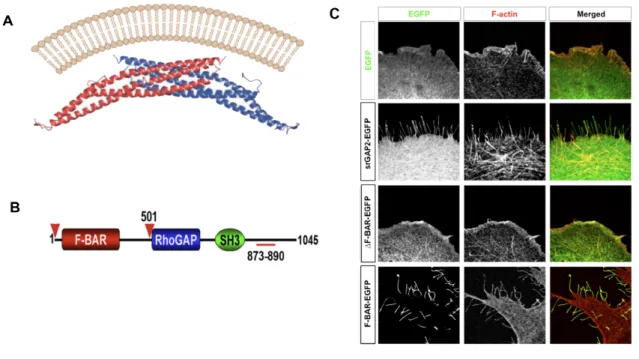
srGAP2 is an F-BAR-containing protein that regulates neuronal morphology
(Guerrier et al. 2009). This protein may act similar to septins by forming complex oligomers
heterodimerize with septin5 and septin11 (Xie et al. 2007), srGAP2 may heterodimerize with
other srGAP family members to achieve its effect. However, unlike other proteins that
contain both a BAR-like and a GTPase domain, the influence of each domain has been
examined independently. Interestingly, while F-BAR domains are structurally predicted to
form membrane invaginations, the F-BAR domain of srGAP2 induces the formation of
filopodia-like membrane protrusions (Guerrier et al. 2009). Overexpression of srGAP2 or its
F-BAR domain only causes increased branching in developing neurons. However, how
srGAP2 affects the membrane at later developmental time points, subsequent to neuronal
migration and synaptogenesis, is unexplored. Yet, because srGAP2 can potently induce the
formation of filopodial-like protrusions, it is reasonable to speculate that the protein may
regulate the formation and morphology of dendritic spines. Therefore Chapter Three of this
thesis will describe our effort to determine how srGAP2 affects dendritic spine morphology,