© 2016. Published by The Company of Biologists Ltd.
The Lhx9-Integrin pathway is essential for positioning of the proepicardial organ
Panna Tandon 2,3,4,*, Caralynn M. Wilczewski 3,4, Clara E. Williams 4, Frank L. Conlon 1-4
(1) University of North Carolina McAllister Heart Institute, UNC at Chapel Hill, Chapel Hill, NC
27599-3280, USA
(2) Department of Biology, UNC at Chapel Hill, Chapel Hill, NC 27599-3280, USA
(3) Department of Genetics, UNC at Chapel Hill, Chapel Hill, NC 27599-3280, USA
(4) Integrative Program for Biological and Genome Sciences, UNC at Chapel Hill, Chapel Hill, NC
27599-3280, USA
*Author for correspondence: Panna Tandon, 220 Fordham Hall, North Medical Drive, Chapel Hill, NC, 27599 ([email protected], tel: 919-962-2138)
Development • Advance article
http://dev.biologists.org/lookup/doi/10.1242/dev.129551 Access the most recent version at
Summary
The development of the vertebrate embryonic heart occurs by hyperplastic growth as well as the incorporation of cells from tissues outside of the initial heart field. Amongst these tissues is the epicardium, a cell structure that develops from the precursor proepicardial organ on the right side of the septum transversum caudal to the developing heart. During embryogenesis cells of the proepicardial organ migrate, adhere and envelope the maturing heart forming the epicardium. The cells of the epicardium then delaminate and incorporate into the heart giving rise to cardiac derivatives including smooth muscle cells and cardiac fibroblasts. Here, we demonstrate that the LIM homeodomain protein Lhx9 is transiently expressed in Xenopus
proepicardial cells and is essential for the position of the proepicardial organ on the septum transversum. Utilizing a small molecule screen we uncovered that Lhx9 acts upstream of Integrin-Paxillin signaling and consistently demonstrate that either loss of Lhx9 or disruption of the Integrin-Paxillin pathway results in mis-positioning of the proepicardial organ and aberrant deposition of extra-cellular matrix proteins. This leads to a failure of proepicardial cells to migrate and adhere to the heart and eventual death of the embryo. Collectively, these studies establish a requirement for the Lhx9-Integrin-Paxillin pathway in proepicardial organ positioning and epicardial formation.
Introduction
Cardiovascular disease is one of the highest causes of mortality and reduced quality of life in the US (Roger et al., 2012). To better optimize therapeutics to preserve and maintain heart physiology it is pertinent that we understand the mechanisms involved in all aspects of heart development, growth and repair. This will further enable us to target resident cardiac cell populations and gene programs for clinical applications.
The heart initially forms in vertebrates as a bilaminar tube comprised of an inner endocardium and outer myocardial layer. At later stages of development a third layer is added to the heart termed the epicardium. In many vertebrates this layer forms from a dynamic precursor cell population, the proepicardial (PE) organ, which arises as a cluster of cells on the right side of the septum transversum adjacent to the heart (Schulte et al., 2007, Jahr et al., 2008, Manner, 1992, Pombal et al., 2008, Serluca, 2008). Whilst FGF and BMP signaling, as well as the transcription factors Snai1 and Twist, have been implicated in PE induction (Schlueter and Brand, 2009, Schulte et al., 2007, Schlueter and Brand, 2013), there is little known about the mechanisms that direct the formation of the PE cell cluster. Once the cluster has formed and is positioned correctly it is induced to bridge towards the developing heart, directed by BMP signaling from the atrioventricular sulcus (AVS) (Ishii et al., 2010). Upon attachment, epicardial cells then traverse over the heart surface as an epithelial-like sheet termed the epicardium.
The epicardial layer is an essential cardiac component as it provides mitogenic stimulation to the developing myocardium (Kang et al., 2008, Lavine et al., 2005, Li et al., 2011, Pennisi et al., 2003, Stuckmann and Lassar, 2002). In addition, the epicardium is the source of epicardial-derived cells (EPDCs) whereby subpopulations of this layer undergo epithelial-mesenchymal transition and invade the underlying myocardium. Here they differentiate into vital cardiac cells such as smooth muscle cells and pericytes of the vasculature and fibroblasts that contribute to the scaffold and support of the heart
(Gittenberger-de Groot et al., 2010, Lie-Venema et al., 2007, Manner et al., 2001, Ratajska et al., 2008, von Gise and Pu, 2012). Current understanding is that EPDC lineage specificity is determined early within the precursor PE cell cluster prior to epithelial-mesenchymal transition (EMT) (Acharya et al., 2012, Guadix et al., 2006, Manner, 1999, Mikawa and Gourdie, 1996, Katz et al., 2012, Wei et al., 2015). In the event of a cardiac infarction or damage in the adult, research has also demonstrated the utility of the epicardium in providing regenerative support and cardiac cells to the damaged tissue (Lepilina et al., 2006, Limana et al., 2010, Winter and Gittenberger-de Groot, 2007, Winter et al., 2009). Therefore, better
comprehension of how this source of pluripotent cells is specified and develops is vitally important to understanding mechanisms of cardiac development, disease and repair.
Here we report that LIM Homeobox transcription factor 9 (Lhx9) is essential for the formation and function of the epicardium. We show that loss of Lhx9 in Xenopus leads to defects in the assembly of the PE cluster and subsequently failed migration and spreading of epicardial cells onto the surface of the developing heart. Using a small pharmacological molecule screen in Xenopus we ascertained that Integrin-Paxillin signaling is required for proper PE clustering, which is confirmed by a significant decrease in epicardial integrin alpha 4 (itga4) expression upon loss of Lhx9 function. Given that interactions between cells and the surrounding ECM are vital for epicardial formation and its role in cardiac repair (Kalman et al., 1995, Nahirney et al., 2003, Pae et al., 2008, Wang et al., 2013, Benesh et al., 2013, Fransen and Lemanski, 1991, Mercer et al., 2013, Rongish et al., 1996, Yang et al., 1995), we further show a novel function for Lhx9 in mediating Integrin-adhesion
mechanisms for correct PE cell clustering. Therefore Lhx9 is vital for the formation of the epicardium and development of the heart.
Results
Lhx9 is expressed in a temporally dynamic pattern during epicardial formation.
The epicardium is a specialized layer of cells surrounding the heart derived from a mesothelial precursor structure, the proepicardial organ (PEO), located on the septum transversum. Animals lacking a functional epicardium die prematurely and display
hemorrhaging hearts with thin ventricular walls (Moore et al., 1998, Lie-Venema et al., 2005, Gittenberger-de Groot et al., 2000, Acharya et al., 2012, Combs et al., 2011). Many
vertebrates show a right side clustering of PE cells during epicardial formation (Viragh et al., 1993, Kalman et al., 1995, Nahirney et al., 2003, Serluca, 2008, Pombal et al., 2008, Jahr et al., 2008). Recently we have demonstrated that the LIM homeodomain transcription factor family lhx9 is transiently expressed in the Xenopus PEO (Tandon et al., 2013). We therefore conducted a detailed analysis to establish the relationship between Lhx9 expression and formation of the PEO on the septum transversum.
The sequence and structure of Lhx9 has been shown to be highly conserved across vertebrates (Bertuzzi et al., 1999, Retaux et al., 1999, Failli et al., 2000, Bachy et al., 2001, Ottolenghi et al., 2001, Alunni et al., 2007, Oshima et al., 2007, Peukert et al., 2011) being comprised of two LIM protein binding domains and a DNA-binding homeodomain (HD). Consistent with other vertebrates we identified two isoform variants of lhx9 in Xenopus, one
with a full length HD (lhx9HD) and one with a truncated HD (lhx9α) generated by alternative splicing of exon 5 (Failli et al., 2000, Molle et al., 2004) (Fig. S1).
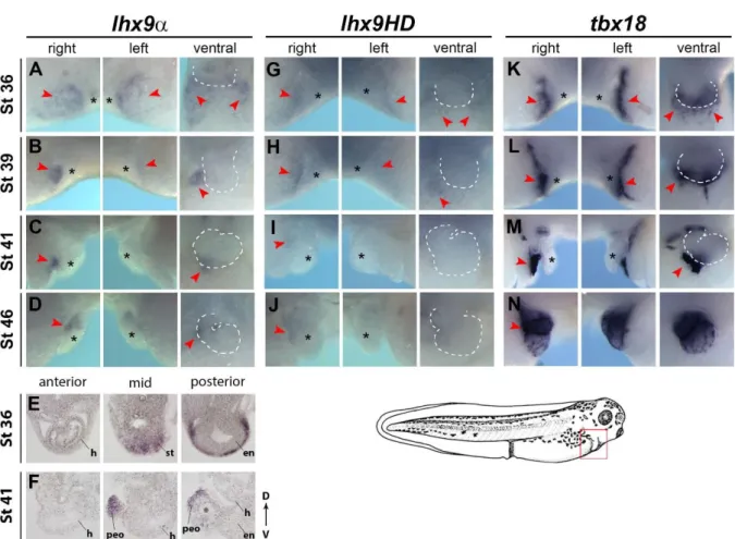
To ascertain the endogenous role of Lhx9 during epicardial development we performed a detailed spatio-temporal analysis of both lhx9 isoforms. Results from these studies show lhx9α is initially expressed bilaterally on the septum transversum prior to PEO formation (Fig. 1A, E, Fig. S2-S4). In all vertebrates, the lateral confinement of the PEO cells to the right side of the embryo can be observed by the expression of the conserved PEO marker tbx18 (Fig. 1K-N, Fig. S3) (Tandon et al., 2013). Consistently we observed a dramatic restriction of lhx9α expression to the right side of the septum transversum (stages 39-46) (Fig. 1B-D, F) and subsequently lhx9α is down regulated, relative to tbx18, as the PEO cells
mature and migrate onto the heart (Fig. 1D, N, Fig. S3) (Tandon et al., 2013). An identical pattern of expression was obtained with an independent probe recognizing the LIM domains of lhx9α and lhx9HD (Fig.1E, F, Fig. S1, Fig. S4, Fig. S11). We further observed a highly similar pattern of expression for lhx9HD in the septum transversum (Fig. 1G-J, Fig. S5) however we note lhx9HD is more weakly expressed and dramatically down regulated upon PE clustering compared to lhx9α (Fig. 1I, Fig. S5). We also note we did not observe
detectable expression of lhx2, a closely related LIM-HD transcription factor to Lhx9, in the developing epicardium at any stage (Fig. S6) (Viczian et al., 2006).Taken together these studies demonstrate lhx9α and lhx9HD isoforms have overlapping dynamic expression during epicardial development and lhx9α is the predominant isoform expressed during PEO
formation.
Lhx9 is required for epicardial formation.
To determine the requirement for Lhx9in the vertebrate PEO we depleted Lhx9 in Xenopus laevis (X. laevis) using Morpholinos (see Supplementary Methods for details).
Depletion of Lhx9 function in Xenopus does not impede early embryonic
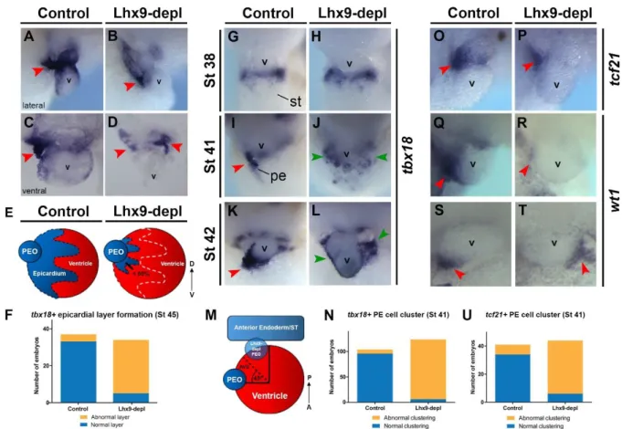
development. However, analysis of the developing heart at tadpole stages revealed a severe epicardial defect. Whilst control embryos displayed PE cells clustering to the AVS on the right side of the heart and a layer of epicardial cells on the ventricle surface (Fig. 2A, C, red arrowheads marking PE cell cluster, tbx18 in situ hybridization), embryos lacking Lhx9 failed to accumulate PE cells to the right-side AVS and displayed a reduced and discontinuous epicardial layer on the ventricular surface (Fig. 2B, D, E, F, red arrowheads).
Lhx9 is required for clustering of proepicardial cells.
Having demonstrated proper epicardial formation requires Lhx9 function and because lhx9
expression is detected on the septum transversum and PE cluster (Fig. 1A-F, Fig. S2), we assessed whether Lhx9 functions during the earlier stages of epicardial specification. At stage 38 the conserved pan-marker of the epicardial lineage, tbx18 was observed on the septum transversum and deemed indistinguishable between control and Lhx9-depleted embryos (Fig. 2G, H). Therefore Lhx9 is not required for specification of the epicardial lineage. However, by stage 41 when PE cells begin to cluster to the right side of embryos (Fig. 2I, red
arrowhead), Lhx9-depleted stage-matched sibling embryos maintained diffuse tbx18
expression bilaterally along the septum transversum (Fig. 2J, green arrowheads, Fig. S9A). By stage 42, the PE cluster had attached to the AVS on the right side of the heart in controls whereas in Lhx9-depleted embryos there was either no detectable cluster and tbx18
expression persisted on the anterior endoderm/septum transversum or the cluster was mispositioned caudal to the heart (Fig. 2L, M, N).
Consistently, we observed the spatial distribution of two independent epicardial markers, tcf21 and wt1, were similarly disrupted in Lhx9-depleted embryos. Both tcf21 and
wt1 expression marks the punctate cluster of PE cells at stage 41 on the right side of the embryo (Fig. 2O, Q, S, red arrowhead), whilst expression of these epicardial markers is reduced and mislocalized in Lhx9-depleted embryos (Fig. 2P, R, T, U, red arrowheads, Fig. S9B). This data collectively implies Lhx9 function is critical for normal clustering of PE cells during early stages of development and in its absence epicardial formation is disrupted.
Proepicardial cell cluster positioning is driven by Integrin-mediated mechanisms.
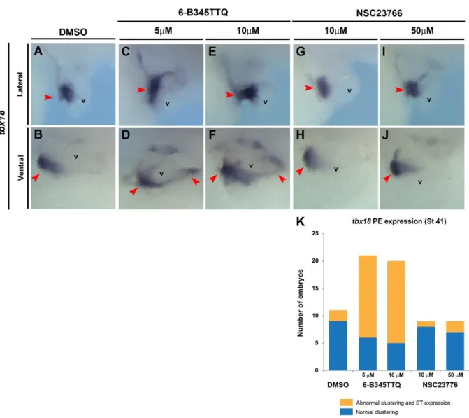
The cellular and molecular mechanisms which lead to the clustering of the PEO on the right side of the septum transversum remain entirely unknown. To assess whether active cell migration is required for asymmetric PE clustering we conducted a screen in wild type embryos with a defined bank of small molecule inhibitors known to disrupt cell migration and adhesion in Xenopus (Harata et al., 2013, Broders-Bondon et al., 2007). Whilst the inhibition of myosin II (Blebbistatin) resulted in tadpoles with severe pericardial edema (possibly due to reduced heart beat) and paralysis, we did observe clustered PE cells (marked by tbx18) to the right of the embryo, albeit due to the pronounced edema we were unable to determine attachment to the heart (data not shown). Inhibition of Rac1 activity (NSC23766 trihydrochloride) which has been extensively shown to disrupt cell migration (Diaz et al.,
2014, Huang et al., 2013) did not affect PE cell clustering compared to controls as depicted by tbx18 expression on the AVS (Fig. 3A, B, G-J). Little tbx18 expression was preserved on the septum transversum in these embryos implying PE clustering is not dependent on
classical cell migration mechanisms involving actin-myosin interactions and GTPase activity. Interestingly, exposure from stages 38-41 when PE cell clustering is occurring, to a small molecule that disrupts Integrin-Paxillin interactions (6-B345TTQ) led to a PE cluster positioning defect phenocopying loss of Lhx9 function. Integrin binds to ECM components and induces the recruitment of proteins, such as Paxillin, to focal adhesions at the site of cell attachment. These protein complexes then function to reorganize the actin cytoskeleton and are essential for the coordinated adhesion and motility of cells through an ECM environment (Bellis et al., 1995). Paxillin is a multi-LIM domain protein has been shown to directly and specifically bind to the cytoplasmic tail of Itgα4 to coordinate cell spreading, adhesion and
migration (Liu et al., 1999, Alon et al., 2005). Whilst some clustering of PE cells was evident in cultured tadpoles, we observed mispositioning of the cluster to the caudal side of the heart as well as persistent tbx18 expression on the septum transversum (Fig. 3C-F, K) as seen in Lhx9-depleted embryos (Fig. 2H, J, L, M). This data therefore suggests Integrin-mediated mechanisms plays an important role in the correct positioning of the PEO cell cluster in developing embryos.
Lhx9 is required for proepicardial clustering via an Integrin-Paxillin interaction.
Our data demonstrate Lhx9 as well as Integrin-mediated cellular functions are essential for the correct clustering and positioning of PE cells. We previously reported lhx9 and itgα4 are co-regulated in response to loss of the epicardial transcription factor Tcf21 (Tandon et al., 2013). Based on these observations we hypothesized Lhx9 acts upstream of itgα4 to correctly position the PEO. Consistent with this hypothesis we find itgα4 expression is detected
subsequent to lhx9 in the clustered PE cells from stage 39-46 (Fig.4A-D, red arrowheads, Fig. S3). We thus assessed the expression of itgα4 in Lhx9-depleted hearts. Consistent with itgα4
acting downstream of Lhx9 we find itgα4 is reduced and misplaced in Lhx9-depleted
embryos (Fig. 4E, F, O, Fig. S8, Fig. S9C). By contrast, the expression of Integrin β1 (Itgβ1) is strongly localized to the epicardium (as marked by tcf21) and appears to be independent of Lhx9 function (Fig. 4G-J’, Fig. S11) therefore enabling its use as a marker of epicardial cells. Due to the clustering defects observed upon reducing the Itgα4-Paxillin interaction
biochemically (Fig. 3) and reduced itgα4 expression in Lhx9-depleted embryos, we sought to determine if Lhx9 is responsible for Integrin-mediated cell adhesion and migration processes
in PE cells. To address this possibility we assayed for phosphorylated Paxillin (Y118) which has been used to demonstrate Integrin activation at focal adhesions and shown to modulate cell spreading, migration and invasion (Bellis et al., 1995, Burridge et al., 1992, Turner, 1991, Nakamura et al., 2000, Lewis and Schwartz, 1998, Zaidel-Bar et al., 2007, Sachdev et al., 2009). Our data indicated a significant decrease in phosphorylated Y118 Paxillin
expression specifically in the attached PE cluster in Lhx9-depleted embryos compared to controls (Fig. 4K-N’, P). Taken together these data strongly imply Lhx9 acts through Itgα4 to correctly position the PEO.
Lhx9-regulated Integrin signaling is essential for correct formation of the epicardial
layer.
Collectively our data demonstrates a role for Lhx9-Itgα4 in the correct positioning and clustering of the PEO. Our data also suggests Integrin activity at focal adhesions is required for the clustering of PE cells. Previous data has implicated Itgα4 as being essential for epicardial development particularly in regulating PE cell clustering, epicardial adhesion and epicardial migration (Sengbusch et al., 2002, Pinco et al., 2001, Yang et al., 1995, Dettman et al., 2003, Dokic and Dettman, 2006, Hirose et al., 2006, Pae et al., 2008, Bax et al., 2010). Integrin heterodimers have been shown to coordinate cytoskeletal interactions and adhesion with the surrounding ECM environment (Gardiner, 2011, Gehler et al., 2013, Huttenlocher and Horwitz, 2011, Manninen, 2015, Wolfenson et al., 2013), pertinent processes for epicardial formation and function (Kalman et al., 1995, Nahirney et al., 2003).
To determine if the depleted adhesive properties of the PEO and its mis-positioning have persistent biological consequence in the development of the vertebrate epicardium and heart, we examined the epicardial ECM environment of control and Lhx9-depleted hearts. Using Itgβ1 as a marker of epicardial cells we analyzed the expression of known Itgα4 ligands and ECM components. Whilst we observed no discernible alterations in the
expression of the Itgα4-Itgβ1 (Very Late Antigen 4, VLA-4) ligand vascular cell adhesion
molecule 1 (vcam1, Fig.S10) in Lhx9-depleted embryos, we did detect a decrease in Fibronectin within the PE cluster as well as in the heart relative to controls (Fig. 5A-E) (Humphries et al., 1995, Wu et al., 1995). This suggests alterations to Integrin function in response to loss of Lhx9 is specific to Itgα4 pathway components. In addition these data
further implies the adhesive qualities and positioning of the PEO is critical for the proper deposition of epicardial ECM.
To address this further we examined the localization of epicardial-ECM markers Laminin and β-dystroglycan. In control embryos we observed weak Laminin deposition surrounding attached PE cells (Fig. 5F, F’, white arrowheads) but a continuous smooth layer of Laminin was present under the mature epicardial layer (Fig. 5F”, white arrowheads). However in Lhx9-depleted embryos, when epicardial cells were present on the myocardial surface, we observed an increased accumulation of Laminin in the basal portion of attached PE cells (Fig. 6G, G', white arrowheads) as well as discontinuous deposits within epicardial cells on the surface of the heart (Fig. 5G”, white arrowheads). To further confirm these findings, we analyzed β-dystroglycan, a transmembrane glycoprotein that attaches cells to the
basement membrane through Laminin binding (Ervasti and Campbell, 1993, Klietsch et al., 1993, Smalheiser and Schwartz, 1987). In Lhx9-depleted embryos β-dystroglycan is reduced or disrupted at the point of contact between the PE cluster and heart surface (as indicated by
tcf21) (Fig. 5J-K’) compared to controls (Fig. 5H-I’). Note β-dystroglycan is prominent in the endocardium of both control and Lhx9-depleted hearts showing this defect is specific to the epicardium (Fig. 5I’, K’, yellow arrowheads). Taken together these results demonstrate PE cell adhesive integrity is required for the proper formation of the epicardial layer.
Collectively, our data are consistent with a role for Lhx9 in the clustering and positioning of the PEO (Fig. 6A-B). Our data further show the ability of Lhx9 to regulate Itgα4 signaling is vital for the correct establishment of clustering, attachment and migration
of PE cells to the heart (Fig, 6A, C, D). These signals are in turn necessary for the correct deposition of ECM components thus, facilitating epicardial cells to form a cohesive layer over the myocardial surface (Fig. 6E, F).
Discussion
Our data collectively shows Lhx9 is essential for proepicardial positioning and that loss of Lhx9 is associated with aberrant deposition of essential ECM components during crucial stages of epicardial clustering and migration. We further observe that loss of Lhx9 correlates with a decrease in Integrin-ECM signaling and the ability of PE cells to form cohesive adhesions downstream of Integrin activity. Because the PE cluster defect in Lhx9-depleted embryos closely resembles the defect caused by disturbing the Itgα4-Paxillin interaction
biochemically, we propose and demonstrate Lhx9 functions to modulate itgα4 expression. Taken together this data establishes a role for Lhx9 in Integrin-mediated cell adhesion and motility crucial for the proper positioning and clustering of PE cells.
Though most vertebrates show a right side clustering of PE cells during epicardial formation (Viragh et al., 1993, Kalman et al., 1995, Nahirney et al., 2003, Serluca, 2008, Pombal et al., 2008, Jahr et al., 2008) it remains to be established if the sidedness of
epicardial formation is essential for its function. In our studies we further find that alterations in the positioning of the epicardium on the septum transversum is essential for the correct attachment of the epicardium to the heart at the AVS. Thus, these imply that alterations in PE clustering predispose an embryo to cardiac defects.
Itga4 and epicardial development
The function of Itgα4 during epicardial development has been characterized in mice mutants
who displayed absence of the epicardial layer and lack of coronary vessels at the AVS (Yang et al., 1995). This was shown to be due to defects in the migratory process (Sengbusch et al., 2002) and as demonstrated by this work. Therefore the role of Itgα4 in epicardial migration appears to be conserved regardless of the mode of epicardial formation; cluster bridge in frog or free-floating cysts in mouse. Our data provides new evidence that the expression of itga4
is restricted to immature PE cells and downregulated as the epicardial layer matures, similar to the hematopoietic lineage (Arroyo et al., 1999, Lobb and Hemler, 1994). Using Itgβ1 as a marker of epicardial cells we analyzed the expression of known Itgα4 ligands and ECM components. We report a down regulation of Fibronectin in embryos lacking Lhx9 but observe no alterations in expression of vcam1, the Itgα4-Itgβ1 ligand. Taken together these
findings demonstrate that loss of Lhx9 functions upstream of itgα4 and that the function of Lhx9 is specific to Itgα4 function.
Lhx9 and Tcf21 during epicardial formation
Our previous studies identified lhx9 and itgα4 as genes upregulated upon Tcf21 loss. The role of Lhx9 in cardiac development to date has been limited to studies on the effect of its
persistent expression upon FOG2 ablation (Smagulova et al., 2008). Whilst these studies provide insight into the effects of potentially prolonged Lhx9 activity, the requirement for Lhx9 has not been addressed. Consistently with our finding on Tcf21, we observe here that depletion of Lhx9 results in decreased itgα4 with a sparse and disorganized epicardial layer on the heart. Given their similar patterns of expression in the developing epicardium, this data suggests therefore that Tcf21 and Lhx9 function coordinately together to enable the correct clustering and migration of PE cells onto the heart surface through Integrin-directed
mechanisms. It is of interesting note that Tcf21 and LIM homeodomain transcription factors
are both found to be essential for the development of the gonad and craniofacial structures (Moncaut et al., 2012, Harel et al., 2012, Cui et al., 2004, Birk et al., 2000) suggesting that the transcriptional targets of these two genes and the pathways they regulate may unveil a conserved mechanism into regulating cell motility and thus, have implications in cancer as well as cardiac and organ development (Vladimirova et al., 2009, Yang et al., 2014, Miller et al., 2014, Weiss et al., 2013, Miller et al., 2013, Zhang et al., 2012, Ye et al., 2012, Richards et al., 2011, Arab et al., 2011).
Materials and Methods
In situ hybridization
Whole-mount in situ hybridization (ISH) was carried out as previously described (Tandon et al., 2013, Harland, 1991, Charpentier et al., 2015), the pericardial cavity membrane in late tadpole stage embryos being removed post-fixation to improve resolution. Embryos were processed for gelatin (30 µm) sectioning as previously reported (Gessert and Kuhl, 2009, Tandon et al., 2013, Charpentier et al., 2015). All probes were previously used (Tandon et al., 2013) or generated by PCR cloning from Xenopus embryonic cDNA (Supplemental Table 1). Information on constructs is available upon request. All phenotypes were quantified and statistically assessed using Fishers Exact test or Chi-Square (Graphpad Prism 6). Figure images show representative phenotypes quantified. For spatiotemporal analysis of lhx9, lhx2, tbx18 and itga4 transcripts in whole wild type embryos (Supplemental Figs. 2-6), multiple focal plane images were compiled using auto-blend function (Photoshop CS4). Original images are available upon request.
Xenopus manipulations
Xenopus embryos were cultured, microinjected and staged according to Niewkoop and Faber (Nieuwkoop and Faber, 1967, Tandon et al., 2013). Xla.Tg(Cardiac-actin:GFP)Mohun
transgenic frogs were used as previously reported (Latinkic et al., 2002, Tandon et al., 2013). The 5’UTR from both short and long X. laevislhx9 alloalleles was cloned and sequenced to
verify design of the translation-blocking Morpholino (MOT). A 5-base mismatch MO was used as a control (Supplemental Table 2). Two transcription-blocking Morpholinos were designed against the splice donor-site of exon 1 from both lhx9 alloallele genomic loci (MO1, Table 2) (Tandon et al., 2012) (Genetools). 30 ng of each MO was injected at the one-cell
stage, with both MO1 being co-injected (Tandon et al., 2013, Tandon et al., 2012). To assess the specificity and efficiency of MOT, 1ng lhx9-5’UTR-GFP RNA (designed against each genomic loci) was co-injected together with the stated concentrations of MO. Stage 11 embryos were collected, lysed and analyzed using western-blot as previously performed (Tandon et al., 2012, Tandon et al., 2013) using GFP antibody (JL8, #632381, Clontech, 1:10,000) and Shp2 antibody (#610622, BD Transduction, 1:2500) as a loading control. To assess the efficiency of MO1, embryos were injected at the one-cell stage, and the hearts from 25 embryos collected at stage 42 from each condition. RT-PCR was performed
(Superscript II, Invitrogen) for lhx9 with gapdh as a loading control (Supplemental Table 1). See also Supplemental Methods.
Immunohistochemistry
Embryos were processed for agarose vibratome sectioning (100 µm) as reported
(Wallingford, 2010, Tandon et al., 2013, Charpentier et al., 2015). Sections were then washed with PBS + 1% Triton X-100, blocked with wash buffer + 10% fetal calf serum and
incubated with primary antibody as previously reported (Langdon et al., 2012, Tandon et al., 2013, Charpentier et al., 2015). Antibodies included mouse anti-Tropomyosin (CH1, DSHB 1:25), rabbit anti-Laminin (L9393, Sigma, 1:100), mouse anti- β-dystroglycan (MANDAG2, 7D11, DSHB, 1:100), rabbit anti-Fibronectin (F3648, Sigma, 1:250), rabbit anti-phospho-Paxillin pTyr118 (44-722G, Invitrogen, 1:100) and mouse anti-Integrin β1 (8C8, DHSB, 1:100). CH1 was deposited to the DSHB by Lin J.J.-C, MANDAG2(7D11) by Morris G.E and 8C8 by Hausen P/ Gawantka V. Sections were then incubated in DAPI/PBS (200 ng/ml) and imaged using a Zeiss LSM700 Spectral Confocal Laser Scanning Microscope, with representative figures compiled using ImageJ and Photoshop. Image fluorescence, gauged by integrated pixel density, was measured using ImageJ and quantified with standard two-tailed student’s t-test (Graphpad).
Live tadpole small molecule culture assay
Xenopus wild type embryos were incubated in the appropriate concentration of small molecule or DMSO control in 0.1 X MBS from stage 38 to stage 41 at room temperature in the dark. Blebbistatin (B0560, Sigma) was used at 10 µM (Straight et al., 2003), 6-B345TTQ (B7438, Sigma) was used at 5 and 10 µM (Kummer and Ginsberg, 2006, Kummer et al., 2010, Hung et al., 2013) and NSC23766 trihydrochloride (SML0952, Sigma) was used at 5
and 50 µM. Embryos were briefly rinsed in 0.1x MBS, fixed in 4% PFA and subjected to in situ hybridization as described.
Funding
We are thankful for funding from the National Institutes of Health (NICHD, R21HD073044to FLC) and the American Heart Association (13POST16950044, postdoctoral fellowship to PT).
Acknowledgements
We would like to thank the faculty of the Microscope Services Laboratory at UNC for help with microscopy imaging. The antibodies against tropomyosin, Integrin β1 and
β-dystroglycan were obtained from the Developmental Studies Hybridoma Bank, created by the NICHD of the NIH and maintained at The University of Iowa, Department of Biology, Iowa City, IA 52242.
Author Contributions
PT developed the concepts, performed experiments and analyzed data for the manuscript as well as prepared and edited the paper prior to submission. CMW and CEW performed experiments and phenotypic analysis, FLC prepared and edited manuscript.
Figures
Figure 1. Spatio-temporal analysis of lhx9 isoforms during Xenopus epicardial
development.
Whole embryo in situ hybridization was performed on Xenopus embryos at stages 36 to 46, using probes specific for lhx9a (A-D), lhx9LIM (E, F), lhx9HD (G-L) and tbx18 (K-N) (See Fig. S2, S3, S4, S5). Views display embryos facing right, left and ventral with anterior to the top. Images show magnified cardiac regions as depicted by red box on tadpole schematic (bottom). Red arrowheads depict staining of septum transversum and PE clusters. Cardiac region is depicted with *. White dashed line outlines heart in ventral views. (E, F) Transverse gelatin section in situ hybridization images of cardiac region demonstrate lhx9LIM expression exclusively in the septum transversum (stage 36) and proepicardial tissue (stage 41). en, endocardium; h, heart; peo, proepicardial organ; st, septum transversum.
Figure 2. Lhx9 is required for proper epicardial layer and PE cluster formation.
Epicardial layer formation (as shown with tbx18) analysis in control (A, C) and Lhx9-depleted (B, D) embryos at stage 45. (A, B) Images show lateral view of cardiac region, dorsal to the top and anterior to right. (C, D) Transverse gelatin sections through
representative embryo cardiac regions showing tbx18 expression, dorsal to the top. PE cell clustering (red arrowheads) and epicardial cell layer on the ventricular surface is shown. (E) Epicardial layer formation defects were quantified as abnormal as ≤ 50% ventricular
coverage as depicted in schematic. (F) Quantification of observed epicardial layer formation defects, as represented in A-D, from three independent experiments, p = <0.0001 by two-tailed Fisher’s exact test. Proepicardial clustering morphology was analyzed using in situ
hybridization for tbx18 in control (G, I, K) and Lhx9-depleted (H, J, L) embryos at stage 38 (G, H), stage 41 (I, J) and stage 42 (K, L). Ventral view showing cardiac region, anterior to the top. Tbx18 expression is detected as a cluster of cells in control embryos on the right of the embryo near the atrioventricular sulcus (I, K, red arrowheads) whereas in Lhx9-depleted embryos, the cluster is either not detected and tbx18 expression remains throughout the septum transversum region or is mis-positioned to the caudal side of the heart (abnormal clustering) (J, L, green arrowheads). (M) Quantification of PE clustering defects is depicted
in the schematic as being abnormal by bilateral tbx18 expression retention on septum
transversum and/or cluster mis-positioning of ≥ 45o caudal to the AVS compared to controls. (N) Quantification of observed clustering phenotype at stage 41 represented in C and D. Data taken from seven independent experiments, p = <0.0001 by two-tailed Fisher’s exact test. (O-T) In situ hybridization analysis for tcf21 (O, P, U) and wt1 (Q-T) proepicardial expression at stage 41. Images depict lateral view of the cardiac region, dorsal to the top and anterior to the right (M-P) or ventral view, dorsal to top (Q, R). Red arrowheads mark clustered PE cells. (U) Quantification of observed phenotypes as depicted in M, data taken from six independent experiments, p = <0.0001 by two-tailed Fisher’s exact test. avs, atrioventricular sulcus; pe/peo; proepicardial organ, st; septum transversum, v; ventricle.
Figure 3. Integrin-Paxillin association is required for PE clustering.
In situ hybridization for tbx18 on whole embryos after incubation in stated concentrations of small molecules between stages 38 and 41. Lateral views (A, C, E, G, I) and ventral views (B, D, F, H, J) of the cardiac region from representative embryos are shown, red arrowheads mark PE cells. (C-F) Images depict maintained tbx18 expression on septum transversum and clustering abnormalities as shown Fig.2M when embryos incubated in 6-B345TTQ. (K) Embryos taken from two independent experiments, p = 0.000248 by Chi Square test.v, ventricle.
Figure 4. Lhx9 regulates Integrin α4-Paxillin signaling in PE cluster.
(A-D) Whole embryo in situ hybridization for itgα4, stages 36 to 46. Lateral views of anterior portion (left panels) with magnified image of cardiac region (right panels). Red arrowhead marks PE cluster, * marks heart. (E, F) In situ hybridization for itgα4 in control (E) and Lhx9-depleted embryos (F) at stage 41, representative cardiac region shown, anterior to right, dorsal to top, red arrowhead marks PE cluster. (O) Quantification of observed itgα4
expression (reduced expression denoting ≤ 50% stain intensity to controls) and clustering defects shown in E, F (see Fig. 2M for phenotype assessment), , embryos taken from six independent experiments, p = <0.0001 by Chi-square. (G-J) Transverse cardiac region agarose sections from control (G, H) and Lhx9-depleted (I, J) stage 41 embryos post-in situ hybridization for tcf21 (G, I), nuclei expression with DAPI (H, J, blue) and Itgβ1
immunohistochemistry (H, J, magenta). White outlines (H, J) indicate PE cluster as depicted by tcf21 and Itgβ1. (K-N) Transverse cardiac region agarose sections from control (K, L) and Lhx9-depleted (M, N) stage 45 Xla.Tg(Cardiac-actin:GFP)Mohun embryos depicting
representative immunohistochemical analysis for DAPI (blue), GFP (green) to mark
cardiomyocytes, Itgβ1 (magenta) to mark epicardial cells and phosphorylated Y118-Paxillin
(red). Magnified images (L, N) from white boxes (K, M). White arrowheads mark PE cluster.
(P) Pixel intensity (integrated density) levels for phosphorylated Y118-Paxillin (M) from five control and ten Lhx9-depleted embryos, p = 0.0134 by two-tailed student t-test.v, ventricle.
Figure 5. Disrupted Lhx9-Integrin signaling alters epicardial ECM environment.
(A-E) Transverse cardiac agarose sections from control (A, B) and Lhx9-depleted (C, D)
Xla.Tg(Cardiac-actin:GFP)Mohun embryos at stage 45. Nuclei stained with DAPI (blue), GFP marking cardiomyocytes (green) and immunohistochemical expression for Itgβ1 (B, D, magenta) to mark epicardial cells, and Fibronectin (B”, D”, red). Threshold binary images (Image J) in B’ and D’ show Itgβ1-positive epicardial cells used to quantify pixel intensity in B” and D”. (E) Pixel intensity (integrated density) levels for Fibronectin from five control
and ten Lhx9-depleted embryos, p = 0.064 by two-tailed student t-test. (F-K) Transverse cardiac agarose sections from control (F, H, I) and Lhx9-depleted (G, J, K) embryos at stage 45. Nuclei stained with DAPI (blue) and immunohistochemical stain for tropomyosin
(cardiomyocytes, F, G, green), laminin (F, G, magenta) and β-dystroglycan (H’-K’, magenta).
Tcf21 (H-K) demonstrates PE cell attachment to the heart. White and red boxes depict
magnified images in ‘ and “ panels. White arrowheads mark PE (F’, G’, I’, K’) and migrating
epicardial cells (F”, G”). Yellow arrowheads mark expression in endocardial tissue (B”, D”, I’, K’). v; ventricle. Representative images from seven (laminin) and six (β-dystroglycan)
embryos per condition, from two independent experiments.
Figure 6. Model depicting role for Lhx9 in epicardial development in Xenopus.
(A) During early tadpole stages the epicardial lineage is determined and marked by
transcription factors tcf21, tbx18, wt1 and lhx9 (blue) as a bilateral population of cells on the septum transversum caudal to the heart (red). (B) Lhx9 functions to drive clustering of cells to form the proepicardial cluster on the right side of the embryo (blue), whereby itgα4 expression is activated. At this stage signaling factors, most likely BMP (yellow arrows), from the heart atrioventricular sulcus (AVS) direct epicardial migration. (C, D)
Lhx9-Integrin-mediated signaling, including focal adhesion (FA) formation and phosphorylation of Paxillin (pPaxillin), allow the PEO bridge (blue) to attach to the heart (red) at the AVS. (E, F) Once the PEO has attached to the heart, deposition of essential ECM components such as Fibronectin and Laminin are required for the epicardial layer to adhere and spread over the heart surface.
References
ACHARYA, A., BAEK, S. T., HUANG, G., ESKIOCAK, B., GOETSCH, S., SUNG, C. Y., BANFI, S., SAUER, M. F., OLSEN, G. S., DUFFIELD, J. S., OLSON, E. N. & TALLQUIST, M. D. 2012. The bHLH
transcription factor Tcf21 is required for lineage-specific EMT of cardiac fibroblast progenitors. Development, 139, 2139-49.
ALON, R., FEIGELSON, S. W., MANEVICH, E., ROSE, D. M., SCHMITZ, J., OVERBY, D. R., WINTER, E., GRABOVSKY, V., SHINDER, V., MATTHEWS, B. D., SOKOLOVSKY-EISENBERG, M., INGBER, D. E., BENOIT, M. & GINSBERG, M. H. 2005. Alpha4beta1-dependent adhesion strengthening under mechanical strain is regulated by paxillin association with the alpha4-cytoplasmic domain. J Cell Biol, 171, 1073-84.
ALUNNI, A., MENUET, A., CANDAL, E., PENIGAULT, J. B., JEFFERY, W. R. & RETAUX, S. 2007. Developmental mechanisms for retinal degeneration in the blind cavefish Astyanax mexicanus. J Comp Neurol, 505, 221-33.
ARAB, K., SMITH, L. T., GAST, A., WEICHENHAN, D., HUANG, J. P., CLAUS, R., HIELSCHER, T.,
ESPINOSA, A. V., RINGEL, M. D., MORRISON, C. D., SCHADENDORF, D., KUMAR, R. & PLASS, C. 2011. Epigenetic deregulation of TCF21 inhibits metastasis suppressor KISS1 in metastatic melanoma. Carcinogenesis, 32, 1467-73.
ARROYO, A. G., YANG, J. T., RAYBURN, H. & HYNES, R. O. 1999. Alpha4 integrins regulate the proliferation/differentiation balance of multilineage hematopoietic progenitors in vivo. Immunity, 11, 555-66.
AVRAHAM, O., HADAS, Y., VALD, L., ZISMAN, S., SCHEJTER, A., VISEL, A. & KLAR, A. 2009.
Transcriptional control of axonal guidance and sorting in dorsal interneurons by the Lim-HD proteins Lhx9 and Lhx1. Neural Dev, 4, 21.
BACHY, I., VERNIER, P. & RETAUX, S. 2001. The LIM-homeodomain gene family in the developing Xenopus brain: conservation and divergences with the mouse related to the evolution of the forebrain. J Neurosci, 21, 7620-9.
BALASUBRAMANIAN, R., BUI, A., DING, Q. & GAN, L. 2014. Expression of LIM-homeodomain transcription factors in the developing and mature mouse retina. Gene Expr Patterns, 14, 1-8.
BAX, N. A., BLEYL, S. B., GALLINI, R., WISSE, L. J., HUNTER, J., VAN OORSCHOT, A. A., MAHTAB, E. A., LIE-VENEMA, H., GOUMANS, M. J., BETSHOLTZ, C. & GITTENBERGER-DE GROOT, A. C. 2010. Cardiac malformations in Pdgfralpha mutant embryos are associated with increased expression of WT1 and Nkx2.5 in the second heart field. Dev Dyn, 239, 2307-17.
BELLIS, S. L., MILLER, J. T. & TURNER, C. E. 1995. Characterization of tyrosine phosphorylation of paxillin in vitro by focal adhesion kinase. J Biol Chem, 270, 17437-41.
BENESH, E. C., MILLER, P. M., PFALTZGRAFF, E. R., GREGA-LARSON, N. E., HAGER, H. A., SUNG, B. H., QU, X., BALDWIN, H. S., WEAVER, A. M. & BADER, D. M. 2013. Bves and NDRG4 regulate directional epicardial cell migration through autocrine extracellular matrix deposition. Mol Biol Cell, 24, 3496-510.
BERTUZZI, S., PORTER, F. D., PITTS, A., KUMAR, M., AGULNICK, A., WASSIF, C. & WESTPHAL, H. 1999. Characterization of Lhx9, a novel LIM/homeobox gene expressed by the pioneer neurons in the mouse cerebral cortex. Mech Dev, 81, 193-8.
BIRK, O. S., CASIANO, D. E., WASSIF, C. A., COGLIATI, T., ZHAO, L., ZHAO, Y., GRINBERG, A., HUANG, S., KREIDBERG, J. A., PARKER, K. L., PORTER, F. D. & WESTPHAL, H. 2000. The LIM homeobox gene Lhx9 is essential for mouse gonad formation. Nature, 403, 909-13.
BLAIR, S. S., BROWER, D. L., THOMAS, J. B. & ZAVORTINK, M. 1994. The role of apterous in the control of dorsoventral compartmentalization and PS integrin gene expression in the developing wing of Drosophila. Development, 120, 1805-15.
BRODERS-BONDON, F., CHESNEAU, A., ROMERO-OLIVA, F., MAZABRAUD, A., MAYOR, R. & THIERY, J. P. 2007. Regulation of XSnail2 expression by Rho GTPases. Dev Dyn, 236, 2555-66.
BURRIDGE, K., TURNER, C. E. & ROMER, L. H. 1992. Tyrosine phosphorylation of paxillin and
pp125FAK accompanies cell adhesion to extracellular matrix: a role in cytoskeletal assembly. J Cell Biol, 119, 893-903.
CHARPENTIER, M. S., TANDON, P., TRINCOT, C. E., KOUTLEVA, E. K. & CONLON, F. L. 2015. A distinct mechanism of vascular lumen formation in Xenopus requires EGFL7. PLoS One, 10,
e0116086.
COMBS, M. D., BRAITSCH, C. M., LANGE, A. W., JAMES, J. F. & YUTZEY, K. E. 2011. NFATC1 promotes epicardium-derived cell invasion into myocardium. Development, 138, 1747-57.
CUI, S., ROSS, A., STALLINGS, N., PARKER, K. L., CAPEL, B. & QUAGGIN, S. E. 2004. Disrupted
gonadogenesis and male-to-female sex reversal in Pod1 knockout mice. Development, 131, 4095-105.
DETTMAN, R. W., PAE, S. H., MORABITO, C. & BRISTOW, J. 2003. Inhibition of alpha4-integrin stimulates epicardial-mesenchymal transformation and alters migration and cell fate of epicardially derived mesenchyme. Dev Biol, 257, 315-28.
DIAZ, J., MENDOZA, P., ORTIZ, R., DIAZ, N., LEYTON, L., STUPACK, D., QUEST, A. F. & TORRES, V. A. 2014. Rab5 is required in metastatic cancer cells for Caveolin-1-enhanced Rac1 activation, migration and invasion. J Cell Sci, 127, 2401-6.
DOKIC, D. & DETTMAN, R. W. 2006. VCAM-1 inhibits TGFbeta stimulated epithelial-mesenchymal transformation by modulating Rho activity and stabilizing intercellular adhesion in epicardial mesothelial cells. Dev Biol, 299, 489-504.
ERVASTI, J. M. & CAMPBELL, K. P. 1993. A role for the dystrophin-glycoprotein complex as a transmembrane linker between laminin and actin. J Cell Biol, 122, 809-23.
FAILLI, V., ROGARD, M., MATTEI, M. G., VERNIER, P. & RETAUX, S. 2000. Lhx9 and Lhx9alpha LIM-homeodomain factors: genomic structure, expression patterns, chromosomal localization, and phylogenetic analysis. Genomics, 64, 307-17.
FERAL, C. C., ROSE, D. M., HAN, J., FOX, N., SILVERMAN, G. J., KAUSHANSKY, K. & GINSBERG, M. H. 2006. Blocking the alpha 4 integrin-paxillin interaction selectively impairs mononuclear leukocyte recruitment to an inflammatory site. J Clin Invest, 116, 715-23.
FRANSEN, M. E. & LEMANSKI, L. F. 1991. Extracellular matrix of the developing heart in normal and cardiac lethal mutant axolotls, Ambystoma mexicanum. Anat Rec, 230, 387-405.
GARDINER, N. J. 2011. Integrins and the extracellular matrix: key mediators of development and regeneration of the sensory nervous system. Dev Neurobiol, 71, 1054-72.
GEHLER, S., PONIK, S. M., RICHING, K. M. & KEELY, P. J. 2013. Bi-directional signaling: extracellular matrix and integrin regulation of breast tumor progression. Crit Rev Eukaryot Gene Expr, 23, 139-57.
GESSERT, S. & KUHL, M. 2009. Comparative gene expression analysis and fate mapping studies suggest an early segregation of cardiogenic lineages in Xenopus laevis. Dev Biol, 334, 395-408.
GITTENBERGER-DE GROOT, A. C., VRANCKEN PEETERS, M. P., BERGWERFF, M., MENTINK, M. M. & POELMANN, R. E. 2000. Epicardial outgrowth inhibition leads to compensatory mesothelial outflow tract collar and abnormal cardiac septation and coronary formation. Circ Res, 87, 969-71.
GITTENBERGER-DE GROOT, A. C., WINTER, E. M. & POELMANN, R. E. 2010. Epicardium-derived cells (EPDCs) in development, cardiac disease and repair of ischemia. J Cell Mol Med, 14, 1056-60. GUADIX, J. A., CARMONA, R., MUNOZ-CHAPULI, R. & PEREZ-POMARES, J. M. 2006. In vivo and in
vitro analysis of the vasculogenic potential of avian proepicardial and epicardial cells. Dev Dyn, 235, 1014-26.
HARATA, A., MATSUZAKI, T., NISHIKAWA, A. & IHARA, S. 2013. The cell sorting process of Xenopus gastrula cells involves the acto-myosin system and TGF-beta signaling. In Vitro Cell Dev Biol Anim, 49, 220-9.
HAREL, I., MAEZAWA, Y., AVRAHAM, R., RINON, A., MA, H. Y., CROSS, J. W., LEVIATAN, N., HEGESH, J., ROY, A., JACOB-HIRSCH, J., RECHAVI, G., CARVAJAL, J., TOLE, S., KIOUSSI, C., QUAGGIN, S. & TZAHOR, E. 2012. Pharyngeal mesoderm regulatory network controls cardiac and head muscle morphogenesis. Proc Natl Acad Sci U S A, 109, 18839-44.
HARLAND, R. M. 1991. In situ hybridization: an improved whole-mount method for Xenopus embryos. Methods Cell Biol, 36, 685-95.
HIROSE, T., KARASAWA, M., SUGITANI, Y., FUJISAWA, M., AKIMOTO, K., OHNO, S. & NODA, T. 2006. PAR3 is essential for cyst-mediated epicardial development by establishing apical cortical domains. Development, 133, 1389-98.
HUANG, X. G., CHEN, Y. Z., ZHANG, Z. T., WEI, Y. T., MA, H. Z., ZHANG, T. & ZHANG, S. C. 2013. Rac1 modulates the vitreous-induced plasticity of mesenchymal movement in retinal pigment epithelial cells. Clin Experiment Ophthalmol, 41, 779-87.
HUMPHRIES, M. J., SHERIDAN, J., MOULD, A. P. & NEWHAM, P. 1995. Mechanisms of VCAM-1 and fibronectin binding to integrin alpha 4 beta 1: implications for integrin function and rational drug design. Ciba Found Symp, 189, 177-91; discussion 191-9.
HUNG, W. C., CHEN, S. H., PAUL, C. D., STROKA, K. M., LO, Y. C., YANG, J. T. & KONSTANTOPOULOS, K. 2013. Distinct signaling mechanisms regulate migration in unconfined versus confined spaces. J Cell Biol, 202, 807-24.
HUTTENLOCHER, A. & HORWITZ, A. R. 2011. Integrins in cell migration. Cold Spring Harb Perspect Biol, 3, a005074.
ISHII, Y., GARRIOCK, R. J., NAVETTA, A. M., COUGHLIN, L. E. & MIKAWA, T. 2010. BMP signals promote proepicardial protrusion necessary for recruitment of coronary vessel and epicardial progenitors to the heart. Dev Cell, 19, 307-16.
IWAI, L. & KAWASAKI, H. 2009. Molecular development of the lateral geniculate nucleus in the absence of retinal waves during the time of retinal axon eye-specific segregation. Neuroscience, 159, 1326-37.
IWASAKI, T., NAKATA, A., MUKAI, M., SHINKAI, K., YANO, H., SABE, H., SCHAEFER, E., TATSUTA, M., TSUJIMURA, T., TERADA, N., KAKISHITA, E. & AKEDO, H. 2002. Involvement of
phosphorylation of Tyr-31 and Tyr-118 of paxillin in MM1 cancer cell migration. Int J Cancer, 97, 330-5.
JAHR, M., SCHLUETER, J., BRAND, T. & MANNER, J. 2008. Development of the proepicardium in Xenopus laevis. Dev Dyn, 237, 3088-96.
KALMAN, F., VIRAGH, S. & MODIS, L. 1995. Cell surface glycoconjugates and the extracellular matrix of the developing mouse embryo epicardium. Anat Embryol (Berl), 191, 451-64.
KANAI, Y., KANAI-AZUMA, M., TAJIMA, Y., BIRK, O. S., HAYASHI, Y. & SANAI, Y. 2000. Identification of a stromal cell type characterized by the secretion of a soluble integrin-binding protein, MFG-E8, in mouse early gonadogenesis. Mech Dev, 96, 223-7.
KANG, J., GU, Y., LI, P., JOHNSON, B. L., SUCOV, H. M. & THOMAS, P. S. 2008. PDGF-A as an epicardial mitogen during heart development. Dev Dyn, 237, 692-701.
KATZ, T. C., SINGH, M. K., DEGENHARDT, K., RIVERA-FELICIANO, J., JOHNSON, R. L., EPSTEIN, J. A. & TABIN, C. J. 2012. Distinct compartments of the proepicardial organ give rise to coronary vascular endothelial cells. Dev Cell, 22, 639-50.
KLIETSCH, R., ERVASTI, J. M., ARNOLD, W., CAMPBELL, K. P. & JORGENSEN, A. O. 1993. Dystrophin-glycoprotein complex and laminin colocalize to the sarcolemma and transverse tubules of cardiac muscle. Circ Res, 72, 349-60.
KUMMER, C. & GINSBERG, M. H. 2006. New approaches to blockade of alpha4-integrins, proven therapeutic targets in chronic inflammation. Biochem Pharmacol, 72, 1460-8.
KUMMER, C., PETRICH, B. G., ROSE, D. M. & GINSBERG, M. H. 2010. A small molecule that inhibits the interaction of paxillin and alpha 4 integrin inhibits accumulation of mononuclear leukocytes at a site of inflammation. J Biol Chem, 285, 9462-9.
LANGDON, Y., TANDON, P., PADEN, E., DUDDY, J., TAYLOR, J. M. & CONLON, F. L. 2012. SHP-2 acts via ROCK to regulate the cardiac actin cytoskeleton. Development, 139, 948-57.
LATINKIC, B. V., COOPER, B., TOWERS, N., SPARROW, D., KOTECHA, S. & MOHUN, T. J. 2002. Distinct enhancers regulate skeletal and cardiac muscle-specific expression programs of the cardiac alpha-actin gene in Xenopus embryos. Dev Biol, 245, 57-70.
LAVINE, K. J., YU, K., WHITE, A. C., ZHANG, X., SMITH, C., PARTANEN, J. & ORNITZ, D. M. 2005. Endocardial and epicardial derived FGF signals regulate myocardial proliferation and differentiation in vivo. Dev Cell, 8, 85-95.
LEPILINA, A., COON, A. N., KIKUCHI, K., HOLDWAY, J. E., ROBERTS, R. W., BURNS, C. G. & POSS, K. D. 2006. A dynamic epicardial injury response supports progenitor cell activity during zebrafish heart regeneration. Cell, 127, 607-19.
LEWIS, J. M. & SCHWARTZ, M. A. 1998. Integrins regulate the association and phosphorylation of paxillin by c-Abl. J Biol Chem, 273, 14225-30.
LI, P., CAVALLERO, S., GU, Y., CHEN, T. H., HUGHES, J., HASSAN, A. B., BRUNING, J. C.,
PASHMFOROUSH, M. & SUCOV, H. M. 2011. IGF signaling directs ventricular cardiomyocyte proliferation during embryonic heart development. Development, 138, 1795-805.
LIE-VENEMA, H., ERALP, I., MAAS, S., GITTENBERGER-DE GROOT, A. C., POELMANN, R. E. &
DERUITER, M. C. 2005. Myocardial heterogeneity in permissiveness for epicardium-derived cells and endothelial precursor cells along the developing heart tube at the onset of coronary vascularization. Anat Rec A Discov Mol Cell Evol Biol, 282, 120-9.
LIE-VENEMA, H., VAN DEN AKKER, N. M., BAX, N. A., WINTER, E. M., MAAS, S., KEKARAINEN, T., HOEBEN, R. C., DERUITER, M. C., POELMANN, R. E. & GITTENBERGER-DE GROOT, A. C. 2007. Origin, fate, and function of epicardium-derived cells (EPDCs) in normal and abnormal cardiac development. ScientificWorldJournal, 7, 1777-98.
LIMANA, F., CAPOGROSSI, M. C. & GERMANI, A. 2010. The epicardium in cardiac repair: From the stem cell view. Pharmacol Ther.
LIU, S., THOMAS, S. M., WOODSIDE, D. G., ROSE, D. M., KIOSSES, W. B., PFAFF, M. & GINSBERG, M. H. 1999. Binding of paxillin to alpha4 integrins modifies integrin-dependent biological
responses. Nature, 402, 676-81.
LOBB, R. R. & HEMLER, M. E. 1994. The pathophysiologic role of alpha 4 integrins in vivo. J Clin Invest, 94, 1722-8.
MANNER, J. 1992. The development of pericardial villi in the chick embryo. Anat Embryol (Berl), 186, 379-85.
MANNER, J. 1999. Does the subepicardial mesenchyme contribute myocardioblasts to the
myocardium of the chick embryo heart? A quail-chick chimera study tracing the fate of the epicardial primordium. Anat Rec, 255, 212-26.
MANNER, J., PEREZ-POMARES, J. M., MACIAS, D. & MUNOZ-CHAPULI, R. 2001. The origin, formation and developmental significance of the epicardium: a review. Cells Tissues Organs, 169, 89-103.
MANNINEN, A. 2015. Epithelial polarity - Generating and integrating signals from the ECM with integrins. Exp Cell Res.
MERCER, S. E., ODELBERG, S. J. & SIMON, H. G. 2013. A dynamic spatiotemporal extracellular matrix facilitates epicardial-mediated vertebrate heart regeneration. Dev Biol, 382, 457-69.
MIKAWA, T. & GOURDIE, R. G. 1996. Pericardial mesoderm generates a population of coronary smooth muscle cells migrating into the heart along with ingrowth of the epicardial organ. Dev Biol, 174, 221-32.
MILLER, C. L., ANDERSON, D. R., KUNDU, R. K., RAIESDANA, A., NURNBERG, S. T., DIAZ, R., CHENG, K., LEEPER, N. J., CHEN, C. H., CHANG, I. S., SCHADT, E. E., HSIUNG, C. A., ASSIMES, T. L. & QUERTERMOUS, T. 2013. Disease-related growth factor and embryonic signaling pathways modulate an enhancer of TCF21 expression at the 6q23.2 coronary heart disease locus. PLoS Genet, 9, e1003652.
MILLER, C. L., HAAS, U., DIAZ, R., LEEPER, N. J., KUNDU, R. K., PATLOLLA, B., ASSIMES, T. L., KAISER, F. J., PERISIC, L., HEDIN, U., MAEGDEFESSEL, L., SCHUNKERT, H., ERDMANN, J., QUERTERMOUS, T. & SCZAKIEL, G. 2014. Coronary heart disease-associated variation in TCF21 disrupts a miR-224 binding site and miRNA-mediated regulation. PLoS Genet, 10, e1004263.
MOLLE, B., PERE, S., FAILLI, V., BACH, I. & RETAUX, S. 2004. Lhx9 and lhx9alpha: differential biochemical properties and effects on neuronal differentiation. DNA Cell Biol, 23, 761-8. MONCAUT, N., CROSS, J. W., SILIGAN, C., KEITH, A., TAYLOR, K., RIGBY, P. W. & CARVAJAL, J. J. 2012.
Musculin and TCF21 coordinate the maintenance of myogenic regulatory factor expression levels during mouse craniofacial development. Development, 139, 958-67.
MOORE, A. W., SCHEDL, A., MCINNES, L., DOYLE, M., HECKSHER-SORENSEN, J. & HASTIE, N. D. 1998. YAC transgenic analysis reveals Wilms' tumour 1 gene activity in the proliferating coelomic epithelium, developing diaphragm and limb. Mech Dev, 79, 169-84.
MORENO, N., BACHY, I., RETAUX, S. & GONZALEZ, A. 2004. LIM-homeodomain genes as
developmental and adult genetic markers of Xenopus forebrain functional subdivisions. J Comp Neurol, 472, 52-72.
MORENO, N., GONZALEZ, A. & RETAUX, S. 2008. Evidences for tangential migrations in Xenopus telencephalon: developmental patterns and cell tracking experiments. Dev Neurobiol, 68, 504-20.
NAHIRNEY, P. C., MIKAWA, T. & FISCHMAN, D. A. 2003. Evidence for an extracellular matrix bridge guiding proepicardial cell migration to the myocardium of chick embryos. Dev Dyn, 227, 511-23.
NAKAMURA, K., YANO, H., UCHIDA, H., HASHIMOTO, S., SCHAEFER, E. & SABE, H. 2000. Tyrosine phosphorylation of paxillin alpha is involved in temporospatial regulation of paxillin-containing focal adhesion formation and F-actin organization in motile cells. J Biol Chem, 275, 27155-64.
NIEUWKOOP, P. D. & FABER, J. 1967. Normal table of Xenopus laevis (Daudin). Amsterdam: North-Holland Publishing.
OSHIMA, Y., NOGUCHI, K. & NAKAMURA, M. 2007. Expression of Lhx9 isoforms in the developing gonads of Rana rugosa. Zoolog Sci, 24, 798-802.
OTTOLENGHI, C., MOREIRA-FILHO, C., MENDONCA, B. B., BARBIERI, M., FELLOUS, M., BERKOVITZ, G. D. & MCELREAVEY, K. 2001. Absence of mutations involving the LIM homeobox domain gene LHX9 in 46,XY gonadal agenesis and dysgenesis. J Clin Endocrinol Metab, 86, 2465-9.
PAE, S. H., DOKIC, D. & DETTMAN, R. W. 2008. Communication between integrin receptors facilitates epicardial cell adhesion and matrix organization. Dev Dyn, 237, 962-78.
PENNISI, D. J., BALLARD, V. L. & MIKAWA, T. 2003. Epicardium is required for the full rate of myocyte proliferation and levels of expression of myocyte mitogenic factors FGF2 and its receptor, FGFR-1, but not for transmural myocardial patterning in the embryonic chick heart. Dev Dyn, 228, 161-72.
PERALTA, M., STEED, E., HARLEPP, S., GONZALEZ-ROSA, J. M., MONDUC, F., ARIZA-COSANO, A., CORTES, A., RAYON, T., GOMEZ-SKARMETA, J. L., ZAPATA, A., VERMOT, J. & MERCADER, N. 2013. Heartbeat-driven pericardiac fluid forces contribute to epicardium morphogenesis. Curr Biol, 23, 1726-35.
PETIT, V., BOYER, B., LENTZ, D., TURNER, C. E., THIERY, J. P. & VALLES, A. M. 2000. Phosphorylation of tyrosine residues 31 and 118 on paxillin regulates cell migration through an association with CRK in NBT-II cells. J Cell Biol, 148, 957-70.
PEUKERT, D., WEBER, S., LUMSDEN, A. & SCHOLPP, S. 2011. Lhx2 and Lhx9 determine neuronal differentiation and compartition in the caudal forebrain by regulating Wnt signaling. PLoS Biol, 9, e1001218.
PINCO, K. A., LIU, S. & YANG, J. T. 2001. alpha4 integrin is expressed in a subset of cranial neural crest cells and in epicardial progenitor cells during early mouse development. Mech Dev, 100, 99-103.
PLAVICKI, J. S., HOFSTEEN, P., YUE, M. S., LANHAM, K. A., PETERSON, R. E. & HEIDEMAN, W. 2014. Multiple modes of proepicardial cell migration require heartbeat. BMC Dev Biol, 14, 18. POMBAL, M. A., CARMONA, R., MEGIAS, M., RUIZ, A., PEREZ-POMARES, J. M. & MUNOZ-CHAPULI, R.
2008. Epicardial development in lamprey supports an evolutionary origin of the vertebrate epicardium from an ancestral pronephric external glomerulus. Evol Dev, 10, 210-6.
RATAJSKA, A., CZARNOWSKA, E. & CISZEK, B. 2008. Embryonic development of the proepicardium and coronary vessels. Int J Dev Biol, 52, 229-36.
RETAUX, S., ROGARD, M., BACH, I., FAILLI, V. & BESSON, M. J. 1999. Lhx9: a novel LIM-homeodomain gene expressed in the developing forebrain. J Neurosci, 19, 783-93.
RICHARDS, K. L., ZHANG, B., SUN, M., DONG, W., CHURCHILL, J., BACHINSKI, L. L., WILSON, C. D., BAGGERLY, K. A., YIN, G., HAYES, D. N., WISTUBA, II & KRAHE, R. 2011. Methylation of the candidate biomarker TCF21 is very frequent across a spectrum of early-stage nonsmall cell lung cancers. Cancer, 117, 606-17.
RICHARDSON, A., MALIK, R. K., HILDEBRAND, J. D. & PARSONS, J. T. 1997. Inhibition of cell spreading by expression of the C-terminal domain of focal adhesion kinase (FAK) is rescued by
coexpression of Src or catalytically inactive FAK: a role for paxillin tyrosine phosphorylation. Mol Cell Biol, 17, 6906-14.
ROGER, V. L., GO, A. S., LLOYD-JONES, D. M., BENJAMIN, E. J., BERRY, J. D., BORDEN, W. B., BRAVATA, D. M., DAI, S., FORD, E. S., FOX, C. S., FULLERTON, H. J., GILLESPIE, C., HAILPERN, S. M., HEIT, J. A., HOWARD, V. J., KISSELA, B. M., KITTNER, S. J., LACKLAND, D. T., LICHTMAN, J. H., LISABETH, L. D., MAKUC, D. M., MARCUS, G. M., MARELLI, A., MATCHAR, D. B., MOY, C. S., MOZAFFARIAN, D., MUSSOLINO, M. E., NICHOL, G., PAYNTER, N. P., SOLIMAN, E. Z., SORLIE, P. D., SOTOODEHNIA, N., TURAN, T. N., VIRANI, S. S., WONG, N. D., WOO, D. & TURNER, M. B. 2012. Heart disease and stroke statistics--2012 update: a report from the American Heart Association. Circulation, 125, e2-e220.
RONGISH, B. J., HINCHMAN, G., DOTY, M. K., BALDWIN, H. S. & TOMANEK, R. J. 1996. Relationship of the extracellular matrix to coronary neovascularization during development. J Mol Cell Cardiol, 28, 2203-15.
ROSE, D. M. 2006. The role of the alpha4 integrin-paxillin interaction in regulating leukocyte trafficking. Exp Mol Med, 38, 191-5.
ROSE, D. M., LIU, S., WOODSIDE, D. G., HAN, J., SCHLAEPFER, D. D. & GINSBERG, M. H. 2003. Paxillin binding to the alpha 4 integrin subunit stimulates LFA-1 (integrin alpha L beta 2)-dependent T cell migration by augmenting the activation of focal adhesion kinase/proline-rich tyrosine kinase-2. J Immunol, 170, 5912-8.
SACHDEV, S., BU, Y. & GELMAN, I. H. 2009. Paxillin-Y118 phosphorylation contributes to the control of Src-induced anchorage-independent growth by FAK and adhesion. BMC Cancer, 9, 12. SCHLUETER, J. & BRAND, T. 2009. A right-sided pathway involving FGF8/Snai1 controls asymmetric
development of the proepicardium in the chick embryo. Proc Natl Acad Sci U S A, 106, 7485-90.
SCHLUETER, J. & BRAND, T. 2013. Subpopulation of proepicardial cells is derived from the somatic mesoderm in the chick embryo. Circ Res, 113, 1128-37.
SCHULTE, I., SCHLUETER, J., ABU-ISSA, R., BRAND, T. & MANNER, J. 2007. Morphological and molecular left-right asymmetries in the development of the proepicardium: a comparative analysis on mouse and chick embryos. Dev Dyn, 236, 684-95.
SENGBUSCH, J. K., HE, W., PINCO, K. A. & YANG, J. T. 2002. Dual functions of [alpha]4[beta]1 integrin in epicardial development: initial migration and long-term attachment. J Cell Biol, 157, 873-82.
SERLUCA, F. C. 2008. Development of the proepicardial organ in the zebrafish. Dev Biol, 315, 18-27. SMAGULOVA, F. O., MANUYLOV, N. L., LEACH, L. L. & TEVOSIAN, S. G. 2008. GATA4/FOG2
transcriptional complex regulates Lhx9 gene expression in murine heart development. BMC Dev Biol, 8, 67.
SMALHEISER, N. R. & SCHWARTZ, N. B. 1987. Cranin: a laminin-binding protein of cell membranes. Proc Natl Acad Sci U S A, 84, 6457-61.
STRAIGHT, A. F., CHEUNG, A., LIMOUZE, J., CHEN, I., WESTWOOD, N. J., SELLERS, J. R. & MITCHISON, T. J. 2003. Dissecting temporal and spatial control of cytokinesis with a myosin II Inhibitor. Science, 299, 1743-7.
STUCKMANN, I. & LASSAR, A. B. 2002. Erythropoietin and retinoic acid signaling in the epicardium is required for cardiac myocyte proliferation. Cold Spring Harb Symp Quant Biol, 67, 45-8. TANDON, P., MITEVA, Y. V., KUCHENBROD, L. M., CRISTEA, I. M. & CONLON, F. L. 2013. Tcf21
regulates the specification and maturation of proepicardial cells. Development, 140, 2409-21.
TANDON, P., SHOWELL, C., CHRISTINE, K. & CONLON, F. L. 2012. Morpholino injection in Xenopus. Methods Mol Biol, 843, 29-46.
TURNER, C. E. 1991. Paxillin is a major phosphotyrosine-containing protein during embryonic development. J Cell Biol, 115, 201-7.
TURNER, C. E. 1998. Paxillin. Int J Biochem Cell Biol, 30, 955-9.
TZCHORI, I., DAY, T. F., CAROLAN, P. J., ZHAO, Y., WASSIF, C. A., LI, L., LEWANDOSKI, M.,
GORIVODSKY, M., LOVE, P. E., PORTER, F. D., WESTPHAL, H. & YANG, Y. 2009. LIM homeobox transcription factors integrate signaling events that control three-dimensional limb
patterning and growth. Development, 136, 1375-85.
UNO, Y., NISHIDA, C., TAKAGI, C., UENO, N. & MATSUDA, Y. 2013. Homoeologous chromosomes of Xenopus laevis are highly conserved after whole-genome duplication. Heredity (Edinb), 111, 430-6.
VICZIAN, A. S., BANG, A. G., HARRIS, W. A. & ZUBER, M. E. 2006. Expression of Xenopus laevis Lhx2 during eye development and evidence for divergent expression among vertebrates. Dev Dyn, 235, 1133-41.
VIRAGH, S., GITTENBERGER-DE GROOT, A. C., POELMANN, R. E. & KALMAN, F. 1993. Early
development of quail heart epicardium and associated vascular and glandular structures. Anat Embryol (Berl), 188, 381-93.
VLADIMIROVA, V., MIKESKA, T., WAHA, A., SOERENSEN, N., XU, J., REYNOLDS, P. C. & PIETSCH, T. 2009. Aberrant methylation and reduced expression of LHX9 in malignant gliomas of childhood. Neoplasia, 11, 700-11.
VON GISE, A. & PU, W. T. 2012. Endocardial and epicardial epithelial to mesenchymal transitions in heart development and disease. Circ Res, 110, 1628-45.
WALLINGFORD, J. B. 2010. Preparation of fixed Xenopus embryos for confocal imaging. Cold Spring Harb Protoc, 2010, pdb prot5426.
WANG, J., KARRA, R., DICKSON, A. L. & POSS, K. D. 2013. Fibronectin is deposited by injury-activated epicardial cells and is necessary for zebrafish heart regeneration. Dev Biol, 382, 427-35. WEI, K., DIAZ-TRELLES, R., LIU, Q., DIEZ-CUNADO, M., SCIMIA, M. C., CAI, W., SAWADA, J., KOMATSU,
M., BOYLE, J. J., ZHOU, B., RUIZ-LOZANO, P. & MERCOLA, M. 2015. Developmental origin of age-related coronary artery disease. Cardiovasc Res, 107, 287-94.
WEISS, D., STOCKMANN, C., SCHRODTER, K. & RUDACK, C. 2013. Protein expression and promoter methylation of the candidate biomarker TCF21 in head and neck squamous cell carcinoma. Cell Oncol (Dordr), 36, 213-24.
WILSON, S. I., SHAFER, B., LEE, K. J. & DODD, J. 2008. A molecular program for contralateral trajectory: Rig-1 control by LIM homeodomain transcription factors. Neuron, 59, 413-24. WINTER, E. M. & GITTENBERGER-DE GROOT, A. C. 2007. Epicardium-derived cells in cardiogenesis
and cardiac regeneration. Cell Mol Life Sci, 64, 692-703.
WINTER, E. M., VAN OORSCHOT, A. A., HOGERS, B., VAN DER GRAAF, L. M., DOEVENDANS, P. A., POELMANN, R. E., ATSMA, D. E., GITTENBERGER-DE GROOT, A. C. & GOUMANS, M. J. 2009. A new direction for cardiac regeneration therapy: application of synergistically acting
epicardium-derived cells and cardiomyocyte progenitor cells. Circ Heart Fail, 2, 643-53.
WOLFENSON, H., LAVELIN, I. & GEIGER, B. 2013. Dynamic regulation of the structure and functions of integrin adhesions. Dev Cell, 24, 447-58.
WU, C., FIELDS, A. J., KAPTEIJN, B. A. & MCDONALD, J. A. 1995. The role of alpha 4 beta 1 integrin in cell motility and fibronectin matrix assembly. J Cell Sci, 108 ( Pt 2), 821-9.
YANG, J. T., RAYBURN, H. & HYNES, R. O. 1995. Cell adhesion events mediated by alpha 4 integrins are essential in placental and cardiac development. Development, 121, 549-60.
YANG, Y. & WILSON, M. J. 2015. Lhx9 gene expression during early limb development in mice requires the FGF signalling pathway. Gene Expr Patterns.
YANG, Z., LI, D. M., XIE, Q. & DAI, D. Q. 2014. Protein expression and promoter methylation of the candidate biomarker TCF21 in gastric cancer. J Cancer Res Clin Oncol.
YE, Y. W., JIANG, Z. M., LI, W. H., LI, Z. S., HAN, Y. H., SUN, L., WANG, Y., XIE, J., LIU, Y. C., ZHAO, J., TANG, A. F., LI, X. X., GUAN, Z. C., GUI, Y. T. & CAI, Z. M. 2012. Down-regulation of TCF21 is associated with poor survival in clear cell renal cell carcinoma. Neoplasma, 59, 599-605. ZAIDEL-BAR, R., MILO, R., KAM, Z. & GEIGER, B. 2007. A paxillin tyrosine phosphorylation switch
regulates the assembly and form of cell-matrix adhesions. J Cell Sci, 120, 137-48.
ZHANG, H., GUO, Y., SHANG, C., SONG, Y. & WU, B. 2012. miR-21 downregulated TCF21 to inhibit KISS1 in renal cancer. Urology, 80, 1298-302 e1.