Contents lists available atScienceDirect
BBA - Biomembranes
journal homepage: www.elsevier.com/locate/bbamem
Synthetic antimicrobial peptides delocalize membrane bound proteins
thereby inducing a cell envelope stress response
Soraya Omardien
a, Jan W. Drijfhout
b, Henk van Veen
c, Soraya Schachtschabel
a, Martijn Riool
d,
Leendert W. Hamoen
e, Stanley Brul
a,⁎,1, Sebastian A.J. Zaat
d,1aSwammerdam Institute for Life Sciences, Department of Molecular Biology and Microbial Food Safety, University of Amsterdam, Amsterdam, The Netherlands bLeiden University Medical Centre (LUMC), Leiden University, Leiden, The Netherlands
cDepartment Central Electron-Microscopy, Academic Medical Centre, Amsterdam, The Netherlands
dDepartment of Medical Microbiology, Amsterdam Infection and Immunity Institute, Academic Medical Centre, University of Amsterdam, Amsterdam, The Netherlands eSwammerdam Institute for Life Sciences, Department of Bacterial Cell Biology and Physiology, University of Amsterdam, Amsterdam, The Netherlands
A R T I C L E I N F O
Keywords:
Amphipathic peptides Cell envelope stress Membrane bound proteins Membrane perturbation Thrombocidin-1 derivatives BPI derivative
A B S T R A C T
Background: Three amphipathic cationic antimicrobial peptides (AMPs) were characterized by determining their effect on Gram-positive bacteria usingBacillus subtilisstrain 168 as a model organism. These peptides were TC19 and TC84, derivatives of thrombocidin-1 (TC-1), the major AMPs of human blood platelets, and Bactericidal Peptide 2 (BP2), a synthetic designer peptide based on human bactericidal permeability increasing protein (BPI). Methods:To elucidate the possible mode of action of the AMPs we performed a transcriptomic analysis using microarrays. Physiological analyses were performed using transmission electron microscopy (TEM),fluorescence microscopy and variousB. subtilismutants that produce essential membrane bound proteins fused to green
fluorescent protein (GFP).
Results:The transcriptome analysis showed that the AMPs induced a cell envelope stress response (cell mem-brane and cell wall). The cell memmem-brane stress response was confirmed with the physiological observations that TC19, TC84 and BP2 perturb the membrane ofB. subtilis. UsingB. subtilismutants, we established that the cell wall stress response is due to the delocalization of essential membrane bound proteins involved in cell wall synthesis. Other essential membrane proteins, involved in cell membrane synthesis and metabolism, were also delocalized due to alterations caused by the AMPs.
Conclusions:We showed that peptides TC19, TC84 and BP2 perturb the membrane causing essential proteins to delocalize, thus preventing the possible repair of the cell envelope after the initial interference with the mem-brane.
General significance: These AMPs show potential for eventual clinical application against Gram-positive bacterial cells and merit further application-oriented investigation.
1. Introduction
Due to an increase in antimicrobial resistance development, effort has been placed on understanding the means by which bacteria acquire resistance and to search for new antimicrobials. To address the latter, antimicrobial peptides (AMPs) have been proposed as a potential novel class of antibiotics [1,2]. AMPs are thought to reduce the chances of resistance development due to their non-specific rapid membrane tar-geting effect [2]. However, the non-specific activity of AMPs can cause an increase in toxicity to mammalian cells [1,2]. Additionally, AMPs have a low bioavailability, are prone to protease degradation, and the
production cost is higher than for classical antimicrobials [1,2]. Fur-thermore, reports have been made of pathogenic bacteria that have shown to develop resistance against natural AMPs [3]. Thus, rational design of AMPs has been employed with the intention of developing cost effective highly active short peptides with simple structures. Naturally occurring peptides or proteins are used as a starting point for peptide design, for example peptidomimetic POL7080 derived from protegrin I (PG-I) [4], semi-synthetic NVB302 derived from deox-yactagardine B [5], P113 derived from histatin 5 [6] and Omiganan derived from indolicidin [7].
To understand the distinctive attributes of AMPs that lead to their
https://doi.org/10.1016/j.bbamem.2018.06.005
Received 8 November 2017; Received in revised form 24 May 2018; Accepted 6 June 2018
⁎Corresponding author.
1Both authors contributed equally. E-mail address:[email protected](S. Brul).
Available online 09 June 2018
0005-2736/ © 2018 The Author(s). Published by Elsevier B.V. This is an open access article under the CC BY-NC-ND license (http://creativecommons.org/licenses/BY-NC-ND/4.0/).
efficient antimicrobial activity, efforts are placed in determining the mode of action of designed peptides. For this study, peptides TC19 and TC84, derived from thrombocidin-1 (TC-1) [8], and the designer pep-tide BP2 [9] were selected. Peptide TC19 and TC84 were derived from the N-terminal end of TC-1 and were modified to improve their anti-microbial activity compared to the native peptide [8]. TC19 and TC84 only differ by one amino acid, i.e. a cysteine (C) was replaced by an alanine (A) near the C-terminal end, resulting in an increase in stability of TC84 in 100% human plasma compared to TC19 [8]. Both TC19 and TC84 showed antimicrobial activity againstStaphylococcus aureus[8]. Peptide TC19 further showed to be a broad spectrum antimicrobial by being active againstStaphylococcus epidermidis,Pseudomonas aeruginosa, and an extended-spectrum beta-lactamase (ESBL)-producingEscherichia coli[8]. TC19 has antifungal activity againstAspergillus nigerand Can-dida albicans, and has anti-biofilm activity againstS. aureus [8]. BP2 was fashioned through molecular modelling and rational design based on the LPS-binding domains of bactericidal permeability increasing protein (BPI) [9]. BP2 has shown to be active against both Gram-po-sitive and -negative bacteria [9–11].
All three peptides are predicted to share common features such as their amphipathic nature and cationic charge but differ in amino acid sequence. Noticeably, TC84 contained one alpha-helix promoting ala-nine residue [12,13] and BP2 two. In this study, we aimed to determine the mode of action of TC19, TC84 and BP2 using the Gram-positive bacteriumBacillus subtilis. We expected to observe a related stress re-sponse between the TC peptides, but different from the response seen upon exposure to BP2. We found TC19, TC84 and BP2 to be active in the micro-molar range against B. subtilis. Our transcriptomic analysis showed that both the cell membrane and cell wall were targeted, which we confirmed during the physiological observations. We found that TC19, TC84 and BP2 perturbed the membrane of B. subtiliscells in a concentration dependent manner without directly damaging the cell wall. This lead to the delocalization of essential proteins involved in cell wall synthesis, cell membrane synthesis and metabolism contributing to the envelope stress. Minor differences in the stress response between the TC peptides and the BP2 peptide were observed.
2. Materials and methods
2.1. AMP information, strains used and the culturing conditions
TC19 (LRCMCIKWWSGKHPK), TC84 (LRAMCIKWWSGKHPK) and BP2 (GKWKLFKKAFKKFLKILAC) were dissolved in 0.01% acetic acid and stored at−20 °C. Stocks were thawed on ice prior to experiments. Bacillus subtilisstrains used in the study can be found in Table S1.B. subtilis cultures were prepared in complete minimal medium (CMM). This medium contained Spizizen's Minimal Medium (SMM), as de-scribed in Anagnostopoulos & Spizizen (1961) [14], with the mod-ifications described in Halbedel et al. (2014) [15]. Pre-cultures were prepared by inoculating a single colony from Luria Broth (LB) solid medium into 5 ml LB medium and culturing overnight. The overnight culture was inoculated into CMM or LB to have an initial optical density at an absorbance of 600 nm (OD600) of 0.05 and subsequently incubated until an OD600of 0.4 to 0.6 (the early exponential growth phase) was obtained. The pre-cultures were diluted for each experiment to an OD600 of 0.2, if not specified otherwise. Culturing was performed at 37 °C under continuous agitation at 200 rpm where appropriate. Cul-turing media were supplemented when required. Information about the medium supplements required for each strain can be found in the Table S1.
2.2. Determination of the minimal inhibitory concentration (MIC) and minimal bactericidal concentration (MBC)
To obtain the lowest concentration necessary to have an inhibitory effect onB. subtilis(strain 168), the MIC was determined. The MBC was
determined to establish whether the AMPs are lethal at concentrations close to the MIC. The MIC was determined by measuring the OD600for 24 h in a microtiter plate reader (Multiskan FC, Thermo Scientific).B. subtiliscells at an OD600of 0.02 (1 × 10⁷CFU/ml) were treated in CMM containing the AMP. A two-fold serial dilution from 56μM to 0.11μM of the AMP was prepared in afinal reaction volume of 150μl in each well. The control consisted of CMM without AMP. The experimental conditions to determine the MBC were similar to the MIC, but after 24 h the culture in every well was plated out onto LB solid medium. The MIC was considered to be the lowest AMP concentration where no out-growth was observed, meaning no change in OD600forB. subtiliscells. The MBC was considered to be the lowest AMP concentration which killed 99.99% of the culture after 24 h. The Student'st-test was applied to determine statistical significance between two groups.
2.3. Assessing the development of resistance to the AMPs
Adaptation or the development of resistance was evaluated by performing an evolutionary study. B. subtilis was inoculating at an OD600 of 0.02 (1 × 10⁷CFU/ml) in CMM with AMPs ranging from 56μM to 0.44μM. Culturing was performed for 24 h and the MICs were determined by measuring the OD600. The lowest concentration that had no detectable growth was noted as the MIC. Cultures that had an OD600 of≥50% of the OD600of the untreated cultures were selected to be re-inoculated in fresh CMM containing the AMPs. Cultures were in-oculated into the fresh medium to afinal OD600of 0.02. Cultures were subjected to repeated exposure to the AMPs for 14 passages. Ciprofloxacin was used as a positive control. Ciprofloxacin is an anti-biotic of thefluoroquinolone class known to target DNA gyrase and topoisomerase IV, thus inhibiting DNA replication [16]. Results were reported as fold change in MIC over number of passages.
2.4. Time-kill assay to observe the killing effect of the AMPs on B. subtilis at concentrations close to the MIC values
To observe the killing effect of the AMPs onB. subtiliscells over time, a time-kill assay was performed using the MIC concentrations determined previously. The cell number for the time-kill assay was increased from 1 × 10⁷CFU/ml, used during determining the MIC, to 1 × 10⁸CFU/ml. A pre-culture was diluted to an OD600 of 0.2 (1 × 10⁸CFU/ml) with CMM and divided into 1 ml aliquots. To each 1 ml aliquot, the AMP was added to reach afinal concentration at the MIC and at values close to the MIC. This was 56μM to 0.11μM for TC19, TC84 and BP2. The control was a culture incubated without AMPs. From each 1 ml reaction, 25μl was removed and added to 25μl of 0.1%w/vpolyanetholesulfonic acid sodium salt (SPS), a polyanionic polymer that neutralizes the cationic AMPs [10, 17]. The samples in SPS were diluted in 0.85% w/v NaCl (saline solution) to obtain a ten-fold serial dilution range from 10−1 to 10−6. From the undiluted sample and dilution series, 10μl was removed and spotted onto LB solid medium. The overnight incubation was performed at 37 °C. The number of CFUs were quantified and the results were expressed as log10CFU/ ml. Aliquots were removed at time points 5, 30, 60 and 120 min. Three biological repeats were performed. Concentrations used in subsequent experiments are based on the time-kill assay results.
2.5. Preparation of RNA for microarray analysis
concentrations caused a one log reduction in CFU/ml during the time kill assay. The cultures were incubated for 5 min and 120 min. SPS was added to have a final concentration of 0.05%w/vand the cells har-vested by centrifuging for 2 min at 10,000 rpm and at 20 °C using the Sorvall RC-6 (Thermo Scientific). Residual medium was removed by centrifuging the pellet for 5 min at 4000 rpm. The pellet was snap frozen using liquid nitrogen for subsequent RNA isolation.
RNA was isolated by initially grinding the frozen pellet with a mortar and pestle. The ground cells were added to occupy about 50μl of a pre-chilled Eppendorf tube. To each Eppendorf tube, 300μl Trizol was added. After incubating the mixture for 5 min at room temperature (20 °C), 60μl chloroform was added and incubation at room tempera-ture was continued for 3 min. The mixtempera-ture was centrifuged for 15 min at 12,000 rpm at 4 °C and the upper aqueous layer removed. This aqueous layer was added to 1 volume 70% ethanol and the mixture transferred to a RNeasy MinElute spin column (Qiagen). Subsequently, the method prescribed by the manufacturer of the RNAease Mini Kit (Qiagen) was followed.
2.6. Synthesis of labelled cDNA, hybridization, and scanning of the DNA microarrays
The RNA concentrations were measured on the NanoDrop ND-2000 (Thermo Scientific). The integrity of the RNA samples was assessed on a 2200 TapeStation system (Agilent Technologies) using the RNA ScreenTape (Agilent Technologies). Per sample, 5μg of total RNA was combined with ArrayControl RNA Spikes (Ambion) and 1μg random octamers (Biolegio), denatured at 65 °C for 10 min and placed on ice-water for 5 min. Subsequently, a first strand master mix was added containingfirst strand buffer (Thermo Fisher Scientific), 0.5 mM dGAC, 0.35 mM dUTP, 0.15 mM dUTP-Cy3 (GE Healthcare) and 200 U SuperScript IV (Thermo Fisher Scientific). This mixture was subse-quently incubated for 5 min at 25 °C, 60 min at 50 °C and 10 min at 80 °C. Finally, NaOH was added to hydrolyse the remaining RNA by heating at 70 °C for 15 min. The reaction was stopped by adding MOPS buffer and the labelled cDNA was purified with the E.Z.N.A. MicroElute RNA Clean-up Kit (Omega Biotek). Dye incorporation and cDNA yield were measured on the NanoDrop ND-2000 yielding a frequency of in-corporation of > 10 pmol/μg.
Each hybridization mixture was made up from 1.1μg Test (Cy3) and 1.1μg Reference (Cy5) sample. Samples were dried and 1.98μl water was added. The hybridization cocktail was made according to the manufacturer's instructions (NimbleGen Arrays User's Guide —Gene Expression Arrays Version 5.0, Roche NimbleGen). To each sample 7.2μl from this mix was added. The samples were incubated for 5 min at 65 °C and 5 min at 42 °C prior to loading. Hybridization samples were loaded onto a 12 × 135 K microarray custom designed againstB. subtilis (Roche NimbleGen). Microarrays were hybridized for 20 h at 42 °C with the NimbleGen Hybridization System (Roche NimbleGen). Afterwards, the slides were washed according to the NimbleGen Arrays User's Guide — Gene Expression Arrays Version 6.0 and scanned with an Agilent DNA microarray scanner G2565CA (Agilent Technologies). Feature extraction was performed with NimbleScan v2.6 (Roche NimbleGen).
2.7. Microarray data extraction and processing
The microarray data were analysed using the R statistical language (https://cran.r-project.org/) with packages made available by the Bioconductor project (https://www.bioconductor.org/). Gene expres-sion values were calculated using the robust multi-array average (RMA) algorithm [18]. The normalized data was statistically analysed for differential gene expression using a mixed linear model with coeffi -cients for Block (random), and each experimental treatment (fixed) [19, 20]. A contrast analysis was applied to compare each exposure, which were 5 or 120 min of treatment with the peptide, with the control, which was the untreated sample. The Fs test statistic [21] was used for
hypothesis testing and the resultingp-values were corrected for false discoveries according to [22]. Genes were considered to be diff eren-tially expressed when the expression ratio exceeded a factor of two and showed a significant difference in log expression ratio (p≤0.05). Identified genes were categorized according to SubtiWiki (http:// subtiwiki.uni-goettingen.de/). The hierarchical clustering was per-formed using the Euclidean distance method and complete agglom-eration method.
2.8. Confirming membrane damage using transmission electron microscopy (TEM)
Physiological changes toB. subtilisafter treatment were observed using TEM. The culture was prepared as mentioned previously in the time-kill assay and harvested after incubating the culture with the AMPs for 5 or 120 min. One volume of Mc Dowell'sfixative was added to the treated culture and the cells were pelleted using centrifugation for 2 min at 10,000 rpm. The supernatant, containing thefixative and medium, was removed and undiluted Mc Dowell'sfixative added to the pelleted cells for preservation until further processing.
Negative staining of the cells was performed using uranyl acetate. In brief, the Mc Dowell's fixative was removed from the sample after centrifugation. The cells were washed once and re-suspended with double distilled water. Carbon coated grids were place on top of a 10μl aliquot of bacterial cell suspension for 2 min and washed 5 times on a drop of distilled water. The grids were, subsequently, placed on top a small drop of 3.5% uranyl acetate for 1.5 min and excess uranyl acetate was removed by holding the grids to afilter paper at an angle of 45°. The grids werefinally dried in the petri dish withfilter paper prior to imaging.
TheB. subtiliscells on the grids were visualized and examined with a FEI Technai-12 Spirit Biotwin transmission electron microscope (FEI, Eindhoven, Netherlands) and micrographs were taken with a Veleta side-mounted TEM camera using Radius acquisition software (Olympus Soft Imaging Solutions, Münster, Germany). Image measurements were performed with processing features within the Radius software package. One biological repeat was performed.
2.9. Membrane perturbation measurement using thefluorescent dye, Sytox Green
2.10. Bacterial cytological profiling using mutants producing green fluorescent protein (GFP) fusion proteins
B. subtilismutants expressing proteins fused to the GFP, were used to determine whether the AMPs caused delocalization of proteins involved in various cellular processes, thus rendering these processes inactive. Culturing was in CMM containing the required supplements for in-duction (Table S1). Treatment with the AMPs was for 5 min while shaking at 37 °C. GFP-fused proteins were visualized using the Nikon Eclipse Ti fluorescence microscope at an excitation wavelength of 395 ± 5 nm and emission wavelength of 509 ± 5 nm. Microscopy slides were prepared by transferring 0.5μl culture onto a thin 1.5%w/v agarose pad on a microscopy slide. These experiments were performed in duplicate. Quantification of images were performed by counting 200 cells of three biological repeats and expressing the results as % cells with delocalized proteins of the total cells counted. The Nikon Eclipse Ti was equipped with an Intensilight HG 130 W lamp, a C11440-22CU Hamamatsu ORCA camera, a CFI Plan Apochromat DM 100× oil ob-jective, an OkoLab stage incubator (Napoli, Italy) and with the NIS elements software version 4.20.01. Microscopy images were analysed in ImageJ/Fiji (http://rbsweb.nih.gov/ij/).
3. Results
3.1. Differences in antimicrobial activity between AMPs are marginal
The MIC and MBC were determined to observe possible differences in activity and to obtain the lowest peptide concentrations necessary to evaluate the mode of action of the AMPs againstB. subtilisstrain 168. The difference between the MIC and MBC values of the AMPs againstB. subtiliswere not significant (Table 1).
3.2. B. subtilis does not adapt or develop resistance to the AMPs
To determine whether B. subtilisadapts to or develops resistance against TC19, TC84 and BP2,B. subtiliswas passaged 14 times through various concentrations of TC19, TC84 and BP2. Ciprofloxacin, afl uor-oquinolone that inhibits DNA gyrase, was tested as a positive control for resistance development. WhereasB. subtilisdeveloped resistance to ci-profloxacin within 10 passages, the strain was unable to grow at a concentration higher than two-fold of the MIC of the peptides, showing that adaption or resistance does not occur against TC19, TC84 or BP2 throughout the 14 passages of the experiment (Fig. 1).
3.3. TC19, TC84 and BP2 kill cells rapidly
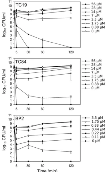
The time-kill assay for TC19, TC84 and BP2 againstB. subtiliscells showed a rapid decline in numbers of colony forming units (CFU) at 5 min at concentrations of 56μM to 7μM for TC19 and TC84, and 3.5μM to 0.44μM for BP2 (Fig. 2) against a cell number 10-fold higher than what was used during the MIC/MBC measurements. Concentra-tions higher than 3.5μM BP2 also caused rapid killing and are not shown. Peptides TC19 and TC84, however, caused a further decline in
Table 1
Minimal inhibitory concentration (MIC) and minimal bactericidal concentra-tion (MBC) of the antimicrobial peptides against B. subtilis strain 168 (1.5 × 10⁶cells/well).
Antimicrobial peptide MIC (μM) MBC (μM)
Mean ± SD Mean ± SD
TC19 7.0 ± 3.3 (n= 6) 7.0 ± 4.6 (n= 3) TC84 7.0 ± 4.2 (n= 6) 3.5 ± 0.2 (n= 3) BP2 3.2 ± 1.5 (n= 6) 3.5 ± 0.2 (n= 3)
nis the number of biological repeats performed.
Fig. 1.Adaptation or resistance development ofB. subtilisafter TC19, TC84 and BP2 treatment using an evolutionary study. Ciprofloxacin was used as a positive control. Induction of adaptation or resistance was performed by repeated ex-posure ofB. subtilisto ¼MIC, ½MIC, 1 × MIC, 2 × MIC and 4 × MIC, of TC19, TC84 and BP2 for 14 passages. The lowest concentration that had no detectable growth after these 14 passages was reported as fold change of the MIC. The highest detectable fold change observed for each of the peptides was two, which was considered not to be adaption or resistance development. An inoculum of 1 × 10⁷CFU/ml was used and culturing was performed for 24 h. Two biological repeats are shown.
CFU upon 30 min of incubation. TC19 showed a further decline at 14μM after 60 min, followed by a complete killing of the culture. BP2 only showed a decline at 5 min with no bacteria surviving at 3.5μM. TC19 and TC84 are thus slower acting compared to BP2. This difference could be due to the difference in net positive charge of TC19 and TC84, which is 4+, compared to that of BP2 which is 7+. Thus, the elec-trostatic interaction of BP2 to the net negatively charged cell envelope ofB. subtilismight be greater in the case of BP2 than with TC19 and TC84. Survival of remaining cells in the incubation could occur due to the reduction of available peptide in the medium, due to protease de-gradation or due to binding of the peptides to cells, cell debris, com-ponents of the medium or to the surface of the microtiter wells (Fig. S1). Concentrations used in the subsequent experiments were based on the time kill assay results.
Aliquots of the culture were taken at 0, 5, 30, 60 and 120 min. Surviving cells were expressed as colony forming units (CFU) per ml. Standard error bars represent three biological repeats. CFU/ml values that were zero were substituted with one, to display the values on the graph.
3.4. B. subtilis transcriptional response after treatment with AMPs reveals cell envelope stress
To elucidate the mode of action of TC19, TC84 and BP2, wefirst performed a transcriptomic analysis ofB. subtiliscells treated with sub-lethal concentrations at 5 and 120 min after peptide addition.B. subtilis differentially expressed the highest number of genes in response to TC19 (Table 2). The majority of the genes differentially expressed after treatment with TC84 were also expressed in response to TC19 (Fig. 3). Five minutes of treatment with BP2 yielded the lowest number of dif-ferentially expressed genes, and most of these genes were also diff er-entially expressed in response to TC19 and/or TC84 (Fig. 3). No genes were differentially expressed after 120 min treatment with BP2. The stress response of TC19, TC84 and BP2 was different from that of known antimicrobials that target cell wall synthesis (D-cycloserine, oxacillin, ristocetin, bacitracin, vancomycin and amoxicillin), the cell membrane (the non-ionic detergent Triton X-114), the cell membrane without permeabilising the membrane (the ionophore monensin) and compounds that target the cell membrane by permeabilising the membrane (gramicidin A and polymyxin B) [23] (Fig. 4). The data show that the stress response of the AMPs was most similar to that of the detergent, Triton X-114. We chose in our analysis to focus on spe-cific regulons to obtain an overview of the response ofB. subtilisto the AMPs. Only key genes that indicate a possible mode of action of the peptides are mentioned. Individual genes might not be differentially expressed by all three peptides.
B. subtilisresponded to TC19, TC84 or BP2 by upregulating genes associated with the following two-component systems (TCSs) and their cognate genes: the liaIH-liaGFSR operon, the bceRS-bceAB TCS-ABC transporters, thepsdRS-psdABTCS-ABC transporters, theyxdJKLMyxeA operon,ytrABCDEFandywoBCDoperons, and theyvrHbregulon (Fig. S2 and Table S2).B. subtilisinduced the expression of genes involved in all of the TCS mentioned in response to a 5 min TC19 exposure. Only the genes associated with the BceRS TCS, the YtrA regulon, the PsdRS
TCS and the YvrHb regulon were differentially upregulated in response to 120 min of treatment with TC19. In response to 5 min of treatment with TC84, genes associated with the LiaRS TCS, BceRS TCS, YxdJK TCS, the YtrA regulator and the YvrHb TCS were responsive. These genes were not upregulated in response to 5 min of treatment with BP2, except for one gene in theytrABCDEFoperon, and two in the YvrHb TCS. None of the TCSs and their cognate genes were differentially up-regulated after 120 min of treatment with TC84 or BP2. When com-paring the stress response with the above-mentioned antimicrobials, only Triton X-114, bacitracin and vancomycin exposed cells showed an induction of expression of the genes associated with the LiaRS TCS [23]. Furthermore, genes associated with the BceRS TCS were only induced by bacitracin and ristocetin, and those associated with the YtrA reg-ulator by vancomycin and ristocetin [23].
The LiaRS TCS is upregulated in response to cell wall synthesis in-hibition but also to membrane perturbation [24–29]. The BceRS TCS, PsdRS TCS and the YtrA regulon are upregulated during cell wall synthesis inhibition [24,25,30–32]. YxdJK TCS are associated with cell membrane perturbation [26]. The YvrHb regulon plays a key role in maintaining the cell envelope integrity by positively regulatingwprA, wapA-yxxG,dltABCDE,sunA,sunT-bdbA-yolJ-bdbB,yvrI-yvrHaand sigX-rsiX, and negatively regulating thelytABCoperon [33,34].
B. subtilisresponded to TC19, TC84 or BP2 by upregulating the expression of genes under control of the extracytoplasmic function (ECF) sigma factors SigM, V, W and X, which play a key role in cell envelope stress response [24–26,31,35–44] (Fig. S2). Genes associated with SigB were also differentially expressed, but most of the genes upregulated after 5 min and 120 min are shared by SigM, W or X (Fig. S3). Downregulated genes associated with SigB were mostly diff eren-tially expressed after treatment with TC19 for 120 min, and the ma-jority of these genes were not regulated by SigM, W or X (Table S3). These genes were involved in multidrug resistance and resistance against ethanol, salt, paraquat, low temperature, peroxide, and other antimicrobial stimuli (Table S3).
For an overview of the genes regulated and differentially expressed by SigM, V, W and X in response to TC19, TC84 or BP2 refer to Table S4. Results on genes relevant for the mode of action are summarized as follows. Genes involved in cell wall stress were upregulated. These were ydaH(also amj),bcrC, the penicillin-binding protein genespbpX and pbpE, and ywaC, the biomarker gene for screening cell-wall active compounds [45]. Similarly, Triton X-114 induced the expression of pbpE, amoxicillin theywaC, vancomycin theydaH, and ristocetin the bcrCandywaC[23]. TheydaHgene encodes a lipid IIflippase involved in the transport of lipid II across the membrane [46] andbcrCencodes
Table 2
The number of differentiallyaexpressed genes after treatment with the AMPs.
TC19 TC84 BP2
5 min 120 min 5 min 120 min 5 min 120 min
Upregulated 187 528 107 36 39 0
Downregulated 7 470 1 4 0 0
a Genes are considered to be differentially expressed when the expression ratio exceeds a factor of two and shows a significant difference in log expression ratio (p≤0.05).
undecaprenyl pyrophosphate phosphatase important for cell wall synthesis and is involved in resistance against bacitracin and paraquat [31,47,48]. Genes involved in cell envelope biogenesis (divIB,mreC, mreD,murBandrodA), and lipoteichoic acid synthesis (yfnI) were also upregulated [49–54]. These responses indicate that the peptides impose cell wall stress on theB. subtiliscells.
B. subtilisalso responded to TC19 and TC84 by upregulating genes involved in modifying the cell surface such as thedltoperon,psd, and oat. DltA, DltB, DltC, DltD and DltE are all involved in the D-alanylation of teichoic acids and lipoteichoic acids [55]. Psd is a phosphatidylserine decarboxylase and is involved in the synthesis of phosphatidylethano-lamine (PE), a zwitterionic phospholipid of the cell membrane [56]. Oat is involved inO-acetylation of peptidoglycan, a modification that has shown to be involved in resistance of Gram-positive bacteria against lysozyme [57].
Fatty acid metabolism was increased in response to exposure ofB. subtiliscells to TC19 by the upregulation ofyrhJ, a cytochrome P450 monooxygenases (CYP) referred to as P450 CYP102A3 for B. subtilis [58–60] (Table S4). CYP102A3 hydroxylates branched chain fatty acids, and the degradation of iso andanteisofatty acids by CYP102A3 has been proposed as a means of altering thefluidity of the cell mem-brane [60,61].B. subtilisalso upregulatedfloT,floA,yuaFandpspAin response to TC19 and TC84. GenesfloT,floAandyuaFand involved in membranefluidity homeostasis andB. subtiliscan alter its membrane fluidity by upregulating these genes. The phage shock homologue and biomarker for membrane distortion, PspA, is involved in stabilizing the membrane [40,62,63]. These responses all indicate a response of the bacteria to cell membrane distortion by the peptides. Interestingly, only treatment with Triton X-114 showed an upregulation of the expression ofyuaFandpspA[23].
Bacterial cells may experience oxidative stress and DNA damage due to cell envelope distortion after treatment with the peptides. Indeed we foundbcrC,yqjL,spxandyjbCupregulated, which are associated with oxidative stress or paraquat resistance [47,64–69]. Similarly,yqjLwas
upregulated by Triton X-114, amoxicillin, vancomycin, ristocetin, and polymycin B, andyjbCby amoxicillin and ristocetin. Transcription of a DNA repair gene,recU [70], and DNA integrity scanning gene,disA, [71] was upregulated. The sigma factor Xpf was differentially upregu-lated after treatment with TC19 and TC84, but not after treatment with BP2.B. subtilishas a suicidal response to DNA-damage by lysing and by producing the phage-like bacteriocin PBSX bacteriophage particles that kill strains non-lysogenic for this phage by damaging the cell wall [72]. Xpf positively regulates the expression of genes associated with PBSX particles production [72]. However, genes regulated by the Xre regulon were also upregulated after treatment with TC19 and TC84. Xre is a transcriptional repressor of Xpf [72]. The combined effects of upregu-lation of Xpf and Xre are unclear.
Finally, the genesfosB,ybfO,ydbS,ydbT,yqeZ,yqfB,sunI,yfhLand yknWXYZreported to be involved in resistance to antimicrobials were upregulated (Table S5). FosB is a metallothiol transferase shown to confer resistance to fosfomycin and to the antimicrobials produced by B. amyloliquefaciens[35,36]. YbfO is similar to an erythromycin es-terase known to be involved in erythromycin resistance [32]. Genes ydbSandydbTare involved in the resistance against the antimicrobials produced byB. amyloliquefaciens[35]. GenesyqeZandyqfBas part of theyqeZyqfAB operon [35] and sunI[73] are involved in sublancin resistance, andyfhLand theyknWXYZoperon in resistance to the B. subtilistoxic protein SdpC [35]. The gene encoding SdpC,yvaY, was not differentially expressed. However, mutants with deletions of genes (ones that are non-essential) mentioned above did not show reduced susceptibility to TC19, TC84 and BP2 (Table S6). Interestingly, Triton X-114 induced the expression of fosB, ybfO, ydbS, yqeZ, yqfB, yfhL, yknWXYZ, but none of the other antimicrobials [23].
In summary, the transcriptomic analysis suggested thatB. subtilis responded to the TC19, TC84 and BP2 exposure by upregulating genes associated with cell membrane distortion and cell wall synthesis. The induction of other gene sets indicates that alteration to the cell envelope likely caused oxidative stress and possibly DNA damage.B. subtilisthus
respond to the changes to the cell envelope by gene expression aimed at changing the cell surface, altering membranefluidity and upregulating genes associated with resistance against known antimicrobials.
3.5. TC19, TC84 and BP2 cause membrane damage
Since the transcriptomic analysis suggested that the cell envelope is targeted by the peptides, we employed TEM to investigate any struc-tural changes that the treatment of TC19, TC84 and BP2 might create after 5 min and 120 min. Lethal concentrations were used, which were 14μM TC19 and TC84, and 3.5μM BP2. Cells treated for 5 min with TC19, TC84 and BP2 stained black with uranyl acetate whereas the untreated cells appeared grey with black uranyl acetate deposited on the surface of the cell (Fig. 5). TC19, TC84 and BP2 did not lyse the cells and no visible cell wall damage was observed at 5 min. In contrast, after 120 min of exposure, disrupted cells were observed (Fig. 5). Cross sections of the cells treated for 5 min showed an irregularly distributed and abnormal nucleoid (Fig. 5, red arrows). Uranyl acetate staining of the cells entirely, implying that membrane perturbation had occurred since the membrane was permeable to the dye. Membrane permeabi-lisation, in a peptide concentration dependent manner, was confirmed using thefluorescent dye Sytox Green (Fig. 6). Thesefindings suggest that the cell membrane is the primary target for the peptides. The cell wall stress response might be a result of cell membrane distortion and
the irregular nucleoid could be a consequence of the loss of cell membrane integrity.
3.6. Delocalization of membrane bound proteins
To further address the molecular events involved in cell envelope homeostasis we analysed in more detail protein localization in the cell membrane starting from the notion that essential cellular processes, such as cell wall and cell membrane synthesis, occur at the plasma membrane [74]. To study the localization of various proteins involved and to aid in the elucidation of their function, we used a panel of mutant strains expressing proteins fused to greenfluorescent protein (GFP), to investigate whether the membrane proteins delocalized after treatment with TC19, TC84 and BP2. TheB. subtilismutants were cul-tured in defined minimal medium (CMM), in which the growth of the bacteria is slow and protein localization might differ from previous reports where rich medium was used (Fig. S4). Defined minimal medium was selected as the exact concentration of each component is known, unlike rich medium.
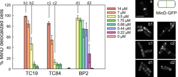
MinD was initially evaluated, where MinD together with MinC forms a complex that inhibits theZ-ring formation [75]. MinD-GFP is known to localize at the septum and at the cell poles [76]. In non-treated cells (Fig. 7, image a) MinD-GFP indeed was localized at the septum, whereas once TC19, TC84 and BP2 were added MinD-GFP
Fig. 5.Transmission electron micrographs (TEM) ofB. subtilisafter peptide treatment.B. subtiliswas treated for 5 and 120 min with 14μM TC19, 14μM TC84 or 3.5μM BP2. Negative staining with uranyl acetate was performed of cells after treatment with the peptides and cross sections were performed of the uranyl acetate stained cells. Negative staining showed cells stained black with uranyl acetate indicating perturbed membranes (treated). Cells with an intact membrane appears grey with black uranyl acetate on the surface of the cell (untreated). Cross sections of the stained cells after treatment for 5 min with TC19, TC84 and BP2 had irregular nucleoids compared to the untreated cells (red arrows). After 120 min of treatment with the peptides the cross sections reveal lysed cells, while all observed untreated cells remained intact. Scale bar of microscopy images represent 1μm.
delocalized. At lethal concentrations, 14μM TC19 (b1) and TC84 (c1), and 3.5μM BP2 (d1), MinD-GFP was evenly distributed throughout the cells. At sub-lethal concentrations, 3.5μM TC19 and TC84, and 0.22μM BP2, the appearance of MinD-GFP was“spotty”. Such a“spotty” ap-pearance of MinD-GFP when delocalized has also been observed forB. subtilistreated with compounds that dissipate the membrane potential, valinomycin or CCCP [76]. However, the even distribution of the MinD-GFP as observed after exposure to the high TC19, TC84 and BP2 con-centrations has never been reported. We suspect that at lethal peptide concentrations, MinD-GFP dissociates from the membrane due to rapid cell death, but at sub-lethal concentrations the protein remains attached but delocalized due to distortion of the membrane. Quantification of the microscopy images showed that MinD-GFP delocalization, both “spotty”and fully delocalized, occurred in a concentration dependent manner. This was the case for all GFP-fusion proteins observed. Therefore, only results for lethal concentrations are shown. We in-vestigated our microarray data to observe whether B. subtilis is re-sponding directly to the delocalization of MinD or its associated protein MinC, but we found no differential expression of MinD or MinC after 5 or 120 min of treatment with the peptides.
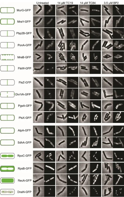
Delocalization or dissociation of the MinD-GFP occurred rapidly (≤ 5 min) (Fig. 7). This was also observed for MreB (Fig. 8). MreB is a cell-shape determinant that forms an important part of the cell wall synthesis machinery and is pivotal in maintaining cell membrane homeostasis [77–79]. Both MinD and MreB requires the presence of a membrane potential for correct localization [76]. The transcriptomic analysis revealed that MreB was not differentially expressed after treatment with TC19, TC84 and BP2 (Table S7). Expression of the genes encoding the MreB homologous proteins Mbl and MreBH [78], how-ever, were upregulated by 1.5 logFC and 4.6 logFC after TC19 treatment for 120 min (Table S7). After treatment with TC84 for 120 min MreBH were upregulated by 1.1 logFC. The upregulation of these genes sug-gests thatB. subtilisrespond to the delocalization of these membrane bound proteins.
The delocalization of MreB, together with the cell wall stress re-sponse, hinted at the possibility that other cell wall synthesis-associated proteins might also be delocalized. MreB forms a complex with MreC, MreD, RodA, MurG, MraY and several PBPs [80–82]. MurG was initially investigated as it has been shown to delocalize after treatment with the lipopeptide daptomycin, the cationic hexapeptide MP196, the cyclic decapeptide gramicidin S and the cyclic hexapeptide cWFW [34,83, 84]. Treatment with TC19, TC84 and BP2 caused a rapid (≤5 min)
delocalization of the MurG-GFP. MurG is a N-acetylglucosamine transferase that catalysis the addition ofN-acetylglucosamine to theN -acetylmuramic acid residue of lipid I, resulting in the formation of lipid II [51, 85]. The transcriptomic analysis revealed that B. subtilis re-sponded to the treatment with TC19 after 120 min by upregulating MurG and genes co-transcribed with MurG, MurB, SpoVE, DivIB and Sbp (Table S7) [23,86]. MurB was differentially expressed in response to TC84 after 120 min. In conclusion, it appears thatB. subtilisadjust the expression of genes associated with MurG functionality in response to MurG delocalization.
Additional proteins involved in cell wall synthesis that delocalized after treatment with TC19, TC84 and BP2 were MraY-GFP, PBP2b-GFP, PonA-GFP and FtsW-GFP. Essential MraY is a phospho-N -acetylmur-amoyl-pentapeptide transferase that catalyses the transfer of the phospho-MurNAc-pentapeptide moiety to undecaprenyl phosphate lo-cated at the membrane, forming lipid I [51,85]. PonA is a class A pe-nicillin binding protein (PBP) with both transglycosylase and trans-peptidase activity [87–90]. MraY and PonA are both dependent on the proper localization of MreB [91, 92]. Penicillin-binding protein 2b (PBP2b) is a class B transpeptidase involved in the cell wall synthesis during cell division [90,93]. PBP2b together with FtsW forms part of the divisome responsible for septal cell wall synthesis [94,95] as well as for the stabilization of theZ-ring [94]. The Z-ring is formed by the polymerization of a tubulin-like protein, FtsZ, into a circular structure at mid-cell [96]. Delocalization of PBP2b, FtsW or FtsZ will prevent proper formation of the Z-ring, which serves as a scaffold for other proteins involved in synthesizing the septum or cell division site [49], and will thus interfere with cell division.
A general range of proteins involved in cell membrane synthesis (PgsA and PlsX), ATP synthesis (AtpA), Krebs' cycle and respiration (SdhA), cell division (FtsZ and DivIVA), transcription (RpoC), transla-tion (RpsB) and DNA repair (DnaN and RecA) were selected to assess whether TC19, TC84 and BP2 affect other cellular functions due to membrane distortion. Delocalization of all proteins involved in cell membrane synthesis, ATP synthesis, Kreb's cycle and respiration, and cell division were observed. However, localization of RpoC and RpsB localized within the cytosol, and of DnaN and RecA, associated with the nucleoid, was not affected (Fig. 8). Clearly the abnormal localization of ATP synthase subunit AtpA is an indication of extensive perturbation of the cell membrane. In normal cells the AtpA complex is uniformly distributed at the membrane and its delocalization has been used pre-viously as an indicator for abnormal membrane curvatures [77].
4. Discussion
TC19, TC84 and BP2 are predicted to share the common features of being cationic and amphipathic peptides and comply with the char-acteristics of their class by distorting the cell envelope ofB. subtilisin a non-specific manner. TC19, TC84 and BP2 were rapidly bactericidal (≤ 5 min). Furthermore, as expected for this class of antimicrobial pep-tides, neither of these three peptides causedB. subtilisto develop re-sistance, as tested by 14 cycles of exposure to the peptides. TC19, TC84 and BP2 caused rapid membrane permeabilisation without causing overt cell lysis, as observed with the TEM and Sytox Green staining images. TC19, TC84 and BP2 at sub-lethal concentrations initiated a stress response distinctively different from known antimicrobials. We
compared the response with responses observed upon treatments that target the cell envelope by inhibiting cell wall synthesis (D-cycloserine,
oxacillin, ristocetin, bacitracin, vancomycin and amoxicillin), distorting the cell membrane (i.e. the detergent Triton X-114), distorting the cell membrane without causing membrane permeabilisation (the ionophore monensin) and those that do permeabilise the membrane (gramicidin A and polymyxin B) [23]. An independent study using a proteomic ap-proach also found that treatingBacillus subtiliswith different membrane active compounds does not necessarily cause a similar stress response [97]. Our AMPs induced a stress response most similar to Triton X-114. Triton X-114 is a non-ionic detergent that causes a phase-separation and is often used to solubilize and separate proteins during extraction [98].
TC19, TC84 and BP2 induced a cell envelope stress response (CESR) by upregulating genes regulated by Sigma factors M, V, W and X and two component regulator systems (TCSs).B. subtilisresponded to TC19 and TC84 similarly as when exposed to the natural cationic alpha-he-lical AMP LL-37 [99], by upregulating genes controlled by the LiaRS, YxdJK, and BceRS TCS regulators [26]. The YxdJK TCS has only been associated with cell membrane perturbation [26], but the LiaRS TCS have been linked with both cell wall synthesis inhibition and membrane perturbation [24–29]. The BceRS TCS and its cognate ABC transporters, BceAB, are upregulated in response to bacitracin and vancomycin, and are associated with cell wall synthesis inhibition [24, 30, 31]. Ad-ditionally,B. subtilisdifferentially upregulated the YtrA regulon in re-sponse to TC19 and TC84. The YtrA regulon has been associated with exposure to cell wall synthesis inhibiting compounds [24, 25, 32]. Thesefindings suggest that TC19 and TC84 target the cell wall archi-tecture and/or synthesis in addition to the cell membrane. However, LL-37 has to our knowledge not been shown to be involved in cell wall synthesis inhibition, and the upregulation of the BceRS TCS suggest that LL-37 is targeting cell wall synthesis. In the case of BP2, few genes were differentially expressed in response to the peptide. We speculate that it is due to the very rapid activity of BP2 that prevents the initiation of a stress response similar to what was observed for TC19 and TC84. The few genes that were upregulated suggested that BP2 might have a si-milar cell envelope target as TC19 and TC84. However, the physiolo-gical analysis showed that no cell wall damage or cell lysis occurred after 5 min of treatment with lethal concentrations of TC19, TC84 and BP2. Instead, the membrane distortion caused by these peptides did cause delocalization of proteins essential for cell wall synthesis, MurG, MraY, MreB, PonA, PBP2b and FtsW, within 5 min of treatment. Ex-posure to LL-37 also caused delocalization of these membrane bound proteins involved in cell wall synthesis (Fig. S5), suggesting that this delocalization may be a general effect of exposure to cationic amphi-pathic antimicrobial peptides acting on the membrane, since delocali-zation of membrane bound cell wall synthesis proteins has also been reported for non-pore-forming linear hexapeptide MP196, lipopeptide daptomycin and non-pore-forming cyclic hexapeptide cWFW [34,83, 84]. The changes which TC19, TC84, BP2 and LL-37 cause to the membrane must be different to those caused by daptomycin, since daptomycin did not affect the localization of integral membrane pro-teins MraY and PBP2b [84]. The activity of TC19, TC84 and BP2 on the cell membrane also caused other proteins involved in cell membrane synthesis and metabolism to delocalize. Delocalization of such proteins prevents their normal functioning [76,100] and will be deleterious for the survival of the cells. The delocalization of membrane proteins in-volved in cell wall synthesis by TC19, TC84, BP2 and LL-37 suggests that these amphipathic AMPs have common elements in their mode of action, some of which might be mediated through a shared secondary structure. LL-37 and BP2 are referred to as alpha-helical [9,99] while we have preliminary indications that at least TC84 acquires some alpha-helical features upon interaction with membrane mimetics (un-published observations).
5. Conclusion
TC19, TC84 and BP2 showed to be promising candidates as anti-microbial agents against Gram-positive bacterial cells as their mem-brane perturbation activity causes interference with various essential cellular processes leading to death. We observed no adaptation or re-sistance development against TC19, TC84 and BP2. No difference in mode of action between TC19 and TC84 was found, but BP2 was more rapidly active compared to TC19 and TC84 which we suspect is due to its higher cationic charge. We employed a transcriptomic approach to elucidate the mode of action and observed that the membrane pertur-bation caused by the cationic amphipathic peptides induced a mem-brane and cell wall stress response. With the aid of the greenfluorescent protein fused to essential proteins bound to the membrane, we were
able to establish that proteins involved in cell wall synthesis are delo-calized. Delocalization will have a deleterious effect on the normal functioning of the proteins. Conclusively, ourfindings have elucidated crucial aspects of the mode of action of TC19, TC84 and BP2. The ob-served lack of readily emerging resistance implies significant potential for further preclinical studies aimed at clinical development.
Transparency document
The http://dx.doi.org/10.1016/j.bbamem.2018.06.005 associated with this article can be found, in online version.
Acknowledgements
The authors acknowledge Edward A. de Koning and Terrens N.V. Saaki for constructing the strains EKB40, and TNVS30D, respectively, and Urša Gubenšek for constructing the strain UG-10 during her Master's internship. We thank the MicroArray Department (MAD) of the University of Amsterdam for their contribution to the transcriptomic analysis. S. Omardien acknowledges the Erasmus Mundus Action 2 program (EMA2) and University of Amsterdam for funding. M. Riool and S. A. J. Zaat are supported by the FP7-HEALTH-2011 grant 278890, BALI–Biofilm Alliance. The authors declare that they have no conflict of interest.
Appendix A. Supplementary material
Supplementary data to this article can be found online athttps:// doi.org/10.1016/j.bbamem.2018.06.005.
References
[1] L. Czaplewski, R. Bax, M. Clokie, M. Dawson, H. Fairhead, V.A. Fischetti, S. Foster, B.F. Gilmore, R.E.W. Hancock, D. Harper, I.R. Henderson, K. Hilpert, B.V. Jones, A. Kadioglu, D. Knowles, S. Ólafsdóttir, D. Payne, S. Projan, S. Shaunak, J. Silverman, C.M. Thomas, T.J. Trust, P. Warn, J.H. Rex, Alternatives to anti-biotics—a pipeline portfolio review, Lancet Infect. Dis. 16 (2016) 239–251,http:// dx.doi.org/10.1016/S1473-3099(15)00466-1.
[2] C. Ghosh, J. Haldar, Membrane-active small molecules: designs inspired by anti-microbial peptides, ChemMedChem 10 (2015) 1606–1624,http://dx.doi.org/10. 1002/cmdc.201500299.
[3] F. Guilhelmelli, N. Vilela, P. Albuquerque, L. da S. Derengowski, I. Silva-Pereira, C.M. Kyaw, Antibiotic development challenges: the various mechanisms of action of antimicrobial peptides and of bacterial resistance, Front. Microbiol. 4 (2013) 1–12,
http://dx.doi.org/10.3389/fmicb.2013.00353.
[4] N. Srinivas, P. Jetter, B.J. Ueberbacher, M. Werneburg, K. Zerbe, J. Steinmann, B. Van Der Meijden, F. Bernardini, A. Lederer, R.L.A. Dias, P.E. Misson, H. Henze, J. Zumbrunn, A. Käch, L. Eberl, K. Riedel, S.J. Demarco, J.A. Robinson,
Peptidomimetic antibiotics target outer-membrane biogenesis inPseudomonas aer-uginosa, Science (80-.). 327 (2010) 1010–1013. doi:https://doi.org/10.1126/ science.1182749.
[5] G.S. Crowther, S.D. Baines, S.L. Todhunter, J. Freeman, C.H. Chilton, M.H. Wilcox, Evaluation of NVB302 versus vancomycin activity in an in vitro human gut model ofClostridium difficileinfection, J. Antimicrob. Chemother. 68 (2013) 168–176,
http://dx.doi.org/10.1093/jac/dks359.
[6] Demegen_Pharmaceuticals, Novel candidiasis therapy, Pharm. Prod. Prod. Profiles (2013) (www.demegen.com/pharma/candidiasis.htm).
[7] M.N. Melo, D. Dugourd, M.A.R.B. Castanho, Omiganan pentahydrochloride in the front line of clinical applications of antimicrobial peptides, Recent Patents on Anti-Infective Drug Discovery 1 (2006) 201–207.
[8] S.A.J. Zaat, P.H.S. Kwakman, J.W. Drijfhout, Thrombocidin-derived Antimicrobial Peptides, WO2015099535, (2015).
[9] P.R. Abraham, B.J. Appelmelk, S.J.H. Van Deventer, Synthetic Peptides with Antimicrobial and Endotoxin Neutralizing Properties for Management of the Sepsis Syndrome, US6624140B1, 2003.
[10] P.H.S. Kwakman, L. De Boer, C.P. Ruyter-Spira, T. Creemers-Molenaar, J.P.F.G. Helsper, C.M.J.E. Vandenbroucke-Grauls, S.A.J. Zaat, A.A. Te Velde, Medical-grade honey enriched with antimicrobial peptides has enhanced activity against antibiotic-resistant pathogens, Eur. J. Clin. Microbiol. Infect. Dis. Off. Publ. Eur. Soc. Clin. Microbiol. 30 (2011) 251–257, http://dx.doi.org/10.1007/s10096-010-1077-x.
[12] I. Zelezetsky, A. Tossi, Alpha-helical antimicrobial peptides—using a sequence template to guide structure-activity relationship studies, Biochim. Biophys. Acta Biomembr. 1758 (2006) 1436–1449,http://dx.doi.org/10.1016/j.bbamem.2006. 03.021.
[13] S. Marqusee, V.H. Robbins, R.L. Baldwin, Unusually stable helix formation in short alanine-based peptides, Proc. Natl. Acad. Sci. U. S. A. 86 (1989) 5286–5290,http:// dx.doi.org/10.1073/pnas.86.14.5286.
[14] C. Anagnostopoulos, J. Spizizen, Requirements for transformation inBacillus subtilis, J. Bacteriol. 81 (1961) 741–746.
[15] S. Halbedel, M. Kawai, R. Breitling, L.W. Hamoen, SecA is required for membrane targeting of the cell division protein DivIVA in vivo, Front. Microbiol. 5 (2014) 58, ,
http://dx.doi.org/10.3389/fmicb.2014.00058.
[16] D.C. Hooper, Mechanisms of action and resistance of older and newerfl uor-oquinolones, Clin. Infect. Dis. 31 (2000) S24–S28.
[17] J. Dankert, J. Van der Werff, S.A.J. Zaat, W. Joldersma, D. Klein, J. Hess, J. Van Der Werff, S.A.J. Zaat, W. Joldersma, D. Klein, Involvement of bactericidal factors from thrombin-stimulated platelets in clearance of adherent viridans streptococci in ex-perimental infective endocarditis, Infect. Immun. 63 (1995) 663–671.
[18] R.A. Irizarry, B. Hobbs, F. Collin, Y.D. Beazer-Barclay, K.J. Antonellis, U. Scherf, T.P. Speed, Exploration, normalization, and summaries of high density oligonu-cleotide array probe level data, Biostatistics 4 (2003) 249–264,http://dx.doi.org/ 10.1093/biostatistics/4.2.249.
[19] R.D. Wolfinger, G. Gibson, E.D. Wolfinger, L. Bennett, H. Hamadeh, P. Bushel, C. Afshari, R.S. Paules, Assessing gene significance from cDNA microarray expres-sion data via mixed models, J. Comput. Biol. 8 (2001) 625–637.
[20] X. Cui, G.A. Churchill, Statistical tests for differential expression in cDNA micro-array experiments, Genome Biol. 4 (210) (2003), http://dx.doi.org/10.1186/gb-2003-4-4-210.
[21] X. Cui, J.T.G. Hwang, J. Qiu, N.J. Blades, G.A. Churchill, Improved statistical tests for differential gene expression by shrinking variance components estimates, Biostatistics 6 (2005) 59–75,http://dx.doi.org/10.1093/biostatistics/kxh018. [22] J.D. Storey, R. Tibshirani, Statistical significance for genomewide studies, Proc.
Natl. Acad. Sci. U. S. A. 100 (2003) 9440–9445,http://dx.doi.org/10.1073/pnas. 1530509100.
[23] W. Eiamphungporn, J.D. Helmann, TheBacillus subtilissigma(M) regulon and its contribution to cell envelope stress responses, Mol. Microbiol. 67 (2008) 830–848,
http://dx.doi.org/10.1111/j.1365-2958.2007.06090.x.
[24] T. Mascher, N.G. Margulis, T. Wang, R.W. Ye, J.D. Helmann, Cell wall stress re-sponses inBacillus subtilis: the regulatory network of the bacitracin stimulon, Mol. Microbiol. 50 (2003) 1591–1604,http://dx.doi.org/10.1046/j.1365-2958.2003. 03786.x.
[25] L.I. Salzberg, Y. Luo, A.B. Hachmann, T. Mascher, J.D. Helmann, TheBacillus subtilis
GntR family repressor YtrA responds to cell wall antibiotics, J. Bacteriol. 193 (2011) 5793–5801,http://dx.doi.org/10.1128/JB.05862-11.
[26] M. Pietiäinen, M. Gardemeister, M. Mecklin, S. Leskelä, M. Sarvas, V.P. Kontinen, Cationic antimicrobial peptides elicit a complex stress response inBacillus subtilis
that involves ECF-type sigma factors and two-component signal transduction sys-tems, Microbiology 151 (2005) 1577–1592,http://dx.doi.org/10.1099/mic.0. 27761-0.
[27] T. Mascher, S.L. Zimmer, T.A. Smith, J.D. Helmann, Antibiotic-inducible promoter regulated by the cell envelope stress-sensing two-component system LiaRS of
Bacillus subtilis, Antimicrob. Agents Chemother. 48 (2004) 2888–2896,http://dx. doi.org/10.1128/AAC.48.8.2888-2896.2004.
[28] A.-B.A.-B. Hachmann, E.R. Angert, J.D. Helmann, Genetic analysis of factors af-fecting susceptibility ofBacillus subtilisto daptomycin, Antimicrob. Agents Chemother. 53 (2009) 1598–1609,http://dx.doi.org/10.1128/AAC.01329-08. [29] B. Hutter, C. Schaab, S. Albrecht, M. Borgmann, N.A. Brunner, C. Freiberg,
K. Ziegelbauer, C.O. Rock, I. Ivanov, H. Loferer, Prediction of mechanisms of action of antibacterial compounds by gene expression profiling prediction of mechanisms of action of antibacterial compounds by gene expression profiling, Antimicrob. Agents Chemother. 48 (2004) 2838–2844,http://dx.doi.org/10.1128/AAC.48.8. 2838-2844.2004.
[30] R. Ohki, K. Giyanto, W. Tateno, S. Masuyama, K. Moriya, N. Ogasawara Kobayashi, The BceRS two-component regulatory system induces expression of the bacitracin transporter, BceAB, inBacillus subtilis, Mol. Microbiol. 49 (2003) 1135–1144,
http://dx.doi.org/10.1046/j.1365-2958.2003.03653.x.
[31] M. Cao, J.D. Helmann, Regulation of theBacillus subtilisbcrC bacitracin resistance gene by two extracytoplasmic function sigma factors, J. Bacteriol. 184 (2002) 6123–6129,http://dx.doi.org/10.1128/Jb.184.22.6123-6129.2002.
[32] M. Cao, T. Wang, R. Ye, J.D. Helmann, Antibiotics that inhibit cell wall biosynthesis induce expression of theBacillus subtilissigma(W) and sigma(M) regulons, Mol. Microbiol. 45 (2002) 1267–1276,http://dx.doi.org/10.1046/j.1365-2958.2002. 03050.x.
[33] M. Serizawa, K. Kodama, H. Yamamoto, K. Kobayashi, N. Ogasawara, J. Sekiguchi, Functional analysis of the YvrGHb two-component system ofBacillus subtilis: identification of the regulated genes by DNA microarray and northern blot analyses, Biosci. Biotechnol. Biochem. 69 (2005) 2155–2169.
[34] K. Scheinpflug, M. Wenzel, O. Krylova, J.E. Bandow, M. Dathe, H. Strahl, Antimicrobial peptide cWFW kills by combining lipid phase separation with auto-lysis, Sci. Rep. 7 (2017) 44332, ,http://dx.doi.org/10.1038/srep44332. [35] B.G. Butcher, J.D. Helmann, Identification ofBacillus subtilissigma(W)-dependent
genes that provide intrinsic resistance to antimicrobial compounds produced by bacilli, Mol. Microbiol. 60 (2006) 765–782, http://dx.doi.org/10.1111/j.1365-2958.2006.05131.x.
[36] M. Cao, B.A. Bernat, Z. Wang, R.N. Armstrong, J.D. Helmann, FosB, a cysteine-dependent fosfomycin resistance protein under the control of sigma(W), an
extracytoplasmic-function sigma factor inBacillus subtilis, J. Bacteriol. 183 (2001) 2380–2383,http://dx.doi.org/10.1128/JB.183.7.2380-2383.2001.
[37] M. Cao, P.A. Kobel, M.M. Morshedi, M.F.W. Wu, C. Paddon, J.D. Helmann, Defining theBacillus subtilissigma(W) regulon: a comparative analysis of promoter consensus search, run-offtranscription/macroarray analysis (ROMA), and transcriptional profiling approaches, J. Mol. Biol. 316 (2002) 443–457,http://dx.doi.org/10. 1006/jmbi.2001.5372.
[38] M. Cao, J.D. Helmann, TheBacillus subtilisextracytoplasmic-function sigmaX factor regulates modification of the cell envelope and resistance to cationic antimicrobial peptides, J. Bacteriol. 186 (2004) 1136–1146,http://dx.doi.org/10.1128/JB.186.4. 1136–1146.2004.
[39] V. Guariglia-Oropeza, J.D. Helmann,Bacillus subtilissigmaV confers lysozyme re-sistance by activation of two cell wall modification pathways, peptidoglycan O-acetylation and D-alanylation of teichoic acids, J. Bacteriol. 193 (2011) 6223–6232,
http://dx.doi.org/10.1128/JB.06023-11.
[40] J.D. Helmann,Bacillus subtilisextracytoplasmic function (ECF) sigma factors and defense of the cell envelope, Curr. Opin. Microbiol. 30 (2016) 122–132,http://dx. doi.org/10.1016/j.mib.2016.02.002.
[41] Y. Luo, K. Asai, Y. Sadaie, J.D. Helmann, Transcriptomic and phenotypic char-acterization of aBacillus subtilisstrain without extracytoplasmic function sigma factors, J. Bacteriol. 192 (2010) 5736–5745,http://dx.doi.org/10.1128/JB. 00826-10.
[42] J. Radeck, G. Fritz, T. Mascher, The cell envelope stress response ofBacillus subtilis: from static signaling devices to dynamic regulatory network, Curr. Genet. (2016) 1–12,http://dx.doi.org/10.1007/s00294-016-0624-0.
[43] A. Rukmana, T. Morimoto, H. Takahashi, N. Ogasawara Giyanto, Assessment of transcriptional responses ofBacillus subtiliscells to the antibiotic enduracidin, which interferes with cell wall synthesis, using a high-density tiling chip, Genes & Genetic Systems 84 (2009) 253–267,http://dx.doi.org/10.1266/ggs.84.253.
[44] P.D. Thackray, A. Moir, SigM, an extracytoplasmic function sigma factor ofBacillus subtilis, is activated in response to cell wall antibiotics, ethanol, heat, acid, and superoxide stress, J. Bacteriol. 185 (2003) 3491–3498,http://dx.doi.org/10.1128/ JB.185.12.3491-3498.2003.
[45] T.L. Czarny, A.L. Perri, S. French, E.D. Brown, Discovery of novel cell wall-active compounds using Pywac, a sensitive reporter of cell wall stress, in the model Gram-positive bacteriumBacillus subtilis, Antimicrob. Agents Chemother. 58 (2014) 3261–3269,http://dx.doi.org/10.1128/AAC.02352-14.
[46] A.J. Meeske, L.-T. Sham, H. Kimsey, B.-M. Koo, C.A. Gross, T.G. Bernhardt, D.Z. Rudner, MurJ and a novel lipid IIflippase are required for cell wall biogenesis inBacillus subtilis, Proc. Natl. Acad. Sci. U. S. A. 112 (2015) 6437–6442,http://dx. doi.org/10.1073/pnas.1504967112.
[47] M. Cao, C.M. Moore, J.D. Helmann,Bacillus subtilisparaquat resistance is directed by sigmaM, an extracytoplasmic function sigma factor, and is conferred by YqjL and BcrC, J. Bacteriol. 187 (2005) 2948–2956,http://dx.doi.org/10.1128/JB.187.9. 2948-2956.2005.
[48] R. Bernard, M. El Ghachi, D. Mengin-Lecreulx, M. Chippaux, F. Denizot, BcrC from
Bacillus subtilisacts as an undecaprenyl pyrophosphate phosphatase in bacitracin resistance, J. Biol. Chem. 280 (2005) 28852–28857,http://dx.doi.org/10.1074/ jbc.M413750200.
[49] P. Gamba, J.W. Veening, N.J. Saunders, L.W. Hamoen, R.A. Daniel, Two-step as-sembly dynamics of theBacillus subtilisdivisome, J. Bacteriol. 191 (2009) 4186–4194,http://dx.doi.org/10.1128/JB.01758-08.
[50] M. Leaver, J. Errington, Roles for MreC and MreD proteins in helical growth of the cylindrical cell wall inBacillus subtilis, Mol. Microbiol. 57 (2005) 1196–1209,
http://dx.doi.org/10.1111/j.1365-2958.2005.04736.x.
[51] J. van Heijenoort, Lipid intermediates in the biosynthesis of bacterial pepti-doglycan, Microbiol. Mol. Biol. Rev. 71 (2007) 620–635,http://dx.doi.org/10. 1128/MMBR.00016-07.
[52] A.O. Henriques, P. Glaser, P.J. Piggot, C.P. Moran, Control of cell shape and elon-gation by the rodA gene inBacillus subtilis, Mol. Microbiol. 28 (1998) 235–247
http://www.ncbi.nlm.nih.gov/pubmed/9622350.
[53] D.J. Scheffers, M.B. Tol, LipidII: just another brick in the wall? PLoS Pathog. 11 (2015) 1–12,http://dx.doi.org/10.1371/journal.ppat.1005213.
[54] I.C. Sutcliffe, Priming and elongation: dissection of the lipoteichoic acid biosyn-thetic pathway in Gram-positive bacteria, Mol. Microbiol. 79 (2011) 553–556,
http://dx.doi.org/10.1111/j.1365-2958.2010.07480.x.
[55] M. Perego, P. Glaser, A. Minutello, M.A. Strauch, K. Leopold, W. Fischer, Incorporation ofD-alanine into lipoteichoic acid and wall teichoic acid inBacillus subtilis: identification of genes and regulation, J. Biol. Chem. 270 (1995) 15598–15606,http://dx.doi.org/10.1074/jbc.270.26.15598.
[56] K. Matsumoto, M. Okada, Y. Horikoshi, Cloning, sequencing, and disruption of the
Bacillus subtilispsd gene coding for phosphatidylserine decarboxylase, J. Bacteriol. 180 (1998) 100–106.
[57] T.D. Ho, J.L. Hastie, P.J. Intile, C.D. Ellermeier, TheBacillus subtilis extra-cytoplasmic functionσfactorσ(V) is induced by lysozyme and provides resistance to lysozyme, J. Bacteriol. 193 (2011) 6215–6222,http://dx.doi.org/10.1128/JB. 05467-11.
[58] O. Lentz, V. Urlacher, R.D. Schmid, Substrate specificity of native and mutated cytochrome P450 (CYP102A3) fromBacillus subtilis, J. Biotechnol. 108 (2004) 41–49,http://dx.doi.org/10.1016/j.jbiotec.2003.11.001.
[59] O. Lentz, A. Feenstra, T. Habicher, B. Hauer, R.D. Schmid, V.B. Urlacher, Altering the regioselectivity of cytochrome P450 CYP102A3 ofBacillus subtilisby using a new versatile assay system, Chembiochem 7 (2006) 345–350,http://dx.doi.org/10. 1002/cbic.200500266.