i
The Effects of a Single Bout of Resistance Exercise on Inflammatory Markers in Breast Cancer Survivors
Jacob M. Allen
A thesis submitted to the faculty of the University of North Carolina at Chapel Hill in partial fulfillment of the requirements for the degree of Masters of Arts in the Department of
Exercise and Sport Science (Exercise Physiology).
Chapel Hill Spring 2013
Approved By:
iii ABSTRACT
JACOB M. ALLEN: The Effects of a Single Bout of Resistance Exercise on Inflammatory Markers in Breast Cancer Survivors
(Under the direction of Anthony C. Hackney, Ph.D., D.Sc.)
This study examined the effect of resistance exercise (RE) on the acute responses of three inflammatory cytokines (Interleukin 6 [IL-6], Interleukin 10 [IL-10], Tumor Necrosis Factor-α [TNF-α]) in breast cancer survivors. Breast cancer survivors (BCS) (n = 4) and healthy age, matched controls (C) (n=8) completed three sets of four exercises at 70% of one-repetition max. Blood samples were collected prior (Pre), immediately after (ImP), 2 hours after (2P) and 24 hours after (24P) exercise bout. No differences were observed between groups for each cytokine at each time point after exercise. However, the RE did cause a significant upregulation in IL-6 at 2P (p=0.027) and a significant downregulation of TNF-α at 24P in both groups (p=0.011). These findings suggest that no difference exists in
iv
ACKNOWLEDGEMENTS
Throughout the course of completing this thesis, I have had the support and
motivation from numerous individuals, and without their help, this study could not have been possible. First and foremost, I would like to express thanks to my advisor, Dr. Anthony Hackney, for his scientific expertise and motivational skills that gave me the guidance and perseverance that I needed throughout this project. I would also like to recognize and thank my thesis committee members, Dr. Claudio Battaglini and Dr. Elizabeth Evans. Dr.
Battaglini was key in participant recruitment, while also providing important direction, expertise and inspiration when needed most. Meanwhile, Dr. Evans provided ample support in reviewing my thesis, as well as critical advice during the formulation of the IRB
v
TABLE OF CONTENTS
Page
LIST OF TABLES……….viii
LIST OF FIGURES………..ix
Chapter I. BASIS FOR STUDY……….1
Introduction..……….………...………..1
Statement of Purpose.……….…………...…………4
Limitations….……….………...4
Delimitations……….……….5
Significance of Study……….………5
II. REVIEW OF LITERATURE………7
Introduction………7
Interleukin- 6………..8
Interleukin-10………...12
Tumor Necrosis Factor-α……….14
Cytokine Interactions and Temporal Responses to Exercise………...15
Acute Inflammatory Responses to Resistance Exercise………..18
Inflammation and Cancer………….………25
The Inflammatory Response to Exercise in Cancer Survivors…...27
vi
II. METHODOLOGY………..31
Subjects………31
Recruitment………..32
Instrumentation………32
General Procedures………..33
Statistical Analyses………..36
Hypotheses………...37
IV. RESULTS………39
Subject Characteristics……….39
Strength Assessments………...39
Plasma Volume Shifts………..40
Plasma Cytokine Concentrations……….40
IL-6………..41
IL-10………41
TNF-α………...41
Cortisol………..………...42
Cortisol vs. IL-6………...42
V. DISCUSSION………..44
Introduction………..……44
IL-6……….….45
IL-10...……….48
TNF-α……….…..49
vii
Implications for Cancer Survivors and Future Directions...54
Limitations of study……….57
VI. SUMMARY, CONCLUSIONS AND RECOMMENDATIONS…………...59
Conclusions………..………..…..60
APPENDICES A. Informed Consent Form (Breast Cancer Survivors)……..……….62
B. Informed Consent Form (Healthy Controls)……….………..70
C. Medical History Questionnaire………..…….78
D. HIPAA……….…...83
E. Consent for Storing Biological Specimens………..…………86
F. Laboratory Visit Appointment Reminder………..…..91
G. Perceived Stress Scale Form………...93
H. Data Collection From-Visits………..…….94
I. Data Collection Form-Visits………...95
J. Information………...96
viii
LIST OF TABLES
Table
1. Subject Characteristics……….………..39
2. Strength Assessments……….40
3. Plasma Volume Shifts………40
4. Cytokine Responses………...41
5. Cortisol Reponses………..42
ix
LIST OF FIGURES
Figure
1. Protocol Timeline………37
2. Blood Draw Timeline………..38
1 CHAPTER I
BASIS FOR STUDY Introduction:
Chronic degenerative diseases, such as cancer, are often characterized by the presence of chronic low-grade inflammation (Lira et al., 2009). Cancer-related inflammation initiates the proliferation and survival of malignant cells, promotes angiogenesis and metastasis, disrupts immune responses, modifies responses to hormones, and ultimately leads to a reduced chance of survival (Mantovani et al., 2008; Pierce et al., 2009). Low-grade
inflammation is also associated with numerous negative symptoms of cancer, including but not limited to: fatigue, cachexia, pain, depression and increased treatment toxicity (Seruga et al., 2008). In addition to the independent action of the disease, chemotherapy and radiation treatments are likely to augment the inflammatory response and thus contribute to the worsening of many symptoms (Balkwill & Mantovani, 2001; Pederson & Saltin, 2006; Seruga et al., 2008). Accordingly, cancer patients and survivors are in need of treatments and therapies that alleviate inflammation as well as the negative ailments associated with the disease (Battaglini et al., 2012).
2
movement and quality of life in breast cancer populations is well founded (Al-Majid & Waters, 2008; Courneya et al., 2003; Courneya et al., 2007).
While most of the current research has focused on aerobic exercise, resistance exercise has also been shown to have encouraging effects in cancer populations. Resistance training can increase lean body mass, as well as improve muscle function and strength in breast cancer survivors (Courneya et al., 2007; Schmitz et al., 2005). Additionally, studies have shown benefits of resistance exercise in halting or even reversing the effects cancer related muscle wasting (Galvao et al., 2006; Segal et al., 2003). Furthermore, resistance training may be effective to ‘prevent and delay’ low grade systemic inflammatory related diseases (Calle & Fernandez, 2010; Phillips et al., 2010). As a result of these encouraging studies, resistance exercise is being implemented more frequently in cancer exercise programs (Battaglini et al., 2006).
In order to optimize the use of resistance exercise in cancer survivors, it is necessary to understand the acute physiological perturbations, as uncovering these phenomena will provide a better understanding to the time course of recovery from exercise. Furthermore, the long-term effect of exercise may to some extent be attributed to the anti-inflammatory response elicited by acute exercise sessions (Mathur & Pederson, 2008). Therefore, the acute inflammatory response to resistance exercise, which has not been investigated extensively in cancer survivors, is an important concept to explore.
3
inflammatory state. Notably, the benefits of exercise are thought to arise partially from a larger systemic anti-inflammatory rather than pro-inflammatory response (Peterson &
Pederson, 2005). The initiation of an anti-inflammatory response, directed predominantly by cytokines, inhibits extensive proliferation of immune cells, impedes the production of
proteins responsible for proteolysis, and ultimately establishes a more optimal milieu for recovery (Peterson & Pederson, 2006). Moreover, the acute anti-inflammatory response has been hypothesized to contribute to the long-term benefits of exercise (Peterson & Pederson, 2006).
The acute effects of exercise, however, are not always encouraging. Researchers have showed that intense exercise, prolonged exercise, or exercise in an immunosuppressed state, can induce a pro-inflammatory state, eventual immunosuppression, and thus inadequate recovery from exercise (Nieman, 1998; Smith, 2000). This phenomenon is partially described by the “open window” theory, which is understood as a period of time after exercise in which an individual may be subject to increased risk of infection due to a depression in immune function (Nieman, 1998). Hence, if subsequent exercise sessions take place during this “open window”, further immunosuppression is likely to occur (Nieman, 1998). Other theories also provided support the possible negative effects of exercise. The overtraining syndrome (OTS), described by ‘excessive training’ and ‘deteriorating results’, supports that exercise in an hyper-inflamed state will initiate a ‘survival’ response rather an ‘adaptive’ response (Smith, 2000). It is hypothesized that OTS is coordinated by pro- and anti-inflammatory cytokines, and that the summation of acute interactions will propagate into lasting effects (Smith, 2000).
4
Therefore, deliberating the interplay between the opposing inflammatory responses in the acute state remains a principal concern in determining the overall response to exercise. In turn, exercise protocols and programs for cancer survivors can be implemented in a more comprehensible and coordinated fashion.
Purpose:
The purpose of this study was to investigate the acute inflammatory response to resistance exercise in breast cancer survivors. To accomplish this, the extent and time course of exercise induced cytokines (IL-6, TNF-α and IL-10) released into the plasma were
observed. These observations will help elucidate not only isolated cytokine changes from exercise, but also the interactions amongst these proteins. To understand how the
inflammatory response may differ in cancer survivors, post-exercise cytokine dynamics were compared between two groups (cancer survivors vs. healthy age-matched participants). Uncovering the acute inflammatory responses may better explain whether there is a difference between healthy controls and cancer survivors in the recovery from exercise. In turn, exercise protocols and programs for cancer survivors can be implemented in a more comprehensible and coordinated fashion. As an exploratory purpose, the effect of resistance exercise on the major catabolic hormone, cortisol, was determined. Cortisol release has also been strongly associated with the cytokine IL-6. Suitably, this study examined any
correlative function that may exist between these endocrine components. Limitations
1. The findings are applicable to post-menopausal women.
5
3. Changes in cytokines were measured only in the blood. There was no other
measurements to verify local cytokine changes. (e.g. Muscle or Leukocyte mRNA) Delimitations
1. Emotional stress levels were assessed with a questionnaire before each exercise session.
2. Subjects were asked to refrain from eating 2 hours prior to exercise session to avoid nutritional alterations in inflammation.
3. There was no strenuous activity within 24 hours of the exercise session.
4. No NSAIDS (non-steroidal anti-inflammatory), caffeine, or alcohol was consumed within 24 hours of exercise session.
5. No subjects with range of motion issues or with biomechanical deficiencies were allowed to participate in the study.
Significance of Study
This study will help to elucidate how inflammation is acutely altered after resistance exercise. Few studies to date have examined these effects in cancer survivors. This is especially significant in cancer survivors who are inclined to chronic inflammation and immunosuppression. If alterations in cytokines and cortisol are evident after the resistance exercise bout, then careful analysis of the type and time course of the
inflammation will help clarify how and when the patients are recovering from the exercise session.
6
state. Thus, if a subsequent exercise or any other strenuous activity were to be undertaken, it may hinder the ability of a participant to recover from the exercise session and/or cause further inflammation or immunosuppression. On the contrary, if an anti-inflammatory or an absence of an inflammatory response is observed, then it may be assumed that the cancer survivors are responding encouragingly to the exercise session.
7 CHAPTER II
LITERATURE REVIEW
Research throughout the past two decades has demonstrated that exercise induces extensive, yet varying changes in the immune system (Nieman, 1994; Pederson & Febbraio, 2008). While moderate exercise may improve immune function, strenuous exercise elicits enhanced recruitment of immune and inflammatory mediators, which over time, may cause a suppression in immunity leading to increased susceptibility to infections (Nieman, 1994; Suzuki et al., 2002). As mediators of these phenomena, cytokines released into the
circulation have been a recent emphasis for researchers. Thus, understanding the interactions between exercise and cytokines provides a unique opportunity to evaluate a portion of the underlying endocrine and immune mechanisms during and after exercise. Moreover, since this investigation was aimed to describe the effects of exercise in cancer survivors,
uncovering how inflammation elicits adverse symptomology and poor prognosis in cancer survivors is an important concept to review.
8
parameters (Abbas et al., 2007; Pederson, 2000). More recently, researchers have linked systemic cytokine production to a multitude of cell types (muscle, adipose and nervous) not associated with the immune system (Pederson & Febbraio, 2008). This extra-immune production is also important to consider when reviewing post-exercise cytokine dynamics and inflammation.
The major cytokines that are released during exercise include, but are not limited to: Interleukin-6 (IL-6), Tumor necrosis factor-α (TNF-α) and Interleukin-10 (IL-10) (Peterson & Pederson, 2005). The production and interactions of these proteins have been discussed extensively in some contexts, yet the relationships have not been thoroughly studied in exercise oncology. In order to better describe the roles of inflammation in exercise and cancer pathology, this review of literature will first attempt to unravel the actions of these signaling components in governing metabolic, endocrine and immune function. Next the effect of exercise on the cytokine dynamics will be elucidated. Finally, this review will attempt to portray these components in the context of cancer pathogenesis.
Interleukin-6
Interleukin-6 (IL-6) is a pleiotropic cytokine that has multiple roles in immunity and metabolism (Ostrowski et al., 1999; Pederson, 2000). The roles of IL-6 are sometimes conflicting. IL-6 has been implicated as both a pro and anti-inflammatory cytokine
(Ostrowski et al., 1999, Peterson & Pederson 2005). Furthermore, the protein has been noted as being essential for muscle synthesis, yet directly implicated in muscle atrophy (Haddad et al., 2004; Serrano et al., 2008). For this review, IL-6 will be discussed for its role in
9
cytokine (myokine) will be introduced. Applications of IL-6 to cancer will be discussed in subsequent sections.
IL-6 has shown to have systemic effects on metabolism as it has shown to increase glycogenesis, glucose uptake, lactate production, fatty acid uptake/oxidation and hepatic glucose production (Glund and Krook, 2008). This metabolic control is thought to arise partially from the interaction between IL-6 and glucocorticoids, namely cortisol (Steensberg et al., 2003). IL-6 can stimulate cortisol production through multiple pathways. IL-6 initiates cortisol production through a direct stimulatory effect on the
hypothalamic-pituitary-adrenocortical (HPA) axis and the adrenal cortex (Schwalbe et al., 1994). Cortisol has a well-documented role as a catabolic hormone; thus has the ability to initiate the depletion of glycogen, fat and protein for use by the cell (Nieman et al., 2004). The interaction between IL-6 and cortisol is important to consider when reviewing IL-6 and its effects on metabolism.
IL-6 also has a major role in the systemic regulation of other cytokines. IL-6 has the ability to inhibit pro-inflammatory cytokines TNF-α and Interleukin-1 (IL-1) in serum (Peterson & Pederson, 2005). Additionally, IL-6 stimulates the production of
10
initial finding suggested that IL-6 may have a governing role in immune function during and after exercise.
Before the early 2000’s, the prevailing theory was that systemic production of IL-6 was a consequence of an immune cell infiltration of damaged skeletal muscle and subsequent release of cytokines into the serum (Nehlsen-Canarella et al., 1997; Nieman et al., 1998). However, others theorized that muscle itself might be responsible for the release of IL-6 (Pederson, 2000). This was confirmed when Steensberg et al. (2000) linked the increase in plasma levels of the cytokine to contracting skeletal muscle. The researchers accomplished this by comparing arterial IL-6 concentrations between an exercising and non-exercising limb. The exercising limb produced significant amounts of IL-6, while the non-exercising limb did not (Steensberg et al., 2000). Since IL-6 is synthesized and released from
contracting muscles, and not from resting muscles exposed to the same endocrine changes, this validates that circulating systemic factors cannot solely explain why contracting muscles synthesize and release IL-6 (Pederson et al., 2007). Thus, it is more probable that local factors in the muscle are responsible for the systemic production of IL-6. Pederson et al. (2004) later termed IL-6 a “myokine”, and suggested that muscle should be considered an endocrine organ.
In addition to its role as a myokine, IL-6 has been implicated as locally acting cytokine governing metabolic control (Pederson et al., 2007). This entails that IL-6 has the ability to initiate changes in the metabolic state in absence of other regulatory hormones. Peterson et al. (2005) demonstrated this by using recombinant IL-6 infusion techniques in healthy older individuals. The team observed increases in fat lipolysis in vivo without
11
IL-6 up-regulation in cultured myotubes, and again observed a significant increase in lipolysis (Peterson et al., 2005). Both of these findings suggest that IL-6 has the ability to induce metabolic events in absence of other major systemic hormones.
IL-6 has also been cited as a direct intramuscular ‘energy sensor’ (Pederson et al., 2004; Steensberg et al., 2003). The mechanism behind ‘energy sensing’ has been suggested to involve a link between IL-6 and adenosine monophosphate kinase (AMPK; Kelly et al., 2004). AMPK has the ability to inhibit malonyl-CoA production which leads to an increase in fat lipolysis and other metabolic regulations (Kelly et al., 2009). The relationship to IL-6 was elucidated by Kelly et al. (2004) when the researchers observed significant increases in AMPK after IL-6 incubation. The researchers supported this claim further when they displayed that IL6 (-/-) knockout mice had reduced activation of AMPK. This relationship between IL-6, AMPK and other energy sensing pathways suggests a novel role for IL-6 in local metabolic control (Kelly et al., 2009).
12
When discussing the production of IL-6, it is important to note that the upstream and downstream intracellular signaling pathways for IL-6 differ significantly between muscle and immune cells. IL-6 signaling in leukocytes is dependent upon activation of the nuclear factor kappa-beta (NF-kB) signaling pathway while different networks of signaling cascades are responsible for intramuscular IL-6 expression and signaling. Thus, IL-6 signaling in
leukocytes induces a pro-inflammatory response linked to a preceding TNF-α signal, whereas IL-6 activation and signaling in muscle is totally independent of a preceding TNF-α response or NF-kB activation (Pederson & Febbraio, 2008). Moreover, since IL-6 is strongly linked to metabolic regulation, it can be assumed that the ‘energy sensing’, metabolic properties of IL-6 preside over the immune regulating properties (Pederson & Febbraio, 2012). Overall, it appears as if the intracellular activation of IL-6 in skeletal muscle is different from that in immune cells, and thus the presence of post-exercise IL-6 in serum is not indicative of a typical pathogenic immune response.
In summary, IL-6 has a strong regulatory effect on metabolic and immune processes in the human body. The ability of the cytokine to act on a systemic and local level suggests a broad and multifaceted role. This is evidenced further in the ability of IL-6 to induce both an anabolic and catabolic state in muscle. IL-6 has been termed a ‘myokine’ and thus
substantially increases in the blood after muscular contraction. Interleukin-10
13
and TNF-α, while concurrently inducing the production of other anti-inflammatory cytokines, such as interleukin 1 receptor antagonist (IL-1-ra; Jankord & Jemiolo, 2004; Peterson & Pederson, 2005; de Waal Malefyt et al. 1991).
IL-10 regulates expression of cytokines, soluble mediators and cell surface molecules, and their ability to activate and maintain immune and inflammatory responses (Moore et al., 2001). The best-characterized IL-10 signaling pathway is the Janus Kinase/signal transducer of activated T- cell (JAK/ stat) pathway. IL-10 and its membrane receptor (IL-10R) engage tyrosine kinases and subsequent phosphorylation of ‘latent’ transcription factors which are thought to compete with pro-inflammatory cytokines working through similar pathways (Moore et al., 2001). Moreover, the ability of IL-10 to neutralize immune cells and cytokines lies partly in the inhibition of NF-kB pathway (Wang et al., 2005). The NF-kB pathway is involved in a multitude of intracellular and paracrine function, one of which is the induction of pro-inflammatory cytokines (Wang et al., 2005). Thus, the ability of IL-10 to inhibit this pathway is of major importance in it’s anti-inflammatory actions.
IL-10 is significant in respect to the anti-inflammatory action of exercise (Peterson & Pederson, 2005). Jankord & Jemiolo (2004) displayed that higher level of physical activity was associated with higher serum levels of IL-10. Additionally, IL-10 mRNA, in
14 Tumor Necrosis Factor-α
Tumor necrosis factor-α (TNF-α) is a pro-inflammatory cytokine that has multiple roles in immunity, inflammation and muscle metabolism (Abbas et al., 2007; Peterson & Pederson, 2005). TNF-α is released from a variety of immune cells, muscle and adipose tissue both at rest, and in response to exercise (Mathur & Pederson, 2007). Recent evidence has emerged to suggest that chronic, low-grade levels of TNF-α have a direct role in the metabolic syndrome and cancer pathogenesis (Balkwill & Mantovani, 2001; Mathur & Pederson, 2007). Furthermore, stressed or injured muscle fibers produce TNF-α, which suggests that the cytokine has important roles in exercise recovery and adaptation.
TNF-α has been implicated in impaired glucose and triglyceride disposal (Moller, 2000). The cytokine exerts its negative effects on muscle and adipose tissue through the inactivation of receptors responsible for transporting glucose and triglycerides into the cell (Moller, 2000). As described by Hotamisligil and Spiegelman (1994), TNF-α is a ‘potent inhibitor’ of the insulin receptor and insulin receptor substrate. Plomgaard et al. (2005) confirmed this phenomenon by displaying that TNF-α directly infused into humans induces insulin resistance in skeletal muscle. The researchers describe impaired phosphorylation of the Akt 160 substrate, a powerful regulator of GLUT 4 signaling, as the primary trigger behind the adverse effects of TNF-α on insulin signaling (Plomgard et al., 2005). TNF-α also increases fatty acid incorporation into diacylglycerol, which may suggest another mechanism for the pathogenic effects of the cytokine (Mathur & Pederson, 2007).
15
proteasome pathway, which enhances degradation of muscle proteins and promotes muscle wasting (Reid & Li, 2001). TNF-α is also thought to inhibit muscle growth factors
responsible for anabolic processes (Reid & Li, 2001).
Overall, TNF-α is a potent inflammatory cytokine with strong associations to metabolic diseases and cancer. The cytokine acts as a powerful stimulator of muscle breakdown and inhibitor of glucose and triglyceride disposal.
Cytokine Interactions and Temporal Responses to Exercise
The post-exercise inflammatory response is coordinated by a time dependent release of cytokines into the blood. These proteins mediate the metabolism, endocrine and immune processes in the time window shortly after exercise, which reflects some of the acute exercise adjustments (Pederson et al., 2007). Furthermore, the repair of damaged skeletal muscle involves immune cells and cytokines, which mediate progression from an inflammatory response to the eventual long-term adaptive response (Tidball, 2005). Therefore,
understanding the short-term dynamics of these inflammatory mediators after exercise is an important concept to consider. The response and interactions between IL-6, IL-10 and TNF-α during and after exercise will be discussed in this section.
16
release is dependent on the intensity and duration of the exercise session (Febbraio et al., 2002; Pederson 2000).
Since contracting skeletal muscle is a principal source of IL-6 found in the plasma, it is not surprising that exercise involving a limited muscle mass may not initiate the cytokine’s response (Hirose et al., 2004; Nosaka and Clarkson, 1996). In contrast, exercise that involves large muscle groups produces the most dramatic plasma IL-6 increases (Nehlsen-Canarella et al., 1997; Nieman et al., 2004). Additionally, Fischer (2006) displayed that exercise duration is the most important factor determining post-exercise plasma IL-6 response. In fact, more than 50% of the variation in plasma IL-6 following exercise can be explained by exercise duration alone (Fischer, 2006).
IL-6 peaks two to eight hours after both aerobic and resistance exercise and precedes that of IL-10 and TNF-α (Louis et al., 2007; Pederson, 2000). As mentioned, IL-6 has the ability to stimulate anti-inflammatory IL-10 and inhibit pro-inflammatory TNF-α after an exercise session (Steensberg et al., 2001; Steensberg et al., 2003). As the initial cytokine upregulated during exercise, IL-6 has been labeled as the possible ‘true exercise factor’ (Pederson, 2004).
IL-10 and other anti-inflammatory cytokines are thought to act in a regulatory fashion post-exercise (Suzuki et al., 2002). IL-10 is directly stimulated from IL-6, and thus peak production occurs after IL-6 (Peterson & Pederson, 2005; Steensberg et al., 2001).
17
TNF-α is a pro-inflammatory cytokine that is also stimulated by muscle contraction (Steensberg et al., 2002). However, the cytokine shows a blunted response after exercise due to the inhibitory effects from IL-6 and IL-10 (Peterson & Pederson, 2005; Starkie et al., 2003). Therefore, the increase in TNF-α is usually minimal following exercise. This is the case unless exercise duration is prolonged or the intensity is severe (Brenner et al., 1999; Ostrowksi et al., 1999).
It is also important to review the post-exercise sources of cytokines. As discussed, IL-6 is released from skeletal muscle and its release precedes that of anti-inflammatory
cytokines IL-10 and IL-ra. Researchers, however, did not find a significant increase in IL- 6 mRNA expression in blood leukocytes following 2 hours of intensive cycling (Nieman et al., 2006). Conversely, the primary anti-inflammatory cytokines do appear to be released from immune cells, as mRNA expression of IL-10 and IL-1RA are significantly upregulated post exercise in blood leukocytes (Nieman et al., 2006). This suggests that post-exercise
concentrations of IL-6 may not be representative of a typical immune response, as IL-6 expression is minimal, if not absent, in immune cells following exercise (Nieman et al., 2006). Moreover, the ability of IL-6 to stimulate blood leukocytes to release
anti-inflammatory cytokines indicates a strong relationship between the contraction of skeletal muscle and the post-exercise anti-inflammatory state.
18 Acute Inflammatory Responses to Resistance Exercise
The up-regulation of cytokines following aerobic exercise is extensive (Ostrowski et al., 1999; Pederson et al., 2000). Resistance exercise also causes cytokine proliferation, but to a lesser degree (Brenner et al., 1999; Izquierdo et al., 2009). This section will discuss some of the studies that have examined how acute resistance exercise acute alters inflammation.
Brenner et al. (1999) compared cytokine changes following all-out exercise, circuit training and prolonged exercise. The investigators only found significant cytokine changes (IL-6 and TNF-α) in the prolonged exercise training. On the contrary, other researchers have found significant changes in cytokines from resistance exercise sessions in shorter resistance exercise sessions (Izquierdo et al., 2009; Louis et al., 2007; Nieman et al., 2004; Smith et al., 2000).
Smith et al. (2000) found significant increases in IL-6 following severe eccentric muscle loading 12, 24 and 72 hours post-exercise. In addition, the investigators found
significant increases in IL-10 ranging from 48 to 144 hours post-exercise. The observation of serum cytokines, especially IL-10, extended well beyond the cessation of exercise, suggests this resistance exercise session may have induced a more long-term anti-inflammatory response. This is evidenced further by a concurrent reduction in the pro-inflammatory cytokine IL-1beta (Smith et al., 2000).
19
release. In support of the latter explanation, Toft et al. note that the aging immune system may be associated with impaired repair mechanisms in the muscle, thus explaining the blunted IL-6 response post-exercise in the elderly subjects (Toft et al., 2002). Others theorize that muscle fiber type may have strong influences on IL-6 production, and the blunted IL-6 response in the elderly is a result of reduced fast-twitch fiber type (Louis et al., 2007) Irrespective of the reasoning, it is clear that age does indeed have an effect on the post-exercise cytokine response.
Nieman et al. (2004) found significant increases in IL-6 and TNF-α immediately after 2 hours of heavy resistance exercise. A major finding from this study is that IL-6 release is not affected by carbohydrate ingestion. This is contrary to previous studies that note significantly greater IL-6 response in subjects that are glycogen depleted (Nieman et al., 2003; Steensberg et al., 2001). The authors also note that ‘unrelenting, high- intensity’ exercise workloads are more likely to induce cytokine responses than exercise regimens with multiple rest intervals (Nieman et al., 2004).
Peake et al. (2006) examined serum cytokine levels in ten healthy untrained men after completion of submaximal and maximal exercise of the elbow flexors. The submaximal session consisted of ten sets of sixty repetitions at ten percent of one repetition maximum (1-RM). The maximal protocol consisted of ten sets of three repetitions at 100 percent of 1-RM. Interestingly, serum concentrations of IL-6 increased after the submaximal workload, but not the maximal workload. The authors note that a minor glycogen depletion from the repeated submaximal contractions may have initiated the IL-6 release into the serum (Peake et al., 2006).
IL-20
6 and IL-10 changes after two loading protocols with the same relative intensity and the same absolute load, before and after a training program. Researchers found significant IL-6
responses after exercising both pre-training and post-training, but only observed significant IL-10 responses after the post-training exercise. Moreover, exercise with the same relative load after the training program enhanced the post-exercise IL-6 response and elicited a significant IL-10 response. This is in contrast to exercise with the same absolute load after the training program, in which neither IL-6 nor IL-10 were significantly upregulated (Izquierdo et al., 2009).
Louis et al. (2007) examined IL-6 and TNF-α dynamics following aerobic and resistance exercise. In contrast to other studies, these researchers observed muscle mRNA cytokine changes rather than blood concentrations. Though the absolute increases in
cytokines were higher when measuring mRNA, the temporal response of cytokines, however, were not dissimilar to other studies examining serum concentrations of cytokines, as IL-6 was highest two to eight hours into recovery after resistance exercise. Concurrently, TNF-α levels peaked between four and eight hours into recovery (Louis et al., 2007).
In a study by Hirose et al. (2004), healthy, untrained males completed two bouts of eccentric action of elbow flexors (6 sets of 5 repetitions at 40 % 1-RM). The authors note that the oscillations in cytokines and other inflammatory mediators were minimal, and
21
Pereira et al. (2012) examined the effect of an acute resistance exercise session in women with metabolic syndrome compared to healthy age matched controls. The subjects (24 sedentary, middle-aged females) performed seven different exercises at 60% of the 1RM and 10 repetitions each set. Though systemic inflammatory markers (IL-6 and TNF-α) were significantly higher or trended higher in the metabolic syndrome group at rest, resistance exercise had little effect on these cytokines in either group. This is in agreement with studies by Buford et al. (2009) and Ferreira et al. (2010) that investigated the acute effect of
resistance exercise in 24 and 14 untrained women respectively, and found no increase in serum cytokine levels after each exercise session.
22
McFarlin et al. (2004) also examined the influence of training status on inflammatory cytokine production. The researchers showed that lipopolysaccharide (LPS) stimulation of cytokines before, immediately, 2 hours after, 6 hours after and 24 hours after resistance exercise in elderly women trended higher in the untrained group vs. trained group. The authors assert that this phenomenon may be caused from a higher expression of Toll like receptor 4 (TLR4) on innate immune cells in sedentary individuals. As a primary receptor to molecular patterns expressed by pathogens and subsequent stimulating factor for
pro-inflammatory cytokines by innate immune cells, TLR4s represent an important element to the pathogen-host interaction. Thus, if TL4R expression is lower, as in the case with trained individuals, the inflammatory response and immune response will be dampened. This is in agreement with Calle and Fernandez (2010) who describe the ‘paradoxical’ phenomenon to training adaptations, in which lower levels of pro-inflammatory cytokines are evident at rest and after exercise in the trained state relative to the pre-trained state. Therefore, it can be assumed that post-exercise cytokine dynamics is highly dependent on training status, and should be an important consideration when interpreting post-exercise inflammatory responses.
23
Plasma TNF-α levels were also shown to increase as a result of an acute resistance exercise bout. However, resting TNF-α levels were shown to significantly decrease (37%) after the 10 weeks of the resistance training (Phillips et al., 2010). Moreover, Phillips et al. (2010) also found a significant decrease in lipopolysaccharide (LPS) stimulated
pro-inflammatory cytokine production after the training program throughout out all exercise time intervals. Overall, these findings suggest that resistance exercise training results in: a
moderated resting inflammatory state, a blunted cytokine response training to acute exercise and a reduced inflammatory reactivity from a foreign pathogen; thus improving overall immune function and reducing risk of inflammatory issues associated with disease (Phillips et al., 2010).
The aforementioned study also supports earlier research by Greiwe et al. (2001) in which the authors suggest the use of resistance exercise as an effective method for alleviating the effects of muscle cachexia. The study by Greiwe et al. (2001) supports this claim by displaying that resistance could increase protein synthesis in concurrence with a decrease in systemic TNF-α levels. Phillips et al. (2010) presents a similar argument by displaying that a decrease in systemic levels of TNF-α was associated with improvements in overall strength. Though these studies do not examine cancer survivors directly, these findings are applicable to two interrelated symptoms that remain prevalent concerns in this population: chronic inflammation and muscle cachexia.
24
exercise bouts, as well as substrate storage and utilization (Pereira et al., 2012). In regards to these aforementioned concepts, it is important to note that the differences between type of muscle and the amount of muscle mass that was exercised differed between the studies. For instance, Louis et al. (2007) used a lower body, isolated, concentric action of the quadriceps muscle, as compared to Smith et al. (2000) that used upper and lower body, multi-jointed, and eccentric action of multiple muscle groups. Furthermore, some researchers used
eccentric muscle loading as the exercise protocol (Hirose et al., 2004; McIntyre et al., 2000; Smith et al., 2000; Toft et al., 2002). These differences in resistance exercise protocols will undoubtedly have an effect on inflammation. As described by Paulsen and Peake (2013) the inflammatory response to resistance exercise is heavily dependent on the specificity of the exercise program. Applying heavy loads with few repetitions and long rest intervals are likely to produce small to moderate changes in hormonal response. However, circuit training, with higher repetitions and short rest intervals are likely to induce changes more similar to aerobic exercise (Paulsen & Peake, 2013). Additionally, the hormonal and inflammatory response is dependent the type and the size of the exercised muscle or muscle groups. For example, exercise that emphasizes large muscle groups and ‘multi-jointed, dynamic movement patterns’ are likely to induce a greater systemic inflammatory response than ‘isolated concentric muscle contractions’ (Paulsen & Peake, 2013).
25
the production of IL-6. Overall, it is apparent that protocols differed between groups when measuring cytokines; and that duration, mode and intensity are important factors to consider when interpreting the results.
Inflammation and Cancer
In 1863, Rudolph Virchow first proposed that there was a significant link between immunity, inflammation and cancer when he observed leukocyte infiltration in tumor cells (Balkwill & Mantovani, 2001). Recently Virchow’s theory has resurfaced, as cancer has been shown to produce a network of pro and anti-inflammatory cytokines that collaborate in cancer progression (Balkwill & Mantovani, 2001). In addition, adverse symptoms have been linked to cancer-related inflammation and the production of cytokines (Seruga et al., 2008). Increased circulating cytokines have been associated with cachexia, fatigue, depression and pain in cancer patients (Bower et al. 2002, Seruga et al. 2008). The likely cause of these symptoms lies in the tendency of the tumor microenvironment to exhibit a pro-inflammatory state (Balkwill & Mantovani, 2001; Bower et al. 2002). Overall, chronic, low grade
production of cytokines including TNF-α, and IL-6 is associated with negative symptoms and poor prognosis (Balkwill & Mantovani, 2001; Bower et al., 2002; Karin & Greten, 2005; Seruga et al., 2008).
TNF-α has an especially integrative role in cancer-induced inflammation (Al-Majid & Waters, 2003; Balkwill & Mantovani, 2001). At high-levels, the cytokine induces an
26
levels of TNF-α can also act as a ‘double-edge sword’; acting in tumor progression while simultaneously initiating apoptosis in other parts of the body (e.g. muscle; Balkwill & Mantovani, 2001, Figueras et al., 2005, Karin & Greten, 2005).
Chronic levels of IL-6 have also been associated in the progression of diseases such as rheumatoid arthritis, diabetes and cancer (Chung et al., 1999; Pradhan et al., 2001). The role of IL-6 in cancer biology is complex. As described by Chung et al. (1999), IL-6 appears to change roles from a ‘paracrine growth inhibitor to an autocrine growth stimulator’ during the progression of cancer. Giri et al. (2001) supported this claim, citing that IL-6 is
upregulated 18-fold in pancreatic cancer cells. This group noted that autocrine signaling, through upregulation of particular receptors and intracellular signaling molecules, are likely the cause of this phenomenon (Giri et al., 2001). Others have argued that IL-6 is not a always a problematic cytokine; and that the elevated levels of IL-6 are only a result of an unbalanced cytokine network (Pederson et al., 2007). This is especially the case in respect to exercise, in which acute elevation of IL-6 concentrations are not associated with poor prognosis
(Pederson et al., 2006.) Pederson’s group goes further by arguing that the post-exercise internal milieu of cytokines, including IL-6 and other myokines, are involved in mediating the beneficial effects of exercise.
27
the production and release of these components after exercise may present an optimal state for adaptation to external stress.
The Inflammatory Response to Exercise in Cancer Survivors
The amount of research examining the effect of effects exercise on cytokine changes is limited in cancer survivors. Studies that have focused on exercise and cytokines in cancer groups have focused on the changes over an extended training period. On the contrary, the acute cytokine dynamics in cancer populations has not been observed thoroughly. This section will examine some of these studies in further detail to further understand the role of exercise on cytokines and inflammation in cancer survivors.
Fairey et al. (2005) was one of the first groups to look at the effects of a long-term exercise intervention on cancer subjects. The researchers examined the effect of an eight-week (three sessions per eight-week) exercise program on cytokines and other hematological variables. Cytokines remained unchanged in these subjects throughout the exercise intervention.
Jones et al. (2012) analyzed changes in IL-6, TNF-α and C-reactive protein before and after a 6-month exercise intervention in breast cancer survivors. The team found no changes in any cytokines. It is important to note, however, that the exercise prescription was outside a controlled setting. When adherence was accounted for (> 80%), the researchers found a significant decrease in IL-6 as a result of the exercise program.
28
showed a significantly lesser cytokine proliferation than the sedentary controls (Sprod et al., 2010).
Hutnick et al. (2005) examined the effect of three and six month exercise program on the change in lymphocyte and IL-6 activation (among other immune markers). The exercise protocol consisted of aerobic and resistance training. In contrast to other studies, in which IL-6 tended to decrease across a training intervention, this group found that exercise induced higher (though non-significant) levels of IL-6 throughout the training protocol. This suggests that IL-6 may be chronically elevated in certain circumstances or under certain exercise programs. It should be noted, however, that unlike Sprod et al. (2010), the study by Hutnick et al. (2005) did not include a control group.
Other research has found no significant changes in cytokines post-training in cancer subjects. Jones et al. (2009) observed effects of pre-surgical exercise training on systemic inflammatory markers among patients with malignant lung lesions. The team found no changes in IL-6, Il-10 or TNF-α (Jones et al., 2009). Gomez et al. (2011) examined the effects of an eight week aerobic and resistance exercise program on a numbers of cytokines in breast cancer survivors. The researchers found no significant change in IL-6 or TNF-α.
One known study, to date, has examined the acute cytokine dynamics in cancer survivors. Galvao et al. (2008) examined both the long term and acute immune and endocrine responses to resistance exercise in prostate cancer patients. With respect to the chronic
29
in cytokines, as both IL-6 and TNF-α increased significantly immediately after four sets of eight isotonic exercises at 6-repetition max. Rest between sets was 1-1.5 minutes, with 2-4 minutes between exercises.
Overall, there is no clear consensus that exercise interventions have a discernable effect on systemic cytokine levels. Sprod et al. (2010) and Greiwe et al. (2001) found significant changes while other studies (Fairey et al., 2005; Galvao et al., 2008; Hutnick et al., 2005; Jones et al., 2009; Jones et al., 2012) did not. It should be noted, however, that some studies showed significant alteration in critical immune parameters from the exercise intervention; Fairey et al. (2005) detected increases in cytotoxic NK activity, and Hutnick et al. (2005) observed rises in CD4+ lymphocyte activity. Thus, it appears as if more studies, perhaps exhibiting longer exercise interventions, need to be compiled to improve the understanding of the effect of extended exercise programs on the long-term immune and inflammatory regulation in cancer survivors. Moreover, the study by Galvao et al. (2008) displays intriguing but still partial results with respect to the acute post-exercise cytokine dynamics in cancer survivors.
Summary
Three particular cytokines that are linked to cancer and exercise were examined in the study. IL-6, which is most extensively upregulated post-exercise, assumes essential roles in metabolic function and immunity in healthy and cancer patients alike. IL-10 is an
30
progression. Cortisol will be examined as a secondary observation in this study. IL-6 and cortisol are linked in important metabolic, endocrine and immune functions, thus defining the correlative function between these two factors during and after exercise is desirable.
Exercise has shown to modulate cytokines in a fairly predictable fashion. IL-6
increases most extensively and it’s release precedes that of IL-10 or TNF-α. IL-6 and its role as a modulatory factor for IL-10 and other anti-inflammatory cytokines, suggests that this cytokine may be the crucial signal in suppressing inflammation. TNF-α is not as extensively upregulatedafter exercise, but if exercise is severe, and immunosuppression is present (e.g. cancer survivors), this cytokine could prove problematic in promoting proper recovery.
31 CHAPTER III
METHODOLOGY Subjects
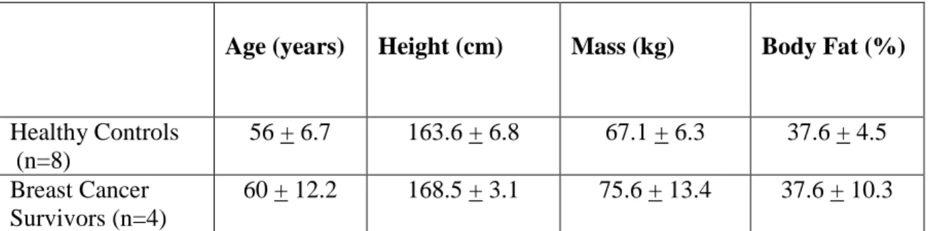
Participants of this study consisted of 4 female breast cancer survivors and 8 healthy age matched (+ 5 years) women. The breast cancer survivor group was comprised of post-menopausal females, 40 - 72 years old. Subjects with stage 1, 2 or 3 invasive breast cancers and within 1-year post treatment were eligible to participate. At the time of the experiment, subjects had no presence of metastatic disease and were in post-menopausal status for at least 1 year. Patients receiving adjuvant hormonal or adjuvant therapy were also eligible. Subjects were not taking any anti-inflammatory medications, and had medical clearance from their oncologist to participate in the study.
The control group consisted of healthy females of 40 to 72 years of age. The control group was also post-menopausal, but had no history of cancer diagnosis or treatment and no limitations in range of motion that would impede upon the ability to perform the prescribed resistance training exercises safely.
Exclusion criteria were set forth by the ACSM guidelines to determine
32
All participants filled out a Medical History Questionnaire and an informed consent, both of which were reviewed prior to participation in the study.
Recruitment
Participants for the breast cancer survivor group were recruited from the Get REAL & HEEL Breast Cancer Program at the University of North Carolina, Chapel Hill.
Participants for the control group were recruited by the posting of flyers at various locations throughout the campus of the University of North Carolina, Chapel Hill, as well as via informational emails sent out to the campus community at the University of North Carolina, Chapel Hill.
Instrumentation
Anthropometric Measures: Resting cardiac function was assessed using a GE CASE Cardiosoft V. 6.6 ECG diagnostic system (Palatine, IL). Height was taken using a
Perspective Enterprises Stadiometer (Portage, MI). Weight was attained via a Detecto Scale (Webb City, MO). A Discovery Dual Energy X-Ray Absorption (DEXA) scanner (Bedford, MA) was used to assess body composition.
33
Blood Analyses: Blood was taken at 4 time intervals throughout the study. The first three were done using a 22 gage Protectiv Plus catheter (Ethicon Endo-Surgery Inc., Cincinnati, OH). The fourth was done using standard venipuncture techniques. Blood was collected in 10.8 mg K2EDTA BD Vacutainers ® (Franklin Lakes, NJ). To analyze for IL-6, IL-10 and TNF-α enzyme linked immunosorbent assays (ELISA) were conducted using Biolegend (San Diego, USA) ELISA kits. Plasma volume shift measurements were performed to account in blood volume changes from pre-exercise to post-exercise. The changes were assessed using the Dill and Costill (1974) Method. Blood cortisol values were determined using a cortisol ELISA Kit from Abnova (Tapei City, Taiwan).
General Procedures:
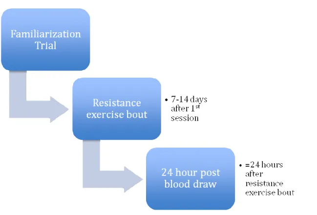
Familiarization Session: Medical history forms, Physical Activity Readiness Questionnaires (PAR-Q’s), and pre-assessment guidelines were administered to subjects prior to their first visit to the Integrative Exercise Oncology Research Laboratory (IEORL) at the University of North Carolina at Chapel Hill (APL). Upon arrival, forms were reviewed for any pre-testing health contraindications. Subjects then signed an informed consent form detailing the experimental protocol and potential risks and benefits associated with
34
consisting of unloaded cycling on a cycle ergometer. Heart rate remained below 40% of heart rate reserve (HRR).
Following the warm-up, subjects were familiarized with the resistance training equipment that was in the experimental protocol by rehearsing each exercise using minimal resistance. Following initial exposure to each of the resistance exercise machines in the same order in which they were used during the experimental protocol, each subject’s one-
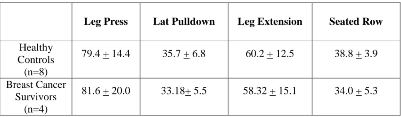
repetition- maximum (1-RM) values was obtained, using the protocol from Levinger et al. (2009). The order of exercises during the experimental protocol was as follows: leg press, latissimus dorsi pull down, leg extension, and seated row. Heart rate was monitored continuously throughout the exercise session.
Upon the completion of the familiarization trial, subjects were asked to rest for 10 minutes, after which heart rate and blood pressure were taken again. If abnormally elevated, subjects were to remain seated until the values decreased. If within normal values, subjects were released from the IEORL.
Experimental Session: Subjects returned to the IEORL between 2 and 14 days following the familiarization session. The extended time window between sessions was to ensure adequate recovery from the previous exercise session. Upon arrival, subjects rested in the seated position for 10 minutes, during which time adherence to the pre-assessment guidelines was reviewed. A Psychological Stress Scale (PSS) evaluation was administered to determine the subjects’ level of psychological stress, which could affect hormone levels during and after the exercise session.
35
the Department of Exercise and Sport Science. Subjects then rested for ten minutes after which blood pressure and heart rate were taken. After the placement of catheter and rest, subjects underwent a warm-up session on an unloaded cycle ergometer for 5 minutes. Heart rate was kept below 40 % of HRR. Following the warm up, the resistance training protocol began. Subjects performed10 repetitions at 70% of 1-RM of each exercise (leg press, lateral pull down, leg extension, and seated row, respectively) with 30-45 seconds of rest in between subsequent exercise and 90-120 seconds of rest between each set. The entire circuit was completed three times for a total of 120 repetitions. Rhythm and timing of each repetition was set at 4 seconds (2 seconds concentric, 2 seconds eccentric) and was kept consistent throughout the exercise protocol and between subjects. Heart rate and RPE was taken following each set to ensure subject safety and monitor their conditions. Subjects were encouraged to consume water at any point during and following the experimental protocol.
Upon completion of the third exercise circuit, subjects were seated and the
36
24-Hour Blood Draw: Subjects returned to the IEORL for the final blood draw 24 hours (+ 30 minutes) following the commencement of the resistance exercise session. Upon arrival to the laboratory, pre-assessment guidelines were reviewed, and the blood sample was taken using a 22 gage needle. After the 24-hour post exercise blood draw subjects had
completed the experimental procedure. The blood-draw timeline is depicted in Figure 2 Statistical analysis-
All data was analyzed using SPSS version 17.0 (Chicago, IL). For comparison of cytokine (IL-6, Il-10 and TNF-α) and cortisol changes between the cancer group and healthy controls, four separate (2X4) mixed-model analysis of variance (ANOVAs) were performed. Statistical significance was set a priori at an α-level of p ≤0.05. The independent variables for this analysis include subject condition (breast cancer survivor and healthy control) and the resistance training intervention. The dependent variables were the baseline values and changes in cytokines: IL-6, IL-10, TNF-α and also cortisol, throughout the time intervals (immediately post-exercise, 2 hours post-exercise, and 24 hours post-exercise). For any significant results, a follow-up Bonferroni post-hoc analysis was used to determine any between or within group differences.
37 Hypotheses:
H1: IL-6 will be significantly upregulated from baseline in the cancer survivors and healthy controls immediately-post exercise and 2 hours post-exercise and return to baseline 24 hours post exercise
H2: IL-10 will be significantly upregulated from baseline at 2 hours post-exercise and 24 hours post-exercise in healthy controls but not in the cancer survivors.
H3: TNF-α will be significantly upregulated 2 hours post exercise and 24 hours post exercise in the cancer survivors but not in the healthy controls.
H4: Peak (%) change (+) in IL-6 will correlate significantly with peak (%) change (+) of cortisol.
39 CHAPTER IV
RESULTS Subject Characteristics:
Four middle-aged female breast cancer survivors and eight, age-matched healthy controls participated in this study (See Table 1 for subject characteristics). Due to recruitment and scheduling difficulties, the number of participants to participate in the study that was proposed at the beginning of the study was not obtainable, leaving the sample size relatively small. Therefore, the results should be interpreted with such limitations in mind. Moreover, difficulties in obtaining blood specimens or adequate amounts of specimen occurred in each study group, resulting in a limited ability to evaluate all biomarkers. Due to this limitation in blood sample amount, only six out of the eight subjects’ blood samples were used for the analysis of cytokines.
Table 1: Subject Characteristics (Mean + SD)
Age (years) Height (cm) Mass (kg) Body Fat (%)
Healthy Controls (n=8)
56 + 6.7 163.6 + 6.8 67.1 + 6.3 37.6 + 4.5 Breast Cancer
Survivors (n=4)
60 + 12.2 168.5 + 3.1 75.6 + 13.4 37.6 + 10.3
Strength Assessments
40
Table 2: Strength Assessment Measures: One-Repetition Maximum (kg) (Mean + SD)
Leg Press Lat Pulldown Leg Extension Seated Row
Healthy Controls
(n=8)
79.4 + 14.4 35.7 + 6.8 60.2 + 12.5 38.8 + 3.9 Breast Cancer
Survivors (n=4)
81.6 + 20.0 33.18+ 5.5 58.32 + 15.1 34.0 + 5.3
Plasma Volume Shifts
Plasma volume (PV) shifts as a result of the experimental trials were calculated from mean Hemoglobin (Hb) and Hematocrit (Hct) values for each time point. The greatest mean PV shifts occurred from pre-trial to post-trial. No differences in PV shift were observed between groups. See Table 3 for PV shifts.
Table 3: Plasma volume changes (∆ % changes) from pre-exercise to immediate
post-exercise; pre-exercise to 2-hour post-exercise and pre exercise to 24 hour post. All values are expressed as mean ± standard deviation (SD).
Plasma Cytokine Concentrations
Table 4 displays the plasma cytokine concentrations for both groups (healthy
controls, cancer survivors) and each time interval after PV shift was accounted for. For the analysis, four separate 2X4 mixed-model analysis of variance (ANOVAs) were performed. A
Trial ∆ Pre- to Post ∆ Pre- to 2 hours Post ∆ Pre- to 24 hours Post
Healthy Controls
(n=8)
-10.53 + 5.26 1.59 + 6.49 -2.95 + 7.46
Breast Cancer Survivors
(n=4)
41
Bonferroni post-hoc was administered to assess any within or between group differences. Significance was set a priori at P<0.05.
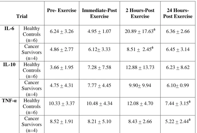
Table 4: Plasma cytokine concentrations (pg/ml) (Mean + SD).
Trial
Pre- Exercise Immediate-Post Exercise
2 Hours-Post Exercise
24 Hours-Post Exercise
IL-6 Healthy Controls
(n=6)
6.24 + 3.26 4.95 + 1.07 20.89 + 17.63δ 6.36 + 2.66 Cancer
Survivors (n=4)
4.86 + 2.77 6.12+ 3.33 8.51 + 2.45δ 6.45 + 3.14 IL-10 Healthy
Controls (n=6)
3.66 + 1.95 7.28 + 7.58 12.88 + 13.73 6.23 + 8.62 Cancer
Survivors (n=4)
4.75 + 4.31 7.77 + 4.45 9.90+ 9.94 6.10+ 0.99 TNF-α Healthy
Controls (n=6)
10.33 + 3.37 10.48 + 4.34 12.08 + 4.70 7.44 + 3.15δ Cancer
Survivors (n=4)
8.52 + 1.91 8.21 + 5.10 8.43 + 2.66 5.22 + 2.44δ δ P<0.05 vs. Pre-exercise (main-effect of time)
IL-6: The analysis revealed a significant main effect of time (p=0.015) when examining the
changes in IL-6 over the time periods. A Bonferoni post-hoc revealed that there was a significant increase of IL-6 in both groups from pre-exercise to 2 hours-post exercise (p=0.027). No differences were observed between groups (p=0.341).
IL-10: The analysis revealed no differences main effect of time (p=0.285) nor differences
between groups (p=0.397).
TNF-α: The analysis revealed a significant main effect of time (p=0.023) when examining
42
significant downregulation of TNF-α from pre-exercise to 24 hours-post exercise (p=0.011). No differences existed between groups (p=0.213)
Cortisol
Table 5. Plasma Cortisol Concentrations (ng/mL). All values are expressed as mean + standard deviation (SD).
Trial
Pre- Exercise Immediate Post Exercise
2 Hours Post Exercise
24 Hours Post Exercise
Cortisol
Healthy Controls
(n=6)
101.8 + 26.0 87.9+ 42.8 126.5+ 128.5 127.2 + 97.1
Cancer Survivors
(n=4)
91.7 + 8.8 61.2 + 8.5 47.6 + 11.3 55.3 + 14.6
*The cortisol responses were not statistically analyzed for significant changes between groups or over time, since this measure was not a primary outcome variable in this study.
Cortisol vs. IL-6
43
Table 6. Average peak percent (%) change for cortisol and Interleukin 6 (Pre-exercise to 2 hours post-exercise). All values expressed as mean + standard deviation (SD).
Cortisol vs. IL-6: There was no relationship (Spearman rho) between peak percent change of
cortisol and IL-6 (p=0.751).
Trial Cortisol IL-6
Healthy Controls (n=6)
-53.7 + 207.1 364.9 + 6.4
Breast Cancer Survivors (n=4)
44 CHAPTER V
DISCUSSION Introduction
The primary purpose of this study was to explore the effects of an acute bout of moderate intensity resistance exercise on inflammatory markers in breast cancer survivors. Contrary to the proposed hypothesis, breast cancer survivors responded similarly to the exercise session compared to the healthy controls. The theorized inflammatory response in cancer survivors, which would have been represented by greater increases in
pro-inflammatory cytokines and a reduced upregulation of anti-pro-inflammatory cytokines relative to the healthy controls, was not evident. To the contrary, the resistance exercise session was shown to reduce the inflammation in both groups, evident by a reduction in TNF-α 24 hours post exercise. Furthermore, and irrespective to any differences between the groups, the exercise session elicited an inflammatory response that was moderately consistent with what has been reported in previous literature (Galvao et al., 2008; Phillips et al., 2010; Toft et al., 2002).
45
each time point, and to present more clearly what the inflammatory response may represent in terms of recovery and adaptation from exercise. Lastly, this discussion will explore the questions that arise from this study, and how those inquiries may be applied to cancer exercise therapy and future research.
Interleukin-6
The response of Interleukin-6 (IL-6) to the resistance exercise session did not differ between groups (p=0.341) and the temporal response was similar to patterns that were
displayed in previous literature (Peake et al., 2006; Phillips et al., 2010; Toft et al., 2002). Of particular note, is that IL-6 was shown to be upregulated at 2 hours post-exercise (p=0.027). This is in agreement with other studies that also displayed the peak upregualtion of IL-6 between two and six hours into recovery (MacIntyre et al., 2000; Peake et al., 2006; Toft et al., 2002).
This pattern of IL-6 dynamics is consistent with the idea that IL-6 is a cytokine that is released directly from muscle, and that complete diffusion out of the muscle will likely occur sometime after the end of the exercise session (Pederson & Febbraio, 2008). Moreover, the degree of IL-6 proliferation is also an important aspect to examine, as variation in IL-6 responses have been reported based on the mode and duration of the exercise task (Nieman et al., 2004; Paulsen & Peake, 2013; Toft et al., 2002). In this present study IL-6 was
46
More specifically, the exercise was administered in a circuit fashion and in a short time window (less than 30 minutes), thus it is possible that the IL-6 produced by the contracting muscle failed to enter into the blood before the immediately post-exercise blood draw.
47
The present study displayed significant IL-6 changes after relatively short exercise session (<30 minutes), therefore exercise duration is unlikely to be a major factor in the IL-6 production. In regards to this concept, other factors of the exercise protocol may have aided in the release of IL-6 from the muscle. As noted by Nieman et al. (2004) IL-6 upregulation during and after exercise is heavily dependent on fuel utilization, particularly on
carbohydrate intake and glycogen storage. Since the present study was administered in a circuit fashion rest intervals were relatively short (30-45 seconds), it is likely that
carbohydrate utilization was much higher relative to a protocol that allows for longer rest intervals. As described by Paulsen and Peake (2013), applying heavy loads with few
repetitions and long rest intervals are like to produce small to moderate changes in hormonal and inflammatory response. However, circuit training, with higher repetitions and short rest intervals are likely to induce changes more similar to aerobic exercise (Paulsen & Peake, 2013). Therefore, the present study displays changes in IL-6 that are most likely indicative of continuous muscle contraction and resultant glycogen depletion.
48
session did not induce a prolonged inflammatory effect or severe muscle damage. This is not surprising, as eccentric action was not a main component of the exercise session; thus it is unlikely that muscle damage was too severe. Moreover, the IL-6 response into recovery is consistent with other studies using similar protocols (Ferreira et al., 2010; Phillips et al., 2010; Prestes et al., 2009).
Interleukin-10
The resistance exercise bout did not induce any significant changes in the major anti-inflammatory cytokine, Interleukin-10 (IL-10), in either group at each time interval
(p=0.226), nor were there any differences between groups (p=0.810). IL-10 did, however, show a trend increasing two hours into recovery with respect to all subjects (main effect). This is revealed when examining individual responses through a Wilcoxon non-parametric statistical test, as eight out of the ten subjects displayed an upregulation of IL-10 in the plasma two hours into recovery compared to baseline (p=0.037). Though only exploratory in nature, the trend found in this analysis is consistent with other literature examining IL-10 responses, in which this major anti-inflammatory cytokine is upregulated 45 minutes to 6 hours after resistance exercise (Hirose et al., 2004; Izquierdo et al., 2010).
49
IL-10 response (see Results Table 4) may be due, in some part, to the varying levels of training status among the participants.
Overall, a major function of IL-10 is to suppress the production of the major pro-inflammatory cytokines (TNF-α, IL-6 and IL-1). Furthermore, others propose that IL-10 is a key component in the repair of muscle damage, and subsequent post-exercise adaptation (Malm et al. 2000). The current data displays that IL-10 concentration trended upward within a short time-window following the cessation of the exercise bout, suggesting that the
response may reflect a role for IL-10 in suppression of inflammation and muscle recovery. However, this last point is speculation since statistical significance in the primary analysis was not found.
Tumor Necrosis Factor-α
Tumor Necrosis Factor-α (TNF-α), the major pro-inflammatory cytokine examined in this study, remained unchanged immediately after exercise and two-hours into recovery, but was significantly downregulated twenty-four hours into recovery in both groups (p=0.011; main effect of time). No differences were observed between groups (p=0.213).
50
Differences, however, do exist between the Hirose et al. (2004) and the current study. Most significantly, the protocol from the current study consisted of exercises that focused on multiple lower and upper body muscle groups with dynamic movement patterns, while the protocol from Hirose et al. (2004) consisted of eccentric action of the elbow flexors with isolated movement patterns. As explained by Paulsen and Peake (2013) the hormonal and inflammatory response is dependent on the type and the size of the exercised muscle or muscle groups. For example, exercise that emphasizes large muscle groups and ‘multi-jointed, dynamic movement patterns’ are likely to induce a greater systemic inflammatory response than ‘isolated muscle contractions’ (Paulsen & Peake, 2013). Moreover, the
reduction in TNF-α in the study by Hirose et al. (2004) was independent of an increase in IL-6, while in the current study it is likely that the upregulation of IL-6 had some effects on the systemic reduction in TNF-α (see Cytokine Interactions and Inflammatory effects of
Resistance Exercise section). Regardless of the differences in these two studies, it remains intriguing that some degree of an anti-inflammatory effect persisted after the bouts of exercise.
Exploring a broader concept, the acute down regulation of TNF-α in this study and