OXIDATIVE STRESS MODULATES RESPONSE TO NON-SURGICAL PERIODONTAL THERAPY
Abdel-ghany Alsaidi
A Master’s thesis submitted to the faculty of the University of North Carolina at
Chapel Hill in partial fulfillment of the requirements for the degree of Masters of Science in the Department of Periodontology
Chapel Hill 2013
Approved by:
iii ABSTRACT
ABDEL-GHANY ALSAIDI: Oxidative Stress Modulates Response To Non-Surgical Periodontal Therapy
(Under the direction of Dr. Steven Offenbacher)
Oxidative stress is an important physiologic modifier of immune and inflammatory
mechanisms. We hypothesized that the elevated total-body oxidative stress, measured
by serum levels of 8-isoprostane, impairs: 1- biofilm IgG response to periodontal therapy
2- clinical parameter responses to periodontal therapy.
108 subjects diagnosed with moderate to severe chronic periodontitis were recruited in
this single-blinded, delayed treatment, randomized, controlled clinical trial. Subjects
were randomly assigned to 2 groups (delayed treatment and combination treatment
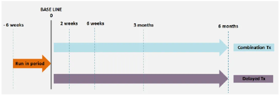
groups). Both groups were clinically evaluated and biological samples collected at minus
6 weeks (run-in period), 0 weeks (baseline- treatment provided to one group only), 2
weeks, 6 weeks, 3 months and 6 months. In addition to probing depth (PD) and bleeding
on probing (BOP) measured at 6 sites per tooth, serum was used for analysis of total
8-isoprostane and IgG antibodies against a panel of 17 common periodontal bacteria.
At the completion of the study, 87 patients had a complete data set at the 6 month
follow –up. Treatment resulted in a transient increase in total IgG levels at 2-weeks
iv
treatment was seen predominantly among subjects with low oxidative stress at baseline
(2.2 fold greater than high oxidative stress group). Furthermore, low oxidative stress
was associated with much better clinical response (PD and BOP) as compared to
subjects with high oxidative stress. These findings suggest that systemic oxidative stress
impairs IgG response and the clinical response to non-surgical periodontal therapy.
v
ACKNOWLEDGMENTS
First of all, praise is due to Allah, The Most Beneficent, The Most Merciful, for all His
guidance and giving while I was preparing this master’s thesis.
I would like to express my deep gratitude to my supervisors Drs. Steven Offenbacher,
Silvana Barros and James Beck for their guidance, suggestions and invaluable
encouragement in my graduate education and throughout the development of this
thesis.
I would like to thank my Mother, Father, Brothers and Sister for their endless love,
encouragement, and all of their spiritual, and financial support throughout my life.
I am also very thankful to Dr. Stephanie Nigro for her friendship, support and love.
Last but not least I would like to thank Mr. Kevin Moss, Drs. Roger Arce, Thiago Morelli,
Julie Marchesan, David Barrow and the members of COSD for all of their indispensable
vi
TABLE OF CONTENTS
LIST OF FIGURES ... viii
LIST OF TABLES ... ix
LIST OF ABBREVIATIONS ... x
Chapters 1. INTRODUCTION AND BACKGROUND ... 1
OXIDATIVE STRESS ... 3
ROS & FREE RADICALS ... 5
ANTIOXIDANTS ... 6
8-ISOPROSTANE ... 9
OXIDATIVE STRESS & PERIODONTAL DISEASE ... 10
2. HYPOTHESES ...0
3. MATERIALS AND METHODS ... 1
Study Population and Power Analyses ... 1
Inclusion and Exclusion Criteria ... 1
Study Design ... 2
Run-in Period & Randomization ... 4
vii
Serum IgG and 8-isoprostane Analyses ... 5
Total 8-isoprostane ... 5
Statistical Analysis ... 18
4. RESULTS ... 8
Study Population Demographics ... 8
Total Serum Biofilm IgG Antibody Response ... 10
IgG Response Stratified on levels of Oxidative Stress ... 12
Changes in Clinical Signs ... 15
Bleeding on probing Stratified on Levels of Oxidative Stress and treatment ... 16
Probing depth Stratified on Levels of Oxidative Stress and treatment ... 17
5. DISCUSSION ... 0
viii
LIST OF FIGURES
Figure 1: The biological effects of small and large shifts in the
balance of activity between reactive oxygen species (ROS) and antioxidants (AO) ... 3
Figure 2: Major cellular sources of ROS in living non-photosynthetic cells ... 9
Figure 3: Study timeline ... 3
Figure 4: Total IgG for Treatment and control groups... 11
Figure 5: Total bacterial IgG in means, divided to 4 groups ... 14
Figure 6: Total IgG change between 2 and 0 week for the Tx and Control groups ... 15
Figure 7: Mean bleeding on probing for all 4 groups in percentages. ... 16
Figure 8: Mean probing depth in millimeters for all 4 groups at 0, 6, 12 and 24-week time points ... 17
Figure 9. Improvement in Extent Bleeding On Probing in Response to Treatment ... 18
ix
LIST OF TABLES
Table 1. Study timeline legend ... 3
Table 2. Subjects’ Demographic ... 9
Table 3. Summary of evaluable subject population ... 10
x
LIST OF ABBREVIATIONS
IRB: Institutional Review Board GCF: Gingival Crevicular Fluid ROS: Reactive Oxygen Species IgG: Immunoglobulin G BOP: Bleeding on Probing
CAL Clinical attachment level measured in millimeters PD: Probing Depth measured in millimeters
PMNs: Polymorphonuclear Leukocytes SD: Standard Deviation
8-Iso: 8-Isoprostane Tx: Treatment
ANOVA:ANalysis Of VAriance between groups APC: Antigen-presenting cell
BMI: Body mass index LPS: Lipopolysaccharide
INTRODUCTION AND BACKGROUND
Periodontitis is a term used to describe an inflammatory process, initiated by the plaque
biofilm, that leads to loss of periodontal attachment to the root surface and adjacent
alveolar bone and which ultimately results in tooth loss unless treated. The
inflammatory and immune responses to the bacteria and also viruses1
that colonize the periodontal and associated tissues involve the systemic circulation
and ultimately the peripheral systems of the body. This creates a complex bi-directional
series of host– microbial interactions involving cellular and humoral factors and
networks of cytokines, chemokines, and growth factors. It is believed that the primary
etiological agent is specific, predominantly gram-negative anaerobic or facultative
bacteria within the sub-gingival biofilm.2, 3
The majority of periodontal tissue destruction is caused by an inappropriate host
response to those microorganisms and their products.3 More specifically, a loss of
homeostatic balance between proteolytic enzymes (e.g. neutrophil elastase) and their
inhibitors (e.g. a1-antitrypsin) and reactive oxygen species (ROS) and the antioxidant
defense systems that protect and repair vital tissue, cell, and molecular components is
believed to be responsible.
The basis for such dysregulation is in part genetic (38–82%), 4 and in part the result of
2
In humans, oxidative stress is thought to be involved in the development of many
diseases or may exacerbate their symptoms. These include cancer, Parkinson's
disease, Alzheimer's disease, atherosclerosis, heart failure, myocardial
infarction, Schizophrenia, Bipolar disorder, fragile X syndrome, Sickle Cell Disease, lichen
planus, vitiligo, autism, and chronic fatigue syndrome.5
A paradigm shift in our understanding of the importance of reactive oxygen and
antioxidant species to human biology over the last decade came from the realization
that vital and ubiquitous transcription factors, such as nuclear factor-kB and activating
protein-1 were redox-sensitive. Large, upward shifts in the pro-oxidant/ antioxidant
ratio intracellularly bring about direct damage to vital biomolecules and structures, cell
membrane damage and dysfunction, and cell death (by necrosis or accelerated
apoptosis), and extracellularly cause direct connective tissue damage (both mineralized
3
Figure 1. The biological effects of small and large shifts in the balance of activity between reactive oxygen species
(ROS) and antioxidants (AO)– adapted from Chapple et al.6 -Reproduced with permission
OXIDATIVE STRESS
Oxidative stress was defined by Sies in 1991 as ‘a disturbance in the pro-oxidant–
antioxidant balance in favor of the former, leading to potential damage’.7
This disturbance in the normal redox state of cells can cause toxic effects through the
production of peroxides and free radicals that damage all components of the cell,
including proteins, lipids, and DNA. Further, some reactive oxidative species act as
cellular messengers in redox signaling. Thus, oxidative stress can cause disruptions in
normal mechanisms of cellular signaling.8
In normal physiology there is a dynamic equilibrium between ROS activity and
antioxidant defense capacity, and when that equilibrium shifts in favor of ROS, either by
a reduction in antioxidant defenses or an increase in ROS production or activty,
4
Chemically, oxidative stress is associated with increased production of oxidizing species
or a significant decrease in the effectiveness of antioxidant defenses, such
as glutathione.9
The effects of oxidative stress depend upon the size of these changes, with a cell being
able to overcome small perturbations and regain its original state. However, more
severe oxidative stress can cause cell death and even moderate oxidation can
trigger apoptosis, while more intense stresses may cause necrosis.10
Production of reactive oxygen species is a particularly destructive aspect of oxidative
stress. Such species include free radicals and peroxides. Some of the less reactive of
these species (such as superoxide) can be converted by oxidoreduction
reactions (Redox) with transition metals or other redox cycling compounds
(including quinones) into more aggressive radical species that can cause extensive
cellular damage.11
The major portion of long-term effects is inflicted by damage to DNA.12
The DNA damage that can be induced by ionizing radiation is similar to oxidative stress,
and these lesions have been implicated in aging and cancer. Most of these
oxygen-derived species are produced at a low level by normal aerobic metabolism. Normal
cellular defense mechanisms destroy most of these ROS and free radicals. Likewise, any
damage to cells is constantly repaired. However, under the severe levels of oxidative
stress that cause necrosis, the damage causes ATP depletion, preventing controlled
5
Thus, Oxidative stress is an important physiologic modifier of immune15 and
inflammatory mechanisms.16
Mediators of oxidative stress or ‘‘redox signaling’’, or both, have been shown to regulate
receptor17 and transcription factor signaling and kinase dependent signaling pathways,18
thereby inducing the expression of key cytokines and inflammatory mediators.19
Oxidative stress can shift the balance of Th1 and Th2 cytokine profiles in in vitro and in
vivo model systems.20 This shift is usually toward increased levels of Th2 cytokines,
suggesting that oxidative stress might modify humoral immune responses to commensal
and pathogenic microorganisms. Clinical conditions associated with increased oxidative
stress in humans (e.g., diabetes, periodontitis, and aging) 21 have been associated with
impaired immune function. Smoking, a recognized risk factor for periodontitis22 has
been associated with depressed levels of total serum IgG2,23 which is a Th1-dependent
IgG antibody subtype, and with decreased serum levels of antibody for selected oral
bacteria.24
ROS & FREE RADICALS
Free radicals are defined as ‘any species capable of independent existence that contain
6
They are, by nature, highly reactive and diverse species, capable of extracting electrons
and thereby oxidizing a variety of biomolecules vital to cell and tissue function, which
not only include oxygen free radicals, but also nitrogen and chlorine species. ROS is a
term that has become more popular because it encompasses other reactive species
which are not true radicals but are nevertheless capable of radical formation in the
intra- and extracellular environments.6
ROS are chemically reactive molecules containing oxygen. Examples
include oxygen ions and peroxides. ROS form as a natural byproduct of the normal
metabolism of oxygen and have important roles in cell signaling and homeostasis.
However, during times of environmental stress (e.g., UV or heat exposure), ROS levels
can increase dramatically.26
This may result in significant damage to cell structures. Cumulatively, this is known
as oxidative stress. ROS are also generated by exogenous sources such as ionizing
radiation, trauma, and smoking.26
ANTIOXIDANTS
They are defined as “those substances which when present at low concentrations,
compared to those of an oxidizable substrate, will significantly delay or inhibit oxidation
7
Given that it is estimated that between 1 billion and 3 billion reactive species are
generated per cell per day, the importance of antioxidant defense systems to the
maintenance of health becomes clear.28
The preventative antioxidants function by enzymatic removal of superoxide and
hydrogen peroxide or by sequestration of divalent metal ions, preventing Fenton
reactions and subsequent hydroxyl radical formation.29
The etiology of Alzheimer's disease progression is still debated; however, increased
oxidative stress is an early and sustained event that underlies much of the neurotoxicity
and consequent neuronal loss. Amyloid beta is a metal binding protein and copper, zinc
and iron promote amyloid beta oligomer formation. Additionally, copper and iron are
redox active and can generate reactive oxygen species via Fenton (and Fenton-like
chemistry) and the Haber-Weiss reaction. Copper, zinc and iron are naturally abundant
in the brain but Alzheimer's disease brain contains elevated concentrations of these
metals in areas of amyloid plaque pathology. Amyloid beta can become pro-oxidant and
when complexed to copper or iron it can generate hydrogen peroxide.
Accumulating evidence suggests that copper, zinc, and iron homeostasis may become
perturbed in Alzheimer's disease and could underlie an increased oxidative stress
8
Lactoferrin is probably more important than transferrin within the periodontal tissues,
given the dominance of the neutrophil infiltrate29 and the recognition of high levels of
lactoferrin within gingival crevicular fluid.30
Antioxidants neutralize free radicals by donating one of their own electrons, ending the
electron-"stealing" reaction. The antioxidant nutrients themselves don't become free
radicals by donating an electron because they are stable in either form. They act as
scavengers, helping to prevent cell and tissue damage that could lead to cellular damage
and disease.
Vitamin E (α-tocopherol) the most abundant fat-soluble antioxidant in the body. One of
the most efficient chain-breaking antioxidants available. Primary defender against
oxidation. Primary defender against lipid peroxidation (creation of unstable molecules
containing more oxygen than is usual).31
Vitamin C ( L-ascorbic acid, or simply ascorbate ) The most abundant water-soluble
antioxidant in the body. Acts primarily in cellular fluid. Of particular note in combating
free-radical formation caused by pollution and cigarette smoke. Also helps return
9
Figure 2: Major cellular sources of ROS in living non-photosynthetic cells
8-ISOPROSTANE
The Isoprostanes are prostaglandin-like compounds formed in vivo from the free radical
10
direct action of cyclooxygenase (COX) enzyme. The isoprostanes possess potent
biological activity, and their potential role in mediating certain aspects of the
detrimental effects of oxidant stress is then examined. In addition, evidence showed
that these biological effects may be mediated through interaction with a unique
receptor. A considerable portion of this commentary deals with the utility of measuring
isoprostanes as markers of oxidant injury both in vitro and in vivo. A number of studies
have shown these compounds to be extremely accurate markers of lipid peroxidation in
animal models of oxidative stress and have illuminated the role of oxidant injury in
association with several human diseases.32
Lipid peroxidation refers to the oxidative degradation of lipids. It is the process in
which free radicals "steal" electrons from the lipids in cell membranes, resulting in cell
damage. Isoprostanes and their metabolites have also been shown to be elevated in the
urine of cigarette smokers, and have been suggested as biomarkers of oxidative stress in
smokers.33
Direct-8-iso-Prostaglandin F2a (d-8-iso PGF2a) is a stable end product of both specific
inflammatory enzymatic pathways and nonspecific mechanisms and reflects total lipid
peroxidation, representing an excellent in vivo marker for oxidative stress.34
OXIDATIVE STRESS & PERIODONTAL DISEASE
The idea that ROS are associated with the pathogenesis of a variety of inflammatory
diseases and have a role (direct or indirect) in tissue damage has become a major area
11
literature. However, supporting evidence for their role in tissue damage is often indirect
and circumstantial.6
The relationship to periodontal disease could be derived from understanding the
mechanism in which ROS can affect tissue damage6
1- Protein damage
2- Lipid peroxidation
3- DNA damage
Bleeding on probing
BOP is a term used referring to bleeding that is induced by gentle manipulation of the
tissue at the depth of the gingival sulcus or pocket, or interface between the gingiva and
a tooth. BOP is a sign of inflammation and indicates some sort of destruction
and erosion.35
Also, since P. gingivalis, a very important periodontal pathogen, has an absolute
requirement for heme (from blood, and cultivated using blood agar plates in vitro) it is
an intriguing relationship between BOP and periodontal disease
Longitudinal studies on BOP are available. At present, bleeding on probing is widely
used as an indication for needed treatment. However, bleeding on probing alone is not
a predictor of elevated risk for future loss of clinical attachment. On the other hand, a
lack of bleeding on probing, especially on two or more occasions, is an excellent
HYPOTHESES
The general hypothesis of this study is that elevated levels of oxidative stress at baseline
(measured by serum 8-isoprostane) will have a detrimental effect on the antibody and
clinical response to periodontal therapy.
We could divide the hypothesis into
1- Systemic oxidative stress, as measured by increasing serum levels of
8-isoprostane, are a significant negative modifier of the serum-antibody responses
to mucosal biofilm microorganisms. Specifically, we hypothesized that increased
oxidative stress would be associated with suppression of the systemic IgG
antibody responses to the indigenous oral biofilm microorganism
2- Systemic oxidative stress, as measured by increasing serum levels of
8-isoprostane, are a significant negative modifier of the clinical response to
non-surgical periodontal therapy as measured of Bleeding on Probing (BOP) and
MATERIALS AND METHODS
Study Population and Power Analyses
This study was approved by the Institutional Review Board at the University of North
Carolina at Chapel Hill (IRB project #2019). A total of 108 systemically healthy subjects
aged between 23-63 years, diagnosed with moderate to severe chronic Periodontitis
were recruited. The sample size estimate was based upon the anticipated decrease in
markers of serum inflammatory response, using CRP as the primary effect size
estimator. (The original investigators anticipated having 90% power to detect a 95%
decrease in serum markers, using CRP estimates as previously reported by D'Aiuto et
al.).37
This report is a secondary analysis of the investigation to assess the relationship
between oxidative stress, serum IgG and clinical outcomes.
Inclusion and Exclusion Criteria
108 subjects were recruited; all were diagnosed with moderate to severe periodontitis
based on the 1999 International Workshop for a Classification of Periodontal Diseases
2
Subjects were excluded if they were diabetic, had any acute infection, history of
antibiotic use in the month prior to the baseline assessments, needed prophylactic
antibiotic coverage or had a history of active periodontal treatment during the year
prior to the clinical evaluation.
Study Design
Subjects were recruited into this single-blinded, delayed treatment, controlled
randomized clinical trial.
They were randomized to one of two treatment arms
1- Group 1: Combined therapy (scaling and root planing plus Oral Hygiene Instructions)
Subjects (Combination Treatment) received scaling and root planing at baseline
(0 week) along with oral hygiene instructions and supportive therapy continued
through the 6 month interval consisting of 1- a dentifrice with sodium fluoride,
with a choice of Crest Cavity Protection or Crest Whitening 2- Daily flossing using
Glide dental floss, 3- use of Crest SpinBrush® and 4- Scope mouthwash
(non-market FDA Monograph-compliant Scope® cetylpyridinium chloride (CPC)
Mouthwash for twice daily rinsing. 53 subjects were randomly assigned to the
this treatment arm and 42 completed the 24 week study.
3
At baseline group 2 (Delayed treatment) received examinations and biological
sampling only with no oral hygiene instructions or scaling or root planing. Upon
completion at 6 months they received the same full treatments as above with
the exception of the mouthwash. 53 subjects were randomly assigned to this
treatment arm and 45 completed the 24 week study.
Figure 3. Study timeline
4 Run-in Period & Randomization of subjects
The run-in period in this study was the 6 weeks period before the clinical trial
commenced (when no treatment was given). It was employed to ensure that
participants were in a stable condition and compliant. It was also used as a washout
period if treatments that participants were using before entering the clinical trial were
discontinued.
Subjects were randomly assigned to each treatment arm, adjusting for sex, age, smoking
status and BMI.
Calibration & Blinding of examiners
In our study, 3 examiners performed the clinical measurements and harvested the blood
samples at the appropriate time-points. The examiners were calibrated for probing
depth, clinical attachment loss measurements and bleeding on probing detection. 98%,
80% and 92% agreements were noted for PD, CAL and BOP respectively in a pre-study
calibration exercise.
The analysis aimed to determine whether periodontal treatment modified the serum
IgG response to oral organisms and the relationship of that response to levels of serum
8-isoprostane.
We hypothesized that individuals with high oxidative stress would have an impaired IgG
response and would potentially have an impaired wound healing response. The clinical
5
Serum IgG and 8-isoprostane Analyses
Following a 6 week run-in period during which no treatment was provided, blood
samples and periodontal parameters were collected from both study groups at 0, 2, 6,
12 and 24 weeks.
Blood samples were collected and processed for measurement of serum IgG levels for
specific oral organisms as described by Singer et al.39
Total IgG directed against oral organisms was computed by summing across all IgG
specific organism which included Porphyromonas gingivalis; Prevotella intermedia;
Prevotella nigrescens; Tannerella forsythensis; Treponema denticola; Fusobacterium
nucleatum; Aggregatibacter actinomycetemcomitans; Campylobacter rectus; Eikenella
corrodens; Parvimonas micra; Veillonella parvula; Capnocytophaga ochracea;
Seleomonas noxia; Actinomyces viscosus; Streptococcus intermedius; Streptococcus
sanguis; and Streptococcus oralis.
Total 8-isoprostane
8-isoprostane is a stable end product of both specific inflammatory enzymatic pathways
and nonspecific mechanisms and reflects total lipid peroxidation, representing an
excellent in vivo marker for total body oxidative stress.39
The d-8-iso PGF2a assay is based on the competition between sample 8-iso-PGF2a and a
fixed amount of alkaline phosphatase (AP)-labeled 8-iso-PGF2a for sites on a rabbit
6
polyclonal antibody becomes bound to the goat anti-rabbit antibody coated onto the
microplate. After a wash to remove excess conjugate and unbound sample, a substrate
solution is added to the wells to determine the bound enzyme activity. Color
development is stopped, and absorbance read at 405 nm. The intensity of the color is
proportional to the amount of AP-PGF2a bound to the well, which is inversely
proportional to the concentration of total 8-iso-PGF2a originally present in the sample.
Standard curves ranged from 100,000 down to 32 pg/ml, and serum samples were
diluted 1:5 to yield values within the working range. Deviations of standard duplicates
ranged from 1.0 to 18.8%, with a mean deviation of 5.9%. Sensitivity limit of detection
was estimated by the manufacturer as 103.2 pg/ml for the 2-h incubation format.
Participants were ranked on the serum concentration of d-8-iso PGF2a. With this
ranking, participants in the top half (above the median) of concentration of d-8-iso were
considered ‘‘High oxidative stress’’ and those in the remaining lower half of
concentration of d-8-iso were considered ‘‘Low oxidative stress”.
Statistical Analysis
Statistical analyses and data management were performed using SAS (SAS Institute,
Cary, NC); statistical significance was set at p≤0.05. Frequency distributions, means,
7
were investigated by using t tests for continuous variables. Multivariable modeling was
performed by using SAS Procedure GLM to calculate least-squared means. 2-way
ANOVA and Graphs were performed and generated using GraphPad Prism™ (2013
RESULTS
Study Population Demographics
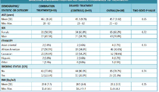
We enrolled and randomized 108 participants which included 55 delayed treatment
control subjects and 53 treatment subjects. The characteristics of this population appear
in Table 2. At enrollment there were no significant differences in the treatment or
delayed treatment groups with regards to age, sex, ethnicity, smoking status and BMI
among the two study groups (table 2), suggesting that randomization was effective in
matching the two groups at baseline. The mean participant age was 46.1 (for the control
group) and 45.3 (for the treatment group) years old with a standard deviation ranged
from 4.4 to 10.3 (Table 3). The majority of the participants were Caucasians (48.6%), and
more female participants were recruited (60.2%). BMI was recorded for all participants
and ranged from 15.8 to 58.3 kg/m2. Statistically significant differences were not found
between the treatment and control arms for the 87 subjects who successfully
9
10
Table 3. Summary of evaluable subject population
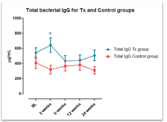
Total Serum Biofilm IgG Antibody Response
Figure 4 demonstrates the levels of total IgG specific for the oral biofilm at baseline, 2, 6,
12 and 24 weeks for both the treated and untreated groups. In the delayed treatment
group there were no changes in total IgG levels over time. However, in the group that
received scaling and root planing plus OHI there was a significant increase in IgG titer at
2 weeks, (p<0.01) as compared to the delayed treatment group, but the levels returned
11
Thus, periodontal therapy was associated with a temporal increase in serum IgG
probably directed against the oral biofilm development.
Thus, the combined therapy was a potent inducer of the acquired immune response
leading to increased titers of immunoglobulins, which is consistent with a protective
role of IgG serum titer.
Figure 4. Total IgG for Treatment and control groups indicating a statistically significant increase in total IgG noted
at 2 weeks for the Tx group (*P<0.01)
There were no significant time or treatment-based changes in levels of 8-isoprostane,
12
IgG Response Stratified on levels of Oxidative Stress
Studies in a large cross-sectional population cohort demonstrated that high oxidative
stress was strongly associated with low total IgG levels.39
We compared antibody changes stratified upon baseline levels of 8-isoprostane
dichotomized at the median (high or low). First, we examined the effect of the baseline
oxidative stress levels on the IgG response to non-surgical periodontal therapy. The IgG
titers for all subjects with 8-iso values above the median (table 2) were included in the
high-oxidative stress sub-group and all the subjects with 8-iso values below the median
(table 2) were included in the low-oxidative stress sub-group, to divide our cohort to 4
groups for statistical analysis
1- High oxidative stress in the combination treatment group
2- Low Oxidative stress in the combination treatment group
3- High Oxidative stress in the delayed treatment group
4- Low Oxidative stress in the delayed treatment group
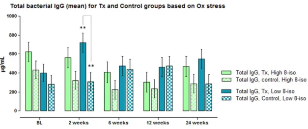
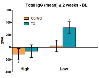
The changes in total oral biofilm IgG levels for the four subgroups are shown in figure 5.
These data demonstrate that the overall increase in total IgG seen in Table 3 above can
be entirely attributable to the increase in IgG titer seen among treated subjects with low
8-isoprostane at baseline. The elevation in IgG titer at 2 weeks within the low oxidative
stress subgroup is approximately 2.2 fold greater, as compared to the high oxidative
13
Figure 6 highlights the two-week change in IgG titer. Since serum IgG is likely to be
protective and a reflection of activation of the acquired protective Th2 response, the
14
Table 4. Summary statistics for the changes in serum 8-isoprostane (in ng/ml) throughout the study for both
groups, means are measured in ng/ml all others pg/ml.
Figure 5. Total bacterial IgG in means, divided into 4 groups (Tx high oxidative stress, Control high oxidative stress,
Tx low oxidative stress and Control low oxidative stress) at all 5 time points.
**shows a statistically significant increase in total IgG count (P<0.01) GROUPS
Visit
Combination Treatment Group Delayed Treatment Group (Control)
N Mean
(SD)
Median Min-Max N Mean (SD) Median Min-Max
Week -6 46 57.5
(99.7)
1061 16-250000 36 49.6 (99.8) 1207 201-250000
Week 0 39 96.7
(12.8)
1415 65-250000 38 72.9 (114.5) 922 101-250000
Week -6 & 0 Mea n
50 78.0
(90.9)
4072 144-250000 49 59.4 (92.0) 1244 104-250000
Week 2 40 88.1
(120.3)
1374 39-250000 39 64 .7 (110.2) 875 66-250000
Week 6 40 75.6
(115.6)
1020 208-250000 39 59.0 (106.0) 1216 228-250000
Week 12 40 57.1
(105.3)
912 9-250000 43 64.6 (110.0) 861 161-250000
Week 24 38 63.2
(195.7)
15
Figure 6. Total IgG change between 2 weeks and 0 time-point for the Tx and Control groups divided based on
oxidative stress levels , a statistically significant increase in total IgG for the low oxidative stress Tx group as
compared to the high oxidative stress. (*P<0.03)
Changes in Clinical Signs
We examined the effects of treatment over time ( in PD, BOP and AL ). There were no
significant changes in CAL, but there were improvements in the Bleeding on Probing
(BOP) and Probing Depth (PD). Further explanation of the choice of PD over CAL for this
study can be found in the discussion section.
It is important to note that no adverse events were reported during the 6 weeks run-in
16
Bleeding on probing Stratified on Levels of Oxidative Stress and treatment
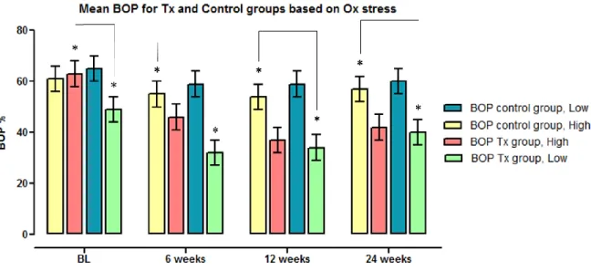
Baseline values and changes in response to therapy in BOP scores are shown in figure 7.
Subjects with low oxidative stress exhibit less BOP at baseline as compared to the
controls with high oxidative stress; this finding was statistically significant (P<0.04). Also,
a significant decrease in mean percentage of BOP was noted in the low oxidative stress
Tx group at 6 weeks as compared to the high oxidative stress Tx group. This difference
persists at 12 and 24 weeks.
Figure 7. Mean extent bleeding on probing for all 4 groups in percentages. Shows a statistically significant decrease
in the percentage of BOP between Tx groups with high ox stress and Tx group with low at baseline (P< 0.04). Similar
17
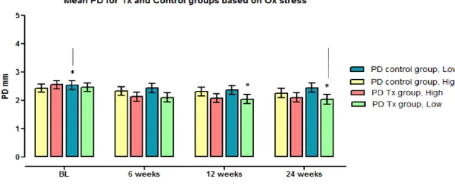
Probing depth Stratified on Levels of Oxidative Stress and treatment
The mean probing depth was analyzed using 2-way ANOVA was performed to determine
the statistical significance. A significant decrease in mean PD was noted for the low
oxidative stress group at 12 and 24 weeks (but not at 6 weeks) as compared to low
oxidative stress controls at base line (Figure 8)
Figure 8. Mean probing depth in millimeters for all 4 groups at 0, 6, 12 and 24-week time points, show a statistically
significant decrease of PD for the low oxidative stress Tx group at 12 and 24 weeks when compared to the low
oxidative stress controls at baseline (P<0.05)
The systemic oxidative stress level is dynamic6 and likely changes for individuals during
the 24 week monitoring period. Although we demonstrated that low levels of oxidative
18
examined whether those individuals with persistently high 8-isoprostane (above the
median) as evaluated at each timepoint differed significantly in the healing response as
compared to those that were consistently low in 8-isoprostane levels, below the
median, as assessed at baseline, 2, 6, 12 and 24 weeks.
Figure 9 shows the clinical changes in BOP for subjects that have consistently low,
consistently high and mixed levels of 8-isoprostane. It can be observed that all
subgroups responded with no statistically significant difference observed.
Figure 9. Mean Bleeding on Probing extent (percentage) in Response to Treatment throughout the study
6 Weeks: L=Low OS at Baseline, H=High OS at Baseline; p-value 0.95, 12 weeks: LL=Low OS at Baseline & at 6
19
p-values are 0.78 and 0.95 for Mix and HH respectively. 24 weeks: LLL=Low OS at Baseline, 6 weeks and 3 months,
HHH=High OS all previous time points. MIX=combination of low or high OS at baseline, 6 weeks and 12 weeks.
p-values are 0.76 and 0.56 for Mix and HH respectively
Figure 10 shows the clinical changes in PD for subjects that have consistently low,
consistently high and mixed levels of 8-isoprostane at 6, 12 and 24 weeks, It can be
observed that LLL group had a better reduction in probing depth mean at 6 months as
compared to the MIX and HHH matched groups P< 0.03 and 0.002 for Mix and HHH
20
Figure 10. Mean Pocket Depth (in millimeters) in Response to Treatment throughout the study
6 Weeks: L=Low OS at Baseline, H=High OS at Baseline; p-value 0.3.
12 weeks: LL=Low OS at Baseline & at 6 weeks, HH=High OS at Baseline & at 6 weeks, MIX=combination of low or
high at baseline or 6 weeks. p-values are 0.41 and 0.19 for Mix and HH respectively
24 weeks: LLL=Low OS at Baseline, 6 weeks and 12 weeks, HHH=High OS all previous time points.
MIX=combination of low or high OS at baseline, 6 weeks and 12 weeks. (*= P=0.02)
DISCUSSION
Several studies have shown that periodontal treatment improves the systemic levels of
antibodies specific to periopathogens.40-42 Also improves the periodontal clinical
parameters used in the diagnosis of periodontal disease.
It was demonstrated throughout multiple reports in the medical literature that
therapies designed to reduce oxidative stress have been related to improved outcomes
in viral infections and improved cell-mediated immune function in animal models and
human clinical trials.43, 44
Thus our goal is to determine the association of systemic levels of oxidative stress with
serum levels of IgG antibody to commensal and pathogenic microorganisms’
response/alteration to periodontal therapy within the periodontal patient population.
The loss of homeostatic balance between proteolytic enzymes (e.g. neutrophil elastase)
and their inhibitors (e.g. a1-antitrypsin) and reactive oxygen species (ROS) and the
antioxidant defense systems to protect and repair vital tissue, cell, and molecular
components is believed to be responsible for periodontal tissue destruction. The basis
for such dysregulation is in part genetic and in part the result of environmental factors
1
Our findings demonstrate that serum concentrations of 8-isoprostane above the median
are related to decreased response of the serum IgG antibodies to oral bacteria. These
findings were controlled for smoking status and other known contributors to oxidative
stress and periodontal severity (e.g., , age and BMI) (Table 1). These findings are in exact
accordance with the Singer, Beck and Offenbacher39 report which was the first to
demonstrate an association between serum 8-isoprostane concentrations and serum
IgG antibody levels to mucosal microbes. Thus, this investigation demonstrates that
systemic oxidative stress consistently suppresses serum IgG antibody responses to total
oral biofilm microbes.
In our study population which could be considered a large representative population
(108 subjects), extremes of oxidative stress have been related to changes in a variety of
immune-response mechanisms.45
The bacteria in our panel of antibodies included gram-negative and gram-positive
bacteria, strict anaerobes, aerobes, and facultative anaerobes, as well as species
associated with periodontal disease and oral health.46
Singer et al.39 showed that serum IgG antibody responses to the oral biofilm microflora
are suppressed in the presence of systemic oxidative stress. Additional studies are
needed to clarify the role of IgG subtype and avidity to these findings.
Singer et al.39 suggested possible studies to explore the clinical impact of oxidative
stress in mucosal disease progression, in the response to therapy, and in the potential
2
Our study investigated the response to non-surgical periodontal therapy in subjects with
high oxidative stress as compared to low oxidative stress subjects; the findings
supported our initial hypothesis that periodontal subjects with high oxidative stress at
baseline will respond less favorably to treatment, as shown on the immunological level
by investigating the total oral biofilm serum IgG titers response.
Although these titers reflect whole-bacterial titers, a high degree of specificity occurs in
organism-specific IgG without significant cross-reactivity across various microbes under
the stringency conditions of the immunobinding.47
Thus, the effect of oxidative stress on IgG responses appears to reflect a suppression of
IgG across a wide range of potential antigenic stimuli.
8-Isoprostane is a stable product of the oxidative metabolism of arachidonic acid,47
and 8-isoprostane concentrations in serum and urine have been used as markers of
systemic oxidative stress,48 and are related to the accumulation of oxidized-LDL.49
The serum concentration of 8-isoprostane has been reported to be related to body
mass index and race.50
To understand the mechanism for the association of 8-isoprostane concentrations with
decreased IgG antibody levels, it will be necessary to define better the specific
mediators of lipid-based redox signaling, specific cellular and molecular targets, and
correlations of IgG subclass antibody levels (and antibody isotypes) to serum
3
It is expected that clinical and mechanistic insights from these future investigations will
enable interventions that better target populations and mechanisms likely to have
impact on the systemic diseases that have been associated with systemic exposure to
the mucosal biofilm microflora.
8-isoprostane concentrations reflect whole-body lipid oxidation, including enzymatic
and nonspecific free-radical lipid oxidative pathways; it is likely that these variables and
physiologic or pathologic factors (or both) that influence oxidative lipid metabolism
contributed to the serum 8-isoprostane concentrations. Singer et al.39 further indicated
that an increased prevalence of deep probing depths (periodontal disease) is associated
with increased (1.6-fold) serum concentrations of 8-isoprostane. The theory that was
proposed to explain this elevation was that the epithelial lining of the periodontal
pocket is adjacent to the subgingival biofilm and is structurally unique without tight
junctions or keratin. Even in health, the epithelial attachment to the teeth is a site that
has a natural IL-8 chemotactic gradient, which brings neutrophils into the periodontal
pocket such that they flush around the teeth and appear in the saliva. Activated
neutrophils within the crevice and saliva may serve to increase salivary 8-isoprostane, as
recently reported by Wolfram et al.51
Thus, the periodontal tissues may represent a source of systemic oxidative stress
4
It is known that the presence of periodontitis is associated with lower serum
concentrations of vitamin C, 52 even after adjusting for smoking, suggesting that
periodontal disease may pose an oxidative stress that consumes ascorbate.
Also, the mucosal tissue wounding associated with deep periodontal pockets has been
associated with an increased occurrence of bacteremia.53
Herzenberg et al.45 found that extreme high levels of oxidative stress have been related
to changes in a variety of immune-response mechanisms.45
It was hypothesized50 that infiltrating neutrophils or macrophages, or both, capable of
mounting an oxidative burst might control the periodontal microflora and thereby limit
consequent serum IgG antibody responses, leading to an inverse relation between
serum 8-isoprostane and IgG antibody levels. An increased presence of periodontal
pockets of PD greater than 5mm would provide a greater reservoir of activated
periodontal PMNs and macrophages and was associated here with increased serum
8-isoprostane levels; however, the greater number of deep pockets also was associated
with significant increases in levels of serum IgG antibodies.
The second part of this study was to look at the clinical response for periodontal
subjects with high oxidative stressto periodontal therapy as evaluated by BOP and PD;
the present study suggests that the outcome in high oxidative stress subjects is less
favorable than in low oxidative stress periodontal subjects.
In this study we mainly looked at the changes in PD even though clinical attachment
5
treatment. However, even if teeth have high CAL, their PD may be less than 3 mm. IgG
titer levels, transient in nature, must be influenced more by the size of the area of
current infection than by the history of tissue destruction. Therefore, we preferred PD
to CAL as a measure of periodontal severity, and particularly as a predictor of systemic
immune response. As expected based on previous reports (Alexander et al., 1996; Behle
et al., 2009), the IgG titers against total oral biofilm bacteria were significantly
decreased by periodontal treatment, corresponding to improvement in periodontal
condition.54
The results suggested that this test is useful for evaluating treatment effects from the
perspective of infection levels, and the test would be useful as a self-evaluation system
for the effects of periodontal treatment.
In our study, both study sub-groups (High and Low oxidative stress with treatment)
showed improvement in PD and BOP after treatment. The pattern, however, was that
subjects with high oxidative stress exhibited less improvement in both parameters than
those with low oxidative stress. The reduction in whole-mouth mean PD was 0.33 mm
vs. 0.50 mm (P<0.01) in high vs. low oxidative stress subjects.
To conclude the major findings in this study are
1- Scaling and root planing results in an increase in 2 week total oral biofilm IgG
6
2- High oxidative stress at baseline was associated with lower IgG antibody at 2
weeks after treatment in subjects with moderate to severe periodontitis.
3- At baseline, subjects assigned to the treatment group who had low oxidative
stress exhibited less BOP as compared to controls with high oxidative stress.
4- Subjects with low oxidative stress responded better to non-surgical periodontal
therapy as measured by mean percentage of BOP reduction at 6, 12 and
24-week time-points and mean PD reduction at 12 and 24 24-weeks as compared to
controls with high oxidative stress.
5- Subjects who presented with consistently low oxidative stress throughout the
study responded better to non-surgical periodontal therapy by measure of PD
reduction observed at 6 months.
This report suggests further studies to investigate the role of oxidative stress as a
modifier of periodontal therapy response in subjects with periodontitis.
These findings suggest possible studies to explore the potential impact of antioxidative
REFERENCES
1. Slots J. Herpes viruses in periodontal diseases. Periodontol 2000 2005;38:33-62.
2. Haffajee AD, Socransky SS. Microbial etiological agents of destructive periodontal diseases. Periodontol 2000 1994;5:78-111.
3. Anonymous Consensus report. Periodontal diseases: pathogenesis and microbial factors. Ann Periodontol 1996;1(1):926-932.
4. Michalowicz BS, Aeppli D, Virag JG, et al. Periodontal findings in adult twins. J Periodontol 1991;62(5):293-299.
5. Palmer RM, Wilson RF, Hasan AS, Scott DA. Mechanisms of action of environmental factors--tobacco smoking. J Clin Periodontol 2005;32 Suppl 6:180-195.
6. Chapple IL, Matthews JB. The role of reactive oxygen and antioxidant species in periodontal tissue destruction. Periodontol 2000 2007;43:160-232.
7. Sies H. Oxidative Stress: Oxidants and Antioxidants. New York: Academic Press. 1991;
8. Halliwell B. Free radicals and antioxidants: updating a personal view. Nutr Rev 2012;70(5):257-265.
9. Schafer FQ, Buettner GR. Redox environment of the cell as viewed through the redox state of the glutathione disulfide/glutathione couple. Free Radic Biol Med 2001;30(11):1191-1212. 10. Lennon SV, Martin SJ, Cotter TG. Dose-dependent induction of apoptosis in human tumour cell lines by widely diverging stimuli. Cell Prolif 1991;24(2):203-214.
11. Valko M, Morris H, Cronin MT. Metals, toxicity and oxidative stress. Curr Med Chem 2005;12(10):1161-1208.
12. Evans MD, Cooke MS. Factors contributing to the outcome of oxidative damage to nucleic acids. Bioessays 2004;26(5):533-542.
13. Lee YJ, Shacter E. Oxidative stress inhibits apoptosis in human lymphoma cells. J Biol Chem 1999;274(28):19792-19798.
8
15. Musher DM, Phan HM, Baughn RE. Protection against bacteremic pneumococcal infection by antibody to pneumolysin. J Infect Dis 2001;183(5):827-830.
16. Langkamp-Henken B, Bender BS, Gardner EM, et al. Nutritional formula enhanced immune function and reduced days of symptoms of upper respiratory tract infection in seniors. J Am Geriatr Soc 2004;52(1):3-12.
17. Cemerski S, Cantagrel A, Van Meerwijk JP, Romagnoli P. Reactive oxygen species
differentially affect T cell receptor-signaling pathways. J Biol Chem 2002;277(22):19585-19593. 18. Peterson JD, Herzenberg LA, Vasquez K, Waltenbaugh C. Glutathione levels in antigen-presenting cells modulate Th1 versus Th2 response patterns. Proc Natl Acad Sci U S A 1998;95(6):3071-3076.
19. Mecocci P, Fano G, Fulle S, et al. Age-dependent increases in oxidative damage to DNA, lipids, and proteins in human skeletal muscle. Free Radic Biol Med 1999;26(3-4):303-308. 20. Beck JD, Slade GD. Epidemiology of periodontal diseases. Curr Opin Periodontol 1996;3:3-9. 21. Quinn SM, Zhang JB, Gunsolley JC, Schenkein HA, Tew JG. The influence of smoking and race on adult periodontitis and serum IgG2 levels. J Periodontol 1998;69(2):171-177.
22. Tangada SD, Califano JV, Nakashima K, et al. The effect of smoking on serum IgG2 reactive with Actinobacillus actinomycetemcomitans in early-onset periodontitis patients. J Periodontol 1997;68(9):842-850.
23. Halliwell B. Reactive oxygen species in living systems: source, biochemistry, and role in human disease. Am J Med 1991;91(3C):14S-22S.
24. Devasagayam T, Tilak JC, Boloor KK, Sane Ketaki S, Ghaskadbi Saroj S, Lele RD. Free Radicals and Antioxidants in Human Health: Current Status and Future Prospects. Journal of Association of Physicians of India October 2004;
25. Halliwell B GJ. Free radicals in biology and medicine. 1989;(Oxford, UK: Oxford University Press,)
26. Ames BN, Shigenaga MK, Hagen TM. Oxidants, antioxidants, and the degenerative diseases of aging. Proc Natl Acad Sci U S A 1993;90(17):7915-7922.
27. Fratelli M, Goodwin LO, Orom UA, et al. Gene expression profiling reveals a signaling role of glutathione in redox regulation. Proc Natl Acad Sci U S A 2005;102(39):13998-14003.
28. Adonogianaki E, Moughal NA, Mooney J, Stirrups DR, Kinane DF. Acute-phase proteins in gingival crevicular fluid during experimentally induced gingivitis. J Periodontal Res
9
29. Brigelius-Flohe R, Traber MG. Vitamin E: function and metabolism. FASEB J 1999;13(10):1145-1155.
30. Morrow JD, Roberts LJ,2nd. The isoprostanes. Current knowledge and directions for future research. Biochem Pharmacol 1996;51(1):1-9.
31. Seet RC, Lee CY, Loke WM, et al. Biomarkers of oxidative damage in cigarette smokers: which biomarkers might reflect acute versus chronic oxidative stress? Free Radic Biol Med
2011;50(12):1787-1793.
32. Schectman G, Byrd JC, Gruchow HW. The influence of smoking on vitamin C status in adults. Am J Public Health 1989;79(2):158-162.
33. Fermin A. Carranza. CARRANZA'S CLINICAL PERIODONTOLOGY. 2002.9th edition:page 447. 34. Haffajee AD, Socransky SS, Lindhe J, Kent RL, Okamoto H, Yoneyama T. Clinical risk indicators for periodontal attachment loss. J Clin Periodontol 1991;18(2):117-125.
35. D'Aiuto F, Parkar M, Andreou G, et al. Periodontitis and systemic inflammation: control of the local infection is associated with a reduction in serum inflammatory markers. J Dent Res 2004;83(2):156-160.
36. Armitage GC. Development of a classification system for periodontal diseases and conditions. Ann Periodontol 1999;4(1):1-6.
37. Singer RE, Moss K, Beck JD, Offenbacher S. Association of systemic oxidative stress with suppressed serum IgG to commensal oral biofilm and modulation by periodontal infection. Antioxid Redox Signal 2009;11(12):2973-2983.
38. Aukhil I, Lopatin DE, Syed SA, Morrison EC, Kowalski CJ. The effects of periodontal therapy on serum antibody (IgG) levels to plaque microorganisms. J Clin Periodontol 1988;15(9):544-550. 39. Ebersole JL, Taubman MA, Smith DJ, Haffajee AD. Effect of subgingival scaling on systemic antibody responses to oral microorganisms. Infect Immun 1985;48(2):534-539.
40. Mooney J, Adonogianaki E, Riggio MP, Takahashi K, Haerian A, Kinane DF. Initial serum antibody titer to Porphyromonas gingivalis influences development of antibody avidity and success of therapy for chronic periodontitis. Infect Immun 1995;63(9):3411-3416.
41. Melhem A, Stern M, Shibolet O, et al. Treatment of chronic hepatitis C virus infection via antioxidants: results of a phase I clinical trial. J Clin Gastroenterol 2005;39(8):737-742.
10
43. Herzenberg LA, De Rosa SC, Dubs JG, et al. Glutathione deficiency is associated with impaired survival in HIV disease. Proc Natl Acad Sci U S A 1997;94(5):1967-1972.
44. Socransky SS, Haffajee AD, Cugini MA, Smith C, Kent RL,Jr. Microbial complexes in subgingival plaque. J Clin Periodontol 1998;25(2):134-144.
45. Sakellari D, Socransky SS, Dibart S, Eftimiadi C, Taubman MA. Estimation of serum antibody to subgingival species using checkerboard immunoblotting. Oral Microbiol Immunol
1997;12(5):303-310.
46. Roberts LJ, Morrow JD. Measurement of F(2)-isoprostanes as an index of oxidative stress in vivo. Free Radic Biol Med 2000;28(4):505-513.
47. Liu ML, Ylitalo K, Salonen R, Salonen JT, Taskinen MR. Circulating oxidized low-density lipoprotein and its association with carotid intima-media thickness in asymptomatic members of familial combined hyperlipidemia families. Arterioscler Thromb Vasc Biol 2004;24(8):1492-1497. 48. Block G, Dietrich M, Norkus EP, et al. Factors associated with oxidative stress in human populations. Am J Epidemiol 2002;156(3):274-285.
49. Wolfram RM, Budinsky AC, Eder A, et al. Salivary isoprostanes indicate increased oxidation injury in periodontitis with additional tobacco abuse. Biofactors 2006;28(1):21-31.
50. Chapple IL, Milward MR, Dietrich T. The prevalence of inflammatory periodontitis is negatively associated with serum antioxidant concentrations. J Nutr 2007;137(3):657-664. 51. Listgarten MA. Pathogenesis of periodontitis. J Clin Periodontol 1986;13(5):418-430. 52. Kudo C, Naruishi K, Maeda H, et al. Assessment of the plasma/serum IgG test to screen for periodontitis. J Dent Res 2012;91(12):1190-1195.