MOLECULAR MECHANISMS IN THE DEVELOPING AND MATURING BRAIN
Ayumi Nakamura
A dissertation submitted to the faculty at the University of North Carolina at Chapel Hill in partial fulfillment of the requirements for the degree of Doctor of Philosophy
in the Curriculum of Neurobiology
Chapel Hill 2016
Approved by:
Mohanish Deshmukh Eva Anton
Mark Zylka
Timothy Gershon
iii
ABSTRACT
Ayumi Nakamura: Molecular Mechanisms in the Developing and Maturing Brain (Under the direction of Mohanish Deshmukh)
Neurons, unlike other cell types, persist throughout the entire lifespan of an
organism. Additionally, neurons use multiple anti-apoptotic brakes during different stages of their life cycle in order to maintain their long-term survival. This dissertation
investigates the role of two well-known anti-apoptotic genes, Bcl-xL and miR-29, which are believed to serve important functions in preventing cell death during embryonic brain development and during the period of postnatal brain maturation, respectively. Using conditional knockout mouse models of Bcl-xL and miR-29, I found that these genes may have additional non-apoptotic roles at different timepoints during the neuron’s life cycle, challenging the previously accepted roles of these genes. First, I found that Bcl-xL, which is believed to be critical for brain development during the embryonic stages, was expressed at low levels in the rapidly dividing neuronal progenitor cells and was thus dispensable for the survival of these cells during embryonic development. In contrast, the early
iv
deleted for miR-29. We and other labs have predicted that the main role of miR-29 is to prevent apoptosis and that deleting miR-29 could lead to widespread neuronal death. Surprisingly, however, miR-29-deficient mice exhibit no signs of cell death in the brain. In contrast, we identified a novel function of miR-29 in governing DNA methylation, an event that has been primarily studied in the context of cancer and whose roles in the brain are just beginning to be uncovered. I have found that miR-29 has an important epigenetic role in the brain via its ability to target the 3’ untranslated region (3’UTR) of a key DNA
methyltransferase known as Dnmt3a. miR-29-deleted brains have increased levels of Dnmt3a, resulting in widespread hypermethylation across the genome. This, in turn, leads to transcriptional repression of multiple neuronal genes in the miR-29 knockout brains, resulting in severe neurobehavioral deficits including hyperactivity, hypersociability, excessive self-grooming and repetitive behaviors. Lastly, using RNAseq and gene
association studies, I found that the pathways that are dysregulated in the miR-29-deleted mice are similar to those that are disrupted in the brains of patients with autism spectrum disorder (ASD), suggesting that miR-29 could be tested as a potential therapy in
v
vi
ACKNOWLEDGEMENTS
The work I describe here was made possible by the support and encouragement of many people. First, I would like to thank my advisor, Dr. Mohanish Deshmukh for not only mentoring me but for also giving me the freedom to pursue new and exciting research directions during my time in graduate school. I would also like to deeply thank my thesis committee members, Eva Anton, Timothy Gershon, Mark Zylka, and Norman Sharpless, for their sound advice and support. I am grateful for the invaluable perspective that they have all brought to these projects, their guidance and their enthusiasm.
I would also like to thank my funding sources, including the National Institute of Child Health and Human Development (NICHD; 1F30HD081865-01) and the National Institutes of Health (NS042197, T32-GM8719 and T32-NS7431).
vii
PREFACE
Chapter Two of this dissertation was previously published. Permission to include the following article was provided by The Journal of Neuroscience.
viii
TABLE OF CONTENTS
LIST OF FIGURES ... xi
LIST OF ABBREVIATIONS ... xiv
CHAPTER ONE: INTRODUCTION ... 1
1.1 An overview of apoptosis ... 1
Roles of the mammalian apoptotic pathway ... 1
The extrinsic pathway of apoptosis ... 2
The intrinsic pathway of apoptosis ... 2
Bax and Bak: The gatekeepers of the mitochondria ... 3
BH3-only proteins: Initial responders to apoptotic stimuli ... 4
Anti-apoptotic Bcl-2 family members ... 4
Events downstream of the release of cyt c ... 5
1.2 The apoptotic pathway is essential for embryonic development and survival ... 6
1.3 Apoptotic mechanisms in the developing CNS... 6
1.4 DNA methylation: An overview ... 9
1.5 Mediators of DNA methylation: The writers, readers and erasers ... 10
The writers ... 10
The readers ... 13
ix
1.6 MicroRNAs and DNA methylation: Focus on miR-29 ... 20
1.7 Non-canonical CH methylation ... 22
Non-CH methylation in pluripotent stem cells ... 22
Non-CH methylation in the maturing brain ... 23
1.8 Figures and Legends ... 25
CHAPTER TWO: BCL-XL IS ESSENTIAL FOR THE SURVIVAL AND FUNCTION OF DIFFERENTIATED NEURONS IN THE CORTEX THAT CONTROL COMPLEX BEHAVIORS ... 49
2.1 Overview ... 49
2.2 Introduction ... 51
2.3 Results and Discussion... 54
2.4 Materials and Methods... 61
2.6 Figures and Figure Legends... 67
CHAPTER THREE: MICRORNA-29 IS AN ESSENTIAL REGULATOR OF DNA METHYLATION DURING BRAIN MATURATION ... 85
3.1 Overview ... 85
3.2 Results ... 87
3.3 Discussion ... 94
3.4 Materials and Methods ... 96
3.6 Figures and Legends ... 106
CHAPTER FOUR: DISCUSSION... 134
5.1 Part I: Summary of findings, clinical relevance and future directions ... 134
x
5.4 Concluding remarks ... 145
xi
LIST OF FIGURES
Figure 1.1: Apoptosis is essential for proper embryonic development ... 25
Figure 1.2: The intrinsic and extrinsic pathways of apoptosis... 27
Figure 1.3: Members of the Bcl-2 family of proteins ... 29
Figure 1.4: Formation of the mammalian apoptosome complex... 31
Figure 1.5: Programmed cell death in the peripheral and central nervous systems ... 33
Figure 1.6: Mechanisms by which DNA methylation induces transcriptional repression ... 35
Figure 1.7: Bisulfite genomic sequencing ... 37
Figure 1.8: Phenotypes of the Dnmt3a and Dnmt3b knockout mice ... 39
Figure 1.9: Mice deficient in both Dnmt3a and Dnmt3b are embryonic lethal ... 41
Figure 1.10: Facial dysmorphic features in patients with DNMT3A overgrowth syndrome ... 43
Figure 1.11: Cytosine methylation and hydroxymethylation are mediated by the DNA methyltransferases and ten-eleven translocation proteins, respectively ... 45
Figure 1.12: Levels of mCG and mCH in the mouse and human frontal cortex across age ... 47
Figure 2.1: Loss of Bcl-xL in the dorsal telencephalon results in microcephaly ... 67
Figure 2.2: Bcl-xL deletion in the brain causes a reduction in cortical thickness by P30 ... 69
Figure 2.3: Bcl-xL deletion leads to cell death in postmitotic neurons ... 71
Figure 2.4: Bcl-xL is critical for the survival of specific populations of cortical neurons in the brain ... 73
xii
Figure 2.6: The apoptotic cell death and microcephaly in Bcl-xLEmx1-Cre mice can
be rescued with co-deletion of Bax and Bak ... 77 Figure 2.7: Loss of Bcl-xL selectively in postmitotic neurons results in reduced
body and brain weights and cell death in the upper regions of the cortex ... 79
Figure 2.8: Bcl-xLNex-Cre mice display self-inflicted skin lesions ... 81
Figure 2.9: Bcl-xLNex-Cre mice exhibit numerous neurobehavioral deficits ... 83
Figure 3.1: Potential microRNA binding sites in the 3' UTR of mouse Dnmt3a ... 106
Figure 3.2: Cerebellar granule neural precursor cells complete their
differentiation into cerebellar granule neurons by P18... 108
Figure 3.3: miR-29 upregulation is essential for brain maturation ... 110 Figure 3.4: Generation of miR-29 mice ... 112 Figure 3.5: Kaplan-Meier survival curve of mice partially deleted for
miR-29 shows dose-dependent effect ... 114 Figure 3.6 miR-29-deficient mouse brains appear grossly normal ... 116 Figure 3.7: miR-29-deficient mouse brains do not exhibit increased apoptosis ... 118 Figure 3.8: Orbitofrontal cortex (OFC) pyramidal neurons did not
differ in basic properties of intrinsic excitability or
current-injected firing ... 120 Figure 3.9: Bisulfite sequencing and gene array analysis reveal
non-canonical DNA methylation and dysregulation
of synapse-associated pathways in the miR-29Nestin brain ... 122
Figure 3.10: miR-29 deficiency in the brain results in upregulation
-catenin pathway ... 124 Figure 3.10: CG methylation is unaltered in miR-29Nestin mice ... 126
Figure 3.11: Altered excitatory/inhibitory balance in miR-29Nestin
mice leads to increased susceptibility to tonic-clonic seizures ... 128 Figure 3.13: miR-29-deficient mice display excessive self-grooming,
xiii
Figure 3.14: Whole-body deletion of miR-29 leads to facial and dorsal
xiv
LIST OF ABBREVIATIONS
ASD: Autism spectrum disorder ASO: Antisense oligonucleotide BBB: Bcl-xL/Bax/Bak
Bcl-2: B- cell lymphoma 2 Bcl-w: B-cell lymphoma w
Bcl-xL: B- cell lymphoma extra large Brn1: Brain 1
CA: Cornu ammonis cC3: Cleaved caspase-3 cDNA: Complementary DNA
CGNP: Cerebellar granule neuron precursor CGN: Cerebellar granule neuron
CNS: Central nervous system
CNTNAP2: Contactin-associated protein-like 2 CTX: Cortex
Cux1: Cut-like homeobox 1 Cyt c: Cytochrome c
DAPI: 4',6-diamidino-2-phenylindole DKO: Double knockout
xv Dnmt3a: DNA methyltransferase 3a
Dnmt3a: DNA methyltransferase 3b E: Embryonic day
E/I: Excitatory/Inhibitory ER: Endoplasmic reticulum FDR: False discovery rate
GABA: Gamma-Aminobutyric Acid HC: Hippocampus
H/E: Hematoxylin/Eosin IHC: Immunohistochemistry KO: Knockout
LNA: Locked nucleic acid mCH: Methyl CH
MBP: Methyl-CpG-binding protein Mcl-1: Myeloid cell leukemia 1 MDS: MeCP2 duplication syndrome MeCP2: Methyl-CpG binding protein miR-29: microRNA-29
miRNA: MicroRNA
MOMP: Mitochondrial outer membrane permeabilization mRNA: Messenger RNA
NeuN: Neuronal nuclei
xvi NGF: Nerve growth factor
NPC: Neural progenitor cell NPY: Neuropeptide Y
OCD: Obsessive-compulsive disorder P: Postnatal day
PBS: Phosphate buffered saline
PCNA: Proliferating cell nuclear antigen PNS: Peripheral nervous system
PPI: Prepulse inhibition
qRT-PCR: quantitative RT-PCR RFP: Red fluorescent protein RNA: Ribonucleic acid
RPS6KA3: Ribosomal protein S6 kinase, 90kD, 3 RTT: Rett syndrome
SAM: S-adenosylmethionine SC: Superior colliculus
Shank3: SH3 and ankyrin repeat protein 3 SVZ: Subventricular zone
Tbr1: T-brain 1
Tet: Ten-eleven translocation TKO: Triple knockout
xvii V1: Primary visual cortex
VZ: Ventricular zone WT: Wildtype
1
CHAPTER ONE: INTRODUCTION
1.1 An overview of apoptosis
Roles of the mammalian apoptotic pathway
Apoptosis, a highly regulated pathway in which a cell is programmed to die, is critical for the maintenance of tissue homeostasis and for the maturation of various organs in the developing embryo (Jacobson et al. 1997). Cell death functions to eliminate cells that are no longer needed by the organism (Danial and Korsmeyer 2004). As a result, strict homeostasis is achieved between newly dividing cells and dying cells. If apoptosis becomes dysregulated, various diseases can result including cancer, autoimmune and inflammatory diseases, as well as neurodegeneration and neuronal injury (Elmore 2007; Fuchs and Steller 2011).
While the apoptotic pathway can be classified into different types, the process can also be categorized into pathological and physiological states (e.g. developmental apoptosis
versus cancer and neurodegeneration). Physiological apoptosis plays a crucial role during
2
characterized in certain mammalian cell lines, exactly how cell death is regulated in the cells of the CNS remains largely unknown.
The extrinsic pathway of apoptosis
In mammalian cells, the two well-known mechanisms of activating apoptosis are via
the extrinsic or intrinsic pathways (Tait and Green 2010). The extrinsic pathway is
engaged in certain cell types including immune cells in order to kill pathogen-infected cells and defective lymphocytes, thereby preventing autoimmunity and the development of tumors (Strasser et al. 2009). The extrinsic pathway involves the binding of ligands (e.g.
Fas, TNF) to death receptors on the plasma membrane. This process leads to the activation of a protein known as caspase-8 and subsequently activates caspase-3, the major
executioner caspase that mediates apoptosis, as discussed below (Tait and Green 2010) (Fig. 1.2). In this pathway, caspase-8 is also able to cleave a pro-apoptotic protein known as Bid into the truncated form of Bid (tBid), which then translocates from the cytosol to the mitochondria to engage the mitochondria to activate apoptosis (Strasser et al. 2009).
The intrinsic pathway of apoptosis
3
cytosol (Saelens et al. 2004). While, for most of history, the mitochondria was referred to as the powerhouse of the cell, it was only in 1996 when Xiaodong Wang’s group made a
seminal discovery that cyt c is released from the mitochondria during cell death (Liu et al. 1996). Although in health cells, cyt c functions as a critical protein in the electron transport chain to carry out mitochondrial respiration to generate ATP via oxidative
phosphorylation, cyt c also plays an essential role in mediating cell death upon its release into the cytosol.
The release of cyt c from the mitochondria is strictly regulated by the Bcl-2 family of proteins (Jiang and Wang 2004). This family is composed of pro-apoptotic and
anti-apoptotic members, which are further sub-classified according to the number of Bcl-2 homology (BH) domains contained within each protein (Fig. 1.3).
Bax and Bak: The gatekeepers of the mitochondria
4
BH3-only proteins: Initial responders to apoptotic stimuli
Another group of pro-apoptotic Bcl-2 family members known as the BH3-only proteins are composed of a single BH3 domain. These BH3-only proteins (Bid, Bim, Puma, Bik, Bad, Bmf, Hrk) respond to various apoptotic stimuli and are classified as either
‘activators’ or ‘sensitizers’ (Lomonosova and Chinnadurai 2008; Chipuk et al. 2010). Activators (Bim, Bid, Puma) function by directly binding to and activating Bax and Bak, while sensitizers (Bmf, Bik, Hrk, Noxa) indirectly activate apoptosis by inhibiting the action of the anti-apoptotic Bcl-2 proteins (Youle and Strasser 2008; Martinou and Youle 2011).
Anti-apoptotic Bcl-2 family members
Finally, the release of cyt c requires not only the activation of the pro-apoptotic proteins Bax and Bak and their activation by the BH3-only proteins but also the inhibition of the anti-apoptotic members (e.g. Bcl-2, Bcl-xL, Bcl-w, Mcl-1), thus preventing cell death by inadvertent activation of a single pathway. These anti-apoptotic proteins normally prevent cell death by inhibiting Bax and Bak activity (Ow et al. 2008; Bogner et al. 2010). Anti-apoptotic Bcl-2 members contain four BH domains, BH1-BH4 (Kroemer 1997). This group of proteins has gained widespread attention in the cancer field where researchers have found that multiple tumors upregulate these anti-apoptotic proteins to evade cell death. Indeed, one of the exciting future directions in personalized medicine is to
5
Events downstream of the release of cyt c
While the release of cytochrome c is a prerequisite for the intrinsic pathway of cell death, it is also considered to be a point of no return by most scientists in the cell death field. However, events after cyt c release are equally important, and any perturbations downstream of the mitochondria have significant effects. Using a cell extract assay similar to the one that enabled the discovery of cyt c in the cell death pathway, Xiaodong Wang’s group identified a protein known as Apaf-1 (apoptotic protease activating factor 1). In fact, the key function of mitochondrially-released cyt c is to bind to and activate Apaf-1 (Danial and Korsmeyer 2004). Apaf-1 contains a WD40 repeat domain that keeps the protein in an inactive conformation (Riedl and Salvesen 2007). The binding of cyt c to the WD40 domain induces a conformational change in Apaf-1 that exposes its caspase activation and
recruitment domain (CARD), resulting in its association with and activation of caspase-9 (Riedl and Salvesen 2007) (Fig. 1.4). The Apaf-1/caspase-9 complex, called the
apoptosome complex, activates caspase-3, which then cleaves specific cellular proteins to induce rapid cell death (Danial and Korsmeyer 2004). Indeed, mutants of Apaf-1 that lack the WD40 domain are constitutively active and can activate caspase-3 in the absence of cyt
c (Hu et al. 1998a; Hu et al. 1998b). In addition, in cell culture models that either inhibit the binding of cyt c to Apaf-1 (e.g. injecting yeast cyt c that cannot bind to Apaf-1) or
6
1.2 The apoptotic pathway is essential for embryonic development and survival
Activation of apoptosis is critical for the development of the embryo, and failure to induce cell death leads to embryonic lethality with significant defects in various organ systems. One organ that critically relies on the cell death program during development is the nervous system (Yuan and Yankner 2000). The striking phenotypes of mutant mice that are defective in apoptosis point to the importance of cell death in the CNS (Kuida et al. 1996; Cecconi et al. 1998; Hakem et al. 1998; Kuida et al. 1998; Woo et al. 1998; Yoshida et al. 1998).Mice that are deficient in Apaf-1, Caspase-9, or Caspase-3 are all embryonic or perinatally lethal and display hyperplasia of CNS tissue that results in exencephaly (Fig. 1.1). In particular, these mice exhibit an enlargement of the mitotic ventricular zone and expansion of the telencephalon (Kuida et al. 1996; Cecconi et al. 1998; Hakem et al. 1998; Kuida et al. 1998; Woo et al. 1998; Yoshida et al. 1998). In addition to these phenotypes in the CNS, mice deficient in the various apoptotic proteins display features such as a delay in the removal of interdigital webs of the forelimbs and hindlimbs that normally occurs in wildtype mice, and thymocytes isolated from these mutant mice also exhibit resistance to various apoptotic stimuli.
1.3 Apoptotic mechanisms in the developing CNS
Apoptosis is especially critical and is tightly regulated in the nervous system of a variety of organisms. Bob Horvitz’s group mapped out the cell fate of every cell in
Caenorhabditis elegans (C. elegans) and found that exactly 131 cells undergo apoptosis
during normal development. What is astonishing is that, of the 131 cells that are
7
1983). These pioneering discoveries in C. elegans underscore the importance of maintaining the proper number of neurons during normal development.
In mammals, the phenomenon of programmed cell death has historically been extensively investigated in the peripheral nervous system (PNS) where trophic factor deprivation is known to activate the mitochondrial pathway to match the number of innervating neurons with the size of its target tissue (De Zio et al. 2005). The apoptotic pathway has been particularly well studied in the sympathetic nervous system, in which sympathetic neurons rely on nerve growth factor (NGF) for survival. In contrast, very little is known about apoptotic mechanisms in the developing CNS. Unlike the PNS, the neurons of the CNS do not depend on a single trophic factor for their survival (Dekkers and Barde 2013; Dekkers et al. 2013) (Fig. 1.5).
8
heterochronic transplantation of larger numbers of precursors would lead to greater numbers of dying interneurons. However, the proportion of cell death was similar across all transplant sizes, challenging the hypothesis that the survival of these CNS interneurons is governed by the neurotrophic theory. These findings have uncovered key features of apoptotic mechanisms that determine how cell death is regulated in the developing mammalian brain.
9
1.4 DNA methylation: An overview
One of the epigenetic modifications that occurs in the mammalian genome is DNA methylation, which involves the addition of a methyl group on to a cytosine adjacent to a guanine (referred to as mCG) (Suzuki and Bird 2008; Smith and Meissner 2013). DNA methylation is a process that is well conserved in plants, fungi and mammals (Chan et al. 2005; Goll and Bestor 2005; Henderson and Jacobsen 2007; Schubeler 2015) and is mediated by the DNA methyltransferase (DNMT) family of proteins (Denis et al. 2011; Jurkowska et al. 2011). DNA methylation is important for processes such as pluripotency and differentiation, embryonic development, genomic imprinting, X chromosome
inactivation, chromatin modification and proper brain maturation (Jones 2012).
DNA methylation is a modification that is generally associated with transcriptional silencing of genes (Suzuki and Bird 2008). At promoter regions, this process promotes transcriptional repression by directly blocking the ability of transcription factors to bind or by indirectly causing transcriptional silencing via the recruitment of methyl-binding
domain-containing proteins and co-repressors such as histone deacetylases, which induce the formation of heterochromatin (Klose and Bird 2006; Lim and Maher 2010) (Fig. 1.6). In addition to promoter regions, gene bodies across the genome are also quite heavily
methylated (Rabinowicz et al. 2003; Eckhardt et al. 2006). What is remarkable is that, unlike the inverse correlation between promoter methylation and gene transcription, the opposite relationship holds true for methylation patterns of gene bodies. Thus, the
10
1.5 Mediators of DNA methylation: The writers, readers and erasers
The writers
The DNMTs are known to catalyze the transfer of a methyl group from
S-adenosylmethionine (SAM) to the C5 position of a cytosine on the DNA, a major process that underlies the epigenetic modification of DNA (Goll and Bestor 2005). To detect the methylation of cytosines throughout the mammalian genome, the gold standard protocol is to use bisulfite genomic DNA sequencing. Treatment of the DNA with sodium bisulfite leads to the deamination of unmethylated cytosine to uracil, while methylated cytosines are not prone to undergoing this chemical conversion (Li and Tollefsbol 2011) (Fig. 1.7). The information in the epigenome can then be sequenced using standard RNAseq analysis or other methods.
The DNMT proteins are often classified into maintenance and de novo DNA
11
methyl groups to cytosines on unmethylated DNA strands during development (Okano et al. 1999).
Analyses of the DNMTs have revealed unique expression patterns of these proteins throughout embryonic and postnatal development and in the later stages of adulthood. Dnmt1 is highly expressed in the embryonic CNS (Goto et al. 1994; Trasler et al. 1996; Inano et al. 2000). Dnmt3b is expressed in the ventricular zone (VZ) during a narrow period between E10.5 and E13.5, but its levels decrease to undetectable levels in the CNS after E15.5 (Feng et al. 2005; Lister et al. 2013a). In contrast, Dnmt3a is expressed in the proliferating neural precursors of the VZ and subventricular zone (SVZ) in the embryo. However, after birth, Dnmt3a becomes predominantly expressed by postmitotic neurons. Its expression in these neurons continues to go up until three weeks of postnatal life, after which it declines to low levels in adulthood (Feng et al. 2005; Lister et al. 2013a). Taken together, the unique expression pattern of the DNA methyltransferases suggest non-overlapping roles in the embryonic and postnatal brain.
To determine whether the DNA methyltransferases are required for mammalian development, several labs have generated global and conditional knockout mice of Dnmt1, Dnmt3a and Dnmt3b. Conditional deletion of Dnmt1 using a Emx1-Cre transgene results in extensive cell death in the dorsal forebrain (Hutnick et al. 2009). Interestingly, although the Dnmt1f/f Emx1-Cre mice are viable and exhibit a normal lifespan, these mice display
multiple neurobehavioral defects including hyperactivity and impaired fear conditioning and learning and memory (Hutnick et al. 2009). While both Dnmt3a+/- and Dnmt3b+/- mice
appear normal and are fertile, Dnmt3a-/- mice exhibit retarded growth after birth and die at
12
Dnmt3b-/- mice are embryonic lethal and exhibit growth impairment and rostral neural
tube defects (Fig. 1.8B). Generation of Dnmt3a-/- Dnmt3b-/- double knockout mice results in
a smaller sized embryo that is also embryonic lethal at E11.5 (Okano et al. 1999) (Fig. 1.9). To specifically examine the consequence of Dnmt3a loss in the CNS, Nguyen et al. (2007) generated a conditional knockout mouse in which Dnmt3a was deleted in the CNS using the Nestin-Cre transgene. Dnmt3af/f Nestin-Cre mice were born at the expected Mendelian
ratio and were healthy at birth, but the males later developed growth retardation, and the mutant mice exhibited shortened lifespan. In addition, although there were no major structural or anatomical defects in the CNS of the Dnmt3af/f Nestin-Cre mice, these mice
exhibited various neurobehavioral deficits, including hypoactivity, reduced exploratory behavior, reduced grip strength and gait abnormalities (Nguyen et al. 2007).
In humans, dysregulated expression of the DNA methyltransferases has been extensively studied in the context of various cancers (Subramaniam et al. 2014). For
example, overexpression of Dnmt1 has been observed in prostate cancer and endometrioid carcinomas and is also associated with poorer tumor differentiation in gastric cancers. Mutations in Dnmt3a are also commonly found in patients with acute myeloid leukemia (Ley et al. 2010; Yan et al. 2011). In addition to the dysregulation of DNA
methyltransferases in cancers, patients with loss of function mutations in Dnmt1 have been found to exhibit hereditary sensory neuropathy with dementia, hearing loss and early lethality (Klein et al. 2011). These patients exhibit both central and peripheral nervous system degeneration. Sensory neuropathy in these patients leads to loss of sensation in the extremities, resulting in infections and thus amputation of their limbs. Mutations in
13
overgrowth syndrome, intellectual disability and facial dysmorphism revealed two Dnmt3a mutations that interfere with domain-domain interactions and histone binding, which result in reduced methyltransferase activity (Tatton-Brown et al. 2014) (Fig. 1.10). Collectively, these studies in mice and humans point to the essential role that the
maintenance and de novo DNA methyltransferases play in the survival of the embryo and the proper development of the brain.
The readers
MeCP2 (methyl-CpG binding protein 2) was initially discovered more than 20 years ago by Bird and colleagues in a search for proteins that bound methylated DNA. MeCP2 is an X-linked gene whose gene product binds to a symmetrical pair of mCG across the genome. More recently, MeCP2 was found to bind to methylated cytosines adjacent to a nucleotide other than guanine (CH methylation, discussed below). MeCP2 functions to transcriptionally repress genes through its recruitment of and interaction with its
corepressors including histone deacetylases and Sin3A proteins at the promoter of target genes (Nan et al. 1998). MeCP2 binds to methylated cytosines via its methyl-CpG-binding domain (MBD) (Nan et al. 1993). The other domain in MeCP2 that is critical for its function is the transcription repression domain (TRD), a region in the protein that has been shown to bind to Sin3A. Sin3A was previously shown to recruit and bind to histone deacetylases HDAC1 and HDAC2 (Hite et al. 2009), which remodel chromatin so that genes are
transcriptionally repressed.
14
expressed in the brain, with its expression being five- to ten-fold higher in neurons than in other cell types. In fact, studies on the timing and distribution of MeCP2 expression revealed that MeCP2 protein expression is highest in the maturing neurons in the CNS. For example, the ontogenetically older structures such as the spinal cord and brainstem
neurons express MeCP2 at an earlier stage before the hippocampus and cerebral cortex, which are structures that develop later in the embryo. In addition, deeper layer neurons in the cortex begin to express MeCP2 earlier than upper layer neurons, consistent with when these neurons are born in the cerebral cortex (Shahbazian et al. 2002a).
Proper neurodevelopment relies on strict regulation of MeCP2 levels in the brain. Mutations in MeCP2 have been shown to cause a variety of syndromes. For instance, missense, frameshift and nonsense mutations in MeCP2 lead to the loss of function of MeCP2 and were shown to cause Rett syndrome (RTT). RTT is a neurodevelopmental disorder first described in 1966 by pediatrician Andreas Rett, who noticed progressive dementia, motor loss and stereotypic hand movements in two young girls who had a normal developmental course for the first 1 to 1.5 years after birth (Rett 1966). The disorder did not receive much attention until 1983 when Swedish neurologist Bengt Hagberg and others noted patients with similar features in Europe and published their findings in the Annals of Neurology (Hagberg et al. 1983). This disorder almost exclusively occurs in females and is one of the most common causes of mental retardation in females. RTT is believed to be sporadic in 99% of cases, and the incidence of this disorder is
15
progressively fail to meet developmental milestones and exhibit features such as microcephaly, scoliosis, seizures, autistic features, gait abnormalities, gradual loss of expressive language and hand use, and stereotypic hand wringing movements. The
spectrum of phenotypes that patients exhibit vary widely from mild mental retardation to the classic features of RTT. In addition to the types of mutations seen in the MeCP2 gene, one explanation for this diversity in clinical features may be attributed to X-chromosome inactivation (XCI). Favorable patterns of XCI or heavily skewed XCI can alleviate the effects of the MeCP2 mutation and thus lead to a milder clinical phenotype in patients (Young and Zoghbi 2004). Mice lacking MeCP2 or those carrying a truncated allele of MeCP2 undergo normal development until 4-6 weeks of age but subsequently develop various
neurobehavioral abnormalities including seizures, hypoactivity, stereotypies, and early death (Chen et al. 2001; Guy et al. 2001; Shahbazian et al. 2002b). Furthermore, in-depth characterizations of mice lacking MeCP2 in GABA-releasing neurons demonstrated that these mice recapitulate many of the features seen in patients with RTT, including repetitive behaviors, increased sociability, and impaired motor coordination (Chao et al. 2010). Extensive molecular analyses revealed that these mice exhibited a reduction in presynaptic GABA release and inhibitory neurotransmission, leading to hyperexcitability in the brain. These findings suggest that a strict balance between excitation and inhibition is necessary, the loss of which results in a dysregulation of normal neurodevelopment.
16
known as Lubs X-linked mental retardation syndrome (Ramocki et al. 2010; Van Esch 2012). MDS is known to occur almost exclusively in males (Van Esch 1993; Villard 2007) and accounts for 1% of unexplained X-linked intellectual disability in males
(Lugtenberg et al. 2008). Females with a duplication of MeCP2 are rarely reported and are usually asymptomatic, possibly due to highly skewed XCI, in which the X chromosome bearing the extra copy of MeCP2 becomes preferentially inactivated (Ramocki et al. 2010). However, mild to severe cognitive impairment was previously described in a subset of female patients (Grasshoff et al. 2011; Scott Schwoerer et al. 2014). Patients with MDS exhibit features such as moderate to severe intellectual disability, autistic features, delayed development, seizures and premature death (Van Esch 2012). Several labs have generated mouse models that recapitulate the clinical features seen in patients with MDS. Bodda et al. generated a transgenic mouse in which MeCP2 was mildly overexpressed (MeCP2WT_EGFP).
These mice displayed increased aggression and were more sensitive to undergoing pentylenetetrazole (PTZ)-induced epileptic seizures (Bodda et al. 2013). Huda Zoghbi’s group generated a mouse model in which the human wildtype MeCP2 protein was overexpressed at approximately two-fold levels in transgenic mice (MeCP2Tg). These
MeCP2Tg mice were developmentally normal until 10 weeks of age but developed a
progressive neurological phenotype, including forepaw clasping, aggression (as
determined by an increased propensity to bite), and kyphosis. Up until 20 weeks of age, MeCP2Tg mice exhibit increased rearing, which is suggestive of reduced anxiety in these
17
(Collins et al. 2004). Overexpression of MeCP2 specifically in neurons (Tau-MeCP2)
recapitulates the features of MDS, including ataxia, impairments in learning and memory, a progressive retardation in growth and body weights, and heightened anxiety, along with defects in excitatory synapse function. Zilong Qui’s group in China developed a transgenic cynomolgus monkey (Macaca fascicularis) overexpressing MeCP2 in the brain. These transgenic monkeys displayed autism-like behaviors with increased repetitive circular motions, increased anxiety and reduced social interaction with both wildtype monkeys and other MeCP2 transgenic monkeys (Liu et al. 2016). Recently, to determine whether the phenotypes in the MeCP2Tg mice are reversible, Sztainberg et al. generated a conditional
MeCP2-overexpressing mouse model to correct MeCP2 levels to wildtype levels and showed that the correction rescued many of the behavioral and electrophysiological features seen in the MeCP2Tg mice. In addition, the authors used an antisense
oligonucleotide (ASO) against MeCP2 and successfully corrected the phenotype in the MeCP2Tg mice with gradual intracerebroventricular infusion of the ASO (Sztainberg et al.
2015). These findings reveal the promising therapeutic approach in successfully delivering ASOs to patients with MDS and reversing their neurological defects.
Taken together, the studies in patients with RTT and MDS and animal models that recapitulate these disorders suggest that maintaining proper levels of MeCP2, particularly in the brain, is essential for normal brain development and maturation, and that too little or too much MeCP2 can have devastating neurological consequences.
18
Although DNA methylation is a relatively stable epigenetic mark, the erasure of DNA methylation has been shown to occur and was also determined to be a stable DNA
modification instead of a transient intermediate. The methylation of cytosines by the DNA methyltransferases generates 5-methylcytosine (5mC), which can further be oxidized by ten-eleven translocation (Tet) proteins into hydroxymethylcytosine (5hmC),
5-formylcytosine (5fC) and 5-carboxylcytosine (5caC) (Wu and Zhang 2011) (Fig. 1.11). These oxidation products are believed to be intermediates involved in DNA demethylation and are thought to contribute to another layer of epigenetic regulation (He et al. 2011; Bender and Weber 2013). Although the existence of hmC was noted more than 40 years ago (Penn et al. 1972), its significance in CNS neurons has only begun to be explored recently. In 2009, Kriaucionis and Heintz revealed the presence of 5hmC in the cerebellar Purkinje neurons of the brain (Kriaucionis and Heintz 2009). Examination of the levels of 5hmC across various stages of development in the cerebellum revealed a progressive increase in 5hmC from the early postnatal period to the adult stage (Song et al. 2011). Using a highly sensitive LC-MS method to detect the levels of hmC across various tissues in mice, Globisch et al. found that, although 5hmC is present across all tissues, the highest levels of 5hmC are detected in the brain and the spinal cord (Globisch et al. 2010). 5hmC levels were also found to inversely correlate with proliferation, as 5hmC levels were high in differentiated cells that exhibited low levels of proliferation (e.g. brain, liver, intestinal villi) and were low in actively proliferating cells of the germ centers in the spleen and the
19
The three mammalian Tet proteins, Tet1, Tet2 and Tet3, are dioxygenases that have all been shown to catalyze the oxidation of 5mC into 5hmC and its subsequent oxidation products. However, the consequence of Tet deficiency in the brain is still unclear. The Tet1 transcript is expressed in hippocampal neurons, and its transcript levels were found to be downregulated by neuronal activity upon KCl-induced cell depolarization in vitro and upon flurothyl-induced seizures in vivo (Kaas et al. 2013). Similarly, Tet3 mRNA was also found to be responsive to neuronal activity in cortical neurons (Li et al. 2014b), suggesting that the activity that helps to shape circuits in the brain leads to increases in hmC levels. To determine the consequence of Tet1 deletion in the brain in vivo, Zhang et al. generated Tet1 knockout mice and found that the mice were viable and fertile and had no growth or
anatomical abnormalities during the early stages of development. However, the Tet1 knockout mice displayed reduced neurogenesis in the adult brain and resulted in a smaller neural progenitor pool in the dentate gyrus of the hippocampus, which was suggested to contribute to their impaired spatial learning and memory.
20
1.6 MicroRNAs and DNA methylation: Focus on miR-29
MicroRNAs are small non-coding RNAs that are evolutionarily conserved.
MicroRNAs were only very recently discovered (the first microRNA was described in 1993) (Lee et al. 1993), but they have gained rapid attention due to their ability to modulate the expression of more than half of the mammalian genome (Bartel 2004). They function by binding to the 3’UTR of target mRNAs and by preventing either the transcription or
translation of these mRNAs . MicroRNAs are 18-22 nucleotides in length and can be present both in intergenic regions as well as in the introns of genes. They are first produced in the cell as primary microRNA transcripts (or pri-miRNAs), which are cleaved by an enzyme known as Drosha into the precursors of microRNAs (pre-miRNAs) (Guo et al. 2010). The pre-miRNAs are then exported into the cytoplasm by the protein exportin. The exported pre-miRNAs are cleaved by the protein Dicer, giving rise to mature microRNAs. The mature miRNAs are then loaded onto a multiprotein complex called RISC (RNA-induced silencing complex), eventually allowing the microRNA to regulate mRNA levels either by the
degradation or translational repression of the mRNA.
Recent work has revealed that microRNAs could play a role in DNA methylation. For example, miR-148 has been shown to target Dnmt3b and Dnmt1 in HeLa cells and in
21
The 29 family consists of three family members, 29a, 29b and 29c (Kriegel et al. 2012). These three members are expressed from two distinct loci as miR-29ab1 and miR-29cb2. Many of the targets as well as the target sites are broadly conserved across different species. According to targetscan.org, miR-29b has at least 2777 transcript sites. Due to its ability to modulate multiple genes and pathways, this microRNA has been widely studied in a variety of contexts including cancer, neurodegeneration, diabetes, and aging (Hebert et al. 2008; Ugalde et al. 2011; Xu et al. 2015). Interestingly, miR-29 has the ability to target two genes that have opposite functions in the same pathway. For example, in certain cancer cell lines, miR-29 is known to target Mcl-1, an anti-apoptotic gene (Mott et al. 2007), thus making this microRNA an attractive molecule for tumor therapy. In contrast, in peripheral neurons, miR-29 is known to target pro-apoptotic BH3-only proteins, thereby preventing cell death (Kole et al. 2011). These findings underscore the contextual and spatial differences of miR-29 function in different cell types.
22
1.7 Non-canonical CH methylation
Although the role of the DNA methyltransferases and MeCP2 in CG methylation had been known for some time, the recently discovery of non-canonical DNA methylation has paved the path for understanding the molecular mechanisms behind the development and maturation of the brain under both physiological conditions and in the context of
neurodevelopmental disorders. While canonical CG methylation (mCG) involves the methylation of a cytosine that is adjacent to a guanine nucleotide, non-canonical cytosine methylation results in the methylation of a cytosine that is adjacent to a non-guanine
nucleotide (referred to as mCH, where H = adenine (A), cytosine (C), or thymine (T)) (Xie et al. 2012; Lister et al. 2013b; Shin et al. 2014a; He and Ecker 2015; Kinde et al. 2015), with mCA being the most predominantly methylated residue in mCH methylation.
Non-CH methylation in pluripotent stem cells
23
Dnmt3a (Ramsahoye et al. 2000). Lister et al. found that, compared to the IMR90 fetal lung fibroblasts in which 0.02% of the genome was methylated in the non-CG context, the DNA of male-derived human ESCs (H1) was found to have high levels of mCH, with nearly a quarter of all methylated sites being non-CG methylated (Lister et al. 2009). In addition to ESCs, induced pluripotent stem cells (iPSCs) are also known to display high levels of mCH. Interestingly, Lister et al. found that iPSCs derived from the IMR90 cells underwent a restoration of non-CG methylation, suggesting that non-CG methylation in the epigenome is reconfigured during the process of differentiation and de-differentiation (Lister et al. 2009).
Non-CH methylation in the maturing brain
Non-CG methylation occurs at very low levels and is virtually undetectable in the embryonic and early postnatal frontal cortex, but its levels gradually increase between birth and the adolescent period and plateaus as the brain undergoes maturation (Fig. 1.12). Similar patterns of mCH accumulation have been found in the dentate gyrus of the
hippocampus in mice (Guo et al. 2014). Consistent with this accumulation of mCH during brain maturation, the levels of Dnmt3a peak at approximately ten days after birth in the mouse frontal cortex and are markedly reduced by postnatal day 20 (Lister et al. 2013b). In fact, this period during which mCH accumulates coincides with the stage of synaptogenesis and synaptic refinement (Lister et al. 2013a).
24
mCA sites within long genes, many of which are associated with neuronal function, and represses their transcription. In both MeCP2 mutant mouse models and postmortem human RTT brains a length-dependent increase in gene expression was observed. Furthermore, Chen et al. found that genes that acquire non-CG methyl marks after birth tend to become dysregulated in mouse models of RTT, suggesting that regulation of mCH by MeCP2 is critical in the postnatal brain. These findings suggest that one of the
mechanisms underlying the pathogenesis in RTT could be the dysregulated expression of long genes in the developing and maturing brain.
25
1.8 Figures and Legends
Figure 1.1: Apoptosis is essential for proper embryonic development.
26
Figure 1.1
Figure adapted from Kuida et al., Cell (1998)
27
Figure 1.2: The intrinsic and extrinsic pathways of apoptosis.
28
Figure 1.2
29
Figure 1.3: Members of the Bcl-2 family of proteins.
30
Figure 1.3
31
Figure 1.4: Formation of the mammalian apoptosome complex.
Normally, in healthy cells that have not released cyt c from the mitochondria, Apaf-1 exists in a monomeric state in which the WD40 repeats fold back to prevent dATP and caspase-9 from binding to the protein domains in Apaf-1. Upon mitochondrial release of cyt c, cyt c
32
Figure 1.4
33
Figure 1.5: Programmed cell death in the peripheral and central nervous systems.
While the apoptotic pathway in the peripheral nervous system is governed by the
34
Figure 1.5
35
Figure 1.6: Mechanisms by which DNA methylation induces transcriptional
repression.
(A) The presence of DNA methylation in the DNA binding sequences of the promoter region prevents the binding of transcription factors and thus mediates gene silencing. (B)
36
Figure 1.6
37
Figure 1.7: Bisulfite genomic sequencing.
After genomic DNA is isolated from cells or tissues, the DNA is treated with sodium
38
Figure 1.7
39
Figure 1.8: Phenotypes of the Dnmt3a and Dnmt3b knockout mice.
(A) An image of wildtype and Dnmt3a-deficient mice at postnatal day 18. Although
Dnmt3a-/- mice are born at the expected Mendelian ratio and appear normal at birth, they
40
Figure 1.8
41
Figure 1.9: Mice deficient in both Dnmt3a and Dnmt3b are embryonic lethal.
42
Figure 1.9
Adapted from Okano et al., Cell (1999)
43
Figure 1.10: Facial dysmorphic features in patients with DNMT3A overgrowth
syndrome.
44
Figure 1.10
45
Figure 1.11: Cytosine methylation and hydroxymethylation are mediated by the DNA
methyltransferases and ten-eleven translocation proteins, respectively.
46
Figure 1.11
47
Figure 1.12: Levels of mCG and mCH in the mouse and human frontal cortex across
age.
48
Figure 1.12
49
CHAPTER TWO: BCL-XL IS ESSENTIAL FOR THE SURVIVAL AND FUNCTION OF
DIFFERENTIATED NEURONS IN THE CORTEX THAT CONTROL COMPLEX BEHAVIORS
2.1 Overview
Apoptosis plays an essential role during brain development, yet the precise mechanism by which this pathway is regulated in the brain remains unknown. In particular, mammalian cells are known to express multiple anti-apoptotic Bcl-2 family proteins. However, the cells of the developing brain could also exist in a primed state where the loss of a single anti-apoptotic Bcl-2 family protein is sufficient to trigger
apoptosis. Here, we examined the critical role of Bcl-xL, an anti-apoptotic protein, during brain development. Using conditional knockout mice in which Bcl-xL is deleted in neural progenitor cells (Bcl-xLEmx1-Cre), we show that the loss of Bcl-xL is not sufficient to trigger
apoptosis in these proliferating progenitors. In contrast, specific populations of postmitotic neurons derived from these progenitors, including upper layer cortical neurons and the CA1-CA3 regions of the hippocampus, were acutely dependent on Bcl-xL. Consistent with this finding, deletion of Bcl-xL selectively in the postmitotic neurons in the brain (Bcl-xL Nex-Cre) also resulted in similar patterns of apoptosis. This Bcl-xL deficiency-induced neuronal
50
51
2.2 Introduction
Regulation of apoptosis is critical for proper embryonic and early postnatal brain development (Raff et al. 1993). The apoptotic pathway in the peripheral nervous system (PNS) is well known to be regulated by target-derived trophic factors that maintain the survival of neurons that are properly innervated at their distal targets (Oppenheim 1991). In contrast, while neurons in the central nervous system (CNS) are known to undergo apoptosis during development (Kuida et al. 1996; Cecconi et al. 1998; Hakem et al. 1998; Yoshida et al. 1998), exactly how apoptosis is regulated in the developing CNS remains unclear. For example, in contrast to PNS neurons, neurons in the developing CNS do not rely on a single trophic factor for their survival (Dekkers et al. 2013). However, increasing evidence indicates that CNS neurons rely more on neuronal activity and proper wiring of synaptic connections to maintain their survival (Dekkers and Barde 2013).
The key regulators of apoptosis are the Bcl-2 family proteins, which contain multiple pro-apoptotic (e.g. Bax, Bak) and anti-apoptotic (e.g. Bcl-2, Bcl-xL, Mcl-1, Bcl-w) proteins (Youle and Strasser 2008; Chipuk et al. 2010). While these pro- or anti-apoptotic proteins are generally thought to have redundant functions, the emerging data points to a level of functional specificity for individual proteins that had not been previously
appreciated. For example, deletion of Bax alone is sufficient to inhibit apoptosis in
postmitotic neurons (Deckwerth et al. 1996; Besirli et al. 2003). In addition, while loss of Mcl-1 results in peri-implantation embryonic lethality (Rinkenberger et al. 2000), Bcl-xL
52
hematopoietic cells, subsequently leading to anemia in these animals; however, another major organ system affected by loss of Bcl-xL is the brain (Motoyama et al. 1995; Roth et al. 1996).
Bcl-xL is known to be expressed in both the embryonic and adult brain (Gonzalez-Garcia et al. 1994; Gonzalez-(Gonzalez-Garcia et al. 1995). Telencephalic neurons deficient in Bcl-xL are more sensitive to apoptosis both in vivo and in vitro (Motoyama et al. 1995; Roth et al. 1996). Moreover, in recent years, there has been increasing attention on the role of Bcl-xL in regulating processes beyond neuronal apoptosis, including neurite outgrowth (Kretz et al. 2004; Park et al. 2015), synaptic plasticity (Jonas et al. 2003; Li et al. 2008; Li et al. 2013) and mitochondrial bioenergetics (Vander Heiden et al. 2001; Chen et al. 2011). Although there has been focus on both the apoptotic and non-apoptotic roles of Bcl-xL in the nervous system, a systematic examination of the consequence of Bcl-xL deficiency in the brain has not been conducted to date.
Here, we generated conditional knockout mice in which Bcl-xL is specifically deleted in the neural progenitor cells (NPCs) of the telencephalon and in the postmitotic cells of the brain. We show that, while loss of Bcl-xL appears to be dispensable for the survival of NPCs, specific populations of postmitotic neurons in the brain critically rely on Bcl-xL for their survival and function. The brains of Bcl-xL-deficient mice displayed severe
microcephaly as a result of increased apoptotic cell death in the neurons of the cortex and hippocampus during the early postnatal stages, a phenomenon that is rescued with
deletion of Bax and Bak. In vivo imaging for visually evoked neural activity in mice deleted for Bcl-xL revealed an abnormally small retinotopic map in the visual cortex that,
53
54
2.3 Results and Discussion
Bcl-xL deletion leads to cell death of newly differentiated neurons
Mice globally deleted for Bcl-xL are embryonic lethal at E13.5 (Motoyama et al. 1995). While Bcl-xL is known to regulate apoptosis in neurons, its essential role in the survival of hematopoietic cells precluded studies that could identify the precise function of Bcl-xL in the nervous system. To critically examine the role of Bcl-xL in the developing brain, we generated conditional knockouts by crossing mice floxed for Bcl-xL with Emx1-Cre mice (Bcl-xLloxP/loxP; Emx1-Cre, hereafter referred to as Bcl-xLEmx1-Cre). To confirm that
Emx1-Cre induces recombination in the NPCs of the dorsal telencephalon (Gorski et al. 2002), we also generated Emx1-Cre; rosa26reporter-tdTomato mice. As expected, there is robust expression of tdTomato in the dorsal telencephalon at E12.5 (Fig. 2.1A). Our results show that Bcl-xL expression is lost throughout all layers of the cortex in the Bcl-xLEmx1-Cre
mice (Fig. 2.1B). A few neurons, including excitatory projection neurons and interneurons, continue to exhibit Bcl-xL immunoreactivity, as Emx1-Cre is known to induce
recombination at an efficiency of ~88% in the cortex and does not induce recombination in interneurons (Gorski et al. 2002) (Fig. 2.1B).
In contrast to mice globally deleted for Bcl-xL, Bcl-xLEmx1-Cre mice were born at the
expected Mendelian ratio and appeared normal at postnatal day 1 (P1) (Fig. 2.1C). At P1, the brains of Bcl-xLEmx1-Cre mice also appeared similar to those of wildtype mice, and there
was no difference in the brain weights between the two genotypes (Fig. 2.1D, E). By P25, however, although Bcl-xLEmx1-Cre mice remained grossly normal, examination of their brains
revealed profound microcephaly (Fig. 2.1F-H). From P1 to P30, Bcl-xLEmx1-Cre mice also
55
cells throughout the cortex at P30 was markedly reduced in the Bcl-xLEmx1-Cre mice (Fig.
2.2A-G).
The fact that the deletion of Bcl-xL in the developing brain did not result in embryonic lethality prompted us to examine Bcl-xL expression in the developing brain. Contrary to our expectation that Bcl-xL is expressed throughout the brain, we found that the expression of Bcl-xL was significantly higher in the non-proliferating cells of the cortex compared to the proliferating progenitors in the subventricular zone (Fig. 2.3A-C). To determine whether the microcephaly observed in Bcl-xLEmx1-Cre mice postnatally was due to
increased apoptosis, we probed the brains for cleaved caspase-3 at multiple time points (E16, P1). Consistent with the expression patterns of Bcl-xL throughout the brain, cell death in the Bcl-xL-deficient embryonic brain occurred primarily in the NeuN-positive differentiated neurons while the proliferating (PCNA-positive) progenitors were largely spared (Fig. 2.3D-H). Even more striking, we found that deletion of Bcl-xL induces cell death and formation of pyknotic nuclei predominantly at P1 in the cortex of the postnatal brain (Fig. 2.4A-D). Analysis of the hippocampus also showed extensive cell death in the CA1-CA3 regions of Bcl-xLEmx1-Cre mice at P1 (Fig. 2.4E, F). Thus, consistent with its pattern
of expression, deletion of Bcl-xL selectively affects the postmitotic neurons rather than their proliferating progenitors.
Bcl-xL deficiency induces cell death predominantly in upper layer cortical neurons
and results in a significantly smaller but functional visual cortex
As the majority of cells that are undergoing apoptosis in Bcl-xLEmx1-Cre mice at P1 are
56
are affected most by Bcl-xL deficiency. By P30, the Bcl-xLEmx1-Cre mice had a significant
reduction in the number of Cux1+ cells in layers II/III and Brn1+ cells in layers II-IV of the
cortex (Fig. 2.4G-J). While deeper layer Tbr1+ neurons also underwent cell death in
Bcl-xLEmx1-Cre mice (Fig. 2.4K, L), cell death was more prominent in the upper layer cortical
neurons.
To examine the physiological relevance of loss of Bcl-xL in the brain, we focused on the primary visual cortex (V1) as a marker for structural and functional organization of the cortex (Fig. 2.5A). Surprisingly, despite the significant reduction in layers II-IV of the cortex,
in vivo imaging of visual responses to a white bar moving either in a horizontal or vertical
direction revealed a functional and retinotopically organized visual cortex in Bcl-xLEmx1-Cre
mice (Fig. 2.5B). Further tests using a vertical grating patch found that, although the neurons appeared to be wired in a topographically correct manner, the size of the visual cortex as measured by the whole V1 area as well as the area representing a 50-degree diameter portion of visual space were both significantly smaller in Bcl-xLEmx1-Cre mice
compared to their wildtype littermates (Fig. 2.5B-D). In fact, as a consequence of the reduced size of the visual cortex, the underlying superior colliculus was fully exposed (Fig. 2.5A). Thus, we were able to image the retinotopic organization of the superior colliculus within the same field-of-view that contained the visual cortex in the Bcl-xLEmx1-Cre mice (Fig.
57
Neuronal death induced by Bcl-xL deficiency is rescued with co-deletion of Bax and
Bak
The key effectors of apoptosis are the pro-apoptotic proteins Bax and Bak, which can be activated either directly or indirectly by the loss of their binding to the
anti-apoptotic proteins such as Bcl-xL (Cheng et al. 2001; Kim et al. 2006; Westphal et al. 2014). Thus, we examined whether the cell death caused by Bcl-xL deficiency is mediated via a Bax- and Bak-dependent apoptotic pathway. To test this, we crossed Bcl-xLEmx1-Cre mice
with BaxloxP/loxP; Bak-/- mice to generate BaxloxP/loxP; Bcl-xLloxP/loxP; Emx1-Cre; Bak-/- triple
knockout mice (triple knockouts hereafter referred to as BBB TKO). First we confirmed that Bcl-xL and Bax underwent recombination in these mice (Fig. 2.6A, B). Bak, which is known to be minimally expressed in postmitotic neurons in the brain (Krajewski et al. 1996), is globally deleted in the BBB TKO mice (Fig. 2.6C). BBB TKO knockouts were born at the expected Mendelian ratio and appeared normal at birth (data not shown). More importantly, the apoptotic cell death observed in Bcl-xLEmx1-Cre mice was completely
rescued in P1 BBB TKO mice (Fig. 2.6D, E). As a result, the cortical thickness and neuronal numbers at P30 were completely restored in these triple knockout mice (Fig. 2.6F-J). Together, these findings show that the neuronal death observed in the brains of Bcl-xL
Emx1-Cre mice can be rescued with deletion of Bax and Bak.
Deletion of Bcl-xL in postmitotic neurons leads to selective neuronal death in the
upper layers of the cortex
58
animal. To specifically investigate the role of Bcl-xL deletion in postmitotic neurons
throughout the brain, we utilized the Nex-Cre mouse model. Nex-Cre directs recombination in the postmitotic neurons at embryonic day E13.5 and spares recombination in neuronal progenitors (Goebbels et al. 2006). Bcl-xLloxP/loxP; Nex-Cre mice (Bcl-xLNex-Cre), similar to
Bcl-xLEmx1-Cre mice, were born at the expected Mendelian ratio. Interestingly, Bcl-xLNex-Cre
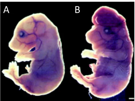
mice displayed smaller body weights at P30 (Fig. 2.7A, B). The brains of Bcl-xLNex-Cre mice
were also smaller compared to their wildtype littermate controls (Fig. 2.7C, D).
Importantly, deletion of Bcl-xL in postmitotic neurons in Bcl-xLNex-Cre mice still resulted in a
cell death phenotype in the brain that was similar to what was observed in the Bcl-xL Emx1-Cre mice, with significantly increased cleaved caspase-3 staining in the upper cortical layers
at P1 (Fig. 2.7E, F). Taken together, our results from both the Bcl-xLEmx1-Cre and Bcl-xLNex-Cre
mice show that the loss of Bcl-xL does not cause widespread apoptosis in the developing brain but instead results in the loss of postmitotic neurons in the upper regions of the cortex during the early postnatal stages.
Loss of Bcl-xL-dependent neurons results in deficits in motor learning, hyperactivity,
and increased risk-taking and self-injurious behaviors
To determine the functional consequence of Bcl-xL deletion in postmitotic neurons, we conducted neurobehavioral assessments of wildtype and Bcl-xLNex-Cre mice starting at
P30. A three-minute tail suspension test revealed that, while wildtype mice exhibited the characteristic flailing of their hindlimbs, Bcl-xLNex-Cre mice displayed hindlimb clasping, an
indicator of generalized neurological dysfunction (Fig. 2.8A, B). The Bcl-xLNex-Cre mice also
59
in approximately 50% of these mice (Fig. 2.8C). These lesions were not a result of fighting with cage mates, as Bcl-xLNex-Cre mice housed alone also developed these lesions. Analysis of
grooming behavior revealed that male Bcl-xLNex-Cre mice spent significantly more time
grooming compared to wildtype controls (Fig. 2.8D). Male Bcl-xLNex-Cre mice also displayed
an altered sensation to pain, as they were significantly more insensitive to noxious heat stimuli on the Hargreaves test (Fig. 2.8E).
In a multi-test behavioral regimen, both male and female Bcl-xLNex-Cre mice displayed
increased locomotor activity in the open field test without changes in rearing movements or time spent in the center region (Fig. 2.9A-C). Interestingly, male Bcl-xLNex-Cre mice also
displayed significantly increased risk-taking behavior as they spent more time in the open arms of the elevated plus maze, suggesting a loss of typical cautionary avoidance of the open areas (Fig. 2.9D, E). The total number of entries in the elevated plus maze was not altered in the knockout mice, indicating that the increased risk-taking behavior in the male Bcl-xLNex-Cre mice could not be attributed to general hyperactivity during the test (Fig. 2.9F).
In contrast to the intact ability for locomotion and rearing movements, the Bcl-xL Nex-Cre knockout mice had profound deficits in motor coordination on an accelerating rotarod.
The Bcl-xLNex-Cre mice failed to show improvement across repeated trials, indicating
impaired motor learning in the rotarod test (Fig. 2.9G). A subset of Bcl-xL-deficient mice without skin lesions (8/16) was further evaluated in the Morris water maze task to
determine the ability of mice to locate a visible escape platform. While all of the wildtype mice demonstrated proficient learning in the visible platform test, Bcl-xLNex-Cre knockout
mice had an overt impairment in reaching the escape platform, with only two Bcl-xLNex-Cre
60
61
2.4 Materials and Methods
Mice. Bcl-xLloxP/loxP mice were generously provided by Dr. You-Wen He (Duke University).
To induce conditional deletion of Bcl-xL, Bcl-xLloxP/loxP mice were crossed with two different
Cre lines, Emx1-Cre and Nex-Cre. Emx1-Cre and tdTomato mice were a kind gift from Dr. William Snider (UNC-Chapel Hill) and Dr. Timothy Gershon, respectively (UNC-Chapel Hill). All other mice were obtained from Jackson Laboratory. Mice were maintained in a 12 hr light, 12 hr dark cycle (lights on at 7AM, lights off at 7 PM). All animal handling and protocols were carried out in accordance with established practices as described in the National Institutes of Health Guide for Care and Use of Laboratory Animals and as approved by the Animal Care and Use Committee at UNC-Chapel Hill.
Surgeries
For intrinsic signal optical imaging, craniotomy was performed on a total of 6 mice at age P29 – 34 (n = 3 wildtype, n = 3 Bcl-xLEmx1-Cre mice). Mice were anesthetized with isoflurane
(5% for induction, 1-2% for surgery, 0.5% for imaging) augmented with chlorprothixene (2.5mg/kg, i.p.). Skull overlaying right visual cortex was exposed, and a custom head-fixing imaging chamber with a 5-mm diameter opening was mounted and secured with
cyanoacrylate glue (Oasis Medical) and dental acrylic (Lang Dental). A 4-mm diameter craniotomy was made within the chamber to expose the visual cortex for imaging. The imaging chamber was then filled with a saline solution containing (in mM) 150 NaCl, 2.5 KCl, 10 HEPES, 2 CaCl2, and 1MgCl2. Physically-activated heat packs (SpaceGel, Braintree
62
Intrinsic signal optical imaging
Custom instrumentation adapted from previous reports (Kalatsky and Stryker 2003; Smith and Trachtenberg 2007) was used. Briefly, two F-mount lenses were used to form a tandem lens macroscope (respective focal lengths of 135 and 50mm; Nikon) which was connected to a Dalsa 1M30 CCD camera (Teledyne DALSA), providing a 4.6mm x 4.6mm field of view. The pial vasculature illuminated (Asahi Spectra low noise halogen source) with green light (550 ± 50nm, Edmund Optics) and imaged through a green emission filter (560 ± 5nm) served as a landmark for depth. From the vasculature, the imaging was focused 600 µm deep into the neocortex where hemodynamic intrinsic signals were imaged with red light (700 ± 38nm, Chroma). Reflected light was then captured through a second red filter (700 ± 5nm, Edmund Optics) at 30 frames/s with custom image acquisition software (code kindly provided by D. Ferster, Northwestern University; with adaptations by J. Stirman, University of North Carolina). Fourier analysis was performed on each pixel column to measure the magnitude and phase of stimulus-evoked signals at the frequency of the periodic visual stimuli (0.125Hz). Area and vertical and horizontal diameters of the entire V1 and a portion of V1 representing 50-degree visual space were measured using ImageJ software (Schindelin et al. 2012).
Visual stimuli
63
viewed by the contralateral eye. A 3° thick drifting white bar sweeping across the monitor once every 8 seconds on a black background (horizontal or vertical) was used for
retinotopic mapping, and a vertical grating patch (50° diameter displayed at the center of the monitor, 2 cycles/s, 0.04 cycle/°) drifting for the last two seconds of an eight-second period was used to measure basic cortical representation of a 50-degree visual space. The periodic motion cycle for both of these stimuli was at 0.125Hz and repeated 50 cycles. The stimulus movies were produced and presented using MATLAB (Pelli 1997) and the
Psychophysics Toolbox (Brainard 1997) and were corrected for three dimensional distortion due to the flatness of the monitor using a custom MATLAB code (code is available online, http://labrigger.com/blog/2012/03/06/mouse-visual-stim/).
Behavioral regimen
Subjects were 16 Bcl-xLNex-Cre (10 males and 6 females) and 19 wildtype controls (10 males
and 9 females). Testing began when mice were approximately 6-7 weeks of age. For each procedure, measures were taken by an observer blind to mouse genotype. Mice were tested in the following assays, with one or two tests per week: elevated plus maze, open field, rotarod, and Morris water maze.
Elevated plus maze. This test was used to assess anxiety–like behavior, based on a