Abstract
Adult neurogenesis is the process of generating functional neurons from neural stem cells
(NSCs) in the adult brain. In humans, this unique form of neuroplasticity is restricted to the
dentate gyrus (DG) of the hippocampus. Unlike developmental neurogenesis, adult neurogenesis
is regulated by local and long distance neuronal circuit activity. However, the identities of
specific cell types, neurotransmitters, and receptors that facilitate this regulation remain largely
unknown. Studies utilizing animal and cell culture models suggest that the neuropeptide
cholecystokinin (CCK) serves as a mitogenic signal for NSCs in the adult brain, but the direct
effects of CCK have remained unclear. We therefore tested the hypothesis that CCK regulates
adult neurogenesis by promoting NSC proliferation. We have found that NSCs express mRNA
encoding the Gq-coupled CCK2 receptor. These receptors were found to be functional after
exhibiting calcium activity following stimulation with the CCK2-receptor selective form of CCK
(CCK8). This latter effect can be blocked by the CCK2 receptor antagonist YM022. In-vivo
chemogenetic stimulation of CCK-releasing cells in the DG produced an increase in NSC
proliferation. However, this increase in NSC proliferation did not result in an increase in total
NSC density, which suggests that CCK promotes NSCs to proliferate asymmetrically rather than
self-renew. In addition, shRNA-mediated knockdown of CCK8 in the DG produced a striking
decrease in NSC proliferation. This information could contribute to facilitating the regeneration
1. Introduction
The discovery of adult neurogenesis has rewritten much of the canonical dogma of
neuroscience; “nerve paths are something fixed, ended, and immutable; everything may die,
nothing may be regenerated1” no longer holds true. However, the mechanisms that govern the
generation of new neurons and their integration into existing neural circuitry are poorly
understood. What is known is that dynamic neural activity regulates the generation of new
neurons and their functional integration into existing circuitry. However, the exact
neurotransmitter systems and receptors that regulate adult neurogenesis remain to be fully
elucidated.
Neurogenesis was originally thought to occur solely during the embryonic and early
postnatal stages of mammalian central nervous system (CNS) development2. Only recently has it
become widely accepted that neurogenesis occurs throughout life, though in restricted regions of
the brain. In humans, this process is limited to the subgranular zone (SGZ) of the dentate gyrus
(DG) in the hippocampus3,4,5—a brain region critical for memory processes and affective state
regulation.
Adult hippocampal neurogenesis recapitulates much of the neuronal developmental
process in the mature CNS: proliferation and fate specification of adult neural precursors,
morphogenesis, migration, axonal and dendritic development, and finally, synapse formation6,7.
Neuronal development culminates in the full integration of new neurons into the existing
Figure 1. Illustration of the five developmental stages during adult hippocampal neurogenesis. Local neuronal activity and afferent inputs play a role in regulating whether these cells
differentiate or self-renew.
However, unlike developmental neurogenesis, adult neurogenesis is finely tuned to
changes in local and afferent neuronal activity (Figure 2). Currently unknown neuronal systems
and pathways relay these changes in activity to NSCs. The lack of knowledge regarding the
mechanisms by which different signaling systems regulate adult neurogenesis has limited the
ability to manipulate the neurogenesis process to determine its specific contribution to brain
functions. Thus, there is a pressing need for basic science research to understand the fundamental
mechanisms involved in this process before any significant progress can be made towards
translational research on adult neurogenesis.
Figure 2. Model of neurogenesis regulation by neuronal activity. (A) Location of the hippocampus in the mouse brain. (B) Neuronal inputs to the hippocampus and from local
Cholecystokinin (CCK) has been identified as one of the most abundant peptides found in
the brain8. Although CCK was originally found in the gastrointestinal tract, one of its molecular
forms, cholecystokinin sulfated octapeptide (CCK8), has been discovered in high levels in the
hippocampus8. CCK acts through two classes of G-protein coupled receptors. CCK2 receptors
represent the predominant subtype in the brain while CCK1 receptors are mainly found in the
gut8,9. Studies have demonstrated that CCK8 binds to CCK2 receptors with great affinity, and
has been implicated in mediating memory processes9,10,11. CCK8 levels and CCK2 receptors have
been found to dramatically decrease in the brain with age11. This effect suggests that CCK8
neurotransmission may play a role in memory impairment found in the aged.
The molecular mechanisms by which CCK is involved in the adult neurogenesis process
are currently being studied. Previous studies have shown that CCK promotes the proliferation
and survival of cultured neuroblasts12 and that CCK2 receptor knock-out mice exhibit reduced
adult neurogenesis13. In addition, CCK promotes dendritic development in-vitro, which can be
blocked by co-incubation with a CCK2 receptor antagonist14. Such studies utilizing animal and
cell culture models have suggested that CCK serves as a mitogenic signal for NSCs in the adult
brain, but the molecular mechanisms and cellular targets underlying the effects of CCK remain
unclear. We hypothesize that CCK regulates adult neurogenesis by promoting NSC proliferation
in-vivo. Identifying the specific components of the CCK-cell system is a primary focus of this ongoing project.
2. Methods
2.1: Mice breeding
For the following experiments, two orthologous mouse lines were bred in the rodent
expressing the cre-recombinase enzyme under the CCK promoter were used in Sections 2.2-2.6.
VGLUT3-Cre mice were used in Section 2.7. Animals were exposed to an alternating 14-hour
light and 10-hour dark cycle with free access to food and water. All mice handling and
experiments were conducted in accordance with the National Institute of Health guidelines and
were approved by the Institutional Animal Care and Use Committee of the University of North
Carolina.
2.2: CCK localization
A cre-dependent adeno-associated virus (AAV) conjugated to the fluorescent reporter
mCherry (AAV-DIO-mCherry) was delivered into the DG of 8-week old CCK-Cre mice by
stereotaxic surgery. The following coordinates (in mm) were used to perform the injection: AP:
-2.0; ML: 1.5; DV: -2.3 from bregma. A Hamilton syringe was used to inject 0.3 µl of the virus
bilaterally at a rate of 0.1 µl per minute. Mice were given 4 weeks to recover after surgery.
Following recovery, animals were injected with a lethal dose of a ketamine cocktail (40 mg/kg
ketamine; 40 mg/mL xylazine) and were then transcardially perfused using 0.1M PBS (pH 7.4)
followed by 4% paraformaldehyde at 4°C. Coronal brain slices (40 µm) of the entire
hippocampus were collected in serial order using a Leica SM2010 R microtome.
2.3: CCK2 receptor in-situ hybridization
Primer Blast was used to design primers to amplify a 1 kB fragment of the CCK2 gene by
the polymerase chain reaction. The following forward and reverse primers were used,
respectively: 5’-ATTTAAGAGCAGTCACCCTCCCG-3’ and
5’-TGAGGGGCAGAAGGAAATCTCTTTAATAGC-3’. The insert sequence was ligated into a
pGEM-4Z plasmid vector containing the promoters for the DNA-dependent SP6 and T7 RNA
EcoRI restriction enzyme. The anti-sense probe was generated using the T7 polymerase.
Digoxigenin-11-UTP was incorporated into the probe to allow for visualization. Frozen sections
of the hippocampus (40 µm) were harvested and mounted on RNase free slides. All hybridization
steps and washes followed that of a previously established protocol15. Visualization of
Digoxigenin-11-UTP labeled RNA probes was achieved by TSA amplification15.
2.4: Calcium imaging
A CCK-Cre mouse was anaesthetized by ketamine overdose and perfused with cutting
buffer (in mM: 110.0 choline chloride, 2.5 KCl, 1.3 NaH2PO4, 25.0 NaHCO3, 0.5 CaCl2.2H2O,
7.0 MgSO4, 20.0 D-glucose, 1.3 sodium L-ascorbate, 3.0 sodium pyruvate, 5.5 kynurenic acid).
The brain was quickly placed in ice-cold cutting buffer solution. Slices (300 μm) were sectioned
using a vibratome (Leica VT1000S) and transferred to a chamber containing artificial
cerebrospinal fluid (in mM: 125.0 NaCl, 2.5 KCl, 1.3 KH2PO4, 1.3 MgSO4, 25.0 NaHCO3, 2.0
CaCl2.2H2O, 1.3 sodium L-ascorbate, 0.6 sodium pyruvate, 10.0 D-glucose, pH 7.4, 32ºC),
bubbled with 95% O2/5% CO2. A bolus-loading protocol16 was followed where SR101 (1 uM)
was added in the calcium indicator dye (Oregon Green 488 BAPTA-1, Invitrogen) to label
hippocampal NSCs. Intracellular calcium change was measured by change in fluorescence. For
each cell (n=4), baseline NSC activity was recorded for 30 seconds using a confocal microscope.
CCK8 (500 nM) was applied to the slices through the perfusion system16 from 30 seconds to 120
seconds while NSC activity continued to be recorded. CCK8 was then removed from the
system, and fluorescence continued to be recorded for an additional 90 seconds. A fresh slice
was taken, and for each cell recorded (n=4), its baseline NSC activity was recorded for 30
seconds. The slice was then pre-incubated with the CCK2-selective antagonist YM022 (2 μm) for
fluorescence was recorded over time. YM022 and CCK8 were removed from the system, and
fluorescence was again measured for an additional 90 seconds.
2.5: Chemogenetic activation of CCK-cells
Utilizing cre-dependent AAV, we selectively infected and expressed the chemogenetic
tool, “Designer Receptors Exclusively Activated by Designer Drugs” (DREADDs), in CCK
releasing interneurons (CCK-cells) of the DG. An excitatory DREADD packaged in an
AAV (AAV-DIO-h3MD-mCherry) was used to increase the activity in CCK-cells. Stereotaxic
surgery was used to bilaterally inject the virus (0.3 µl) directly into the DG of 8-week old
CCK-Cre experimental mice (n=5) at the following coordinates: AP: -2.0; ML: 1.5; DV: -2.3 from
bregma. A fluorescently tagged control virus (AAV-DIO-mCherry) was similarly injected into
the DG of 8-week old CCK-Cre control mice (n=3). Mice were given 4 weeks to recover.
Clozapine-N-Oxide (CNO) administered through the drinking water (0.25 mg/mL) was given to
the mice to activate the cre-dependent DREADD construct. Following 4 days of
DREADD-induced activation of CCK-cells, the DNA (thymidine) analogue EdU (4 mg/kg, every 2 hours,
for 4 times) was injected to label proliferating cells. Animals were transcardially perfused with
0.1M PBS (pH 7.4) followed by 4% paraformaldehyde at 4°C to preserve the tissue for
histology. Coronal brain slices of the hippocampus (40 µm) were taken in serial order.
2.6.1: Construction of AAV-CCK-shRNA
CCK synthesis was disrupted using an AAV-expressing shRNA targeted to CCK. A 15
base pair sequence in the coding region of the CCK gene
(5’-GAACTCGCCAAGCCAGCTGATTTC-3’) was identified as the target region17.
CCK-shRNA was designed using previously published criteria17. A negative control virus was
(5’-CGGAATTTAGTTACGGGGATCCAC-3’) that had no known sequence similarities17.
2.6.2: Western blot
A western blot was performed to determine the effectiveness of AAV-mediated
knockdown of CCK. Cre mice (n=4) stereotaxically received three injections of
CCK-shRNA at 1 uL each at the following coordinates (in mm): AP: -2.0; ML: 1.5; DV: -2.3, -2.1,
-1.15 from bregma. Scramble-shRNA (1 uL) was similarly injected into the DG of the CCK-Cre
control mice (n=3). After 4 weeks, mice were anaesthetized by ketamine overdose and
transcardially perfused as previously described. The hippocampus was acutely dissected out
from the whole brain and flash frozen in liquid nitrogen. Lysate was prepared from the tissue16,
and a western blot was performed using standard procedures with anti-CCK8 (rabbit, 1:1000
dilution) and anti-GAPDH (Abcam, rabbit, 1:1000 dilution) antibodies. Signal was detected
using an Odyssey Imager (LI-COR Biosciences). Analysis utilized ImageJ to measure
densitometry as a ratio of CCK8 to GAPDH total protein expression.
2.6.3: Chemogenetic activation of CCK-cells following CCK knockdown
CCK-Cre mice were given three injections of either CCK-shRNA (n=4) or
scramble-shRNA (n=3) as described above in Section 2.6.2, and were given 4 weeks to recover.
Following recovery, stereotaxic surgery was used to bilaterally inject the excitatory DREADD
(AAV-DIO-h3MD-mCherry) directly into the DG of both groups of mice as described above in
Section 2.5. Mice were given 4 weeks to recover. CNO was administered through the drinking
water (0.25 mg/mL) to activate the DREADD construct. Following 4 days of DREADD-induced
activation of CCK-cells, EdU (4 mg/kg, every 2 hours, for 4 times) was injected to label
4% paraformaldehyde at 4°C. Coronal brain slices of the hippocampus (40 µm) were taken in
serial order.
2.7: Chemogenetic activation of VGLUT3-expressing CCK-cells
At 8 weeks, injection methods described above in Section 2.5 were repeated where
VGLUT3-Cre experimental mice (n=4) were injected with AAV-DIO-h3MD-mCherry.
VGLUT3-Cre control mice (n=4) were injected with AAV-DIO-mCherry. The mice were given
4 weeks to recover and were then administered CNO (0.25 mg/mL) through the drinking water
for 4 days. Animals were subsequently perfused as described previously. Coronal brain slices of
the hippocampus (40 µm) were taken in serial order.
2.8: Immunohistochemistry, confocal imaging, and quantification
To visualize the localization of the virus in Section 2.2, immunostaining was performed
with the following antibodies: anti-mCherry (Abcam, mouse, 1:250 dilution) to label CCK-cells,
anti-CCK8 (rabbit, 1:250 dilution) to label CCK8, and anti-GFAP (Abcam, chicken, 1:250
dilution) to label NSCs. DAPI (4',6-diamidino-2-phenylindole) (Invitrogen, 1:125 dilution) was
used to label cell nuclei. Tissue from Section 2.3 was stained with anti-GFAP (Abcam, chicken,
1:250 dilution) and DAPI (Invitrogen, 1:125 dilution). In Sections 2.5 and 2.6.3,
immunostaining was performed on every seventh brain section of the hippocampus. An antigen
retrieval protocol18 was performed by microwaving sections in boiling citric buffer for 7
minutes. EdU labeling19 was completed using either Life Technologies Alexa Fluor 488 azide
(for Section 2.5) or Alexa Fluor 594 azide (for Section 2.6.3). Anti-Nestin (Aves, chicken, 1:250
dilution) was used to label NSCs. For Section 2.7, the following antibodies were used:
Images were acquired on an Olympus Fluoview FV1000 confocal microscope with a 40X
objective lens using multitrack configuration. To analyze the proliferation of NSCs, the
incorporation of the proliferation marker into Nestin+ NSCs was quantified. To analyze the total
proliferation in the DG, all cells that incorporated the proliferation marker were quantified. To
analyze the total NSCs located in the DG, all Nestin+ cells were quantified. Densitometry
measurements were taken using ImageJ. The experimental group in each experiment were
compared to their respective control group. All analyses were performed by investigators blind
to experimental conditions. Statistical analyses were performed using Student’s t-test.
3. Results
3.1: CCK localization
Immunohistochemistry was used to locate CCK-cells in the DG. Images revealed that
CCK-cells were localized in the SGZ of the hippocampus (Figure 3A-B, 3F-G). Verification of
the somatic targeting patterns of CCK-cells was confirmed by immunohistochemistry against
CCK8 (Figure 3C, 3H). Staining revealed that local CCK-cell projections ramify the NSC rich
Figure 3. (A-C): Single-channel Z-stack images of the mouse DG (gray = DAPI). CCK-cells were labeled with mCherry (red) through stereotaxic delivery of a cre-dependent AAV-virus in CCK-Cre mice. Specific targeting of CCK-cells was confirmed by immunohistochemistry against CCK8 (green). (D) Composite overlay of images from A-C. (E) mCherry-labeled CCK fibers (red) densely innervated the soma of DG cells (blue) immediately proximal to radial processes (white) coming from the SGZ. (F-H) Single-channel high-resolution confocal images of somatic targeting patterns of mCherry labeled CCK-cells (red) co-stained for CCK8 (green) in the subgranular and granular zone of the DG (gray=DAPI). (I) Composite overlay of images from F-H.
3.2: In-situ hybridization
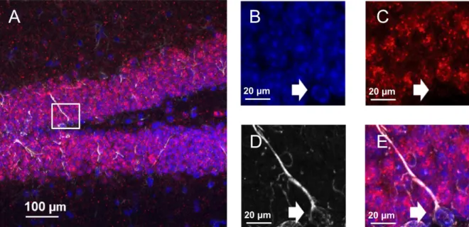
In-situ hybridization was used to determine whether NSCs possess CCK2 receptor mRNA. Probes were correctly synthesized as shown by the colocalization of the CCK2 probe
and GFAP+ NSCs (Figure 4). These images revealed that NSCs in the DG express CCK2
Figure 4. (A) CCK2 receptor mRNA probes (red) localized in the mouse hippocampus. (B-D) Single-channel Z stack images of cell nuclei labeled with DAPI (blue), CCK2 mRNA (red) colocalizing to GFAP+ labeled NSC (white). (E) Composite overlay of images B-D.
3.3: Calcium imaging
To investigate whether the CCK2 receptors identified on NSCs were functional, calcium
imaging was used to test how these receptors responded to the presence of CCK8. Intracellular
calcium change, represented by change in fluorescence, was measured over time. Following
induction with CCK8 (500 nM), SR101-labeled NSCs exhibited a significant increase in average
intracellular calcium response (25.81% ± 6.674, p=0.0306; Figure 5A) in comparison to its
baseline. Additionally, it was found that the CCK8-induced calcium response could be blocked
by pre-incubation with the CCK2-selective antagonist YM022, as demonstrated by the lack of
significant fluorescence change (-3.136% ± 2.513, p=0.3006; Figure 5B). These results reveal
Figure 5. (A) Application of CCK8 produced an increase in intracellular calcium release in SR101-labeled NSCs (n=4) as a result of CCK2-receptor activation. (B) This effect was blocked by the application of the CCK2-antagonist YMO22 in NSCs (n=4).
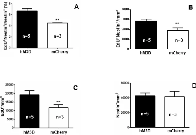
3.4: Chemogenetic activation of CCK-cells
To quantify the effects of DREADD activation of CCK-cells, we measured the
percentage of proliferating NSCs (number of proliferating NSCs/total number of NSCs), the
density of NSC proliferation (number of proliferating NSCs/volume of DG), the total
proliferation density (number of proliferating cells/volume of DG), and the total NSC density
(number of NSCs/volume of DG) in both the experimental and control groups. CCK-cell
activation significantly increased the percentage of proliferating NSCs in comparison to the
control group, in which mice were subjected to AAV-DIO-mCherry injections, and therefore, did
not have their CCK-cells activated upon CNO administration (6.709 ± 0.4001 vs. 4.489 ±
0.07500, p=0.0061; Figure 6A). In addition to an increase in the percentage of proliferating
NSCs, CCK-cell stimulation significantly increased the density of proliferating NSCs (2833 ±
203.6 vs. 1859 ± 297.4, p=0.0311; Figure 6B) as well as the total proliferation in the DG (19303
± 2326 vs. 11732 ± 1734, p=0.0645; Figure 6C). However, CCK-cell activation did not result in
a significant change in the total NSC density (42776 ± 3871 vs. 41595 ± 7041, p=0.8766; Figure
6D).
Figure 6. (A) DREADD activation of CCK-cells in the DG produced an increase in the percentage of proliferating NSCs (proliferating NSCs/total NSCs) in the DG. (B) CCK-cell stimulation increased the density of proliferating NSC (proliferating NSC/volume of DG). (C) CCK-stimulation increased total proliferation density in the DG (proliferating cells/volume of DG). (D) Activation of CCK-cells did not produce a significant increase in total NSC density (total NSCs/volume of DG). Error bars represent S.E.M., and ** denotes significance at p < 0.05.
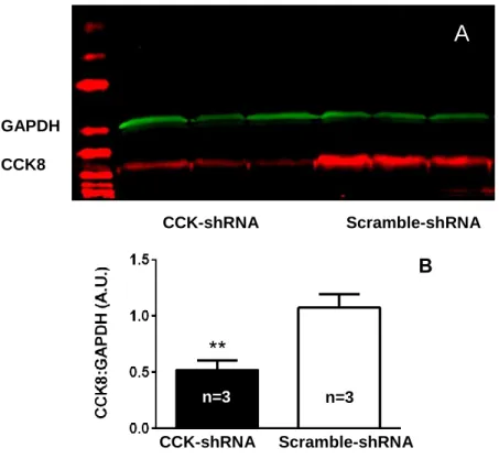
3.5.1: Western blot
A western blot was performed to test the effectiveness of AAV-shRNA mediated
knockdown of CCK8 (Figure 7A). The results indicated a 50% decrease in CCK8 concentration
present in the cells compared to the scramble-shRNA (0.5193 ± 0.08697 vs. 1.075 ± 0.1211,
p=0.0204; Figure 7B). In addition, the concentration of a control protein GAPDH was measured,
and no reduction of protein concentration between the experimental and control groups was
Figure 7. (A) Western blot revealing the effective knockdown of CCK8. (B) CCK-shRNA mediated 50% knockdown of CCK8. Error bars represent S.E.M., and ** denotes significance at p < 0.05.
3.5.2: Chemogenetic stimulation of CCK-cells following CCK8 knockdown
After the effectiveness of AAV-shRNA mediated knockdown of CCK8 was confirmed,
the effects of CCK8 knockdown on NSC proliferation were investigated. Quantification of
proliferating NSCs in the DG following knockdown and DREADD activation of CCK-cells
revealed a significant reduction in the percentage of proliferating NSCs in comparison to the
scramble-shRNA mice (2.356 ± 0.7147 vs. 5.767 ± 0.1965, p=0.0107; Figure 8A). Furthermore,
in comparison to the scramble-shRNA mice, CCK8 knockdown significantly reduced both the
density of proliferating NSCs (478.7 ± 113.8 vs. 1016 ± 90.16, p=0.0176; Figure 8B) and the
total proliferation in the DG (5301 ± 747.5 vs. 9452 ± 1162, p=0.0251; Figure 8C) following
CCK-cell activation. However, the decrease in proliferating NSCs did not translate to a
significant change in total NSC density (21317 ± 1708 vs. 17345 ± 1410, p=0.1530; Figure 8D).
GAPDH
CCK8
CCK-shRNA Scramble-shRNA
B
**
n=3 n=3
A
CCK-shRNA Scramble-shRNA
Figure 8. (A) shRNA-mediated knockdown of CCK8 reduced the percentage of proliferating NSCs (proliferating NSCs/total NSCs) following DREADD activation of CCK-cells in the DG. (B) CCK8 knockdown decreased the density of proliferating NSCs (proliferating NSC/volume of DG). (C) CCK8 knockdown reduced the total proliferation density in the DG (proliferating cells/volume of DG). (D) Knockdown of CCK8 did not produce a significant change in total NSC density (total NSCs/volume of DG). Error bars represent S.E.M., and ** denotes significance at p<0.05.
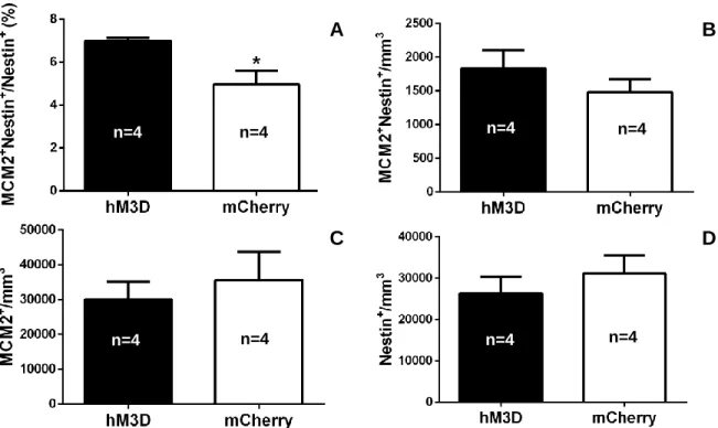
3.6: Chemogenetic activation of VGLUT3-expressing CCK-cells
CCK-cells exhibit heterogeneous attributes such as targeting different regions of the DG
and expressing different subtypes13. One subset of CCK-cells expresses vesicular glutamate
transporter 3 (VGLUT3), and densely innervates the SGZ13. Therefore, we investigated if
selectively activating VGLUT3 CCK-cells was sufficient in recapitulating the observed effect of
increasing NSC proliferation in the DG.
Similar to the results found in Section 3.4, DREADD-activation of VGLUT3 CCK-cells
significantly increased the percentage of proliferating NSCs in comparison to the mice who
received the AAV-DIO-mCherry injection and whose CCK-cells were not activated (6.986 ± A
**
**
C D
CCK-shRNA Scramble-shRNA CCK-shRNA Scramble-shRNA
n=4
n=4 n=3 n=4 n=3
n=3
**
CCK-shRNA Scramble-shRNA CCK-shRNA Scramble-shRNA
n=4 n=3
0.1455 vs. 4.958 ± 0.6373, p=0.0201; Figure 9A). However, unlike the results obtained in
Section 3.4, there was no significant change seen in the total NSC proliferation density (1833 ±
269.5 vs. 1483 ± 192.8, p=0.3320; Figure 9B) or total proliferation density in the DG (30044 ±
5152 vs. 35545 ± 8182, p=0.5900; Figure 9C). No change in total NSC density in the DG was
observed (26308 ± 4109 vs. 31111 ± 4347, p=0.4540; Figure 9D), which was similar to what we
observed in Section 3.4.
Figure 9. (A) DREADD activation of all VGLUT3 CCK-cells in the DG produced an increase in the percentage of proliferating NSCs (proliferating NSCs/total NSCs). (B) Activation of
VGLUT3 CCK-cells did not produce a change in NSC proliferation density (proliferating NSC/volume of DG). (C) VGLUT3 CCK-cell activation did not produce a change in total proliferation density (proliferating cells/volume of DG). (D) DREADD activation did not produce a change in total NSC density (total NSCs/volume of DG). Error bars represent S.E.M., and * denotes significance at p<0.05.
4. Discussion
CCK, a neuropeptide expressed at high levels in the brain, has been implicated in
regulating various physiological functions, including adult neurogenesis. Despite its relevance,
n=4 n=4
D
A C
A
n=4 n=4
A
A
B
A
n=4
little is known about the intercellular mechanisms mediating CCK’s function in this regulation.
Therefore, the purpose of this ongoing study is to elucidate the role of the CCK-cell system in
regulating adult neurogenesis. Preliminary results have indicated that CCK-cell projections are
closely associated with NSCs. Therefore, we have proposed that CCK released from nearby
synapses reaches CCK2 receptors located on NSCs. We hypothesized that CCK functions to
increase the proliferation of adult NSCs in the DG. To test this hypothesis, we used
combinations of immunohistochemistry and chemogenetic tools to manipulate CCK activity
in-vivo. DREADDs allowed in-vivo activation of CCK-cells without the risk of light-induced phototoxicity that often accompanies other methods such as optogenetics. As of yet, no similar
study has taken a chemogenetic approach to study activity-dependent neurogenesis.
We have shown that NSCs possess CCK2 receptors and that these receptors are likely to
be functional. Chemogenetic activation of CCK-cells in two orthologous mouse lines increased
the percentage of NSC proliferation. However, this effect did not translate to an increase in the
total NSC population. shRNA-mediated knockdown of CCK8 was found to reduce the
percentage of NSCs proliferating in the DG. This effect did not result in a decrease in the total
NSC population.
4.1: NSCs likely express functional CCK2 receptors
In-situ hybridization results indicated that NSCs expressed CCK2 receptor mRNA. Furthermore, calcium-imaging data suggested that these receptors were likely
functional as demonstrated by the significant increase in intracellular calcium release that
followed the stimulation of these receptors by CCK8. This increase in intracellular calcium in
NSCs was relayed as change in fluorescence. The observed calcium response was blocked after
induced by CCK8. Only one mouse was used in this calcium imaging experiment. Therefore,
repeat experiments with larger sample sizes are necessary to ensure this effect is consistent
among CCK2 receptors in other CCK-Cre mice.
In addition to repeated calcium imaging experiments, patch clamp electrophysiology will
be used to further study CCK2 receptor activation. A drawback to calcium imaging is that there
can be poor signal to noise ratios due to the use of chemical dyes. Additionally, indirect signals
due to sodium influx during action potentials and synaptic activity can make calcium transients
difficult to distinguish from background noise. Patch clamp recording will reduce some of those
variables to retest CCK2 receptor activity.
4.2: Chemogenetic activation of CCK-cells promotes the proliferation of NSCs
DREADD stimulation of CCK-cells in the DG produced a significant increase in both the
percentage and density of NSC proliferation. The increase in percentage of proliferating NSCs
following CCK-cell activation supports our hypothesis that CCK promotes the proliferation of
NSCs. Proliferating NSC density measurements provided additional confirmation of this effect.
Interestingly, we did not observe a change in the total density of NSCs following CCK-cell
activation, but we did see a significant increase in the total density of proliferating cells in the
DG. The lack of change in total NSC density suggests that CCK may stimulate NSCs to
proliferate asymmetrically by differentiating instead of dividing into another NSC. The
significant increase in total proliferating cell density also suggests that CCK may regulate other
cells in the DG, such as neuroblasts or immature neurons, to differentiate or self-renew. Future
studies will investigate later time points to study the role of CCK in cell differentiation and
determining the fate-choice, survival, and integration of immature neurons into the surrounding
4.3: Loss of CCK8 reduces NSC proliferation and total proliferation in the DG
Western blot analysis revealed that the AAV-expressing shRNA was successful at
knocking down 50% of CCK8 synthesis. This loss in CCK8 resulted in a striking reduction of
both the percentage and density of NSC proliferation after CCK-cell activation. These results
thus show that knocking down CCK8 synthesis prevents the induction of NSC proliferation by
CCK-cell activation. This further supports the role of CCK in promoting NSC proliferation. In
addition, we did not observe a change in the total NSC density, but we did see a significant
decrease in the total proliferating cell density in the DG. The data also provide further support
that CCK may promote the asymmetrical proliferation of NSCs and play a role in regulating the
proliferation of other cell types in the DG. Since CCK-cells can release several molecular forms
of CCK that have affinity to the CCK2 receptor8,9, future studies will investigate whether
knocking down other forms of CCK is sufficient in recapitulating this effect of reducing NSC
proliferation. This will help determine whether or not this effect is specific to CCK8.
4.4: Chemogenetic activation of VGLUT3CCK-cells promotes the proliferation of NSCs Due to the fact that a heterogeneous population of cells releases CCK, we sought to
identify the specific components of the CCK-cell system to better understand how this system
may be involved in adult neurogenesis. Previous studies have indicated that a subset of
CCK-cells express VGLUT313,20. Therefore, we proposed that VGLUT3 CCK-cells were potential
candidates in producing the observed effect of CCK on NSC proliferation.
Results showed that similar to DREADD activation of all CCK-cells in the DG,
activation of VGLUT3 CCK-cells produced the same effect of significantly increasing the
percentage of NSC proliferation. This indicated that activation of the VGLUT3-expressing
providing support for our central hypothesis. While we did not observe an increase in the
density of proliferating NSCs, this experiment was a secondary measure and may reflect the
reduced number of CCK-cells recruited in this experiment. Since VGLUT3 CCK-cells represent
a subset of the entire CCK-cell population, activation of VGLUT3 CCK-cells for this short
duration may not have been sufficient to fully expand this proliferating population to the point of
a significant difference.
4.5: Future directions
In summary, these data strengthen the implication of CCK as being a positive regulator of
adult neurogenesis. We will continue examining the significance of these findings using
behavioral assays to observe the importance of this system in specific neurogenesis-associated
tasks. In addition, later time-points of adult hippocampal neurogenesis will be investigated to
probe the role of CCK in regulating cell-differentiation and fate-choice, survival, and dendritic
and synaptic integration. This information could potentially contribute to future treatment
programs for neurodegenerative diseases by utilizing CCK-receptor agonists or antagonists and
facilitating the regeneration of neurons after brain injury. Understanding the role of local
signaling in promoting proliferation, survival, and integration of newborn neurons into existing
neural circuitry is necessary for the future of such promising therapies.
5. Acknowledgments
First and foremost, I would like to thank my graduate student mentor and co-investigator,
Reid Olsen, for his assistance and unwavering support during the past three years. Reid
collected the calcium imaging and CCK-cell activation data (Sections 3.3 and 3.4). For these
parts, I only assisted in performing the experiments. Thank you to Dr. Alison Xie for her help
Beinfeld at Tufts University and Dr. Andrea Varro at Liverpool University, respectively. Lastly,
I would like to express my gratitude to Dr. Juan Song and all of the members of the Song Lab for
facilitating such a positive learning environment for me in the lab.
This research is supported by Reid’s NRSA, the Summer Undergraduate Research
Fellowship from the Office of Undergraduate Research at UNC, and the Taylor Honors
Fellowship sponsored by Honors Carolina and William W. and Ida W. Taylor.
6. References
1. Cajal, Santiago Ramón. Degeneration & regeneration of the nervous system. Vol. 1.
Oxford University Press, Humphrey Milford, 1928.
2. Ge, Shaoyu, Dennis A. Pradhan, Guo-li Ming, and Hongjun Song. "GABA sets the tempo
for activity-dependent adult neurogenesis." Trends in neurosciences 30, no. 1 (2007): 1-8.
3. Ming, Guo-li, and Hongjun Song. "Adult neurogenesis in the mammalian central nervous
system." Annu. Rev. Neurosci. 28 (2005): 223-250.
4. Bordey, Angélique. "Adult neurogenesis: basic concepts of signaling." Cell Cycle 5, no. 7
(2006): 722-728.
5. Spalding, Kirsty L., Olaf Bergmann, Kanar Alkass, Samuel Bernard, Mehran Salehpour,
Hagen B. Huttner, Emil Boström et al. "Dynamics of hippocampal neurogenesis in adult
humans." Cell 153, no. 6 (2013): 1219-1227.
6. Ming, Guo-li, and Hongjun Song. "Adult neurogenesis in the mammalian brain:
significant answers and significant questions." Neuron 70, no. 4 (2011): 687-702.
7. Song, Juan, Chun Zhong, Michael A. Bonaguidi, Gerald J. Sun, Derek Hsu, Yan Gu,
Konstantinos Meletis et al. "Neuronal circuitry mechanism regulating adult quiescent
neural stem-cell fate decision." Nature 489, no. 7414 (2012): 150-154.
8. Langmesser, Sonja, Maria I. Cerezo‐Guisado, Maria J. Lorenzo, Luis J. Garcia‐Marin,
and Maria J. Bragado. "CCK1 and 2 receptors are expressed in immortalized rat brain
neuroblasts: intracellular signals after cholecystokinin stimulation." Journal of cellular
biochemistry 100, no. 4 (2007): 851-864.
9. Daugé, Valérie, Angélique Sebret, Françoise Beslot, Toshimitsu Matsui, and Bernard P.
Roques. "Behavioral profile of CCK2 receptor-deficient
mice."Neuropsychopharmacology 25, no. 5 (2001): 690-698.
10. Greenstein, R. J., M. M. Ybanez, R-L. Zhang, and W. A. Bauman. "Is aging
preprogrammed? Observations from the brain/gut axis." Mechanisms of ageing and
development 61, no. 2 (1991): 113-121.
11. Harro, Jaanus, and Lars Oreland. "Age-related differences of cholecystokinin receptor
binding in the rat brain." Progress in Neuro-Psychopharmacology and Biological
Psychiatry 16, no. 3 (1992): 369-375.
neural progenitors in the adult mouse forebrain." Proceedings of the National Academy of Sciences 105, no. 9 (2008): 3610-3615.
13. Somogyi, Jozsef, Agnes Baude, Yuko Omori, Hidemi Shimizu, Salah El Mestikawy,
Masahiro Fukaya, Ryuichi Shigemoto, Masahiko Watanabe, and Peter Somogyi. "GABAergic basket cells expressing cholecystokinin contain vesicular glutamate
transporter type 3 (VGLUT3) in their synaptic terminals in hippocampus and isocortex of
the rat." European Journal of Neuroscience19, no. 3 (2004): 552-569.
14. Menon, Shalini, Nicholas Patrick Boyer, Cortney Chelise Winkle, Leslie Marie McClain,
Christopher Carey Hanlin, Dharmendra Pandey, Simon Rothenfußer, Anne Marion Taylor, and Stephanie Lynn Gupton. "The E3 Ubiquitin Ligase TRIM9 Is a Filopodia Off
Switch Required for Netrin-Dependent Axon Guidance." Developmental cell 35, no. 6
(2015): 698-712.
15. Stuber, Garret D., Alice M. Stamatakis, and Pranish A. Kantak. "Considerations when
using cre-driver rodent lines for studying ventral tegmental area circuitry." Neuron 85,
no. 2 (2015): 439-445.
16. Xie, Alison X., Kelli Lauderdale, Thomas Murphy, Timothy L. Myers, and Todd A.
Fiacco. "Inducing plasticity of astrocytic receptors by manipulation of neuronal firing
rates." Journal of visualized experiments: JoVE 85 (2014).
17. Arey, Rachel N., J. F. Enwright, Sade M. Spencer, Edgardo Falcon, Angela R. Ozburn,
Subroto Ghose, Carol Tamminga, and Colleen A. McClung. "An important role for Cholecystokinin, a CLOCK target gene, in the development and treatment of manic-like
behaviors." Molecular psychiatry 19, no. 3 (2014): 342-350.
18. Bonaguidi, Michael A., Michael A. Wheeler, Jason S. Shapiro, Ryan P. Stadel, Gerald J.
Sun, Guo-li Ming, and Hongjun Song. "In vivo clonal analysis reveals self-renewing and
multipotent adult neural stem cell characteristics." Cell 145, no. 7 (2011): 1142-1155.Jao,
Cindy Y., and Adrian Salic. "Exploring RNA transcription and turnover in vivo by using
click chemistry." Proceedings of the National Academy of Sciences 105, no. 41 (2008):
15779-15784.
19. Salic, Adrian, and Timothy J. Mitchison. "A chemical method for fast and sensitive
detection of DNA synthesis in vivo." Proceedings of the National Academy of
Sciences 105, no. 7 (2008): 2415-2420.
20. Fremeau, Robert T., Jonathon Burman, Tayyaba Qureshi, Cindy H. Tran, John Proctor,
Juliette Johnson, Hui Zhang et al. "The identification of vesicular glutamate transporter 3
suggests novel modes of signaling by glutamate." Proceedings of the National Academy