CHARACTERIZING CALCIUM-ACTIVATED CHLORIDE SECRETION: IMPLICATIONS FOR SEX-LINKED VARIATIONS IN CYSTIC FIBROSIS
JOHN THOMAS SHERIDAN
A dissertation submitted to the faculty of the University of North Carolina at Chapel Hill in partial fulfillment of the requirements for the degree of Doctor of Philosophy in the
Department of Cell and Molecular Physiology.
Chapel Hill 2013
Approved by:
ii
ABSTRACT
JOHN THOMAS SHERIDAN: Characterizing Calcium-Activated Chloride Secretion: Implications for Sex-Linked Variations in Cystic Fibrosis
(Under the direction of Dr. Robert Tarran)
Cystic Fibrosis (CF) is a genetic disease that is caused by mutations in the gene encoding the Cystic Fibrosis Transmembrane Conductance Regulator (CFTR), the main Cl- channel in human airways, and is characterized by an inability to regulate salt
transport in epithelial tissues. In CF lungs, the airway surface liquid (ASL) dehydrates and mucus accumulates, resulting in mucus plugging. This obstructs respiration and leaves CF patients prone to infection. However, the Ca2+-activated Cl- channel (CaCC) is still present and represents an alternative channel to regulate the ASL. Anocatmin 1 (Ano1) was only recently identified as an essential subunit of CaCC and very little is known about Ano1 structure and regulation. In our first study, we utilized several microscopy and biochemistry based techniques to determine the quaternary structure of Ano1 at the plasma membrane. Ano1 was tagged with either mCherry or GFP to conduct Förster resonance energy transfer (FRET) experiments, which revealed that Ano1 oligomerizes prior to reaching the plasma membrane. Additionally, altering the cytoplasmic concentration of Ca2+ did not affect FRET, suggesting a static interaction. Several biochemical assays revealed that Ano1 exists as a dimer.
iii
iv
DEDICATION
v
ACKNOWLEDGEMENTS
I would first like to acknowledge and thank my advisor, Dr. Robert Tarran. Through his mentorship and guidance, I have gained independence and confidence in myself. I would also like to thank my committee members, Drs. Kathleen Caron, Richard Cheney, Michael Goy, and Richard Boucher for their input, advice, and guidance. My graduate experience would not have been the same without each member of the Tarran lab. Their enthusiasm, encouragement, support, and camaraderie are greatly
appreciated. I would especially like to thank Julie Rasmussen, Dr. Carey Hobbs, and Dr. Alaina Garland, for their advice and guidance throughout the years. I would also like to acknowledge and thank the staff members in the Department of Cell and Molecular Physiology and the Cystic Fibrosis Center for all of their help.
Next, I would like to acknowledge my early mentors. My professors and advisors at Dickinson College, Drs. John Henson and David Kushner, were especially influential on my decision to pursue a career in science.
During my time in Chapel Hill, I have made so many great friends that are like a second family. There are too many to list here, but I thank each and every one of you. I could not have made it through graduate school if I didn’t have your support.
Finally, I would like to thank my family for believing in me throughout graduate school. My sister, Megan Sheridan, and my aunt, Beverly Balfour, have motivated and supported me every step of the way. My parents, Tom and Linda Sheridan, are my biggest fans and have been an essential source of strength. Their love and
vi
TABLE OF CONTENTS
LIST OF TABLES ... viii
LIST OF FIGURES ... ix
LIST OF ABBREVIATIONS ... x
CHAPTER 1. INTRODUCTION ... 1
1.1 Anatomy and physiology of the airways ... 1
1.2 Airway surface liquid ... 2
1.3 Calcium signaling ... 3
1.4 Store-operated calcium entry ... 5
1.5 STIM1 ... 6
1.6 STIM1 phosphorylation ... 8
1.7 Orai1 ... 10
1.8 STIM1-mediate regulation of Orai1 ... 11
1.9 CaCCs ... 12
1.10 The anoctamin family ... 14
1.11 Ano1 structure and function ... 14
1.12 Cystic fibrosis ... 17
1.13 Sex differences in airway diseases ... 18
1.14 Estrogen receptors ... 21
1.15 Estrogen inhibits CaCC ... 22
vii
2. CHARACTERIZATION OF THE OLIGOMERIC STRUCTURE
OF THE Ca(2+)-ACTIVATED Cl- CHANNEL Ano1/TMEM16a ... 30
2.1 Overview ... 30
2.2 Introduction ... 31
2.3 Materials and methods ... 32
2.4 Results ... 36
2.5 Discussion ... 41
3. 17β-ESTRADIOL PREVENTS PHOSPHORYLATION OF STIM1: IMPLICATIONS FOR STORE-OPERATED CALCIUM ENTRY AND CHRONIC LUNG DISEASES ... 54
3.1 Overview ... 54
3.2 Introduction ... 55
3.3 Materials and methods ... 56
3.4 Results ... 60
3.5 Discussion ... 65
4. GENERAL DISCUSSION ... 81
4.1 Overview of results ... 81
4.2 Chapter 2 overview: Ano1 oligomerization ... 82
4.3 Chapter 3 overview: E2 inhibits STIM1 ... 83
4.4 Future work ... 85
4.5 Clinical implications ... 90
4.6 Conclusion ... 92
viii
LIST OF TABLES
Table 2.1: Primers used in RT-PCR to detect endogenous anoctamin
ix
LIST OF FIGURES
Figure 1.1: Regulation of ASL ... 25
Figure 1.2: The Store-Operated Ca2+ Entry Pathway ... 26
Figure 1.3: Functional domains of STIM1 ... 27
Figure 1.4: Predicted topology of Ano1... 28
Figure 1.5: Effect of E2 on ASL Hydration ... 29
Figure 2.1: Ca2+-activated Cl- Currents induced by fluorescent protein-tagged mouse Ano1 constructs are identical to wild type Ano1 ... 46
Figure 2.2: eGFP linked to mCherry with 5 glycines (eGFP-5G-mCherry) undergoes near-maximal FRET ... 48
Figure 2.3:Ano1-eGFP and Ano1-mCherry undergo specific FRET in the plasma membrane ... 49
Figure 2.4: Ano1-eGFP and Ano1-mCherry co-immunoprecipitate ... 50
Figure 2.5: Ano1-eGFP and Ano1-mCherry FRET is not affected by changes in intracellular Ca2+ ... 51
Figure 2.6: Ano1-eGFP and Ano1-mCherry FRET is not dependent on an intact actin cytoskeleton ... 52
Figure 2.7: Determination of Ano1 subunit stoichiometry by chemical cross-linking and non-denaturing gel electrophoresis ... 53
Figure 3.1: STIM1-dependent Ca2+ influx in HBECs is inhibited by E2... 70
Figure 3.2: E2 inhibits thapsigargin-induced STIM1 translocation in HBECs ... 72
Figure 3.3: E2/ESR1 inhibits SOCE and STIM1 oligomerization in HEK293T cells ... 73
Figure 3.4: E2 inhibits STIM1 aggregation and redistribution ... 74
Figure 3.5: E2/ESR1 inhibits STIM1 mobility ... 75
Figure 3.6: E2 does not alter EB1 mobility ... 77
Figure 3.7: Inhibition of STIM1 phosphorylation decreases STIM1 mobility ... 78
x
LIST OF ABBREVIATIONS
A2BR adenosine-2B receptor
aa amino acid
ADO adenosine Ano1 anoctamin 1
ASL airway surface liquid ASM airway smooth muscle AT1 alveolar type I
AT2 alveolar type II
ATP adenosine triphosphate Ca2+ calcium
CaCC calcium-activated chloride channel CAD CRAC activation domain
CCb9 coiled-coil domain containing region b9 CF cystic fibrosis
CFTR cystic fibrosis transmembrane conductance regulator
Cl- chloride
COPD chronic obstructive pulmonary disease CRAC calcium release-activated calcium DAG diacyl glycerol
DDM n-dodecyl β-D-maltoside
DFDNB 1,5-difluoro-2,4-dinotrobenzene DTT dithiothreitol
xi ENaC epithelial sodium channel EMSA electromobility shift assay ER endoplasmic reticulum ERM ezrin-radixin-moesin ESR1 estrogen receptor alpha ESR2 estrogen receptor beta
FRET Förster resonance energy transfer GPCR G-protein coupled receptor
HBEC human bronchial epithelial cell IL-8 interleukin 8
IP3 inositol 1,4,5-triphosphate
IP3-R inositol 1,4,5-triphosphate receptor
K lysine
K+ potassium
Kd dissociation constant MCC mucociliary clearance
Na+ sodium
OASF orai1 activating small fragment PCD primary ciliary dyskinesia PCL periciliary layer
PDZ zonula occludens-1
PIP2 phosphatidylinositol 4,5-bisphosphate PLC phospholipase c
SAM sterile alpha motif
Ser serine
xii SLPI secretory leucoprotease inhibitor SOAR stim1-orai1 activation region SOCs store-operated channels SOCE store-operated calcium entry S/P serine-proline
STIM1 stromal interaction molecule 1
TM transmembrane
TMEM16A transmembrane protein 16A TRP transient receptor potential UTP uridine triphosphate
2
Chapter I
Background and Introduction
1.1 Anatomy and Physiology of the Airways
The physiological role of the lung is to oxygenate the blood through gas exchange. The lung is subdivided into two zones: the conducting zone and the respiration zone. As air enters the respiratory system it passes through the branching network of the
conducting zone, which is comprised of the trachea, bronchi, and bronchioles. Here, the air is warmed and sterilized to prevent infection. The air then enters the respiratory zone where gas exchange occurs across alveoli.
All sections of the airways are lined with epithelia that protect the body from the outside world. The conducting airways are lined with a pseudostratified epithelium that contains several cell types, including ciliated epithelial cells, goblet cells, and basal cells that work in coordination to establish and maintain a protective layer that traps inhaled pathogens and particles [1, 2]. The trapped pathogens are then removed from the airways by mucociliary clearance (MCC) or cough clearance. MCC is a mechanical process where cilia beat in coordination to propel mucus up the airway tract to be
2
carbon dioxide is released from hemoglobin and diffuses into the alveoli, where it can be exhaled [4]. AT2 cells are cuboidal and secrete surfactant. Surfactant is a substance rich in proteins and lipids that reduces alveolar surface tension to facilitate inflation[5].
1.2 Airway Surface Liquid
The layer of fluid that lines the airway epithelium is known as the airway surface liquid (ASL). The ASL contains two distinct layers: (i) the mucus layer and (ii) the “watery” periciliary layer (PCL). The latter is an ideal environment for ciliary beating [6]. However, recent studies have shown that the PCL also contains mucins that are
tethered to the cilia [7]. The mucus layer sits atop the cilia and forms a gel that is comprised of several mucins, with MUC5AC and MUC5B predominating [8]. ASL hydration must be properly maintained to function correctly. This achieved by regulating secretion and absorption of salts via several ion channels.
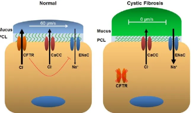
In human airways, the Cystic Fibrosis Transmembrane Conductance Regulator (CFTR) is the main chloride (Cl-) channel that regulates ASL hydration (Fig. 1.1). When CFTR is activated by an elevation in cAMP, Cl- and bicarbonate are secreted into the ASL [9, 10]. Activation of CFTR also inhibits sodium (Na+) absorption through the epithelial Na+ channel (ENaC) [9]. This creates osmotic pressure, which promotes movement of water into the ASL, thus providing hydration. ENaC is a heterotrimer and is comprised of three separate subunits termed: α, β, and γ [11]. To activate ENaC and initiate Na+ absorption, proteases residing in the ASL cleave ENaC, inducing a
conformational change that opens the channel [11]. ENaC can also be regulated by phosphatidylinositol 4,5-bisphosphate (PIP2). PIP2 directly binds to β-ENaC and
3
intracellular Ca2+. When activated, CaCC secretes Cl- and may be able to regulate the ASL [13-15].
Under normal physiological conditions, PCL height is maintained at ~7 μm [9, 14, 16, 17]. This is the optimal height for mucociliary clearance, because outstretched cilia tend to measure 7 μm [16]. These PCL height measurements were performed in vitro, but it is reasonable to assume that similar PCL heights would occur in vivo. Conversely, the mucus layer height varies considerably, with in vivo measurements ranging from 7 to 70 μm [14]. In the event that PCL height decreases, the airway epithelium will respond to increase the height back to 7 μm. This is achieved by stimulating a G-protein coupled receptor (GPCR) with the nucleotide adenosine (ADO) to activate adenylyl cyclase. Adenylyl cyclase raises intracellular cAMP levels, which ultimately activates CFTR to secrete Cl- and raise PCL height [9, 14, 16]. Activation of CFTR also inhibits ENaC through an unknown mechanism to decrease Na+ absorption [18]. Conversely, if the PCL contains excess fluid and height is elevated, the epithelial cells respond by transporting extracellular Na+ into the cell via ENaC. Excess fluid likely decreases CFTR activity by lower the concentration of ADO in the ASL. As a result, ENaC activity
increases and excess water is removed to lower PCL height [17]. PCL/ASL hydration is a regulation balancing act. If ion transport is obstructed, the ASL can dehydrate, which can disrupt MCC. This situation occurs in the genetic disease cystic fibrosis, which will be addressed in detail in a subsequent section.
1.3 Calcium Signaling
4
sequestering Ca2+ to internal stores and by regulating the influx of extracellular Ca2+ [20]. The endoplasmic reticulum (ER) is one of the primary intracellular Ca2+ stores and
actively internalizes cytoplasmic Ca2+ through the Sarco/Endoplasmic Reticulum Calcium transport ATPase (SERCA) pump. When a cell is under resting conditions, there is a constant leakage of Ca2+ from the ER, which is then transported back into the ER via the SERCA pump to control cytoplasmic Ca2+ levels [21-23]. This leakage of Ca2+ from the ER can be easily exploited to deplete ER Ca2+ with a drug called
thapsigargin [21]. Thapsigargin is a SERCA pump inhibitor, which prevents the transport of Ca2+ into the ER. Thus, in the presence of thapsigargin, ER Ca2+ can be depleted without signaling through a GPCR.
5
1.4 Store-Operated Calcium Entry
SOCE was first described in 1989 [20, 26] and has been extensively studied subsequently. Shortly after the discovery of SOCE, a membrane current known as the Ca2+ release-activated Ca2+ current (I
CRAC) was identified [27]. ICRAC has been the most studied aspect of SOCE, but it wasn’t until after the year 2000 that the key signaling elements leading up to ICRAC were discovered [28, 29]. It is now understood that SOCE and ICRAC require two protein families: the stromal interaction molecule (STIM) proteins (STIM1 and STIM2) and the Orai proteins (Orai1, Orai2, and Orai3). The STIM proteins are Ca2+ sensing proteins that span the membrane of the ER and Orai proteins are the pore-forming subunit of the CRAC channel [30].
In addition to Orai, members of the transient receptor potential (TRP) superfamily have long been thought of as store-operated channels (SOCs). However, only Orai1 has been proven to conduct ICRAC [29, 31]. The TRPC (Canonical; TRPC1-TRPC7) family within the TRP superfamily has been extensively studied. Overexpression of TRPC1 and 4 have been shown to increase store-operated entry [32-34], while
6
to modulate ion permeability [43]. Thus, additional experiments need to be performed in order to better understand this process.
1.5 STIM1
The STIM proteins are ubiquitously expressed in vertebrates [44]. STIM1 expression is generally higher than STIM2 expression [44], except in the brain where STIM2 predominates [45]. The essential role of STIM1 in SOCE was initially discovered in Drosophila S2 cells after RNAi-mediated knockdown of Drosophila Stim significantly reduced SOCE [46]. Since then, human STIM1 has been shown to be essential for SOCE [47]. Conversely, the exact role of STIM2 in SOCE is not fully understood. However, there is mounting evidence that STIM2 may be important for maintaining resting cytosolic Ca2+ homeostasis and ER Ca2+ levels, rather than a role in SOCE [48-51].
Structurally, human STIM1 is a 685 amino acid protein that contains a single transmembrane pass (Fig. 1.3). The N-terminal domain of STIM1 resides in the ER lumen, while the larger C-terminal domain resides in the cytoplasm. The N-terminus contains two distinct domains: an EF-hand, which senses changes in ER Ca2+, and a sterile alpha motif (SAM) domain, which mediates oligomerization. The C-terminus of STIM1 is comprised of several domains. Following the transmembrane domain are two coiled-coil domains that contain the STIM1-Orai1 activation region (SOAR), which is followed by the serine-proline (S/P) rich region, and the lysine rich region. The C-terminus of STIM1 also contains an ezrin-radixin-moesin (ERM) domain that
7
formation and translocation to the ER-plasma membrane junction where it induces the influx of Ca2+ through Orai1. Initially, STIM1 was thought to traffic from the ER to the plasma membrane where it induced extracellular Ca2+ influx [53, 54]. However, multiple studies have since shown that SOCE only requires STIM1 that is localized to the ER [47, 55, 56].
This entire process has been extensively studied and the role that each domain of STIM1 plays is now becoming clearer. Arguably, the most important domain is the EF-hand, which senses Ca2+ depletion. The STIM1 EF-hand binds Ca2+ with unusually low affinity and has a dissociation constant (Kd) of ~0.2-0.6 mM [57]. The ER lumen of eukaryotes are predicted to contain ~0.2-2 mM free Ca2+ [57]. This high level of lumenal ER Ca2+ may help explain why the EF-hand binds Ca2+ with low affinity, since the main of role STIM1 as an ER Ca2+ sensor is to induce the influx of extracellular Ca2+ when ER levels fall. It is physiologically beneficial for the cell to sense minor depletions in ER Ca2+ to ensure that enough Ca2+ is stored for a myriad of signaling events. Upon ER Ca2+ depletion, the EF-hand and nearby SAM domain undergo conformational changes that promote oligomerization of multiple STIM1 proteins. Oligomerization is completely dependent on N-terminal interactions. Previous studies have shown that the N-terminus of STIM1 is capable of self-assembling into oligomers, indicating that oligomerization of the EF-SAM domains initiates SOCE [57, 58].
8
that the cytoskeleton may play a role in STIM1 redistribution [61]. Under resting
conditions, STIM1 is known to co-localize with α-tubulin, taking on a tubular appearance similar to the microtubule network [61]. The tubular appearance of STIM1 can be abolished by depolymerizing the microtubule network with nocodazole, which forces STIM1 to take on an ER distribution pattern. There have been conflicting reports on whether STIM1 association with microtubules is important for SOCE. Initial studies suggested that disrupting the microtubule network inhibited ICRAC after ER Ca2+ depletion [61]. However, others have addressed the same issue and have concluded that
disrupting the microtubule network does not inhibit SOCE [52, 62, 63]. It is now understood that STIM1 interacts with microtubules through the microtubule-plus-end tracking protein EB1 [52]. When ER Ca2+ stores are full, STIM1 appears as comets that track the growing ends of microtubules. This association is believed to be important for remodeling the ER and may play a role in ER Ca2+ uptake [52].
1.6 STIM1 Phosphorylation
In recent years, extensive research has been conducted to investigate the role of phosphorylation on STIM1 function. The C-terminus of STIM1 contains the S/P rich region that contains several serine residues that are targets for phosphorylation [64, 65]. However, additional phosphorylation sites outside of this region have been identified [64, 66]. It now appears that the functional impact of phosphorylation wholly depends on the site and can be employed to both enable and inhibit SOCE[64-67].
9
indicator of ERK1/2 activity, was significantly increased after ER Ca2+ depletion. Additionally, they observed a significant increase in phosphorylation of
immunoprecipitated STIM1 after ER Ca2+ depletion. Importantly, STIM1 phosphorylation was abolished when all three serine phosphorylation sites were mutated to alanines, which was associated with a decrease in SOCE in cells expressing the triple point mutant. Taken together, the authors concluded that STIM1 phosphorylation on Ser 575, Ser 608, and Ser 621 was required to activate SOCE [64].
STIM1 phosphorylation has also been shown to inhibit SOCE. It has been documented extensively that SOCE is strongly inhibited during mitosis [68-72]. However, the underlying mechanism had never been identified. In a recent study, researchers discovered that STIM1 phosphorylation strongly increased during mitosis, as indicated by a phospho-specific antibody. They specifically identified Ser 486 and Ser 668 as STIM1 phosphorylation sites that inhibit SOCE during mitosis [65]. Consequently, STIM1 was excluded from the mitotic spindle during mitosis, because phosphorylation of STIM1 disrupted binding to the microtubule plus-end tracking protein EB1 [67]. Mutation of the STIM1 phosphorylation sites to alanines prevented EB1 dissociation and STIM1, along with the ER, redistributed to the mitotic spindle.
However, the functional significance of disrupting STIM1-EB1 dissociation during mitosis is unknown [67].
STIM1 has also been shown to undergo tyrosine phosphorylation. A recent study immunoprecipitated STIM1 from platelets and found that after depleting ER Ca2+ with thapsigargin, STIM1 was rapidly phosphorylated on tryosine residues as indicated by a phospho-tyrosine specific antibody [66]. Additionally, blocking tyrosine
10
it was essential for SOCE in platelets. Thus, the exact role tyrosine phosphorylation plays in STIM1 function and SOCE remains to be determined.
1.7 Orai1
Orai1 is 301 amino acids in length and is predicted to contain four
transmembrane domains with the N- and C-termini located within the cytoplasm [73]. The N-terminus of Orai1 contains a proline/arginine rich region, while the C-terminus contains a putative coiled-coil that is conserved between Orai family members [74, 75]. Orai1 localizes to the plasma membrane and exists as dimer under resting conditions [76]. Following ER Ca2+ depletion and redistribution of STIM1 to the ER-plasma
membrane, Orai1 clusters with STIM1 into puncta [76]. Several studies have shown that Orai1 dimers oligomerize into tetramers following ER Ca2+ depletion [76-78]. Moreover, it has been suggested that Orai1 tetramers interact with two STIM1 molecules to form the functional CRAC channel [77]. However, others have proposed the formation of higher order oligomers[79]. The recently determined crystal structure of Orai from Drosophila melanogaster indicated that Orai assembles into a hexamer [80].
Structural studies have also shown that the first transmembrane segment in each Orai1 subunit forms the ion pore [79-81]. Amino acids lining the pore strongly select for Ca2+ and do not allow the passage of other divalent cations, such as Ba2+ and Cd2+. Furthermore, Orai1 selectivity for Ca2+ is 1000 times greater than selectivity for
11
mutant Orai1 was able to conduct a large current at positive voltages and was constitutively active, even in the absence of STIM1. It is believed that the third transmembrane segment is in a conformation that places negatively charged residues near Orai1’s selectivity filter that facilitates inward Ca2+ current. Furthermore, the third transmembrane segment may also stabilize Orai1’s closed state [84].
1.8 STIM1-Mediated Regulation of Orai1
Whether STIM1 directly interacts with Orai1 has remained controversial. In multiple studies, Förster resonance energy transfer (FRET) has been utilized to study the STIM1-Orai1 interaction [85-87]. There is little FRET between STIM1 and Orai1 under resting conditions. However, there is a significant increase in FRET after ER Ca2+ depletion [85-87]. Additionally, STIM1 has been shown to co-immunoprecipitate with Orai1 [66, 88]. These results suggest that STIM1 and Orai1 are interacting. However, FRET only indicates proximity between two proteins and does not prove an interaction. While co-immunoprecipitation experiments do prove an interaction, they cannot
distinguish between a direct or indirect interaction.
12
plasma membrane[91]. Mutating the SOAR domain prevents activation of Orai1; however, it does not disrupt STIM1-Orai1 clustering [60, 91], which indicates that co-clustering alone is not sufficient to activate Orai1, implying the existence of another regulatory mechanism.
Furthermore, the C-terminus of Orai1 contains a coiled-coil that has been shown to be essential for the STIM1-Orai1 interaction. Studies have shown that mutating a single leucine (aa 273) within the coiled-coil of Orai1 was sufficient to disrupt signaling [74, 75]. The mutant Orai1 was unable to interact and co-cluster with STIM1 and remained inactive following ER Ca2+ depletion [74, 75]. Details regarding the role of Orai1’s N-terminus are also becoming apparent. A recent study suggested STIM1 activates Orai1 in a stepwise manner [92]. The authors of this study proposed that STIM1 initially binds the C-terminus of Orai1, which enables STIM1 to bind the N-terminus of Orai1 and induces a conformational change [92]. Taken together, it is now apparent that STIM1 and Ora1 directly interact and this interaction is dependent on specific sequences in both STIM1 and Orai1.
1.9 CaCCs
CaCCs were first identified over 30 years ago when researchers observed a Ca2+-dependent Cl- current in the rabbit colon [93]. Since then, CaCCs have been identified in almost every tissue and cell type [94-96]. CaCCs have been shown to play essential roles in many physiological processes, including secretion of fluid from
epithelial cells, regulating smooth muscle contraction, olfactory signal transduction, phototransduction, and action potentials in neurons [95, 97].
13
activate CaCC. However, with the addition of a permissible amount of Ca2+, a
membrane depolarization will further increase CaCC activity. At high concentrations of intracellular Ca2+, CaCC is fully activated and a change in the membrane potential will not further modulate activity [96, 99].
Despite numerous studies on functional CaCC activity, the molecular subunit of the channel remained unknown until recently. Numerous proteins were proposed as potential CaCCs, such as CLCA [100], bestrophins [101, 102], and tweety [103]. However, when expressed in cells, the electrophysiological properties generated by these proteins did not match the classical CaCC characteristics, such as outward rectification and voltage sensitivity [104], suggesting that the true CaCC subunit had not yet been discovered. It wasn’t until 2008, when three laboratories independently
identified Anoctamin 1 (Ano1)/transmembrane protein 16A (TMEM16A) as CaCC [105-107].
The three laboratories that simultaneously identified Ano1 utilized different techniques that all lead to the conclusion that Ano1 is CaCC. (i) Caputo and colleagues used a functional genomics approach to identify Ano1 [105]. It was previously observed that IL-4 exposure upregulated Ca2+-dependent Cl- secretion in human bronchial
14
Through various overexpression and siRNA knockdown experiments they identified Ano1 and concluded that it is the subunit of CaCC [107].
1.10 The Anoctamin Family
The Anoctamin family of proteins contains 10 members with Ano1 being the most frequently studied. All eukaryotes express Anoctamin proteins [108]. However, only higher order eukaryotes, such as mammals, express all 10 members. In mammals, Ano1 and 2 are closely related with a sequence homology of ~60% [97]. There is significant sequence variability between Ano1 or 2 and other family members,
suggesting a possible divergence in function [109]. Only Ano1, Ano2, and Ano6 have been shown to form Cl- channels [105-107, 110, 111]. There have been conflicting reports describing cellular localization of the other family members. Tian et al. found that all Anoctamin family members except Ano8, 9, and 10 reach the plasma membrane in HEK293 and FRT cells [112]. Additionally, previous reports have suggested that Ano5 and 7 can reach the plasma membrane [113, 114]. Conversely, a recent study from Duran et al. found no surface expression of Ano3-7 in HEK293 cells and they did not generate Cl- currents when tested by whole cell patch clamping [115]. They went on to speculate that Ano3-7 may reside in intracellular organelles, such as the ER, but it is unclear whether they are actually CaCCs.
1.11 Ano1 Structure and Function
15
may be involved in gating [107]. However, a recent study suggested that the predicted model may not be completely accurate [116]. The authors of this study proposed that the fourth extracellular loop is actually intracellular and may contain a Ca2+-binding site. Their model still predicted the presence of a re-entrant loop; however they suggest that it is located between transmembrane domains 6 and 7 [116].
Interestingly, the gene encoding Ano1 can be alternatively spliced to generate several Ano1 isoforms (Fig 1.4) [117]. Ano1 contains four alternative segments dubbed a, b, c, and d. Segments a and b are located in the N-terminus of Ano1 while the c and d segments are in the first cytoplasmic loop. The alternative splicing pattern of Ano1 varies across human tissues and organs. Some tissues express multiple Ano1 isoforms, while other tissues exclusively express a single isoform [117]. Alternative splicing has also been shown to impact function. For example, inclusion of segment b decreases Ca2+ affinity, which translates to a decrease in channel activity when analyzed by patch-clamping. Furthermore, the currents generated in cells expressing the ac-Ano1 variant are much larger than those seen in cells expressing abc-Ano1 [117]. This suggests that alternative splicing is a mechanism that can be exploited to regulate channel properties, such as Ca2+ sensitivity.
16
for sensing Ca2+ [119]. To date, all human tissues examined that express Ano1 contain the c splice variant segment, suggesting that this segment is essential for function. However, the ab-Ano1 splice variant is still functional, but has different
electrophysiological properties than abc-Ano1 [117]. Outward rectification is a characteristic of abc-Ano1, but this property is abolished when the c segment is
removed. Furthermore, the ab-Ano1 splice variant is able to generate large currents at both negative and positive membrane potentials in the presence of a permissible amount of Ca2+ [117].
Alternatively, it has been suggested that Ano1 does not directly sense Ca2+, but is mediated through an ancillary protein. A study from 2011 suggested that Ano1 physically interacts with the Ca2+ binding protein calmodulin and that this interaction controls Ano1 function [120]. The authors of this study provided evidence that
calmodulin interacts with the N-terminus of Ano1 on a sequence that overlaps with the b splice variant region. However, they noted that the ac-Ano1 splice variant, which lacks the b segment, was still activated by a rise in intracellular Ca2+, though not to the same extent as the abc-Ano1 splice variant [120]. This is in direct conflict with previously published data that suggested that the ac-Ano1 splice variant is more Ca2+ sensitive [117].
Ano1 is present in the plasma membrane. However, little is known about the trafficking or protein interactions of Ano1. To better understand this process, a group purified Ano1 from HEK cells and performed mass spectroscopy to identify Ano1’s interactome [121]. This process identified hundreds of binding partners, including the ERM proteins: ezrin, radixin, and moesin, which are associated with the actin
17
indicated by whole-cell patch clamping. Overall, their data suggests that the ERM proteins and potentially other regulatory proteins associated with the actin cytoskeleton may be involved in regulating Ano1 gating, trafficking, or organizing Ano1 into a
signaling network [121].
Together, it is now apparent that regulation of Ano1 is more complex than originally thought and that multiple mechanisms may be utilized. Alternative splicing appears to add another layer of complexity and may account for the diverse
characteristics of Ano1 that have been observed in various tissues [96].
1.12 Cystic Fibrosis
Cystic fibrosis (CF) is an autosomal recessive genetic disease that affects multiple organs, including the liver, pancreas, reproductive tract, and the lungs. CF is characterized by an inability to properly regulate ion transport, especially Cl-, across epithelia. CF is caused by mutations in the cftr gene and as a result, CFTR-mediated Cl- secretion is disrupted. There are five classes that CFTR mutations generally fall into: (i) defective synthesis, (ii) defective processing, (iii) defective regulation, (iv) defective ion transport, and (v) reduced cell surface abundance [122-124]. More than 1400 mutations in CFTR have been identified. However, by far the most common mutation in humans is ΔF508, which accounts for over 70% of all CFTR mutations [125]. In the lungs, the loss of CFTR-mediated Cl- secretion disrupts ASL regulation. The absence of CFTR also upregulates ENaC, leading to Na+ hyperabsorption [126, 127]. As a result, the ASL dehydrates and proteins in the mucus layer concentrate, which collapses the cilia, impairing MCC [127, 128] (Fig. 1.1). Goblet cells continue to release mucins into the airways, which eventually form plugs that block air flow and impede respiration [129]. Obstruction of the airways ultimately results in chronic infection, inflammation, and
18
While patients with CF lack CFTR, they retain expression of CaCC. CaCC may preserve ASL hydration, at least until a catastrophic event, such as an infection, occurs that decreases CaCC activity [13-15, 130]. It is for this reason that CaCC has attracted attention as a pharmacological target. CaCC is activated in response to an elevation in intracellular Ca2+, which is achieved by stimulating P2Y
2 with ATP or UTP. This also inhibits ENaC due to a reduction in PIP2 levels. However, both ATP and UTP are readily broken down by ectonucleotidases residing on the cell surface, undergoing rounds of hydrolysis until only inosine remains [131]. Pharmacological agents have been
developed that are hydrolysis-resistant and longer lived than ATP and UTP to increase CaCC mediated Cl- secretion [132]. However, they have not proven to be effective in clinical trials [132]. Therefore, it would be beneficial to develop compounds that can directly target and activate CaCC.
1.13 Sex Differences in Airway Diseases
It has been extensively documented that sex can greatly affect lung health and disease. Several studies have shown that female CF patients suffer more severely than their male counterparts. The incidence of CF is not sex linked [133]. However, females have a significantly higher mortality, a shorter life span, and exhibit an increased rate of lung function decline [134, 135]. Recent studies have shown that acute exacerbations, i.e. a sudden decline in lung function, are cyclical and track the menstrual cycle [136]. Specifically, female CF patients are most at risk of experiencing an acute exacerbation when blood estrogen levels are elevated, during the peri-ovulatory phase. This risk significantly decreases once estrogen levels decline.
19
increased antibiotic resistance. Eventually P. aeruginosa will undergo mucoid
conversion, which is characterized by excessive production of alginate that surrounds and protects the bacteria. Biofilms are extremely resistant to antibiotics and will persist indefinitely. The process of mucoid conversion has been shown to be affected by estrogen. Elevations in estrogen increase alginate production and promote biofilm formation [136]. CF females may also be inherently prone to infection. Estrogen has been shown to upregulate production of secretory leucoprotease inhibitor (SLPI), which inhibits interleukin-8 (IL-8) and could prevent an immune response to a pathogen [137].
20
shown to act as a bronchodilator in human ASM cells by decreasing intracellular Ca2+ levels [146], suggesting that estrogen may be airway protective in asthma. However, there are many other characteristics of asthma that may be affected by estrogen, including ASM hyperplasia and hypertrophy, goblet cell hyperplasia, and inflammation.
Chronic obstructive pulmonary disease (COPD) also disproportionately affects women [142]. Dyspnea, or shortness of breath, is the main characteristic of COPD and is due to emphysema and narrowing of the airways. Historically, COPD was considered a male disease that was a result of smoking. However, the number of COPD cases in women has been increasing and now equals those in men [142, 147]. Additionally, the number of deaths attributed to COPD is now greater in women than in men [147]. There are numerous factors that could be involved in this discrepancy, including the effect of smoking on female airways. Research has shown that women are more susceptible to the adverse effects of cigarette smoke[147, 148]. However, in non-smokers, nearly 80% of early onset COPD cases were found to occur in women [149]. Thus, there are likely sex specific factors that affect COPD.
Occurrence and prognosis of lung cancer may also be affected by sex hormones. More women are killed every year by lung cancer than ovarian, uterine, and breast
cancer combined [150]. The number of lung cancer cases continues to climb in women, while in men the number of cases is declining. This may be partially due to smoking habits, because the number of female smokers peaked later than that seen in males [151]. Additionally, it has been suggested that women are more susceptible to cigarette smoke [152]. However, in nonsmokers, studies have shown that women are three times more likely to be diagnosed with lung cancer than men [153, 154]. This suggests that exposure to sex hormones may be a risk factor. The majority of women diagnosed with lung cancer are diagnosed with adenocarcinoma (a type of non-small cell lung
21
156]. Additionally, women who have longer reproductive periods are at an increased risk of adenocarcinoma [157]. In women that are diagnosed with adenocarcinoma, a higher level of circulating estrogen has been associated with shorter survival [158].
1.14 Estrogen Receptors
There are three known estrogen receptors: estrogen receptor alpha (ESR1), estrogen receptor beta (ESR2), and G-protein coupled receptor 30 (GPR30). ESR1 and ESR2 are the classical estrogen receptors that translocate from the cytoplasm to the nucleus upon activation where they act as transcription factors[159]. Upon binding estrogen, the receptor dimerizes and binds to an estrogen response element in the nucleus. Co-activator proteins are then recruited and gene transcription is initiated [159]. GPR30, on the other hand, is a GPCR that does not translocate to the nucleus [160]. GPR30 resides in both the plasma membrane and intracellular compartments. When stimulated, GPR30 can activate many different signaling cascades [161].
While the canonical estrogen receptors (ESR1 and ESR2) are generally
22
described [165, 166]. This suggests estrogen receptors do much more than regulate gene transcription and may play an important role in cell function and health.
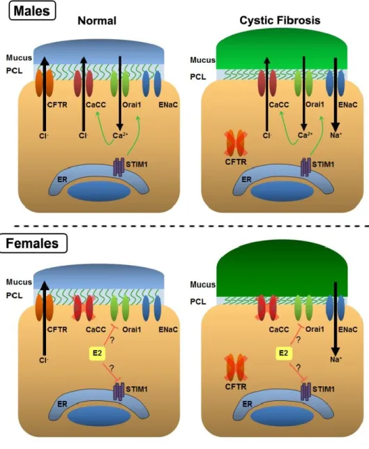
1.15 Estrogen Inhibits CaCC
A previous publication from the Tarran lab determined that acute exposure to estrogen can inhibit CaCC in the airways [167]. Nasal potential differences were performed to measure CaCC activity in vivo. Elevated levels of circulating estrogen corresponded with a decrease in CaCC activity, while CFTR and ENaC were unaffected. While CF airways lack CFTR, they still retain the ability to partially regulate ASL height through CaCC. Normal tidal breathing causes ATP to be released from the epithelia that induces the SOCE pathway and ultimately activates CaCC. To test whether estrogen inhibits CaCC by impacting the SOCE pathway, they performed Ca2+ imaging studies. They specifically observed a decrease in the influx of Ca2+, while ER Ca2+ was
unaffected. Furthermore, Ussing chamber experiments confirmed that estrogen was not directly impacting CaCC. Exposing HBECs to ionomycin, which bypasses the SOCE pathway to directly increase [Ca2+]
i, had no effect on CaCC activity, while the UTP response was significantly inhibited. Also, only cells expressing ESR1 were affected, while those expressing ESR2 showed no decreases in Ca2+ influx. Physiologically, this corresponds with a decrease in ASL height. Therefore, in female patients with CF, losing CaCC mediated Cl- secretion during peak estrogen levels may have detrimental consequences.
23
activity is decreased. Nevertheless, CFTR is still present and ASL hydration is not affected. In patients with CF, who lack CFTR, CaCC represents the only Cl- channel that can regulate the ASL. While the ASL in CF males is ultimately dehydrated due to Na+ hyperabsorption, the presence of CaCC may help alleviate or delay some of the symptoms associated with CF. However, in CF females, estrogen-mediated inhibition of CaCC may leave them at a distinct disadvantage. This may help explain why CF
females suffer more severely and have a shorter life span than CF males.
1.16 Goals and Hypotheses tested by the studies presented in this
dissertation
The aim of the studies presented in this dissertation was to contribute to the understanding of the physical and functional aspects of the CaCC subunit Ano1. It was not until recently that Ano1 was identified as an essential subunit of CaCC [105-107], so very little is known about the structure and function of the channel. In order to fully understand how Ano1 functions it is first necessary to know whether Ano1 oligomerizes. Therefore, in Chapter 2, we sought to test the hypothesis that Ano1 exists as an
oligomer. We utilized a combination of biochemical and microscopy based assays to examine the quaternary structure of Ano1.
24
impact their function. We propose three distinct mechanisms that could be utilized by estrogen to disrupt SOCE. Acute exposure to estrogen could (i) inhibit STIM1
25
26
Figure 1.2. The Store-Operated Ca2+ Entry Pathway. A, Under resting conditions, cytoplasmic Ca2+ levels are low and ER Ca2+ stores are full. Thus, STIM1 is inactive and extracellular Ca2+ is not transported into the cell through Orai1. B, When a cell is
27
Figure 1.3. Functional domains of STIM1. STIM1 is 685 amino acids in length and contains several known domains. The N-terminus contains the EF hand (aa 67-96) to sense ER Ca2+ levels. The SAM domain (aa 132-200) is important for oligomerization after ER Ca2+. Following a single transmembrane (TM) domain (aa 215-234) is the ERM domain (aa 251-535), which contains two coils (aa 251-389). The second coiled-coil is encompassed by the SOAR domain (aa 344-442). SOAR mediates interaction of STIM1 with Orai1 to induce Ca2+ influx. The C-terminus of STIM1 contains the S/P rich region (aa 600-629) that contains several known serine phosphorylation sites and the K rich region (aa 672-685) that stabilizes localization of STIM1 near the plasma
28
Figure 1.4. Predicted Topology of Ano1. Ano1 ispredicted to contain 8
29
__________________________
This Research was originally published in the Journal of Biological Chemistry. John T. Sheridan1, Erin N. Worthington1, Kuai Yu, Sherif E. Gabriel, H. Criss Hartzell and Robert Tarran. Characterization of the oligomeric structure of the Ca(2+)-activated Cl- channel Ano1/TMEM16A. J Biol Chem. 2011 Jan 14;286(2):1381-8. © the American Society for Biochemistry and Molecular Biology.
1These authors contributed equally to this work. 31
Chapter II
Characterization of the Oligomeric Structure of the Ca(2+)-Activated Cl-
Channel Ano1/TMEM16A
2.1 Overview
31
oligomeric nature of Ano1 is an important step in the development of therapeutic drugs that could be useful in the treatment of cystic fibrosis.
2.2 Introduction
Ca2+-activated Cl- channels (CaCCs) play essential roles in many physiological processes including epithelial secretion [94], olfactory transduction [110, 169, 170], photoreceptor adaptation [171], regulation of smooth muscle contraction [172], neuronal and cardiac action potential waveform and firing frequency [173], and nociception [174]. Because of their physiological significance, CaCCs have attracted attention for decades [175, 176]. However, their molecular composition has remained elusive until recently when three laboratories independently identified members of the Anoctamin 1 (Ano1) family, also known as the Transmembrane Protein 16 (TMEM16) family, as essential subunits of CaCCs [105-107]. Ano1/TMEM16A is highly expressed in secretory
epithelial tissues including ductal glands, superficial epithelia of the airway, and oviduct, where it has been implicated in calcium-dependent chloride secretion [105, 107, 177-179]. Ano1 has attracted particular attention in the airways, because it has been suggested that activating this channel might be therapeutically beneficial for treating cystic fibrosis (CF) patients [178]. The cystic fibrosis conductance regulator (CFTR) chloride channel is absent or defective in CF epithelia, leaving Ano1 as a potential alternative pathway to effect transepithelial Cl- secretion.
32
photoreceptor synapses [110, 171, 182]. With the possible exception of Ano8 and Ano10, anoctamins are also predicted to have a re-entrant loop between
transmembrane domains 5 and 6 that may contribute to Cl- selectivity [183, 184]. A common property of ion channels is their oligomeric nature, which may be homo- or hetero-oligomeric [185]. For example, activation of the MthK/Ca2+-activated K+ channel from Methanobacterium thermoautrophicum, involves oligomerization of 4 subunits [186] and it has been suggested that the mammalian Maxi-K K+ channel is activated by a similar mechanism [187]. Activation of CFTR Cl- channels involves dimerization of nucleotide binding domains [188], although whether CFTR enters the plasma membrane as a monomer or a dimer remains controversial [189, 190]. A first step in understanding how Ano1 is gated and regulated requires knowing whether the channel exists as a monomer or an oligomer and, if it oligomerizes, the number of interacting subunits. Since little is known about how Ano1 forms a Cl- conducting channel in the apical membrane, we have used biochemical techniques and Förster resonance energy transfer (FRET)-based approaches to investigate its subunit assembly.
2.3 Materials and Methods
Cell Culture. Human excess donor lungs and excised recipient lungs were obtained at the time of lung transplantation from portions of main stem or lumbar bronchi and cells were harvested by enzymatic digestion as previously described under a
33
supplemented with 10% fetal bovine serum and 1X penicillin/streptomycin solution. HEK293 cells were typically used 2-3 days after seeding on 30 mm glass coverslips. Cultures that were ~75% confluent were transfected for 4-6 hours using Lipofectamine 2000 (Invitrogen) as per the manufacturer’s instructions and allowed to incubate in 5% CO2 at 37°C overnight before use.
Ano1 Constructs and RT-PCR. Mouse Ano1 C-terminally fused with enhanced GFP (Ano1-eGFP) was kindly provided by Dr. Uhtaek Oh (Seoul National University, Korea; variant containing a and c alternative splice sequences, UniProtKB accession number Q8BHY3.2). eGFP is in the pEGFP vector (Clontech). The Ano1-mCherry construct was made by replacing eGFP with monomeric Cherry (Ano1-mCherry, Clontech). In both constructs, the fluorescent tag was separated from the C terminus of Ano1 by a 17 amino acid linker RILQSTVPRARDPPVAT to ensure that the fluorescent protein did not affect Ano1 function as previously described [191]. HA-tagged P2Y2 receptor construct was donated by Kendall Harden (Department of Pharmacology, UNC, Chapel Hill, North Carolina, USA)[192]. Endogenous expression of all Ano family members (Ano1-10) was determined by RT-PCR using primers for Ano1 as listed in Table 1.
34
Acceptor-photobleaching FRET. FRET was performed using a Leica SP5 confocal microscope with a 63X glycerol immersion objective. The donor (eGFP) was excited at 488 nm and emission collected from 495 nm to 549 nm and the acceptor (mCherry) was excited at 561 nm and emission collected from 580 nm to 654 nm. FRET was measured by the acceptor photobleaching method and analyzed using ImageJ (NIH Freeware). The FRET efficiency (%E) was calculated as: ((donorpostbleach
-donorprebleach)/donorpostbleach)*100. Measurements of Ca2+
i. All cultures were loaded with Fura-2-AM (5 µM at 37ºC for 40 min) and imaged with a 40 x 1.2 NA H2O objective on a Nikon Ti-S microscope. Ca2+
i measurements were obtained using an Orca camera (Hamamatsu) and Fura-2 fluorescence was acquired alternately at 340 and 380 nm (emission >450 nm) using Ludl filter wheels with Compix sPCI software. At each excitation wavelength (340 or 380 nm), background light levels were measured by exposing cells to digitonin (15 μM) and MnCl2 (10 mM) and subtracting the emission from the corresponding signal measured in Fura-2–loaded cells before taking the ratio (340/380) [15].
Imaging of the actin cytoskeleton. For labeling of actin, HEK293 cells grown on glass coverslips. Cells were incubated with 1 µM cytochalasin D or vehicle (0.1%
DMSO) for 30 min and were then fixed in 4% PFA for 10 min at room temperature. After washing 3 x in PBS, cells were permeabilized by a 10 min exposure to 1% Triton X. After a 3 x wash, cells were incubated with Alexa488 phalloidin (Invitrogen) for 30 min,
followed by deep red HCS cell mask (Invitrogen) for 30 min as per the manufacturer’s instructions. Fixed cells were then imaged immediately using the Leica SP5 confocal microscope.
35
buffer (0.09% NP-40, 50 mM Tris-HCl, pH 7.4, 10 mM NaMoO4, 150 mM NaCl)
supplemented with 1X complete EDTA-free protease inhibitors (Roche). Lysates were then centrifuged at 16,000 x g for 10 min at 4°C and the supernatants were collected. Protein concentrations were determined by the BCA protein assay kit (Pierce) and equal amounts of protein (800 µg) were diluted to 1.6 µg µl-1 with NP-40 lysis buffer in spin columns (Pierce) and mixed with anti-mCherry monoclonal antibody (Clontech) and rotated at 4°C for 2 hours. Protein A/G agarose beads (30 µl, Pierce) were added to lysate and rotated at 4°C for 4 hours. Beads were washed three times with NP-40 lysis buffer by centrifugation at 1500 x g for 2 minutes at 4°C and protein was eluted by boiling in 2X concentrated sample buffer (1% SDS, 5% glycerol, 25 mM Tris-HCl, pH 6.8, 0.01% bromophenol blue, 0.5% 2-mercaptoethanol). Protein was resolved on a 3-8% gradient NuPAGE Tris-acetate gels (Invitrogen) and transferred to nitrocellulose using iBlot (Invitrogen). Immunoblots were incubated overnight at 4°C with the either anti-mCherry monoclonal antibody (Clontech, 1:1000) or rabbit polyclonal anti-eGFP antibody (Abcam, 1:1000) in 2% fish gelatin blocking buffer. Proteins were detected using SuperSignal West Pico Chemiluminescent Substrate kit (Pierce) or using Goat anit-Rabbit IRDye 800CW antibody (1:10,000) in 2% fish gelatin blocking buffer and analyzed with the Odyssey IR imaging system (LI-COR Biosciences).
36
incubated at 70°C for 10 min. Protein was resolved on a 3-8% NuPAGE Tris-acetate gels and analyzed by western-blotting.
NativePAGE. Cells were sonicated in 600 l NativePAGE 4X sample buffer containing 2% n-dodecyl β-D-maltoside (DDM) (Invitrogen). Lysate was centrifuged at 16,000 x g for 10 min at 4°C. Supernatant was collected and 10 g of protein was treated with varying concentrations of SDS and treated with 0.5% G-250 (Invitrogen). Protein was resolved on NativePAGE Novex 4-16% Bis-Tris gel and detected by western-blotting.
Electromobility Shift Assay. Whole cell HEK293 lysate expressing Ano1-eGFP and Ano1-mCherry was obtained for electromobility shift assay (EMSA) analysis. Whole cell lysate containing 7.5 µg of protein was prebound with anti-mCherry at 4 °C for 1 hour. Prebound lysate was then subjected to non-denaturing PAGE. Protein shift was resolved by western-blotting with anti-GFP.
Statistical Methods. All data are presented as the mean ± SE for n number of cells. Each transfection was repeated on at least three separate days. Differences between means were tested for statistical significance using paired or unpaired t tests or their non-parametric equivalent as appropriate to the experiment. From such
comparisons, differences yielding p ≤ 0.05 were judged to be significant.
2.4 Results
37
were endogenously expressed, suggesting that these additional Ano family members are not capable of forming a CaCC in these cells.
To probe the quaternary structure of Ano1, we fused eGFP and mCherry to the C-termini of Ano1. We then verified that these FRET constructs could still be activated by intracellular Ca2+ to conduct Cl-. With nominally zero intracellular Ca2+, both tagged constructs and the untagged wild type Ano1 failed to display significant currents (< 0.05 nA; Fig 2.1E). However, in the presence of 1 μM free intracellular Ca2+, whole cell currents were typically >5nA for all Ano1s (Fig. 2.1B-E). Importantly, the currents from both constructs exhibited outward rectification and time dependence that was typical of CaCC currents at this Ca2+ concentration and was identical to untagged Ano1 (Fig. 2.1B-E) [104]. These results demonstrate that attaching eGFP or mCherry had no obvious effect on Ano1 function.
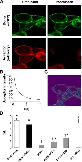
Ano1-eGFP and Ano1-mCherry undergo FRET in the plasma membrane of
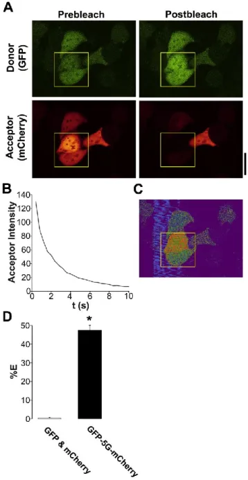
HEK293 cells and airway epithelia. We first used FRET to search for Ano1-Ano1 interactions since this method allows for live-cell quantification. As a positive FRET control, we directly linked eGFP to mCherry by five glycines (in the absence of Ano1) and measured the % FRET with this construct in HEK293 cells using the acceptor photobleaching approach [195]. Typical acceptor photobleaching of this FRET pair is shown in Fig. 2.2A, B. We were able to measure 48 ±1% FRET with this construct (Fig. 2.2C, D), which is close to the maximal FRET that is measurable between eGFP and mCherry (50%) [196], suggesting that we can accurately measure FRET with our system. In contrast, unlinked eGFP and mCherry did not undergo FRET when co-expressed in HEK cells (Fig. 2.2D).
38
close enough to interact. To determine whether Ano1 channels associate before they reach the plasma membrane, we measured Ano1 FRET ~15 h post transfection when the majority of Ano1 is located intracellularly. Intracellular Ano1-eGFP - Ano1-mCherry FRET was also ~8% (Fig. 2.3D), suggesting that Ano1 oligomers assemble before reaching the plasma membrane.
We failed to detect any FRET between eGFP alone and Ano1-mCherry,
suggesting that the Ano1 FRET was due to the specific assembly of Ano1 subunits. To test for the possibility that the plasma membrane of a HEK293 may become crowded following transient transfections, forcing an intimacy that would not normally occur, we co-expressed Ano1-mCherry and eGFP-tagged A2B adenosine receptor (A2BR-eGFP), a G-protein coupled receptor that resides in the plasma membrane but has not
previously been shown to interact with Ano1. In this case, there was a small, but significant, amount of FRET (2%), which was significantly less than the 8% FRET seen between Ano1 subunits (Fig. 2.3D). Co-expressed Ano1-mCherry and eGFP-tagged CFTR also returned moderate FRET (~3.5%) that was also significantly less than the 8% FRET seen between Ano1 subunits (Fig. 2.3D). Together, these low levels of FRET are perhaps indicative of the restricted 2-dimensional diffusion that limits distances between two proteins that do not physically interact in the plasma membrane.
To demonstrate that Ano1-Ano1 FRET was not influenced by the expression system used, we co-expressed the Ano1 constructs in single primary human bronchial epithelial cells. We again observed close to 8% FRET in these cells, suggesting that the Ano1 interaction is an intrinsic property of Ano1, rather than an intimacy forced by the HEK293 expression system (Fig. 2.3D).
Ano1 FRET is due to a protein-protein interaction. To confirm that FRET between Ano1-eGFP and Ano1-mCherry was due to a physical interaction, we
39
against eGFP and mCherry (Fig. 2.4A). As a negative control, we immunoprecipitated Ano1-mCherry from HEK293 cells co-expressing A2BR-eGFP or eGFP and probed with an eGFP antibody (Fig. 2.4A). No bands were detected after blotting for eGFP in control lanes, indicating that these proteins do not interact with Ano1. Western blotting
confirmed that both antibodies are specific for eGFP/mCherry respectively and neither detected the other fluorescent protein (Fig. 2.4B).
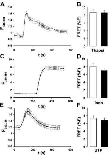
Ano1 FRET is not altered by changes in intracellular Ca2+ or actin cytoskeleton disruption. We next tested whether changes in intracellular Ca2+ could alter Ano1 FRET. Intracellular Ca2+ was raised with 2 µM thapsigargin [21], 100 µM UTP [15], or 10 µM ionomycin [197]. Cells treated with UTP were also transfected with an HA-tagged P2Y2 receptor construct (P2Y2R-HA) to ensure a consistent UTP response. HEK293 cells displayed a robust increase in the fura-2 emission ratio after all three treatments (Fig. 2.5A, C, E). However, these maneuvers failed to affect Ano1 FRET (Fig. 2.5B, D, F), suggesting that Ano1-Ano1 interactions are not influenced by changes in intracellular Ca2+.
Epithelial ion channels have been shown to be linked to the cytoskeletal
infrastructure by postsynaptic density 95 discs large zonula occludens-1 (PDZ) binding motifs and these interactions can be ablated by disrupting F-actin polymerization with cytochalasin D [198]. However, a 30 min treatment with 1 µM cytochalasin D, which extensively disassembled the cytoskeleton, had no effect on Ano1 localization to the plasma membrane or Ano1-Ano1 FRET, suggesting that oligomerization of Ano1 is not contingent on binding to the actin cytoskeleton (Fig. 2.6).
Biochemical evidence that Ano1 is a dimer. In order to investigate the
40
monomer. However, after treating whole cell lysate with the cross-linker 1,5-difluoro-2,4-dinotrobenzene (DFDNB), an additional band was observed which corresponded to an Ano1-eGFP dimer (~260 kDa). Two faint higher-order bands were also visible.
However, the molecular weight of these bands was harder to ascertain and they may represent non-specific crosslinking of Ano1 to other membrane proteins.
We next performed non-denaturing PAGE on lysate obtained from HEK293 cells to validate the cross-linking experiments (Fig. 2.7B). Cells were lysed by 2% DDM and sonication in the absence of SDS. The majority of Ano1 appeared as a dimer with no presence of higher order bands. Inclusion of SDS induced the emergence of a ~117 kDa band indicating that Ano1 had been fully denatured and that we were observing the monomeric protein. This band was slightly smaller than the predicted size of Ano1-eGFP (~130 kDa), but is consistent with an apparent reduction in protein size seen under non-reducing conditions [199]. To ensure that we were observing Ano1-eGFP dimers and not Ano1-eGFP in association with an unidentified endogenous protein, we performed an electromobility shift assay (EMSA) by exposing lysate from HEK293 cells expressing Ano1-eGFP and Ano1-mCherry to an mCherry antibody before
41
2.5 Discussion
We examined the stoichiometry of Ano1 using biophysical and biochemical approaches. We used HEK293 cells since they do not endogenously express Ano1 (Fig. 2.1A) and do not have a spontaneous CaCC, even with 1 µM Ca2+ in the patch pipette (Fig. 2.1E). For our studies, we C-terminally labeled murine Ano1 with either eGFP or mCherry. To ensure that these fluorescent proteins did not interfere with Ano1 function, we included a 17 amino acid linker between Ano1 and eGFP or mCherry. Importantly, these fluorescent proteins had no effect on Ano1-mediated Cl- currents, suggesting that their use in this endeavor was valid (Fig. 2.1).
We chose eGFP/mCherry over the more commonly used CFP/YFP FRET pair since CFP photobleaches easily, which reduces its ability to accept photons during FRET, while eGFP is significantly more photostable [200]. This approach was confirmed using a FRET positive construct where the acceptor was linked to the donor by 5
glycines (Fig. 2.2). The theoretical maximum FRET that can occur between two
fluorescent proteins (including CFP, eGFP, mCherry and YFP) is 50%, which is limited by the size of the fluorescent proteins themselves [196]. Using CFP linked to YFP, we measured 38% ± 3 FRET (n=6). In contrast, we measured 48% ± 1 FRET (n=14) with eGFP linked to mCherry, confirming previous reports that the latter FRET pair is significantly more suited for confocal microscopy (Fig. 2.2) [200].
42
ENaC is a hetero-multimer that returns between 5-15% FRET between different ENaC subunits [201]. ENaC is also known to interact with CFTR [202] and FRET was 7% between CFTR and all three ENaC subunits [202]. A study by Kerschensteiner et al. used acceptor-photobleaching FRET to determine the stoichiometry of voltage-gated K+ (Kv) channels that are known to be heteromeric oligomers [203]. They obtained ~25% FRET efficiency when Kv2.1-CFP and Kv9.3-YFP were expressed compared to a ~10% FRET efficiency when Kv2.1-YFP and Kv9.3-CFP were expressed. Thus, the 8% FRET that we record between Ano1 subunits is comparable to both FRET seen both between ion channel subunits and between distinct ion channels. Perhaps, surprisingly, while CFTR has previously been shown to negatively regulate CaCC [204, 205], we detected only minimal FRET between CFTR and Ano1 (Fig. 2.3), suggesting that regulation of Ano1 by CFTR is not due to a direct interaction. Since we found Ano1-Ano1 FRET to be 8% when measured intracellularly (Fig. 2.3), it is likely that Ano1 subunits come together en route to the plasma membrane. It should be noted that we did not differentiate between intracellular compartments during our analysis and subunits may oligomerize as early as the ER or as late as endosomes.
43
intracellular Ca2+ levels (Fig. 2.5), suggesting that Ano1 oligomerization is fixed and independent of Ano1’s gating ability, which is Ca2+-dependent [105-107].
Chemical-cross linking and non-denaturing PAGE assays indicated that Ano1 is a dimer (Fig. 2.7). A distinct dimer band was observed following chemical-cross linking (Fig. 2.7A), which was not observed under control conditions. Additional higher order complexes were also observed after cross linking. However, these may be proteins that are non-specifically cross linked to Ano1-eGFP. Following non-denaturing PAGE a strong dimer band was also observed (Fig. 2.7B). This dimerization was only disrupted with 3% SDS (Fig. 2.7B). Higher order oligomers were not detected by non-denaturing PAGE, suggesting those observed following cross linking are not true oligomers. We next performed EMSA analysis to determine if the dimer band consists of two Ano1 proteins or Ano1 with an unidentified endogenous protein (Fig. 2.7C). This approach revealed a shift of Ano1-eGFP to a higher molecular weight when prebound with an anti-mCherry antibody, confirming that eGFP was present as a dimer with Ano1-mCherry and not with any other protein. Since only Ano1 is present under
non-denaturing conditions, it is likely that the co-immunoprecipitation is indicative of a direct interaction between Ano1 subunits (Fig. 2.4). A portion of Ano1-eGFP did not shift to a higher molecular weight. This is likely due to the fact that HEK293 cells transfected with Ano1-eGFP and Ano1-mCherry should yield three distinct populations of Ano1 dimers, consisting of eGFP/eGFP, eGFP/mCherry, and
Ano1-mCherry/Ano1-mCherry.
44
45
Table 2.1
Primers used in RT-PCR to detect endogenous anoctamin expression in HEK293 cells
Gene Sense Antisense
Ano1 GCGTCCACATCATCAACATC ATCCTCGTGGTAGTCCATCG
Ano2 TGCCTACCACTACCGGAAAC ACTTCTTTGCAATGCTGCCT
Ano3 AAACCTGAACCACATCAGCC TCTTCCCAAAAAGAAAGCGA
Ano4 CATGGGAAGTCCTTGGAAGA GCCATTGGTAAGCAAACGAT
Ano5 ACACTTCACCAGAATTGGGC GAAGCTGCTGCTGTTCCTCT
Ano6 CAGTTTGGGTTCGTCACCTT AGTACGGGTTTCCCTTGCTT
Ano7 CTACTCCTGCCGGTTCAGAG GTTCCTGCGTGGGTATGTCT
Ano8 ACTTCGCTCTGCTCCTCAAG CTTCATGACGTTGTTGGGTG
Ano9 TGGAGATCAGCACCTGTGAG CGAAGTTCACGATTCGGATT
47
Figure 2.1. Ca2+-activated Cl- Currents induced by fluorescent protein-tagged
mouse Ano1 constructs are identical to wild type Ano1. HEK293 cells were
transiently transfected and subjected 1 day later to whole cell patch clamp recording. A, total RNA was collected from untransfected HEK293 cells and RT-PCR was performed to determine which Ano proteins are endogenously expressed. Ano5, 6, and 10 were strongly expressed while Ano2 and 4 were mildly expressed. Ano1, 3, 7, 8, and 9 were not detectable. B, Wild type mouse Ano1 (variant containing a and c alternative splice sequences, accession Q8BHY3.2). C, Ano1 tagged with eGFP on the C-terminus. D, Ano1 tagged with mCherry on the C-terminus. E, Steady-state current-voltage
48
49
Figure 2.3. Ano1-eGFP and Ano1-mCherry undergo specific FRET in the plasma membrane. A, Typical confocal micrographs of eGFP (green, donor) and Ano1-mCherry (red, acceptor) before and after photobleaching of Ano1-Ano1-mCherry. B, Graph showing typical acceptor photobleaching over 10 s. C, Resultant FRET image calculated from A. D, Mean FRET efficiency (%E). Open bars, plasma membrane FRET in
HEK293 cells (n=56) and human bronchial epithelial cells (n=13). Closed bar,
50
Figure 2.4. Ano1-eGFP and Ano1-mCherry co-immunoprecipitate. A, HEK293 cells were transfected with lipofectamine alone, or with Ano1-mCherry and either Ano1-eGFP, A2B-R-eGFP, eGFP. Input represents 5% of total protein used in
51
Figure 2.5. Ano1-eGFP and Ano1-mCherry FRET is not affected by changes in intracellular Ca2+. A, typical fura-2 emission ratio observed following exposure to 2 µM thapsigargin (n=10). B, mean FRET (%E) from Ano1 in the plasma membrane of HEK293 cells after vehicle (open bars; n=25) and following 5 min exposure to 2 µM thapsigargin (closed bars; n=27). C, typical fura-2 emission ratio observed following exposure to 1 µM ionomycin (n=10). D, Mean FRET (%E) from Ano1 in the plasma membrane of HEK293 cells after vehicle (open bars; n=20) and following 5 min exposure to 1 µM ionomycin (closed bars; n=21). E, typical fura-2 emission ratio observed
52
Figure 2.6. Ano1-eGFP and Ano1-mCherry FRET is not dependent on an intact actin cytoskeleton. Left, Confocal micrographs of Ano1-mCherry in HEK293 cells (red) before and after disruption of actin cytoskeleton (phalloidin/green) with 1 µM
53