THE ROLE OF NMDA RECEPTORS IN THE DORSAL HIPPOCAMPUS IN COCAINE-PAIRED CONTEXTUAL MEMORY RECONSOLIDATION
Kati L. Healey
A thesis submitted to the faculty of the University of North Carolina at Chapel Hill in partial fulfillment of the requirements for the degree of Master of Arts in the Department
of Psychology (Behavioral Neuroscience)
Chapel Hill 2014
Approved by:
Kathryn Reissner
Rita A. Fuchs-Lokensgard Todd Thiele
iii ABSTRACT
KATI L. HEALEY: The Role of NDMA Receptors in the Dorsal Hippocampus in Cocaine-Paired Contextual Memory Reconsolidation
(Under the direction of Kathryn Reissner and Rita A. Fuchs-Lokensgard)
v
ACKNOWLEDGEMENTS
TABLE OF CONTENTS
LIST OF TABLES………...……….………...…viii
LIST OF FIGURES………...…………...….…...……...ix
LIST OF ABREVIATIONS………..…..x
CHAPTER 1: INTRODUCTION……….…………..….………….…...1
Context-Induced Reinstatement of Drug Seeking………...2
The Neural Circuitry of Drug-Memory Reconoslidation………3
N-methyl-D-aspartate Receptors and Memory Processes………..4
Hypotheses and Predictions………5
CHAPTER 2: METHODS………..………...………….…...…...7
Subjects……….…….…..….………..…...…..7
Procedures……….…….…..….………...……7
Experiment 1a..………..…..……….……….…10
. Experiment 1b..………....………...……....…11
Experiment 1c..………....………..……….…...……...….12
Experiment 2 …...………..………12
Data Analysis……….…...………..……...…...12
CHAPTER 3: RESULTS………..………..…….…………...…....……...…14
Histological Analysis………..…….………...………...…....14
vii
Experiment 1c..…...……….………..……...…….17
Experiment 2……….…….…………18
CHAPTER 4: DISCUSSION……….………..……..……...….19
LIST OF TABLES
Table 1 - Descriptive statistics for instrumental behavior
during self-administration training, extinction
ix
LIST OF FIGURES
Figure 1 - Schematics depicting cannula placement………..……25 Figure 2 - NMDA receptor antagonism following memory
retrieval of a context-drug associative memory alters
subsequent cocaine seeking behaviors...…….………...………26 Figure 3 - NMDA receptor antagonism in the DH is dependent on the
memory retrieval of the cocaine-context associated memory…………...28 Figure 4 - NMDA receptor antagonism in the SSTR directly after
cocaine-context associated memory retrieval did not alter
subsequent cocaine seeking behaviors.…….………....30 Figure 5 - NMDA receptor blockade in the dorsal hippocampus does
LIST OF ABBREVIATIONS AND SYMBOLS
ANI Anisomycin
ANOVA Analysis of variance
APV (2R)-amino-5-phoshonovaleric acid BLA Basolateral amygdala
DH Dorsal hippocampus
IKK IκB kinase
NMDA N-methyl-d-aspartate
SSTR Somatosensory trunk region VEH Vehicle
* Significant difference relative to extinction context # Significant difference relative to all other time intervals
CHAPTER 1: INTRODUCTION
Context-induced reinstatement of drug seeking
Drug addiction is a disorder that is chronic and characterized by high rates of relapse. Exposure to environmental stimuli that have previously been paired with a drug of abuse can lead to drug craving (Foltin & Haney, 2000) and relapse (Everitt, Dickinson, & Robbins, 2001), indicating the importance of contextual drug cues. Because substance abuse disorders are a growing problem, it is important to investigate new treatment options that disrupt the cue-drug associations that lead to craving and subsequent relapse. It is theorized that, when a memory is retrieved, it becomes labile and must be re-stabilized to return to long-term memory. This process is referred to as memory reconsolidation (Nader, Schafe, & LeDoux, 2000a; Tronson & Taylor, 2007). If an associative memory is not reconsolidated properly, the associations can weaken. Recent research indicates that reactivated memories undergo processes that result in their reconsolidation into long-term memory that are distinctly different from those involved in initial memory consolidation (Lee & Hynds, 2012; Tronson & Taylor, 2007). Thus, understanding the unique mechanisms of drug memory reconsolidation could lead to methods for disrupting maladaptive associations between contextual cues and drugs of abuse (Milton & Everitt, 2010), and this could be a powerful treatment tool for addiction.
context (A) to be trained to self-administer drug rewards over multiple sessions. After context-response-drug associations are established, the animals are placed in another, distinctly different context (B) where drug is not available and the instrumental behavior learned in context A extinguishes. To assess whether the drug-associated environment acquired motivational effects, the animals are returned to context A where context-induced goal-seeking behavior is assessed in the absence of drug reinforcement during a reinstatement test. Our modified version of this ABA model to study drug memory reconsolidation includes a brief re-exposure to the drug-paired context prior to the test of reinstatement. This brief session is designed to trigger the retrieval and reconsolidation of context-drug memories and creates an opportunity for experimental manipulation of memory reconsolidation.
3 The neural circuitry of drug-memory reconsolidation
N-methyl-d-aspartate receptors and memory processes
N-methyl-d-aspartate (NMDA) receptors play a central role in learning and memory processes in the brain (Collingridge, 1987). The NMDA receptor is a ligand- and voltage- gated ion channel (Johnson & Ascher, 1987;Mayer, Westbrook & Guthrie, 1984) composed of four subunits; two GluN1 subunits and one or two GluN2 subunits GluN2A-D (Furukawa et al., 2005). The DH is very rich in NMDA receptors (Olverman, Jones and Wakins, 1984), specifically those that contain the GluN2 subunits GluN2A or GluN2B (Monyer et al., 1994). A number of studies have implicated NMDA receptors in Pavlovian memory reconsolidation in various brain regions (Alaghband et al., 2014; Liddie & Itzhak, 2014; Chia & Otto, 2013; Alaghband et al., 2013; Wu et al., 2011) and in instrumental memory reconsolidation, such as food-reward memory (Garcia-Delatorre et al., 2014; Lee and Everitt, 2008), odor-reward memory (Rorras-Garcia et al., 2005), and drug-reward memory (Tedesco et al., 2014; Milton et al., 2008; Sadler et al., 2007).
5
the role of NMDA receptors in appetitive instrumental memory reconsolidation in the DH has not been assessed.
Further support for the role of NMDA receptors in memory reconsolidation comes from studies that provide evidence that downstream elements of NDMA receptor-mediated signaling pathways are involved in context-drug memory reconsolidation in the DH. Specifically, recent studies implicated the role of the IκB kinase (IKK) pathway in aversive memory reconsolidation (Lee & Hynd, 2013; Boccia et al., 2007). Consistent with this, IKK inhibition in the DH disrupted a fear memory reconsolidation (Lee & Hynd, 2013) and IKK or nuclear factor κB (a downstream effector of IKK) inhibition disrupted the reconsolidation of an inhibitory avoidance task (Boccia et al., 2007). Furthermore, systemic NMDA receptor antagonism, systemic NMDA receptor subunit GluN2B inhibition, and systemic inhibition of downstream signaling pathways of the NMDA receptor neuronal nitric oxide synthase (nNOS) and mitogen-activating extracellular kinase attenuated cocaine-associated CPP memory (Liddie and Itzhak, 2014). These findings point to the possibility that DH NMDA receptors and their downstream targets may be involved in appetitive instrumental memory reconsolidation as well.
Hypotheses and predictions
CHAPTER 2: METHODS AND MATERIALS Subjects
Male Sprague-Dawley rats (Harlan: 200-250 grams) were single-housed in a vivarium under reversed 12-hr light/dark cycle (lights off at 7pm). Rats were habituated to the
vivarium for 5 days. Rats were fed ad libitum, kept under temperature controlled conditions (68-70°), and had unlimited access to water throughout the experiment. After the habituation period, rats were placed on a diet of 20g of rat chow per day for the remainder of the study. The housing and treatment of animals followed the “Guide for the Care and Use of
Laboratory Rats” (Institute of Laboratory Animal Resources on Life Sciences, National Research Council, 1996) and were approved by the University of North Carolina Institutional Animal Care and Use Committee.
Food Training
programmed consequences. Food training sessions were at least 16 hours overnight, until rats met a criterion of 100 presses on the active lever per session.
Surgery
Prior to surgery, rats were anesthetized with ketamine hydrochloride and xylazine (66.6 mg/kg and 1.33 mg/kg, intraperitoneal, i.p, respectively). Chronic indwelling catheters were made in house and implanted into the right jugular vein for the administration of
intravenous (IV) cocaine, as described previously (Fuchs et al., 2006). The catheter ran subcutaneously and exited the back between the shoulder blades where it was capped by sealed Tygon tubing and a protective screw cap (PlasticsOne). After catheter surgery, the rats were placed into a stereotaxic instrument (Stoelting, Wood Dale, IL) and 26-gauge stainless steel guide cannulae (Plastics One) were aimed bilaterally at the DH (angled by 15° laterally; -3.4 mm AP, ±3.1 mm ML, -2.15 mm DV, relative to bregma) or the anatomical control region, the trunk region of the somatosensory cortex (SSTR; angles by 15° laterally; -3.4 mm AP, ±3.1 mm ML, -0.8 mm DV, relative to bregma). Guide cannulae were sealed with screw caps (PlasticsOne) to prevent occlusion and infection.
9
heparinized saline (70 U/mL). Tests of catheter patency was performed periodically with propofol (1 mg/0.1 mL, i.v.), a short acting barbiturate that causes a rapid loss of muscle tone when administered intravenously.
Cocaine self-administration
Cocaine self-administration training was conducted in standard operant-conditioning chambers (Med Associates Inc.) set up to form two distinct contexts. Context A contained a continuous red house light on the wall across from the levers, intermittent pure tone (80 dB, 1kHZ; 2s on, 2s off), pine scent, a wire mesh floor (26 cm x 27 cm), and a stimulus light above the inactive lever (5s on). Context B contained an intermittent white stimulus light above the inactive lever (2s on, 4s off), a continuous pure tone (75 dB, 2.5 kHz, vanilla scent, a bar floor, a slanted ceramic tile wall (19 cm x 27 cm). These stimuli were not presented during food training.
Extinction training
Rats received seven daily 2-h extinction training sessions in a distinctly different context than the self-administration training context. During the extinction sessions, active and inactive lever presses were recorded but did not have any programmed consequences. After the fourth extinction session, rats were adapted to the intracranial microinfusion procedure. To do so, stainless steel injection cannulae were inserted into the guide cannulae. The injection cannulae extended 1mm below the tip of the guide cannulae. The rats were held by the experimenter for 4 minutes; however, no fluid was infused.
Experiment 1a. Effects of NMDA antagonism in the DH following cocaine memory
reactivation
Experiment 1a examined whether intra-DH administration of the NMDA receptor antagonist, APV, following cocaine memory reactivation would impair context-response-cocaine
memory reconsolidation. On the day after extinction training, animals were placed in the previously cocaine-paired context for a 15 minute memory reactivation session to initiate destabilization and memory reconsolidation (Fuchs et al. 2009). During the session, the levers were extended and responses were recorded; however, lever presses had no programmed consequences.
Intracranial microinfusion and additional extinction training
11
shown to disrupts the expression of drug context-induced reinstatement of cocaine-seeking behavior after infusion into the DH (Xie et al., 2013). Following the infusion, rats received additional daily 2-h extinction training sessions in the extinction context until they reached the extinction criterion (a minimum of two consecutive days with ≤ 25 active lever responses per session).
Test of drug context-induced cocaine-seeking behavior
Twenty-four hours after the extinction criterion was reached, rats received a 1-h test session. During the test session, lever responses were assessed in the previously cocaine-paired context without cocaine reinforcement. Active and inactive levers were recorded but had no programmed consequences. Reinstatement was defined as a significant increase in active lever responding in the cocaine-paired context on the test day relative to responding during the first hour of the last extinction session in the extinction context.
Experiment 1b. Effects of NMDA antagonism in the DH following cocaine memory
reactivation
above both the active and inactive levers, a continuous complex tone (80) d B; alternating between 1, 1.5 and 2.5 kHz at 1 s intervals), citrus-scented air freshener strip (4.5 x 2 cm, Locasmarts LLC., Ormond Beach, FL), and a ceramic tile floor (26 cm x 27 cm).
Experiment 1c. Effects of NMDA antagonism in the SSTR following cocaine memory
reactivation
Experiment 1c investigated whether the effects of NMDAR antagonism observed in Experiment 1a were dependent on the DH. Experimental procedures were identical to those in Experiment 1a except that the guide cannulae were directed at the SSTR. The SSTR is the brain region most likely to receive off target effects of APV, due to possible diffusion of the drug from the DH dorsally, along the cannula shaft.
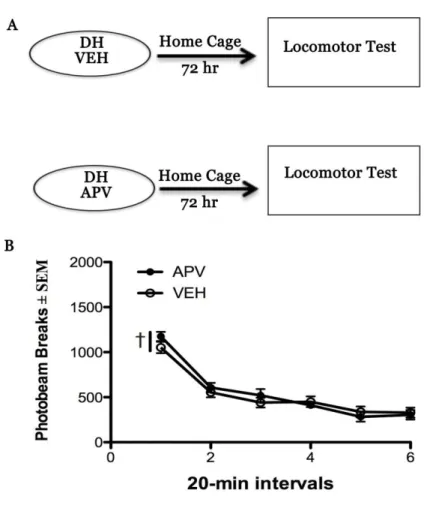
Experiment 2. Effects of NMDA antagonism in the DH on locomotor activity
Intracranial manipulations can have an effect on instrumental cocaine-seeking due to a general effect on motor activity. To assess this, animals were microinjected with either VEH or APV into the DH. Seventy-two hours later (to match the treatment to testing interval in Experiment 1), the rats were placed in a novel Plexiglas chamber (42 x 20 x20 cm high) where locomotor activity was assessed for 2h. Locomotor activity was evaluated by a computerized activity system (San Diego Instruments, San Diego, CA) monitoring
photobeam break, as described previously (Fuchs et al., 2007). Five animals received both APV and VEH treatment in a counter balanced design, with at least 3 days between
13 Data analysis
CHAPTER 3: RESULTS Histology
Schematics illustrating cannula placements are included in Fig. 1. The target brain regions were defined as the dorsal hippocampus (DH) and the somatosensory cortex trunk region (SSTR). Only data from rats with the correct placements were included in the statistical analyses. The resulting Ns per group for vehicle (VEH) and drug-treated (APV) groups were as follows, experiment 1a and 2 – VEH (n = 7), APV (n = 10), experiment 1b – VEH (n = 10), APV (n = 10), and experiment 1c – VEH (n = 4) and APV (n = 3).
Behavioral history
15
NMDAR antagonism in the DH following cocaine memory reactivation blocks subsequent
drug context-induced cocaine-seeking behavior
There was no pre-existing difference between the subsequent APV- and VEH-treated groups in active or inactive lever responding during cocaine self-administration training (day x treatment interaction, F(9,135)=0.58-0.89, p=0.54-0.89; treatment main effect, F(1,15)
=0.15-2.75, p=0.12-0.70; day main effect, F(9,135)=1.58-1.64, p=0.11-0.15). Cocaine intake
gradually increased across time (day main effect, F(9.135)=5.41, p<0.001), but there was no
difference between the groups in cocaine intake (day x treatment interaction or main effect of treatment (F(1-9,15-135)=0.19-1.57, p=0.13-0.89; Fig. 2C). Responding gradually declined when
reinforcement was removed during extinction training (day main effects, F(6,90)=7.46-11.041, p<0.05, Tukey’s test, Day 1> Day 2-7, p<0.05), but there was no pre-existing difference between the groups in active or inactive lever responding during extinction training (treatment by day interaction, F(6,90)=0.183-1.825, p=0.103-0.981; treatment main
effects, F(1,15)= 1.11-4.16, p=0.06-0.31; Fig. 2A-B). Finally, there was no pre-existing
difference between VEH/APV-treated groups in active lever responding during the 15-min cocaine memory reactivation session (t(15)=-0.182, p=0.86; Fig. 2B).
APV administration into the DH following the cocaine-memory reactivation session altered subsequent drug context-dependent cocaine-seeking behavior (ANOVA context x treatment interaction, F(1,15)=16.73, p<0.001; treatment main effect, F(1,15)=4.09, p=0.06;
context main effect, F(1,15)=39.18, p<0.001; Fig. 2D). The group that received VEH injection
into the DH following cocaine memory reactivation showed enhanced active lever
the cocaine-paired context (Tukey’s test, p<0.05), but not in the extinction context.
Furthermore, the APV-treated group failed to show increased active lever responding in the cocaine-paired context relative to the extinction context (Tukey’s test, p>0.05). There was not difference between the groups in inactive lever responding in either context (all treatment and context main effects and interaction, F(1,15)=2.55-3.84, p=0.07-0.13; Fig. 2E).
NMDAR antagonism in the DH in the absence of cocaine memory reactivation fails to
alter subsequent drug context-induced cocaine-seeking behavior
As in Experiment 1a, active and inactive lever responding was stabile during cocaine self-administration training (day main effect, F(19,235)=0.87-1.44, p=0.18-0.56), while active
and inactive lever responding decreased when drug reinforcement was removed during extinction training (active lever main effect of day: F(6,90)=5.64-8.42, p<0.01-0.001, Tukey’s
test, Extinction Day 1> Day2-7, p<0.05; Fig. 3B). Importantly, there was no pre-existing difference between the treatment groups that later received APV or VEH in active lever responding during cocaine self-administration training or extinction training (day x treatment interaction, F(6-9,90-135)=1.00-1.16, p=0.334-1.00; treatment main effect, F(1,15)=0.006-0.39,
p=0.54-0.94). Similarly there was no difference between the groups in cocaine intake (day x treatment interaction and main effect of treatment, F(1-9,15-135)=0.59-0.98, p=0.20-0.46).
Lastly, there was no difference in responding between the groups during the 15-minute reactivation session (t(15)=-1.37, p=0.19).
17
context at test relative to responding in the extinction context (ANOVA context main effect F(1,15)=17.23, p<0.001; Fig. 3D). However, the groups did not differ in active lever
responding in either context (treatment x context interaction, F(1,15)=0.70, p=0.416; treatment
main effect, F(1,15)=1.47, p=0.161). Inactive lever increased upon exposure to the
cocaine-paired context relative to the extinction context ( context main effect, F(1,15)=7.03, p<0.02),
but the groups did not differ in inactive lever responding in either context (treatment x context interaction and treatment main effect, (F(1,15)=0.71-1.47, p=0.24-0.71; Fig. 3E).
NMDAR antagonism in the SSTR following cocaine memory reactivation fails to alter
subsequent drug context-induced cocaine-seeking behavior
As in Experiments 1a and 1b, there was no pre-existing difference between the groups that later received APV or VEH into the SSTR in active or inactive lever responding during cocaine self-administration training (day x treatment, F(9,45) =1.21-1.79, p=0.10-0.31;
treatment main effect F(1,5)=0.20-1.15, p=0.33-0.68; day main effect, F(9,45)=0.98-0.46,
p=0.47-0.90; Fig. 4B). Similarly, there was no difference between the groups in cocaine intake (treatment x day interaction and main effects, F(1-9.5-45)=0.09-1.57, p=0.16-.77; Fig.
4C). Active lever responding declined when drug reinforcement was removed (day main effect, F(6,30)=3.29, p<0.02; Fig. 4B); however, there was no difference between the groups in
active lever responding during extinction training (treatment x day interaction and treatment main effect, F(1-6,5-30)=0.96-2.27, p=0.19-0.47; Fig. 4B). Furthermore, inactive lever
91-6,5-30)=0,19-1.76, p=0.14-0.68, Fig. 4B). There was no difference in responding during the 15
minute reactivation session between VEH/APV-treated animals (t(5)=2.03, p=0.10).
Unlike in Experiment 1a, APV administration into the SSTR following re-exposure to the cocaine-paired context failed to alter subsequent context-dependent cocaine seeking at test (Fig. 4D). Exposure to the cocaine-paired context failed to increase active or inactive lever responding in either group relative to responding in the extinction context (context main effect (F(1,5)=2.04-3.79, p=0.11-0.21; Fig. 4D-E). Furthermore, there was no difference
between the groups in responding on the active or inactive lever in either context (treatment x context interaction, F(1,5)=0.44-5.04, p=0.08-0.54; treatment main effects, F(1,5)=0.02-0.98,
p=0.77-0.89; Fig. 4D-E).
NMDA antagonism did not have a protracted effect on motor activity
General motor activity was assessed 72 hours after infusion with either VEH or APV into the DH of a subset of rats (VEH, n = 9, APV, n = 10). Locomotor activity decreased as the animals habituated to the chamber (time main effect, F(5,85)=80.41, p<0.001, Tukey’s test
Interval 1>Interval 2-6; Fig. 5C). However, there was no difference in locomotor activity between the treatment groups (treatment x time interaction, F(5,85)=1.17, p=0.33; treatment
19
CHAPTER 4: DISCUSSION
Results from this study indicate that NMDA receptor antagonism in the DH following contextual cocaine-memory reactivation attenuates subsequent cocaine-seeking behavior compared to VEH. This is consistent with the established importance of the DH in memory reconsolidation, and expands on the current literature by demonstrating that the NMDA receptor in the DH is an integral component of the signaling mechanisms of memory reconsolidation in a memory reactivation dependent manner.
An important aspect of these findings is that NMDA receptor-dependent
reconsolidation in the DH is memory reactivation-dependent. Specifically, APV infusion immediately after re-exposure to the cocaine-paired context resulted in impaired
reinstatement behavior, consistent with a weakened context-response-cocaine memory trace. In contrast, APV infusion immediately after exposure to a novel, unpaired context failed to alter reinstatement behavior in the cocaine-paired context. This indicates that
In the anatomical control experiment, APV was infused into the SSTR directly after re-exposure to the cocaine-paired context. This manipulation failed to attenuate subsequent cocaine-seeking behavior, suggesting that impairment in cocaine seeking in the group that received APV into the DH following cocaine memory reactivation was not due to APV diffusion dorsally. However, the sample sizes were small in the anatomical control experiment, which limits confidence in these findings. Furthermore, reinstatement
responding was overall very low (i.e., below the extinction criterion) and may have precluded the observation of attenuation in reinstatement due to a floor effect. Weak responding was in part due to a bacterial infection that affected the colony. Overtly sick animals were
eliminated from the study, but the abnormally low reinstatement responding suggests that some of the remaining animals were also sick.
The disruption in operant drug-reward memory reconsolidation shown in this study expands upon previous literature indicating that NMDA receptors are an important
component of signaling pathways responsible for Pavlovian fear and drug-reward memory reconsolidation (Garcia-Delatorre et al., 2014; Liddie & Izthak, 2014; Alaghband et al., 2011; Wu et al., 2011; Milton, 2008). Further, these results establish that NMDA receptor activation within the DH specifically is important for operant drug-reward memory reconsolidation, corroborating results from previous studies on Pavlovian fear and drug-reward memory reconsolidation (Lee & Hynd, 2012).
21
past memory reactivation studies, manipulations were administered prior to the memory reactivation session (Lee & Hynd, 2013; Hellemans, Everitt & Lee, 2006; Lee, Milton & Everitt, 2006). While in those studies it is unclear whether the manipulation affected memory retrieval, destabilization, or reconsolidation, the effects were ascribed to impairment in memory reconsolidation. For example, it has been shown that pharmacological NMDAR blockade in the DH has to be applied prior to (but not after) retrieval in order to disrupt fear memories (Lee & Hynd, 2013). Similarly, NMDAR antagonism in the nucleus accumbens has to be induced before memory retrieval (but not after) in order to block subsequent morphine conditioned place preference (Wu et al., 2012). These results are not consistent with an NMDA antagonist-induced impairment in memory reconsolidation per se. Thus, it appears that the contribution of NMDA receptors to memory reconsolidation depends on memory type and brain region.
Future studies will need to assess the possible contributions of NMDA receptors in the DH to memory retrieval or destabilization. Some evidence has suggested that these contributions are NMDA receptor subunit specific at least in the BLA. Consistent with this, a recent study has shown that selective antagonism of GluN2A- and GluN2B- containing NMDA receptors disrupts memory reconsolidation and memory destabilization, respectively (Milton et al., 2013). Administration of a GluN2A-preferring NMDA antagonist prior to auditory fear cue re-exposure attenuated auditory fear memory 24 hours later without having an acute effect on auditory freezing behavior during the memory reactivation session.
Conversely, intra-BLA injection of GluN2B-preferring antagonist prior to memory
prevented anisomycin-induced impairment in memory reconsolidation. These findings suggested that GluN2B subunit containing NMDA receptors play a role in the destabilization of fear memories in the BLA during after memory retrieval. Similar differences in the contribution of GluN2A- and GluN2B –containing NMDA receptors in the DH may also be present, but this remains an empirical question given that the DH is distinctly different in many respects from the BLA (Ramirez et al., 2009).
The present study leads to questions about downstream mechanisms by which DH NMDA receptors regulate contextual memory reconsolidation. A potential downstream target of NMDA receptors in the hippocampus is the IκB kinase (IKK-NF-κB) pathway, one of two major kinase cascades associated with the NMDA receptor, the other being the MEK-ERK cascade. There is evidence that receptors containing GluN2A-containing NMDARs and GluN2B-containing NMDARs have different downstream signaling mechanisms (Chen et al., 2007) and this leads to the possibility of specific NMDA receptor subtypes having distinct contributions to memory reconsolidation. A recent study by Lee & Hynd (2013) found that the IKK pathway but not the MEK-ERK pathway was involved in the
reconsolidation of a conditioned fear memory in the DH. The contribution of the IKK pathway in the DH extends to other forms of aversive memory reconsolidation, as another study has demonstrated that IKK and nuclear factor-κB inhibition in the DH similarly disrupted the reconsolidation of inhibitory avoidance memory (Boccia et al., 2007).
However, it remains to be investigated whether appetitive memory reconsolidation in the DH is mediated by the IKK pathway.
23
studies have also implicated ERK in memory reconsolidation of both Pavlovian CPP memory (Liddie & Itzhak, 2014; Valjent et al., 2006) and hippocampal recognition memory (Kelly, Laroceh & Davis, 2003). GluN2B subunit containing NMDA receptors are linked to the MEK-ERK pathway (Krapivinsky et al., 2003), and systemic GluN2B subunit antagonism disrupted cocaine CPP memory reconsolidation (Liddie & Itzhak, 2014). However, the contribution of the MEK-ERK pathway to memory reconsolidation may be limited to the BLA, as intra-hippocampal MEK inhibitor UO126 did not attenuate fear response in a fear conditioning memory reconsolidation experiment (Lee & Hynds, 2013). Thus, future studies will need to determine whether the MEK/ERK signaling pathway in the DH plays a role in appetitive instrumental contextual memory reconsolidation.
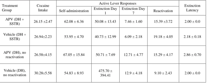
Table 1. Cocaine intake (mean mg/kg per session ± SEM), active lever responses (mean ± SEM), and extinction latency (mean number of days needed to reach the extinction criterion ± SEM). Active lever responses are reported for cocaine
self-administration training (mean of last 3 days of training), extinction training (the first and last day of training), and for the 15-minute memory reactivation session. The extinction criterion was ≤25 active lever responses on two consecutive extinction training
Treatment Group
Cocaine Intake
Active Lever Responses
Extinction Latency Self-administration Extinction Day
1
Extinction Day
7 Reactivation
APV (DH +
SSTR) 26.15 ±2.47 62.08 ± 6.36 50.08 ± 13.43 7.46 ± 1.60 15.39 ±3.72 2.00 ± 0.0
Vehicle (DH +
SSTR) 26.94±2.23 53.95 ± 4.70 40.73 ± 12.99 6.09 ± 2.18 19.18 ± 4.05 2.18 ± 0.18
APV (DH), no
reactivation 26.58±4.15 67.05 ± 15.84 50.71 ± 7.69 12.71 ± 4.77 15.29 ± 4.17 2.86 ± 0.70
Vehicle (DH),
no reactivation 30.28±5.58 54.83 ± 8.93
475.70 ±
394.41 12.9 ± 4.18 9.10 ± 2.43 2.00 ± 0.0
25
27
29
31
Figure 5. NMDA receptor blockade in the dorsal hippocampus does not alter general motor activity relative to control animals. A. Schematic depicting Experiment 2. 5 Animals received intra-DH infusions of VEH and APV in a counterbalanced order. An additional 9 animals received either VEH or APV, and only one test of locomotion. Animals were injected with either VEH or APV and returned to their home cage for 72 hours and then given a test of locomotion. There was no significant difference in the groups on locomotor activity determined by photo beam breaks (NVEH=9, NAPV=10). There was a significant main effect by time
REFERENCES
Alaghband Y, O’Dell SJ, Azamia S, Khalaj AJ, Guzowski JF, & Marshall JF (2014). Retrieval-induced NMDA receptor-dependent Arc expression in two models of cocaine-cue memory. Neurobiol Learn Mem 16:79-89 [epub ahead of print]
Alaghband Y & Marshall JF (2013). Common influences of non-competitive NMDA
receptor antagonists on the consolidation and reconsolidation of cocaine-cue memory. Psychopharmacology(Berl) 226(4):707-719
Boccia M, Freudenthal R, Blake M, de la Fuente V, Acosta G, Baratti C, Romano A (2007). Activation of hippocampal nuclear factor-kappa B by retrieval is required for memory reconsolidation. J Neurosci 27(49):13436-13445
Chen Q, He S, Hu XL, Yu J, Zhou Y, Zhen J, Zhang S, Zhang C, Duan WH, Xiong ZQ (2007). Differential roles of NR2A- and NR2B-containing NMDA receptors in activity-dependent brain0derived neurotrophic factor gene regulation and limbic epileptogenesis. J Neurosci 27:542-552
Chia C & Otto T (2013). Hippocampal Arc (Arg3.1) expression is induced by memory recall and required for memory reconsolidation in trace fear conditioning. Neurobiol Learn Mem. 106:48-55
Collingridge G (1987) Synaptic plasticity: the role of NMDA receptors in learning and memory. Nature 330:604-605
Everitt, B. J., Dickinson, A., & Robbins, T. W. (2001). The neuropschological basis of addiction. Brain Res Brain Res Rev, 36(2-3), 129-138.
Foltin, R. W., & Haney, M. (2000). Conditioned effects of environmental stimuli paired with smoked cocaine in humans. [Clinical Trial Controlled Clinical Trial Research
Support, U.S. Gov't, P.H.S.]. Psychopharmacology (Berl), 149(1), 24-33.
33
Fuchs, R. A., Evans, K. A., Ledford, C. C., Parker, M. P., Case, J. M., Mehta, R. H., & See, R. E. (2005). The role of the dorsomedial prefrontal cortex, basolateral amygdala, and dorsal hippocampus in contextual reinstatement of cocaine seeking in rats. [Research Support, U.S. Gov't, P.H.S.]. Neuropsychopharmacology, 30(2), 296-309. doi: 10.1038/sj.npp.1300579
Furkukawa H, Singh SK, Mancusso R & Gouaux E (2005). Subunity arrangement and function in NMDA receptors. Nature 438:185-192
Garcia-Delatorre P, Perez-Sanchez C, Guzman-Ramos K & Bermudez-Rattoni F (2014). Role of glutamate receptors of central and basolateral amygdala nuclei on retrieval and reconsolidation of taste aversion memory. Neurobiol Learn Mem. 111:35-40
Hellemans KG, Everitt BJ & Lee JL (2006). Disrupting reconsolidation of conditioned withdrawal memories in the basolateral amygdala reduces supression of heroin seeking in rats. J Neurosci 26(49): 12694-12699
Johnson JW & Ascher P. Glycine potentiates the NMDA response in cultured mouse brain neurons. Nature, 325:529-531
Kantak, K. M., Black, Y., E., V., Green-Jordan, K., & Eichenbaum, H. B. (2002). Dissociable effects of lidocaine inactivation on the rostral and caudal basolateral amygdala on the maintenance and reinstatement of cocaine-seeking behavior in rats. Journal of Neuroscience, 22, 1126-1136.
Kelley, J. B., Anderson, K. L., & Itzhak, Y. (2007). Long-term memory of cocaine-associated context: disruption and reinstatement. Neuroreport, 18(8), 777-780.
Kelly A, Laroche S, & Davis S (2003). Activation of mitogen-activated protein
kinsase/extracellular signal-regulated kindase in hippocampal circuitry is requred for consolidation and reconsolidation of recognition memory. J of Neuro 23(12):5354-5360
Lee, JL, Everitt, BJ, & Thomas, KL (2004). Independent cellular processes for hippocampal memory consolidation and reconsolidation. Science, 304(5672), 839-843.
Lee, JL, & Hynds, RE(2013). Divergent Cellular Pathways of Hippocampal Memory Consolidation and Reconsolidation. Hippocampus,23(3):233-244
Lee JL, Milton AL, & Everitt BJ (2006). Cue-induced cocaine seeking and relapse are reduced by disruption of drug memory reconsolidation. J Neurosci
Lee, JL, Milton, AL, & Everitt, BJ(2006a). Cue-induced cocaine seeking and relapse are reduced by disruption of drug memory reconsolidation. [Research Support, Non-U.S. Gov't]. J Neurosci, 26(22), 5881-5887. doi: 10.1523/JNEUROSCI.0323-06.2006
Lee, JL, Milton, AL, & Everitt, BJ(2006b). Reconsolidation and extinction of conditioned fear: inhibition and potentiation. [Research Support, Non-U.S. Gov't]. J Neurosci, 26(39), 10051-10056. doi: 10.1523/JNEUROSCI.2466-06.2006
Liddie S & Itzhak Y (2014). Variations in the stimulus salience of cocaine reward influences drug-associated contextual memory. Addiction Biology [epub ahead of print]
Liu YN, Yang X, Suo ZW, Xu YM & Hu XD (2014). Fyn kinase-regulated NMDA receptor- and AMPA receptor-dependent pain sensitization in spinal dorsal horn of mice. Eur J Pain, 18(8):1120-1128
Mayer M, Westbrook GL & Guthrie PB (1984). Voltage-dependent block by Mg2+ of NMDA responses in spinal cord neurones. Nature 309: 261-263
Meil WM, & See RE (1997). Lesions of the basolateral amygdala abolish the ability of drug associated cues to reinstate responding during withdrawal from self-administered cocaine. [Research Support, Non-U.S. Gov't]. Behav Brain Res, 87(2), 139-148.
35
Milton AL, & Everitt BJ(2010). The psychological and neurochemical mechanisms of drug memory reconsolidation: implications for the treatment of addiction. Eur J Neurosci, 31, 2308-2319.
Monyer H, Nail B, Laurie DJ, Sakmann B, and Seeburg PH (1994). Developmental and Regional Expression in the Rat Brain and Functional Properties of Four NMDA Receptors. Neuron 12:529-540
Nader K, Schafe GE, & LeDoux JE(2000a). The labile nature of consolidation theory. Nat Rev Neurosci, 1(3): 216-219
Nader K, Schafe GE, Le Doux JE (2000b). Fear memories require protein synthesis in the amygdala for reconsolidation after retrieval. Nature, 406(6797):722-726
Nicolle MM, Bizon JL, & Gallagher M (1996). In vitro autoradiography of ionotropic glutamate receptors in hippocampus and striatum of aged Long-Evans rats: relationship to spatial learning. Neuroscience 74(3): 741-756
Pandis C, Sotiriou E, Kouvaras E, Asprodini E, Papatheodoropoulos C, & Angelatou F. Differential expression of NMDA and AMPA receptor subunits in rat dorsal and ventral hippocampus. Neuroscience, 140(1): 163-175
Praxinos G & Watson C (1997). The rat brain in stereotaxic coordinates. Academic Press New York, NY
Prybylowski K, Chang K, Sans N, Kan L, Vicini S, Wenthold RJ (2005). The synaptic localization of NR2B-containing NMDA receptors is controlled by interactions with PDZ proteins and AP-2. Neuron 47:845-857
Ramirez, DR, Bell, GH, Lasseter, HC, Ziaohu, X, Traina, SA, & Fuchs, R. A. (2009). Dorsal hippocampal regulation of memory reconsolidation processes that facilitate drug context-induced cocaine-seeking behavior in rats. Eur J Neurosci, 30: 901-912.
Roche KW, Standley S, McCallum J, Dune Ly C, Ehlers MD, Wenthold RJ (2001).
Sanchez, H, Quinn, JJ, Torregrossa, MM, & Taylor, JR (2010). Reconsolidation of a cocaine-associated stimulus requires amygdalar protein kinase A. J Neurosci, 30(12), 4401-4407
Suzuki, A.,Josselyn, SA, Frankland, PW, Masushige, S, Silva, SJ, & Kida, S (2004). Memory Reconsolidation and Extinction Have Distinct Temporal and Biochemical Signatures. The Journal of Neuroscience, 24(20), 4787-4795.
Tedesco V, Mutti A, Auber A, & Chiamulera C. Nicotine-seeking reinstatement is reduced by inhibition of instrumental memory reconsolidation. Behav Pharmacol, 25(8):725-31
Tronson, N. C., & Taylor, J. R. (2007). Molecular mechanisms of memory reconsolidation. [Research Support, Non-U.S. Gov't Research Support, U.S. Gov't, P.H.S. Review]. Nat Rev Neurosci, 8(4), 262-275.
Valjent E, Corbille AG, BertranGonzalez J, Herve D, & Girault JA (2014). Inhibition of ERK pathway or protein synthesis during reexposure to drugs of abuse erases previously learned place preference. PNAS 103(8):2932-2937
Wells, A. M., Arguello, A. A., Xie, X., Blanton, M. A., Lasseter, H. C., Reittinger, A. M., & Fuchs, R. A. (2012). Extracellular Signal-Regulated Kinase in the Basolateral
Amygdala, but not the Nucleus Accumbens Core, is Critical for Context-Response-Cocaine Memory Reconsolidation in Rats. Neuropsychopharmacology.
Wells, A. M., Lasseter, H. C., Ziaohu, X., Cowhey, K. E., Reittinger, A. M., & Fuchs, R. A. (2011). Interaction between the basolateral amygdala and dorsal hippocampus is critical for cocaine memory reconsolidation and subsequent drug context-induced cocaine-seeking behavior in rats. Learning and Memory, 18, 693-702.
Wu, Y., Yonghui L., Gao J., & Sui N. (2012). Differential effect of NMDA receptor