OXIDATIVE DAMAGE TO GUANINE IN DNA CAUSED BY REACTIVE OXYGEN SPECIES
Wenjie Ye
A dissertation submitted to the faculty of the University of North Carolina at Chapel Hill in partial fulfillment of the requirements for the degree of Doctor of Philosophy in the
Department of Environmental Sciences and Engineering, Gillings School of Global Public Health.
Chapel Hill 2008
Approved by:
ii © 2008 Wenjie Ye
iii
ABSTRACT
WENJIE YE: Oxidative Damage to Guanine in DNA Caused by Reactive Oxygen Species (Under the direction of Dr. Louise M. Ball)
Oxidative damage to DNA, a factor in cancer, mutation, and aging, is attributed to reactive oxygen species (ROS). The less well characterized ROS, organic peroxyl radicals and peracid are present during lipid peroxidation and also produced by peroxidases from organic hydroperoxides. Peracetic acid is also formed in mitochondria. Guanine (Gua) is the nucleobases most susceptible to oxidation due to its lowest electron potential. The study described here focuses on Gua oxidation by epoxidizing reagents including peroxyl radicals and organic peracids. Dimethyldioxirane (DMDO), peracetic acid and m-chloroperbenzoic acid selectively oxidizes the guanine moiety of dGuo, dGMP and dGTP to 5-carboxamido-5-formamido-2-iminohydantoin (2-Ih). Structures were established on mass spectrometry and NMR studies. Labeling studies support a mechanism involving initial epoxidation of the guanine 4-5 bond and contraction of the pyrimidine ring by a 1,2-migration of the guanine
carbonyl C6 to form a transient dehydrodeoxyspiroiminodihydantoin followed by hydrolytic ring opening of the imidazolone ring. The 2-Ih is shown to be a major transformation in the oxidation of the single-stranded DNA 5-mer d(TTGTT) and the 5-base pair duplex
d[(TTGTT)·(AACAA)]. 2-Ih has not previously been reported as an oxidative lesion in DNA. Consistent with the proposed mechanism, no 8-oxoguanine was detected as a product of the
iv
donors. The 2-Ih base thus appears to be a pathway-specific lesion and holds promise as a potential biomarker.
v
ACKNOWLEDGEMENTS
I would like to acknowledge the advice and guidance of Dr. Louise M. Ball, committee chairman. I also thank the members of my Ph.D. committee, Dr. Karupiah Jayaraj, Dr. Stephen Chaney, Dr. Karl Koshlap, Dr. Carol Parker for their guidance and suggestions, especially Dr. Avram Gold for all his advice and encouragement. I also thank the members of the superfund groups in ESE, who contributed to the development of this work, particularly Dr. Ramiah Sangaiah, Diana E. Degen, Dr.Gunnar Boysen, Lina Gao and Leonard B. Collins, without whose knowledge and assistance this study would not have been successful.
I acknowledge the Superfund Basic Research Program for the financial support for this project.The important enzymes for this study were provided by Lannes Steven E. Ealick‘s Lab. in Department of Chemistry and Chemical Biology of Cornell University and Pierre Alexandre Kaminski in Institute Pasteur in Paris. I appreciate their support.
vi
TABLE OF CONTENTS
LIST OF TABLES ... xi
LIST OF SCHEMES ... xii
LIST OF CHART ...xiii
LIST OF FIGURES ... xiv
LIST OF ABBREVLATIONS ...xiviii
Chapter I. Literature Review ... 1
1.1 Oxidation pressure in vivo ... 1
1.1.1 Sources of the endogenous and exogenous oxidizing species ... 2
1.1.1.1 Sources of the endogenous oxidizing species ... 2
1.1.1.2 Sources of the exogenous oxidizing species ... 2
1.1.2 Types of ROS ... 3
1.1.2.1 Superoxide Radical ... 3
1.1.2.2 Hydrogen Peroxide ... 3
1.1.2.3 Hydroxyl Radical ... 4
1.1.2.4 Hypochlorous Acid ... 4
1.1.2.5 Alkoxyl Radicals/Peroxyl Radicals ... 4
1.1.2.6 Organic hydroperoxide... 5
vii
1.1.3 Lipid peroxidation ... 5
1.2 Reactions between oxidizing species and guanine, guanosine and deoxyguanosine ... 6
1.2.1 Oxidation of Guanine by hydroxyl radical ... 7
1.2.2 Oxidation of Guanine by superoxide anion ... 8
1.2.3 Oxidation of Guanine by singlet oxygen ... 8
1.2.4 Oxidation of Guanine by peracyl and peroxyl radicals ... 8
1.2.5 Oxidation of Guanine by metal complexes ... 10
1.2.6 Oxidation of Guanine DMDO ... 10
1.3 Basic information of the N2,3-εGuanine ... 12
1.3.1 Formation of the etheno nucleobase in vivo ... 12
1.3.2 Significance of N9-(β-D-2-deoxyribofuranosyl)-N2,3-ethenoguanine ... 13
1.3.3 Synthesis of N9-(β-D-2-deoxyribofuranosyl)-N2,3-ethenoguanine ... 14
II. OXIDATION OF GUANINE BY DMDO... 27
2.1 Abstract ... 27
2.2 Introduction ... 28
2.3 Materials and methods ... 30
2.3.1 Nuclear magnetic resonance and mass spectrometric analyses ... 30
2.3.2 Chemicals ... 31
2.3.3 Synthesis of [4-13C]- and [7-15N]Gua ... 31
2.3.4 Oxidation of Gua ... 32
2.3.5 Oxidation of d(TTGTT) by DMDO ... 33
2.3.6 Digestion of oxidized d(TTGTT) ... 33
viii
2.3.8 Oxidation of d[(TTGTT)·(AACAA)] ... 34
2.3.9 Oxidation of dGuo by Peracetic Acid ... 34
2.3.10 Oxidation of dGuo by m-CPBA ... 35
2.3.11 Oxidation of dGuo by 18O2-m-CPBA ... 35
2.3.12 Incorporation of 18O into 2-Ih from H218O ... 35
2.3.13 Oxidation of dGuo by DMDO ... 36
2.3.14 Separation of diastereomers 1a and 1b ... 37
2.3.15 Oxidation of dGMP by DMDO... 37
2.3.16 Oxidation of dGTP by DMDO ... 38
2.4 Results ... 39
2.4.1 Oxidation of guanine ... 39
2.4.2 Oxidation of d(TTGTT)... 43
2.4.3 Oxidation of d[(TTGTT)·(AACAA)] ... 45
2.4.4 Oxidation of dGuo by peracids ... 45
2.4.5 18O Labeling reactions ... 46
2.4.6 Oxidation of dGuo by DMDO ... 47
2.4.7 Oxidation of dGMP by DMDO ... 49
2.4.8 Oxidation of dGTP by DMDO... 51
2.5 Discussion ... 52
2.5.1 Oxidation of Guanine ... 52
2.5.2 Oxidation of DNA ... 52
2.5.3 Oxidation of dGuo by peracids ... 54
ix
2.5.5 Structural Analysis of the DMDO Oxidation of dGuo, dGMP and dGTP
... 56
III INVESTIGATION OF RIBOSYLATION ROUTES TO 8,9-DIHYDRO-9-OXO-3-(β-D-2-DEOXYRIBOFURANOSYL)-IMIDAZO[2,1-b]PURINE (N2, 3-edGUO) ... 99
3.1 Abstract ... 99
3.2 Introduction ... 100
3.3 Materials and methods ... 103
3.3.1 Instrumentation ... 103
3.3.2 Chemicals ... 103
3.3.3 Enzymes ... 104
3.3.4 Synthesis of 8,9-dihydro-9-oxoimidazo[2,1-b]purine (N2, 3– ethenoguanine) ... 104
3.3.5 Unambiguous synthesis of 8,9-dihydro-9-oxo-3-(2-deoxy-β-D-ribofuranosyl)-imidazo[2,1-b]purine ... 105
3.3.6 Chemical Glycosylation of 8,9-dihydro-9-oxoimidazo[2,1-b]purine (N2, 3-ethenoguanine) ... 108
3.3.7 Enzymatic glycosylation of N2,3 –ethenoguanine ... 110
3.4 Results and Discussion... 111
3.4.1 Enzymatic glycosylations ... 112
3.4.2 Chemical glycosylation ... 115
3.4.3 Unambiguous synthesis of 8,9-dihydro-9-oxo-3-(2-deoxy-β -D-ribofuranosyl)-imidazo[2,1-b]purine ... 115
IV FUTURE RESEARCH ... 135
4.1 Oxidation of Guanine by Epoxidizing Reagents ... 135
x
4.1.2 Whether the DNA lesion, 2-Ih, exist in mitochondrial DNA? ... 138 4.2 Synthesis of N9-(β-D-2-deoxyribofuranosyl)-N2,3-ethenoguanine ... 140
4.2.1 Can we increase the overall yield of N9-β-deoxyribosyl-N2,3-εdGuo? ... 140 4.2.2 Whether structures of enzymatic products can provide a tool to
xi
LIST OF TABLES Table
1.1 Process leading to generation of ROS ... 26
2.1 Proton signal assignments and NOESY interactions for 1a ... 94
2.2 Proton signal assignments and NOESY interactions for 1b ... 94
2.3 Proton signal assignments and NOESY interactions for 1a′... 95
2.4 C,H cross peaks resolved in the HMBC spectrum of 1a ... 95
2.5 C,H cross peaks resolved in the HMBC spectrum of 1b ... 96
2.6 C,H cross peaks resolved in the HMBC spectrum of 1a ... 96
2.7 NOESY cross peaks for 2a ... 97
2.8 NOESY cross peaks for 2b ... 97
2.9 Multiple-bond (x) and one-bond (y) C,H cross peaks for 2a ... 98
xii
LIST OF SCHEMES Scheme
1.1 Generation of oxidizing species in vivo ... 17
1.2 Oxidation of Guanine by hydroxyl radical ... 18
1.3 Oxidation of Guanine by superoxide anion... 19
1.4 Mechanism of oxidation of guanine by singlet oxygen ... 20
1.5 Enzymatic oxidation of vinly chloride to 2-chlorooxirane and rearrangement to ClCH2CHO and design of experiments for in situ destruction of reactive intermediates ... 21
1.6 Possible Mechanism of Formation of N2,3-εGua from 2-Substituted Oxiranes ... 21
1.7 Kuśmierek‘s Synthetic Routes of N2,3-εdGuo ... 22
1.8 Khazanchi‘s Synthetic Routes of N2,3-εdGuo ... 23
2.1 Synthetic scheme of labeled Guanine ... 59
2.2 Mechanism of guanine epoxidation by DMDO or peracid ... 58
3.1 Synthesis of N2, 3–εGua and chemical and enzymatic glycosylation of N2, 3–εGua ... 117
3.2 Designed route for chemical synthesis of 8,9-dihydro-9-oxo-3-(β -D-2-deoxyribofuranosyl)-imidazo[2,1-b]purine via cycloaddition of bromoacetaldehyde to O6-benzyl-protected dGuo... 118
xiii
LIST OF CHARTS Chart
xiv
LIST OF THE FIGURES Figure
1.1 A Hypothetical Scheme for carcinogenic factors leading to endogenous
species and exocyclic DNA-base damage ... 24
1.2 Suggested mechanism for the formation of etheno adducts from DNA nucleosides and LPO products such as trans-4-hydroxy-2-nonenal derived from PUFAs, as exemplified fro linoleic acid, a ω-6 PUFA (dR=deoxyribose) ... 25
2.1 Full scan ESI-MS+ of NA-2-Ih ... 60
2.2 ESI-MS/MS+ of NA-2-Ih ... 61
2.3 1H NMR (500 MHz, DMSO-d6) of (a) NA-2-Ih, (b) [7-15N]-2-Ih, and (c) [5-13 C]-2-Ih ... 62
2.4 DQF COSY spectrum of NA-2-Ih ... 63
2.5 Proton-decoupled 13C NMR (125 MHz, DMSO-d6) of (a) NA-2-Ih, (b) [7-15N]-2-Ih, and (c) [5-13C]-2-Ih ... 64
2.6 HMBC spectrum of NA-2-Ih ... 65
2.7 NOESY spectrum of NA-2-Ih ... 66
2.8 13C NMR (125 MHz, 9:1 H2O/2H2O) of NA-2-Ih ... 67
2.9 1H NMR (500 MHz, D2O) of oxidized d(TTGTT) in the H1′ - H9 range ... 68
2.10 Negative ion MAL DI-TOF mass spectrum of total reaction mixture of oxidized d(TTGTT) showing unoxidized 5-mer (m/z 1482) and product at +34 mass units (m/z 1516) ... 69
2.11 Negative ion ESI-MS of oxidized 5-mer ... 70
2.12 Negative ion ESI-MS/MS of ion m/z 1516 of oxidized 5-mer ... 71
2.13 Positive ion ESI-MS of oxidized 5-mer ... 72
xv
2.15 HPLC trace (detector at 260 nm) of reaction mixture from DMDO
oxidation of 5-mer ... 74
2.16 Positive ion ESI-MS/MS of m/z 1540 ([MNa]+) from peak at 9.5 min in HPLC of oxidized 5-mer ... 75
2.17 HPLC (UV detector set at 252 nm) of digest of oxidized 5-mer ... 76
2.18 HPLC traces (detector set at 230 nm) of reaction mixtures from dGuo oxidation by (a) peracetic acid and (b) m-CPBA, following extraction of spent oxidant ... 77
2.19 (a) Negative ion ESI-MS showing 16O/18O distribution in 2-Ih base from DMDO oxidation of Gua in 1:1 mixture of H216O/H218O, (b) Negative ion ESI-MS/MS of molecular ion of natural abundance 2-Ih and (c) Negative ion ESI-MS/MS of molecular ion 18O-labeled 2-Ih showing loss of the label with the formamide moiety ... 78
2.20 Positive ion ESI-MS of 2-Ih-dR from oxidation of dGuo with (a) natural abundance m-CPBA and (b) 18O2-m-CPBA, indicating retention of 18O label in the product ion at m/z 143 from loss of deoxyribosylformamide ... 79
2.21 1H NMR (500 MHz, D2O) of mixture of 2-Ih-dR diastereomers and rotamers 1a, 1b, 1a′, 1b′ from the oxidation of dGuo by DMDO ... 80
2.22 ROESY spectrum of diastereomer mixture from oxidation of dGuo ... 81
2.23 HMBC spectrum of diastereomer mixture from the oxidation of dGuo ... 82
2.24 HSQC spectrum of diastereomer mixture from oxidation of dGuo ... 83
2.25 ROESY spectrum of diastereomer 1a ... 84
2.26 ROESY spectrum of diastereomer 1b ... 85
2.27 1H NMR (500 MHz, D2O) of mixture of 2-Ih-dRP diastereomers and rotamers 2a, 2a′, 2b, 2b′ from the oxidation of dGMP by DMDO ... 86
2.28 ROESY spectrum of diastereomer mixture from oxidation of dGMP ... 87
2.29 ROESY NMR spectrum of the H1′ - H9 region of the mixture of diastereomers and rotamers from the oxidation of dGMP by DMDO ... 88
2.30 HMBC spectrum of diastereomer mixture from oxidation of dGMP ... 89
xvi
2.32 13C NMR spectrum (125 MHz, D2O) of diastereomer mixture from
oxidation of dGMP ... 91 2.33 1H NMR of the H1‘– H9 region of the product mixture from oxidation of
dGTP ... 92 2.34 HMBC spectrum of oxidation mixture of dGTP in the C1– H9 region ... 93 3.1 1H NMR of 8,9-dihydro-9-oxo-3-(β
-D-2-deoxyribofuranosyl)-imidazo-[2,1-b]purine ... 120
3.2 1H NMR of 8,9-dihydro-9-oxo-1-(β -D-2-deoxyribofuranosyl)-imidazo-[2,1-b]purine ... 121
3.3 1H NMR of 8,9-dihydro-9-oxo-7-(β -D-2-deoxyribofuranosyl)-imidazo-[2,1-b]purine ... 122
3.4 Exact Mass of 8,9-dihydro-9-oxo-1-(β
-D-2-deoxyribofuranosyl)-imidazo[2,1-b]purine(K+) ... 123 3.5 Exact Mass of 8,9-dihydro-9-oxo-7-(β
-D-2-deoxyribofuranosyl)-imidazo[2,1-b]purine(K+) ... 124 3.6 1H NMR of minor product at 18 min. Possible structure:
8,9-dihydro-9-oxo-3-(α-D-2-deoxyribofuranosyl)-imidazo-[2,1-b]purine ... 125 3.7 Exact Mass of minor product at 18 min. Possible structure:
8,9-dihydro-9-oxo-3-(α-D-2-deoxyribofuranosyl)-imidazo[2,1-b]purine(K+) ... 126 3.8 HPLC trace monitored at 260 nm, of mixture from ribosylation of N2
,3-εGua by L. fermentum transribosylation at pH 7.5 ... 127 3.9 HMBC NMR spectrum (DMSO-d6) of 8,9-dihydro-9-oxo-7-(β
-D-2-deoxyribofuranosyl)-imidazo[2,1-b]purine spanning the region of
H1‘-enthenoguanine interaction ... 128 3.10 NOESY NMR spectrum (DMSO-d6) of 8,9-dihydro-9-oxo-7-(β
-D-2-deoxyribofuranosyl)-imidazo[2,1-b]purine spanning the region of
H1‘-enthenoguanine interaction ... 129 3.11 HMBC NMR spectrum (DMSO-d6) of 8,9-dihydro-9-oxo-7-(β
-D-2-deoxyribofuranosyl)-imidazo-[2,1-b]purine spanning the region of
xvii
3.12 NOESY NMR spectrum (DMSO-d6) of 8,9-dihydro-9-oxo-7-(β -D-2-deoxyribofuranosyl)-imidazo[2,1-b]purine spanning the region of
H1‘-enthenoguanine interaction ... 131 3.13 HMBC NMR spectrum (DMSO-d6) of minor product at 18 min. Possible
structure: 8,9-dihydro-9-oxo-3-(α
-D-2-deoxyribofuranosyl)-imidazo-[2,1-b]purine spanning the region of H1‘-enthenoguanine interaction ... 132
3.14 NOESY NMR spectrum (DMSO-d6) of minor product at 18 min. Possible structure: 8,9-dihydro-9-oxo-7-(β
-D-2-deoxyribofuranosyl)-imidazo[2,1-b]purine spanning the region of H1‘-enthenoguanine interaction ... 133
3.15 NOESY NMR spectrum (DMSO-d6) of 8,9-dihydro-9-oxo-3-(β -D-2-deoxyribofuranosyl)-imidazo-[2,1-b]purine spanning the region of
xviii
LIST OF ABBREVLATIONS
DGh Dehydroguanidinohydantoin
dGuo 2‘-Deoxyguanosine
dGMP 2‘-Deoxyguanosine-5‘-monophosphate dGTP 2‘-Deoxyguanosine-5‘-triphosphate DRTases N-Deoxyribosyltransferases
-CH2- Methylene
dIz 2-Amino-5-(2-deoxyribosylamino) imidazolone
DMDO Dimethyldioxirane
dZ 2,2-Diamino-5-(2-deoxyribosylamino)-5(2H)-oxazolone
ETC Electron transport chain
Gh Guanidinohydantoin
Gua Guanine
Guo Guanosine
H2O2 Hydrogen Peroxide
HOCl Hypochlorous acid
2Ih 5-Carboxamido-5-formamido-2-iminohydantoin
mCPBA m-Chloroperbenzoic acid
MS Mass Spectrometry
N2, 3-εdGuo 8,9-Dihydro-9-oxo-3-(β-D-2-deoxyribofuranosyl)-imidazo[2,1-b]purine
xix N2, 3-εGua N2,3-ethenoguanine
NA Nature abundance
NMR Nuclear Magnetic Resonance
NO Nitric oxide
O2•- Superoxide
ONOO- Peroxynitrite
PDT Purine deoxyribosyltransferase
RO Alkoxyl
ROO Peroxyl
ROOH Organic hydroperoxides
ROS Reactive oxygen species
SOD Superoxide dismutase
Sp Spiroiminodihydantoin
I. Literature Review
1.1 Oxidation pressure in vivo
Oxidative stress is a physiological condition that occurs when there is a significant imbalance between production of reactive oxygen species and antioxidant defenses. In humans, oxidative stress is involved in many diseases, such as atherosclerosis, Parkinson's disease and Alzheimer's disease[Valko M. et al, 2005], but it may also be important in prevention of aging by induction of a process named mitohormesis. ROS can be beneficial, as they are used by the immune system as a way to attack and kill pathogens. ROS are also used in cell signaling. This is dubbed redox signaling. A particularly destructive aspect of oxidative stress is the production of ROS, which include free radicals and peroxides. Some of the less reactive of these species (such as superoxide) can be converted by
oxidoreduction reactions with transition metals or other redox cycling compounds
2
1.1.1 Sources of the endogenous and exogenous oxidizing species 1.1.1.1 Sources of the endogenous oxidizing species
The production of oxidizing species occurs through a variety of endogenous processes which was summarized in the following paragraphs and Table 1.1[Williams, G. and Jeffrey, A.2000], [Frenkkel, 1992].
Oxidizing species are even produced daily during normal respiration by the mitochondrial respiratory chain and during the production of energy by each and every mitochondrion. Therefore, one may consider oxidative stress a necessary outcome of physiological functions such as respiration, digestion, and metabolism.
NADPH oxidase, the best characterized source of ROS, several other enzymes may contribute to ROS generation, including nitric oxide synthase, lipoxygenases,
cyclo-oxygenases, xanthine oxidase and cytochrome P450 enzymes [Williams, G. and Jeffrey, A.2000].
In addition, metals and certain fatty substances can also cause oxidative stress. Metals (i.e., iron copper) acting as pro-oxidants and fatty substances (carbon-based molecules reacting with oxygen) undergoing lipid peroxidation are prime examples in which oxidative stress is accentuated.
1.1.1.2 Sources of the exogenous oxidizing species
Exogenous sources, like radiation, ozone, xenobiotics etc. [Williams, G. and Jeffrey, A., 2000], also can increase the level of the ROS. One cause of oxidative stress includes the redox cycling of toxins called xenobiotics. This occurs during drug or contamination
3
substances, particularly iron, chromium, cobalt(II), and nickel salts in the presence of hydrogen peroxide, have been long recognized as forming oxidized bases in DNA [Klein et al.,1991; Nackerdien et al., 1991; Standeven et al., 1991].Other cause of increased free radical generation occurs if the toxin increases damage by interfering with the antioxidant defenses. Finally, the toxin can also stimulate more free radical generation by damaging the mitochondrial electron transport system.
1.1.2 Types of ROS
1.1.2.1 Superoxide Radical
The O2•- is created by the reduction of oxygen to form superoxide (O2 - e = O2-). This oxygen-centered free radical is formed by autoxidation reactions or by the
mitochondrial electron transport chain (ETC). It is not reactive unless it comes in contact with other radicals, such as nitric oxide (NO), or certain metals, such as iron. O2
-undergoes dismutation in the presence of superoxide dismutase (SOD) to form H2O2 (2 O2 -+ 2 H+ = H2O2 + O2). This radical is highly selective and has beneficial and damaging effects to tissues. It is considered a vital part of the cellular signaling pathways that provide important genetic information [Salo D.C., et al, 1990].
1.1.2.2 Hydrogen Peroxide
4
O2- can accelerate the Fenton reaction, giving an O2--assisted Fenton reaction (H2O2 + O2 -(Fe/Catalyst) = OH- + OH + O2) [Halliwell B. 2003].
1.1.2.3 Hydroxyl Radical
This more potent hydroxyl radical (•OH) is often generated during the Fenton reaction, forming peroxynitrite (ONOO-) from NO acid reacting with oxygen and from ionizing
radiation. This radical attacks and damages most cellular components [Mandelker L., 2008].
1.1.2.4 Hypochlorous Acid
Hypochlorous acid (HOCl) is highly reactive and lipid soluble. It is formed from H2O2 and the enzyme myeloperoxidase. It is especially dangerous to protein constituents, in which it can oxidize and damage biomolecules [Mandelker L., 2008].
1.1.2.5 Alkoxyl Radicals/Peroxyl Radicals
5 1.1.2.6 Organic hydroperoxides
Organic hydroperoxides (ROOH) Formed by radical reactions with cellular components such as lipids and nucleobases, which will be discussed in detail at following text (3.1.3 Lipid peroxidation).
1.1.2.7 Peracid
Peracids (RC(O)OOH) may arise biologically during lipid peroxidation, through formation of
triplet excited ketones and aldehydes by the Russell mechanism [Russell, G. et al, 1957] followed by
β-cleavage and coupling with O2, by peroxidase-catalyzed autoxidation of aldehydes [Adam, W.,
Kurz, A. et al, 1999], or by aldehyde oxidation catalyzed by transition metals [Nam, W. et al, 1996].
In mitochondria, three ThDP-dependent enzymes (S. typhimurium ALS II, Baker‘s yeast pyruvate decarboxylase and Zymomonas mobilis pyruvate decarboxylase) have been shown to catalyze the formation of peracetic acid [CH3C(O)OOH] from pyruvate and O2 under certain conditions [Abell, L. M. et al, 1991], [Bunik, V. I. et al, 2007], an observation that is
significant because mitochondrial DNA repair capability appears to decrease with age [Ledoux, S. P. et al, 2007], [Croteau, D. L. et al, 1999] and accumulated of mutations are implicated in age related
neuropathology and the ageing process in general [Dimauro, S. et al, 2005].
1.1.3 Lipid peroxidation
Lipid peroxidation refers to the oxidative degradation of lipids. It is the process whereby free radicals "steal" electrons from the lipids in cell membranes, resulting in cell damage. This process proceeds by a free radical chain reaction mechanism. It most often affects polyunsaturated fatty acids, because they contain multiple double bonds in between which lie methylene (-CH2-) groups that possess especially reactive hydrogens. Lipid
6
by metabolism of exogenous chemicals, has been recognized since the 1980s as an important contributor of reactive oxygen species involved in genotoxic endpoints. The primary species generated during lipid peroxidation are lipid hydroperoxides. From polyunsaturated lipids with 3 or more double bonds, cyclic peroxides and endoperoxides can also be generated [Yin, H. et al, 2005].
The hydroperoxides can yield alkylperoxyl and alkoxy radicals via redox reactions mediated by transition metals both within and external to the cellular environment [Aoshima, H. et al, 1997] (Scheme 1.1, eq 1, 2).
Peroxyl radicals also result from the coupling of allyl radicals with molecular oxygen following H• abstraction from lipids in the initiation step of lipid peroxidation (Scheme 1.1. , eq 3, 4). In addition, a variety of oxy radicals and other oxidizing species are generated during the peroxidation process. The reactions are summarized in (Scheme 1.1, eq 5–10). Coupling of peroxyl radicals in a chain terminating reaction can yield singlet oxygen or triplet excited ketones and aldehyded [Miyamoto, S. et al, 2006], [Miyamoto, S. et al, 2003] (Scheme 1.1 ,eq 6, 7) by the Russell mechanism[Russell, G. A. et al,1957]. The triplet ketones can, in turn, undergo α-cleavage to acyl radicals (Scheme 1.1 ,eq 8), and subsequent coupling with O2 leads to peracyl radicals (Scheme 1.1. , eq 9) and peracids (Scheme 1.1, eq
10) [Adam, W. et al, 1999], [Adam, W. et al ,2001], [Adam, W., Nau, W.M. et al, 2001] .
1.2 Reactions between oxidizing species and guanine, guanosine and
deoxyguanosine.
ROS can induce plenty of covalent modification to DNA, which encompass
7
cross-link [Evans, M.D. et al, 2004]. Since the focus of this proposal is damage to guanine,
the Background review will not be concerned with the other kinds of damage to the DNA.
Oxidation of dGuo by hydroxyl radical, one-electron abstraction and 1O2 has been the focus
of considerable work and the results covered in several recent reviews [Neeley W. L. et al,
2006], [Tudek, B. et al, 2003].
1.2.1 Oxidation of Guanine by hydroxyl radical
Products of reaction with hydroxyl radical are postulated to result from initial
addition to the base, summarized in Scheme 1.2. Addition at C8 leads to 8-oxodGuo following oxidation or to a formamido pyrimidine (FAPyG) following reduction. Hydroxyl
radical can also add to the C4-C5 bond. Addition at C4 is calculated by several methods to be
favored over addition to C5 [Colson, A. et al, 1997]. Dehydration of the neutral radical
adduct at C4, followed by oxidation, leads to 2-amino-5-(2-deoxyribosylamino) imidazolone
(dIz) and subsequently to 2,2-diamino-5-(2-deoxyribosylamino)-5(2H)-oxazolone (dZ).
In addition to hydroxyl radical, one-electron abstraction from dGuo is accomplished
by other radical species, including NO•, SO4•/-, and a variety of transition metal complexes
that have been investigated as probes for one-electron oxidation or as potential antineoplastic
drugs. (CrV[Sugden, K. D. et al, 2002], CrVI, MnII, FeIII, CoII, NiII and CuII [Choi, S. et al,
2004]) and ionizing radiation. The radical cation resulting from one-electron oxidation of
dGuo deprotonates at physiological pH to a neutral radical with substantial spin density
8 1.2.2 Oxidation of Guanine by superoxide anion
Addition of superoxide anion (O2•/-) to the C8 or C5 followed by protonation gives
the corresponding hydroperoxides. The C8-hydroperoxide leads to 8-oxodGuo under
reducing conditions and the C5-hydroperoxide leads to dIz and dZ (Scheme 1.3. ). Reaction of the Gua radical at C8 or C5 with O2, gives the corresponding peroxyl radicals and,
following hydrogen abstractions, the C8 or C5 hydroperoxides, which follow the respective
pathways summarized in Scheme 1.3.
1.2.3 Oxidation of Guanine by singlet oxygen
dGuo undergoes a 2 + 4 cycloaddition with singlet oxygen to form a transient
4,8-endoperoxide, which rearranges to 8-hydroperoxy-dGuo. The hydroperoxide then reacts via
two pathways (Scheme 1.4.) : (A) reduction to 8-oxodGuo and a 2+ 2 addition of a second
molecule of 1O2 to form the 4,5-endoperoxide [Neeley W. L. et al, 2006]which rearranges to
5-hydroperoxy-8-oxodGuo leading to dZ, via dehydroguanidinohydantoin (DGh); (B)
decomposition to 8-oxodehydro-dGuo followed by addition of water leading to Sp and Gh
through the transient 5-hydroxy-8-oxodG. It may be remarked that formation of the
endoperoxides is formally a two-electron oxidation.
Recently, a diminoimidazole has been reported as a minor product [Suzuki, T. et al,
2003]directly from the reaction of dGuo with 1O2.
1.2.4 Oxidation of Guanine by peracyl and peroxyl radicals
While research efforts have focused on the damage caused by the hydroxyl radical,
1
9
produce ROS (Scheme 1.1. , eq 4, 6, 7, 9, 10) have received less attention. The proposed research is directed at elucidating the role of peracids in ROS-mediated DNA damage,
through the characterization of base modifications and mechanisms of oxidation.
Oxidation of dGuo by a mixture of peracyl and peroxyl radicals generated either by
photolysis of ketones or thermolysis of dioxetanes [Adam, W. et al, 2001] or by peroxyl
radicals generated from hydroperoxides by the action of Coprinus or horseradish peroxidases
[Adam, W. et al, 2000] yielded Sp (incorrectly identified as 4-OH-8-oxodGuo) and
―guanidine releasing‖ products (not characterized further). 8-oxodGuo was identified in trace
quantities, but was determined to be further oxidized under the reaction conditions, implying
that it was an important intermediate[Adam, W. et al, 1995]. Interestingly, dGuo 4,
5-epoxide is a transient in the proposed mechanism [Adam, W. et al, 2001]. In the horseradish
peroxidase-catalyzed oxidation of dGuo by isobutanal, the active oxidants were concluded to
be the peracid and peracyl radical[Adam, W. et al, 1999]. Peroxyacyl radicals and peracids
are also generated during the autoxidation of aldehydes. In support of this conclusion, m
-chloroperbenzoic acid (mCPBA) alone or in the presence of the enzyme efficiently oxidized
dGuo. dZ and dIz were the only products identified in this work, although they accounted for
at most 50% of the dGuo consumed. Under the experimental conditions, no 8-oxodGuo was
detected. mCPBA has been reported to modify dGuo and dAdo residues in ssDNA and to
generate modified nucleotides having altered moblilities from dGMP and dAMP[Jacobsen, J.
S. et al, 1986]. However, the investigators did not characterize either the DNA lesions or the
modified bases. Although reaction schemes have been proposed for the oxidation of dGuo by
peroxyacyl/peroxyl radicals, the mechanisms of oxidation are completely speculative. No
10 1.2.5 Oxidation of Guanine by metal complexes
In addition to two-electron oxidations of dGuo by peracids and 1O2, a two-electron
oxidation of dGuo mediated by an oxoruthenium complex has been reported and of dGMP by
PtIV(1,2-diaminocyclohexane). The latter oxidation yielded 8-oxodGuo via intramolecular
donation of a phosphate oxygen to C8. Of interest with regard to the work proposed here is
the oxidation of guanine in short dsDNA by a MnIII(porphyrin)/KHSO5 system. On the basis
of mass spectrometric analysis of the modified DNA, the major product corresponded to a
gain of 34 amu by Gua (G + 34), and was tentatively identified as
5,8-dihydroxy-7,8-dihydroGua via two-electron oxidation of Gua and deprotonation to a cation [Vialas C. et al,
2000]. Mechanistic studies of Foote [McCallum J.E.B et al., 2004], [Sheu C. et al, 1993],
[Sheu C. et al, 1995]on the transients generated during oxidation of Gua and 8-oxoGua by
1
O2, argue strongly against the likelihood of the proposed structure because of the lability of
5-hydroxy-substituted guanine and 8-oxoguanine. An alternative structure for the G + 34
product is 4-carboxamido-4-formamido-2-iminohydantoin, identified by our laboratory as the
two-electron oxidation product of Gua by dimethyldioxirane [Ye W. et al, 2006]. As
discussed in the following section.
1.2.6 Oxidation of Guanine DMDO
Dimethlydioxirane (DMDO) is a three-membered cylic peroxide that efficiently
transfers oxygen and reacts selectively [Adam W. et al., 1989]. The preparation method of
DMDO reported in the literature yields a 0.1M solution [Adam W. et al., 1987]. DMDO
demonstrates efficient transfer of oxygen through a single concentrated step [Edwards et al.,
11
research of the DNA oxidation, not only because of the reasons mentioned above, but also
the characteristic is low temperature, no residue of oxidant, short time and mild pH value
which mimics the environment in vivo and provides the convenience for purification and
analysis.
Oxidation of Gua and dGuo by DMDO has been studied with regard to its use in
DNA sequencing by selectively introducing piperidine-labile lesions at guanine in both ss-
and dsDNA[Davies J. R. et al, 2002], [Davies J. R. et al, 1990]. Following 2-minutes of
DMDO treatment and appropriate workup, samples of ss- and ds-DNA underwent selective
strand breakage at the guanine sites observed with the Maxam-Gilbert procedure, indicating
efficient and selective oxidation of Gua bases. Preliminary oxidations of Gua and dGuo were
carried out to characterize the lesions; however, the reaction mixtures were worked up by
heating for 5 h at 85 oC prior to chromatographic isolation of products [Davies J. R. et al,
1990], [Ye W. et al, 2006]. Guo and dGuo both yielded
4-amadinocarbamoyl-5-hydroxyimidazole, but the product from dGuo was substituted at C2 with the
deoxyribose-derived trihydroxy-n-butyl group. Both products are at the same oxidation level as the
starting bases. Evidently, prolonged heating of the crude reaction mixture resulted in the
reduction and, in the case of dGuo, rearrangement. The investigators proposed that DMDO
initially epoxidized the C4-C5-bond of dGuo, by analogy with DMDO epoxidation of C5-C6
of the pyrimidine ring of 5′-O-trityl-dThyd [Lupatelli, P. et al, 1993].
Since peracids and the peroxyacyl and peroxyl radicals can epoxidize double bonds
[Reed G.A. et al, 1990], [Dix T.A. et al, 1983] [Lang B. et al, 1986], we propose DMDO as a
12
of dGuo.
1.3 Basic information of the
N
2,3-
ε
Guanine
1.3.1 Formation of the etheno nucleobase in vivoAmong exocyclic DNA adducts, ethenobases have been most widely studied in the last 33 years, as this class of DNA lesions is formed by many genotoxic chemicals including the human carcinogen vinyl chloride (VC) and the multi-species carcinogen urethane [Bartsch H. et al, 1994]. Ethenobases were first described by Kochetkov et al. [Kochetkov N. K. et al, 1971] who identified them as reaction products of 2-chloroacetaldehyde with adenine and cytosine. Interest in the ε-lesions was renewed in 1975 when it was found that they were generated in vitro by the vinyl chloride metabolites, chloroethylene oxide and
2-chloroacetaldehyde (Scheme 1.5.) [Barbin A. et al, 1975], [Laib R.J. et al, 1977], [Laib R.J. et al, 1978]. Using replication and transcription fidelity assays and ε -modified oligo- or polynucleotides, it was established that εdA and εdC have miscoding or ambiguous base pairing properties [Barbin A.et al, 1981], [Hall J.A. et al, 1981], [Spengler S. et al, 1981] and thus could be involved in the mutagenic and carcinogenic effects of VC [IARC, 1979], [Barbin, H. et al, 1986].
In the 1990s, ε -adducts have received renewed attention, because background levels of etheno adducts have been detected in tissues from unexposed humans and rodents,
13
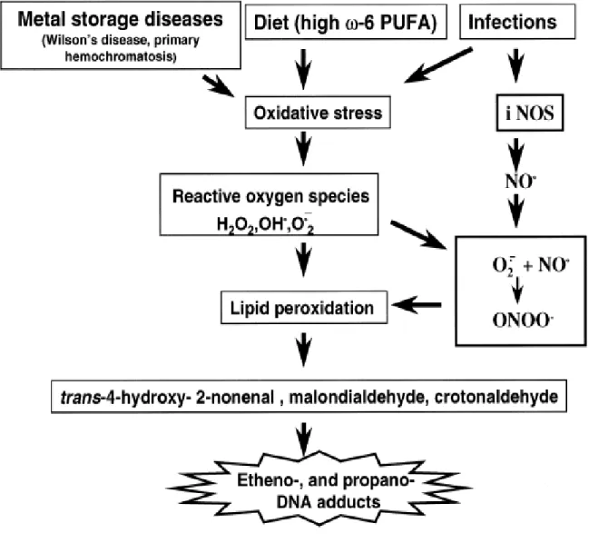
Subsequently, it was shown that high intake of dietary ω-6 –polyunsaturated fatty acids by female volunteers greatly increased LPO-derived etheno-DNA adducts in white blood cells in vivo [Fang J.L., Vaca C.E. et al, 1996]. Further, elevated levels of ε -adducts were found in hepatic DNA from patients and rodents with genetic predisposition to oxidative stress, lipid peroxidation and increased risk of liver cancer due to metal storage disease [Nair J., Sone H., et al 1996]. Also, during inflammatory processes a cascade of reactive
Oxygen/nitrogen intermediates can be generated, that could lead directly to oxidative DNA damage and/or to formation of ε -adduct via reaction of bifunctional 4-hydroxyalkenals and epoxides derived from LPO (Figure 1.2)
1.3.2 Significance of N9-(β-D-2-deoxyribofuranosyl)-N2,3-ethenoguanine
N9-(β-D-2-deoxyribofuranosyl)-N2,3-ethenoguanine(8,9-dihydro-9-oxo-3-(β -D-2-deoxyribofuranosyl)-imidazo[2,1-b]purine) is a one of highly mutagenic etheno DNA adduct both in vitro and in vivo research [Bartsch H., Barbin A. et al, 1994], [Kochetkov N.K. et al, 1971], [Nair J., Barbin A.et al, 1999]. 8,9-dihydro-9-oxo-3-(β -D-2-deoxyribofuranosyl)-imidazo[2,1-b]purine in DNA is therefore of considerable interest as a biomarker of
14
[Scheller, N. et al, 1995] was reported, using a GC-MS method. However, the same
laboratory reported that εG formed from endogenous sources occurred at a much higher level in 10/10 human liver samples (means of 3 and range from 0.7–7 per 107 G) [Swenberg, J.A. et al,1995].
1.3.3 Synthesis of N9-(β-D-2-deoxyribofuranosyl)-N2,3-ethenoguanine
Quantitative analysis of DNA adducts requires both unambiguously characterized standards and on occasion, labeled isotopomers. Preparation of certain classes of deoxynucleoside adducts can be problematic because instability of intermediates under conditions of established synthetic routes, deglycosylation at low pH or elevated temperature often present particular difficulties. Chemical synthesis of 8,9-dihydro-9-oxo-3-(β -D-2-deoxyribofuranosyl)-imidazo[2,1-b]purine has proven to be challenging precisely because of the reported lability of the glycosidic bond under conditions generally applicable to chemical synthesis [Barbin A., Brĕsil H.et al, 1975], [Laib R.J., Bolt H.M., 1977], [Laib R.J., Bolt H.M., 1978].
15
1% if we try to use the labeled dGuo as the starting material to synthesize the internal standard for quantitative analysis.
Johnson‘s Group [Khazanchi R. et al, 1993] described a route starting in the riboside series. O6-Benzylguanosine when treated with bromoacetaldehyde under conditions of continuous buffering gives the N2,3-etheno derivative in 48% yield. The 3',5'-O-(1,1,3,3-tetraisopropyldisiloxa-l,3-diyl) derivative when allowed to react with phenyl
chlorothionoformate led to the corresponding 2' ester. 2‘-Deoxygenation of 10 by the Barton procedure then afforded 65 % of 11, and deprotection of the latter (BuN+F-) gave 12
quantitatively. Catalytic hydrogenation of 12 then produced pure N2,3-εdGuo in 86% yield.(see scheme 1.8.). The overall yield of this route is still lower than 5%. The synthetic route of this procedure is too long which will lead the synthesis of internal standard
unpractical because of the high price of the labeled starting material.
N-Deoxyribosyltransferases (DRTases) [Anand, R. et al, 2004] catalyze the transfer
of a 2‘-deoxyribosyl group from a donor deoxynucleoside to an acceptor nucleobase. DRTases can be divided into two classes on the basis of their substrate specificity. The DRTase I class (also called purine deoxyribosyltransferase (PDT)) is specific for the transfer of deoxyribose between two purines. The DRTase II class (also called nucleoside
deoxyribosyltransferase ) catalyzes the transfer of deoxyribose between either purines or pyrimidines, but has a strong preference for deoxypyrimidines as the donor. DRTase was applied in the synthesis of N9-(β-D-2-deoxyribofuranosyl)-N2,3-ethenoguanine in
16
17
18
19
20
21
Scheme 1.5 Enzymatic oxidation of vinly chloride to 2-chlorooxirane and rearrangement to ClCH2CHO and design of experiments for in situ destruction of reactive intermediates
22
23
24
25
26
Table 1.1 Process leading to generation of ROS [Williams, G. and Jeffrey, A.2000], [Frenkkel, 1992]
1. Cellular Respiration
—Mitochondrial electron transport —Hexose monophosphate shunt
2. Biosynthetic and biodegrading processes of normal intermediary metabolism
—Arachidonic acid metabolism
—b-Oxidation of high-molecular-weight fatty acids (fatty acid CoA oxidase) —Amino acid oxidation (D-amino acid oxidase, tyrosine oxidase, etc.) —Iron metabolism
—Ascorbic acid biosynthesis (L-gulonolactone oxidase: absent inhumans) —Polyamine oxidation
—Steroidogenesis
—Purine oxidation (urate oxidase: absent in humans, xanthine oxidase) 3. Biotransformation of xenobiotics
—Microsomal electron transport (cytochrome P450 and b3) —Other mixed function oxidases
—Peroxidative oxidation (myeloperoxidase, prostaglandin H synthetase) 4. Activation of phagocytic cells by natural stimuli
—Peripheral blood leukocytes —Tissue macrophages
II. Oxidation of Guanine by Epoxidizing Reagents
2.1 Abstract
Oxidative damage to DNA, of concern as a factor in cancer, mutation, and aging, is attributed to reactive oxygen species. Organic peroxyl radicals, known to be present during lipid peroxidation and also products of the action of peroxidases on organic hydroperoxides, have received much less attention. Guanine (Gua) is the most susceptible to oxidation due to the lowest electron potential in all of nucleobases. This chapter focuses on Gua oxidation by epoxidizing reagents. The structure of the product of Gua oxidized by epoxidizing model agent, dimethyldioxirane (DMDO), 5-carboxamido-5-formamido-2-iminohydantoin (2-Ih, Chart 2.1) was established on the basis of mass spectrometry and NMR studies on 2-Ih and
its isotopomers generated by the oxidation of [4-13C] and [7-15N] guanine. The mononeric species dGuo, dGMP and dGTP were oxidized to study the reaction mechanism and to characterize fully the oxidation products. The DMDO selectively oxidizes the guanine
moiety of dGuo, dGMP and dGTP to 2-Ih (see Chart 2.1 and Scheme 2.2), peracetic acid and m-chloroperbenzoic acid also oxidize the base moiety of dGuo to 2-Ih. The presence of the glycosidic bond results in the stereoselective induction of an asymmetric center to give a
mixture of diastereomers, with each diastereomer in equilibrium with a minor conformer
through rotation about the formamido C-N bond. Labeling studies with 18O2-m-CPBA as
oxidant and with DMDO as oxidant in the presence of H218O, in concert with the study of the
28
initial epoxidation of the guanine 4-5 bond and contraction of the pyrimidine ring by a 1,2-migration of the guanine carbonyl C6 to form a transient
dehydrodeoxyspiroiminodihydantoin followed by hydrolytic ring opening of the imidazolone
ring. The 2-Ih is shown to be a major transformation in the oxidation of the single-stranded DNA 5-mer d(TTGTT) by m-CPBA and DMDO and in the oxidation of the 5-base pair duplex d[(TTGTT)·(AACAA)] with DMDO. 2-Ih has not previously been reported as an oxidative lesion in DNA. The lesion is stable to DNA digestion and chromatographic purification suggesting that 2-Ih may be generated as a stable lesion in vivo in DNA by peracids and possibly other epoxidizing agents. Consistent with the proposed mechanism, no 8-oxoguanine was detected as a product of the oxidations of the oligonucleotides or
monomeric species mediated by the monooxygen donors. The 2-Ih base thus appears to be a pathway-specific lesion and holds promise as a potential biomarker.
2.2 Introduction
29
mutagenic [Neeley W. L et al, 2006],. By contrast, DNA damage by initial two-electron processes, whether direct or via rapid sequential one-electron oxidations, has only recently come into focus [Neeley W. L. et al, 2006], [Dedon, P. C. et al, 2008], [Gedick, C. M.et al, 2002], [Hong, I. S. et al, 2007], [Xu, X. et al, 2008]. The project is examining oxidation of DNA by peracids and by dimethyldioxirane (DMDO) as a model congruent in mechanism to peracids [Bach, R.D. et al] [Porter, N. A. et al]. These compounds function as monooxygen donors by formally concerted, two-electron oxidations. Peracids may arise biologically during lipid peroxidation, through formation of triplet excited ketones and aldehydes by the Russell mechanism [Russell, G. et al, 1957] followed by β-cleavage and coupling with O2, by peroxidase-catalyzed autoxidation of aldehydes [Adam, W., Kurz, A. et al, 1999], or by aldehyde oxidation catalyzed by transition metals [Nam, W. et al, 1996]. In mitochondria, 2-oxoacid decarboxylases have been shown to generate peracids under certain conditions
[Abell, L. M. et al, 1991], [Bunik, V. I. et al, 2007], an observation that is significant because mitochondrial DNA repair capability appears to decrease with age [Ledoux, S. P. et al, 2007], [Croteau, D. L. et al, 1999] and accumulated of mutations are implicated in age related neuropathology and the ageing process in general [Dimauro, S. et al, 2005].
In both single and double stranded DNA, guanine appears to be the predominant
target of both peracids and DMDO [Adam, W., Kurz, A. et al, 1999],[Jacobsen, J. S. et al, 1986], [Davies, J. R. et al, 1990]. The action of m-CPBA on DNA appears to be associated with damage targeted to purines that is strongly blocking to replication, particularly in loop
30 2.3 Materials and methods
2.3.1 Nuclear magnetic resonance and mass spectrometric analyses
NMR spectra were recorded on a Varian Inova NMR spectrometer operating at for 500 MHz 1H and at 125 MHz for 13C. 13C shifts were obtained from HSQC and HMBC spectra 1JC-H were derived from unsuppressed 1-bond coupling in the heteronuclear shift correlation spectra. Low resolution electrospray ionization-mass spectrometry (ESI-MS) and tandem mass spectrometry (ESI-MS/MS) were performed on a Finnigan DECA ion trap system. Liquid chromatography ESI-MS (LC-ESI-MS) and LC-ESI-MS/MS were performed on a Finnigan TSQ Quantum system. Exact mass measurements were acquired on an IonSpec HiRes QFT ion cyclotron resonance mass spectrometer (Lake Forest, CA) equipped with a 9.4 T superconducting magnet and a Waters/Micromass Z-spray source. Samples were injected at a flow rate of 500 nL/min. The instrument was employed in the positive ion and broadband modes with a probe voltage of 3.2 kV, a cone voltage of 45 V, an accumulation in Q3 of 5000 ms, and quadrupole ion guide burst optimized to transmit ions in the 200 m/z mass range. Matrix-assisted laser desorption/ionization-MS (MALDI-MS) data were acquired on a Bruker Ultraflex II MALDI-time of flight (TOF)/TOF mass spectrometer (Bruker Daltonics, Billerica, MA). Samples were dissolved in 50:50 MeOH: 0.1% TFA, and MALDI spectra were acquired in the negative ion reflectron mode, using α -cyano-4-hydroxy-cinnamic acid as the matrix. Spectra were acquired over the mass range 500 to 2500 Da, using ACTH(1-17) and ACTH(18-39) as calibration standards.
2.3.2 Chemicals
31
purchased from Sigma-Aldrich (Milwaukee, WI) were used as received. DMDO was generated by potassium monopersulfate oxidation of acetone and was analyzed by iodometric titration immediately prior to use [Murray, R. W., and Jeyaraman, R., 1985]. d(TTGTT) and d(AACAA) were synthesized by the Oligonucleotide Synthesis Core Facility at UNC –
Chapel Hill and purified by HPLC on a reverse phase Vydac C18 250 x 4.6mm column eluted with a linear gradient 8 to 15% MeOH in water over 10 min at flow rate of 1ml/min monitored at a detector wavelength of 260nm.
18O2-m-CPBA was synthesized as follows (personal communication, Spiro, T. G.). A flask containing methylene chloride (35 mL) and m-chlorobenzaldehyde (3 mL) was degassed by 3 freeze-thaw cycles on a high-vacuum manifold, frozen in liquid nitrogen under vacuum (1x10⁻³ mm) and then 18O2 (500 mL, >95 %) from a break-seal vessel was condensed into the reaction flask. The reaction flask was isolated, removed from the vacuum line and irradiated with a Fisher Biotech UV lamp at 365 nm with vigorous stirring for 8 h at -20 oC. Following transfer to a round bottom flask, the reaction mixture was reduced in volume under aspirator pressure to 10 mL and the precipitate collected by filtration and washed with cold hexane, yielding 700 mg 18O2-m-CPBA, activity 55 % by iodometric titration.
2.3.3 Synthesis of [4-13C]-and [7-15N]Gua
Syntheses of [4-13C]Gua and [7-15N]Gua were based on Scheme 2.1 [Scheller, N. et al,
1995], using the appropriately labeled synthons. For [4-13C]Gua, Scheme 2.1 was followed in
its entirety starting with [2-13C]bromoacetic acid to give the 13C-labeled product in 21%
32
starting point and Na15NO2 as the labeled synthon to give the 15
N-labeled product in 30% overall yield.
2.3.4 Oxidation of Gua
To a suspension of Gua (15 mg, 0.1 mmol) in 2 mL of distilled H2O, 2 mL of a solution of NaOH (1% w/v) and 14.2 mg of Na2CO3 were added with stirring until the Gua completely dissolved. The solution was diluted with an additional 6 mL of distilled H2O and cooled to 0 °C in an ice bath, and 1 mL of a 0.11 mM solution of DMDO was added with stirring. After 30 min, the reaction was allowed to warm to ambient temperature (final pH 7.5) and adjusted with acetic acid to pH 7.0. The solvent was removed under a stream of Ar, and the solid residue was lyophilized overnight to give 81.1 mg of a mixture of 2-Ih and salts as an off-white powder. HPLC separation of a 13.6 mg aliquot of the mixture on a 250 mm × 9.4 mm ZORBAX C8 column eluted isocratically with 70% H2O: 30% MeOH gave pure 2-Ih (retention time, ~8.6 min) in the amount of 2.2 mg (71%, calcd from aliquot). UV: ~max (H2O) 229 nm. ESI-MS, natural abundance isotopomer (NA-2-Ih) (Figure 2.1): m/z 186 [MH]+. ESI-MS/MS+ (Figure 2.2), NA-2-Ih: m/z 186 [MH]+, 169 [MH -NH3]+, 158 [MH
-CO]+, 141 [MH -formamide]+. ESI-MS, [7-15N]-2-Ih: m/z 187 [MH]+. ESI-MS/MS [7-15
N]2-Ih: m/z 187 [MH]+, 169 [MH -15NH3] +
, 159 [MH -CO]+, 142 [MH-formamide]+. ESI-MS, [4-13
C]2-Ih: m/z 187 [MH]+. ESI-MS/MS, [4-13C]2-Ih: m/z 187 [MH]+, 170 [MH -NH3] +
, 159
[MH -CO]+, 142 [MH -formamide]+. High-resolution mass determined as the protonated
dimer by ESI FT-ICR: calcd for [C5H7N5O3]2H +
, 371.11760; found, 371.11783 (mass error)
33
2.3.5 Oxidation of d(TTGTT) by DMDO
d(TTGTT) (1 μmol) in 0.5 mL of 0.1 M bicarbonate buffer (pH 8.1) was cooled in an ice bath and treated with 80 μL of 0.09 M DMDO in acetone for 30 min and total reaction mixture was then lyophilized. The total crude reaction mixture was analyzed by MALDI-TOF and positive- and negative ion ESI-MS. The reaction mixture was then separated by HPLC on a Vydac C18 250 x 2.1 mm, 5 μm column under the following conditions: 0 to 10 % MeOH in 10 mM TEAA over 20 min at a flow rate of 1 mL/min and showed one major peak at 9.5 min. ESI-MS data on the collected peak were acquired by loop injection under the following conditions: 25 % methanol/75 % 10 μM aqueous ammonium formate (pH 6.0) at an injector flow rate of 50 μL/min.
2.3.6 Digestion of oxidized d(TTGTT)
34
2.3.7 Oxidation of d(TTGTT) by m-CPBA
To d(TTGTT) (3.8mg, 2.29μmol) dissolved in 5 mL 0.1 mM ammonium acetate buffer at pH 4.65, 0.5 mL of a methanol solution of m-CPBA (5.1 mg, 22.9 μmol) was added dropwise with stirring. After 48 hours, the reaction mixture was extracted with 3 x 5 mL dichloromethane. The aqueous portion was lyophilized and the residue analyzed by ESI-MS/MS following desalting by ZIPTIP C18 (Millipore Corp., Billerica, MA).
2.3.8 Oxidation of d[(TTGTT)·(AACAA)]
One μmole of d(AACAA) in 500 μL of 50 mM NaCl and 0.1 M NaHCO3 (pH 6.5) was mixed with 0.9 μmole d(TTGTT) in 465 μL of 50 mM NaCl and 0.1 M NaHCO3 (pH 6.5), heated to 90 oC and slowly cooled in to 0 oC (estimated Tm of duplex, 12 oC [Kibbe, W. A., 2007]). The duplex was oxidized and digested as described above. The mixture obtained from lyophilization of the Centricon-10 filtrates was separated on an Econosphere C8 column (9.4 x 250 mm) eluted at 2 mL/min using a gradient of 0% to 30% MeOH in water over 20min.
2.3.9 Oxidation of dGuo by Peracetic Acid
35
collected and characterized by NMR and ESI-MS. A yield of 2-Ih = 25 % was estimated from the chromatographic trace at 230 nm by comparing the peak areas (adjusted for ε230) (2-Ih)/( Sp-dR + Sp + 2-Ih + dGuo).
2.3.10 Oxidation of dGuo by m-CPBA
dGuo (0.1 mmol) and 0.1 mmol m-CPBA in 10 mL 9:1 aqueous buffer (0.1 M NH4OAc, pH 4.5)/methanol was stirred at ambient temperature with a further addition of 0.1 mmol m-CPBA at 36 h. After 72 h, m-CBA and residual m-CPBA were extracted with 3 × 5 mL CH2Cl2 and the aqueous reaction layer lyophilized overnight. The resulting residue was separated by semi-preparative HPLC on an Econosphere C8 column (9.4 250 mm) eluted isocratically at 2mL/min with 10% methanol in deionized water. Peaks at 7 min (2-Ih-dR /Ih, 3:1, 80 % resolved) and 15.8 min (dGuo) were collected. A combined yield Ih-dR + 2-Ih = 79 % was determined from the chromatographic trace at 230 nm by comparing the peak areas (adjusted for ε230) (2-Ih-dR + 2-Ih)/( 2-Ih-dR + 2-Ih + dGuo).
2.3.11 Oxidation of dGuo by 18O2-m-CPBA
Oxidation of dGuo with 18O-m-CPBA was performed as described above, except that 18O2-m-CPBA was used in place of natural abundance m-CPBA.
2.3.12 Incorporation of 18O into 2-Ih from H218O
Gua (0.001 mmol) was suspended in 0.2 mL H2O and dissolved by slow addition of 0.2 mL 1 % (w/v) aqueous NaOH. Then 0.4 mL H218O was added, the reaction mixture was cooled in an ice bath and 0.15 mL 0.1 M DMDO in acetone was added. After stirring for 30 min, the solvents were evaporated under a stream of Ar, and the solid analyzed by ESI-MS.
36
dGuo (0.1 mmol) was dissolved in 10 mL NaHCO3 buffer (pH 8.1) cooled in an ice bath. To this solution was added with stirring DMDO in acetone (1.2 mL, 0.10 M) and after 30 min, acetone was evaporated under a stream of Ar. The remaining aqueous solution was lyophilized overnight to give a mixture of products and salts as an off-white powder. The diastereomers were desalted by semi-preparative HPLC on an Econosphere C8 column (9.4 x 250 mm) eluted isocratically with 30% MeOH in water at 2 mL/min and the product mixture collected as a single peak at retention time ~8.25 min. UV: λmax (H2O) 230 nm. Positive ion ESI-MS: m/z 302 ([MH]+). Positive ion ESI-MS/MS: m/z 302 ([MH]+), 284 ([MH – H2O]+), 186 ([MH – deoxyribose]+), 141 ([MH – deoxyribose – formamide]+). Positive ion high resolution Fourier transform-ICR-ESI-MS (as protonated dimer): calcd for [C10H15N5O6]2H+, 603.21229, found 603.21511. 1H NMR (500 MHz, D2O, 5 oC): 1a, 8.59 (s, 1H, H9), 5.62 (ψt, 1H, J= 7.06 Hz, H1‘), 4.35 (m, 1H, H3‘), 3.93 (m, 1H, H4‘), 3.82, 3.72, m, H5‘, H5‖ overlapping with other isomers), 2.61 (m, >1H, H2‖, overlapping with H2‖-1b), 2.31 (m, 1H, H2‘) ppm; 1b, 8.65 (s, 1H, H9), 5.57 (ψt, 1H, J= 6.61 Hz, H1‘), 4.43 (m, 1H, H3‘), 3.98 (m, 1H, H4‘), 3.70 – 3.64, m, H5‘, H5‖, overlapping with other isomers), 2.61 (m, >1H, H2‖, overlapping with H2‖-1a), 2.45 (m, 1H, H2‘) ppm; 1a’, 8.36 (s, 1H, H9), 5.72 (1H, H1‘), 4.39 (m, 1H, H3‘), 2.59 (m, 1H, H2‖) ppm; 1b’, 8.31 (s, 1H, H9), 5.68 (1H, H1‘), 4.46 (m, 1H, H3‘) ppm. 13C NMR (125 MHz, D2O, 5 oC) 1a, 164.2 (C6), 163.8 (1JC9-H9 = 206.3 Hz, C9), 85.5 (1JC1‘-H1‘ = 170.4 Hz, C1‘), 83.3 (1JC4‘-H4‘ = 148.0 Hz, H4‘), 76.2 (C5), 67.8 (1JC3‘-H3‘ = 152.8 Hz, H3‘), 58.9 (C5‘), 37.2 (1JC2‘-H2‘‘ = 148.5 Hz, C2‘) ppm; 1b, 163.8 (1JC9-H9 = 207.0 Hz, C9), 85.2 (1JC1‘-H1‘ = 171.6 Hz C1‘), 84.1 (1JC4‘-H4‘ = 149.2 Hz, H4‘), 76.7 (C5), 68.1 (1JC3‘-H3‘ = 152.3 Hz, H3‘), 64.1 (C5‘), 35.5 (1JC2‘-H2‘‘ = 138.4 Hz, C2‘) ppm.
37
Diastereomers 1a and 1b from the oxidation of dGuo described above were separated by semi-preparative HPLC on an AQUASIL C18 column (10 x 250 mm) eluted isocratically with 1.5 % acetonitrile in 17 mM ammonium acetate. Peaks were collected at 8.84 min (1b) and 9.40 min (1a). Proton shifts of 1a and 1b observed in 1H NMR, NOESY and ROESY spectra (500 MHz, D2O, 5 oC) were identical to those assigned to 1a and 1b in the mixture above.
2.3.15 Oxidation of dGMP by DMDO
38
H5‘,H5‖), 2.41 (m, 1H, H2‖), 2.17 (m, 1H, H2‘) ppm; 2b’: 8.32 (s, 1H, H9), 6.01 (dd, 1H, , J ~ 4.8, 9.4 Hz, H1‘), 4.31 (m, 1H, H3‘), 3.88 (m, 1H, H4‘), 3.73 – 3.56 (m, overlapping with other isomers, H5‘,H5‖), (H2‖, H2‘ not resolved) ppm. 13C NMR (125 MHz, D2O, 5 oC) 2a: 180.5 (C4), 171.9 (C2), 167.1 (C6), 164.6 (1JC9-H9 = 206.9 Hz, C9), 88.3 (C1‘), 87.7 (3JP-C4‘ = 8.6 Hz, C4‘), 79.0 (C5), 71.8 (C3‘), 64.2 (3JP-C5‘ = 4.5 Hz, C5‘), 42.0 (1JC2‘-H2‘‘ = 136.8 Hz, C2‘) ppm; 2b: 181.9 (C4), 171.8 (C2), 167.3 (C6), 166.5 (1JC9-H9 = 207.9 Hz, C9), 88.7 (C1‘), 86.3 (3JP-C4‘ = 9.0 Hz, C4‘), 79.6 (C5), 71.7 (C3‘), 64.4 (3JP-C5‘ = 4.2 Hz, C5‘), 39.0 (C2‘) ppm; 2a’: 181.8 or 182.6 (C4), 172.1 (C2), 167.5 (C6), 165.6 (1JC9-H9 = 208.2 Hz, C9), 88.6 (C1‘), 85.9 (3JP-C4‘ = 8.7 Hz, C4‘), 78.7 (C5), 71.5 (C3‘), 65.3 (3JP-C5‘ = 4.6 Hz, C5‘) ppm, C2‘, not resovled; 2b’: 182.4 (C4), 171.4 (C2), 163.8 (C9), 85.4 (3JP-C4‘ = 7.9 Hz, C4‘), 84.7 (C1‘), 79.0 (C5), 72.3 (C3‘) ppm, C2‘, C5‘ signals could not be resolved.
2.3.16 Oxidation of dGTP by DMDO
dGTP (0.05 mmol) was dissolved in 0.1 mM NaHCO3 buffer (pH8.1) at 0 oC. To this solution was added 0.75 mL of 0.081 M DMDO in acetone with stirring. After stirring 30 min at 0 oC, acetone was removed under a stream of Ar and the reaction mixture lyophilized and stored at -80 oC. The product mixture was characterized without further purification. Negative ion ESI-MS: m/z 584 ([MNa2 – H]-), 562 ([MNa – H]-), 540 ([M – H]-), 460 ([M – H2PO3]-], 387 ([MNa – HP2O7]-). 1H NMR (500 MHz, D2O, 0 oC) 8.45 (s), 8.43 (s), 8.42 (s), 8.18 (s), 5.90 (m), 5.75 (m), 5.59 (m), 4.51 (m), 4.48 (m), 4.36 (m), 4.24 (m), 4.16 (m), 4.04 – 3.83 (m), 2.60 – 2.44 (unresolved), 2.32 (m), 2.17 (m) ppm.
2.4 Results
39
The structure of 2-Ih has been established on the basis of mass spectrometry and
NMR studies on NA-2-Ih and its isotopomers generated by the oxidation of [4-13C] and [7-15
N]Gua, which yield [5-13C]-2-Ih and [7-15N]-2-Ih, respectively. By ESI-MS in the positive mode, the protonated molecular ion of NA-2-Ih was observed at m/z 186, a gain of 34 amu
relative to Gua (Figure 2.1). In the ESI-MS/MS+ of NA-2-Ih (Figure 2.2) and [7-15N]2-Ih, a
product ion at m/z 169 in both fragmentation patterns indicated loss of NH3 and 15
NH3,
respectively, from the carboxamido group, as expected for 15N at position 7. The
high-resolution mass of NA-2-Ih, obtained as the protonated dimer [M2H] +
by HR ESI/
FT-ICRMS+ , corresponds to the required composition [C5H8N5O3]2H +
.
By comparison of 1H NMR spectra acquired on NA-2-Ihin 2H2O and DMSO-d6, a single nonexchangeable proton resonance was identified at 7.94 ppm (DMSO-d6) (Figure
2.3a). Assignment of this signal to the 5-formyl CH is confirmed by the 1H,13C heteronuclear single quantum coherence (HSQC) spectrum, in which a single cross-peak is observed between the nonexchangeable proton and a carbon signal at 161.5 ppm in the region reported for formamido carbon [Ferris, T. D. et al, 1997], [Breitmaier, E. et al, 1973], and therefore assigned to C9 (data not shown). Relatively sharp proton signals present in DMSO-d6 at 7.19, 7.29, and 8.77 ppm (Figure 2.3a) represent three protons undergoing slow exchange with residual H2O at ambient temperature.
On the basis of the strong cross-peaks observed in the double quantum-filtered COSY (DQFC) spectrum (Figure 2.4) between the signals of the slowly exchanging protons at 7.19 and 7.29 ppm, these resonances are assigned to nonequivalent 5-carboxamido protons NHa
and NHb, confirmed in the proton spectrum of [7-15
40
89.7 and 88.8 Hz, respectively. The splittings are identical to 1JN-H coupling reported for the amido protons of acetamide, propiolamide, and formamide, with the smaller coupling constant assigned to NHb trans to the carbonyl oxygen [Ferris, T. D. et al, 1997]. Magnetic nonequivalence of NHa and NHb is expected because of hindered rotation around the amido
C-N bond [Ferris, T. D. et al, 1997]. In the 1H NMR spectrum of [5-13C]-2-Ih(Figure 2.3c), a
three-bond coupling with 3JC-H ) 7.1 Hz is resolved for the signal assigned as NHb providing further confirmation of amido proton non-equivalence. Additionally, a three-bond coupling
with 3JC-H 6.3 Hz (Figure 2.3c) is resolved for the signal assigned as H9 consistent with structure 2-Ih.
In the proton-decoupled 13C NMR spectrum of NA-2-Ih(Figure 2.5a), carbon signals at 181.3 and 172.9 ppm and a quaternary carbon at 74.3 ppm have shifts similar to the iminohydantoin ring carbons of the Gua oxidation product spiroiminodihydantoin [Adam,
W.et al, 2002], [Niles, J. C. et al, 2001]. By comparison with the 13C NMR chemical shifts reported for neutral 5,5-disubstituted iminohydantoin bases [Olofson, A. et al, 1998], the signals at 181.3 and 172.9 ppm can be assigned to C4 and C2, respectively, and the upfield signal at 74.3 ppm can be assigned to the quaternary carbon C5, which is the only carbon in
an sp3 hybridization state. The resonances at 172.9, 167.4, and particularly 161.5 ppm display broadening and some structure, the possible origins of which are discussed below.
The 13C NMR spectrum of the 15N isotopomer of 2-Ih (Figure 2.5b) is consistent with
41
C(=O)-N coupling in amides [Stothers, J. B., 1972 ] and the signal can therefore be assigned to carboxamido carbon C6. The splitting of 7.8 Hz is in the range reported for two-bond C-N couplings [Otting, G., 1996] consistent with the assignment of the signal at 74.3 ppm to the quaternary carbon of 2-Ih made above. In agreement with the single peak observed in HSQC spectrum, the remaining carbon signal at 161.5 ppm belongs to formamido C9.
The weak off-diagonal peak x (approximately aligned with H8) is due to a minor impurity with signals that are overlapping H8 and the broad exchangeable NH proton resonance of 2-Ih between 8.2 and 8.5 ppm. The exchange-broadened peaks on the diagonal are not observed at the contour level of the DQF COSY spectrum.
In the 13C NMR spectrum of 2-Ihfrom oxidation of [4-13C]Gua (Figure 2.5c), the 13C
label appears as the quaternary carbon C5. The 13C-13C couplings in Figure 2.5c are in accord
with the structure [5-13C]-2-Ih and support carbon assignments made from the 13C NMR
spectra of the NA and 15N isotopomers. The 38.3 Hz coupling of the signal at 181.3 ppm
assigned to C4 is in good agreement with 1JC-(C=O) coupling reported for five-membered cyclic ketones, and the 55.1 Hz coupling of the signal at 167.4 ppm assigned to C6
corresponds to 1JC-(C=O) reported for acyclic amides [Stothers, J. B., 1972]. As in NA-2-Ihand
[7-15N]-2-Ih, resonances assigned to C2 and C9 are broadened, with the result that 2J
coupling with 13C5 is not resolved.
The 1H,13C heteronuclear multiple bond correlation (HMBC) spectrum (Figure 2.6)
shows cross-peaks expected from the 1H and 13C assignments. Formamido carbon C9 at
42
This coupling is in the range reported for 1JC-H of the formamido group [Ferris, T. D.et al, 1997], [Breitmaier, E. et al, 1973] and is significantly smaller than one-bond C,H coupling of ≥ 200 Hz reported for intact imidazole rings at C8 of purines (pH 5.6) [Read, J. M., and Goldstein, J. H., 1965]. A two-bond coupling is also observed between the C9 and the slowly exchanging proton at 8.77 ppm, which is accordingly assigned to the formamido H8. Quaternary carbon C5 at 74.2 ppm has the expected cross-peaks with H9, H8, and carboxamido H7b. In accord with hindered rotation about the carboxamido C-N bond and
different HNCC torsion angles, as well as the coupling pattern observed in the 1H NMR spectrum (Figure 2.3c), no cross-peak is present between C5 and H7a. Carboxamido carbon C6 at 167.2 ppm has cross-peaks with NHa and NHb as well as with H8. C4 has a cross-peak only with H8, since the other slowly exchanging protons and nonexchanging H9 are four bonds distant. No cross-peaks were observed for C2, which is separated by four bonds from the nearest slowly exchanging proton.
In the NOESY spectrum of NA-2-Ih (Figure 2.7), the expected cross-peaks were observed between the nonexchangeable formyl H9 and the slowly exchanging protons formamido H8 and carboxamido H7a and H7b.In addition, exchange cross-peaks were observed between the exchangeable protons and between the exchangeable protons and the water. On the basis of exchange cross-peaks with H2O, three broad signals around 9.2, 8.1, and 7.8 ppm in Figure 2.3a can be identified with NH1, NH3 and the exocyclic imino-NH, although specific assignments are not possible.
In the 13C NMR of NA-2-Ih‚HCl recorded in 9:1 H2O/ 2
43
As indicated above, the 13C NMR signals of C2, C6, and C9 show broadening and/or
structure, with C9 being most significantly affected. In the 13C NMR spectrum of the
hydrochloride salt, the C9 resonance displayed well-resolved structure (Figure 2.8), with one predominant narrow line flanked by low intensity peaks (spread over ~ 0.5 ppm) over which well-resolved multiplets were superimposed.
The pattern observed is similar to patterns reported in 13C NMR spectra of amides and is ascribed to a distribution of rotational conformers [Johnston, E. R. et al, 2000], [Bulai, A. et al, 1997], [Bulai, A. et al, 1996], [Hamilton, J. G. et al, 1976]. Analogous rotational isomerism has been observed in formamidopyrimidine derivatives of dGuo [Gates, K. S. et al, 2004], [Tomasz, M. et al, 1987]. In 2-Ih, steric hindrance and H-bonding between the gem
substituents on C5 and the ring carbonyl oxygen are expected to contribute to multiple conformers. Equilibration between carbonyl and hydrated forms of the formamido substituent could cause additional complication of the C9 signal. For the spectrum of
NA-2-Ih‚HCl recorded in H2O/ 2
H2O, further complexity is anticipated from 2
H isotope effects and 13
C,2H coupling in the partially 2H-exchanged compound [Reuben, J., 1985], [Garcia-Martín, M. L. et al, 2002].
2.4.2 Oxidation of d(TTGTT)
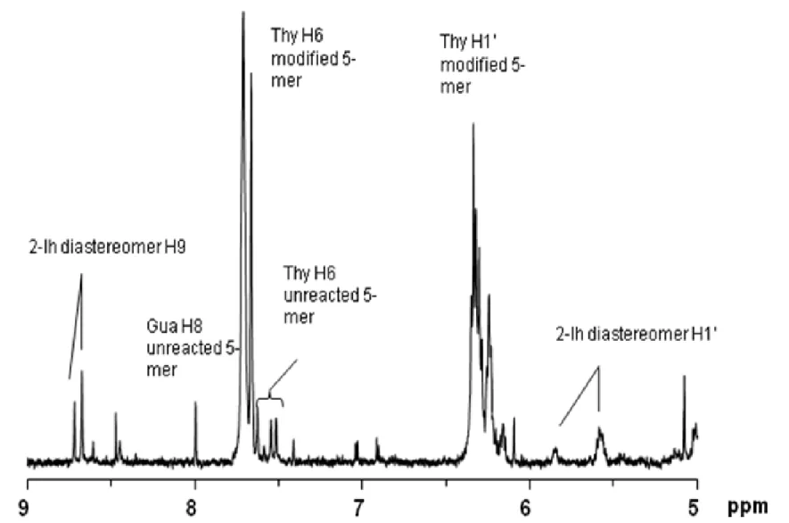
44
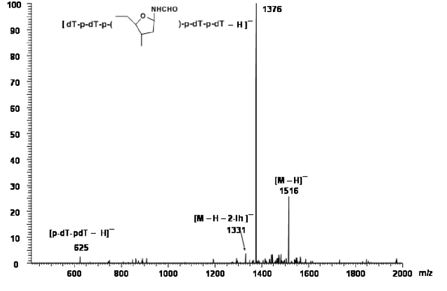
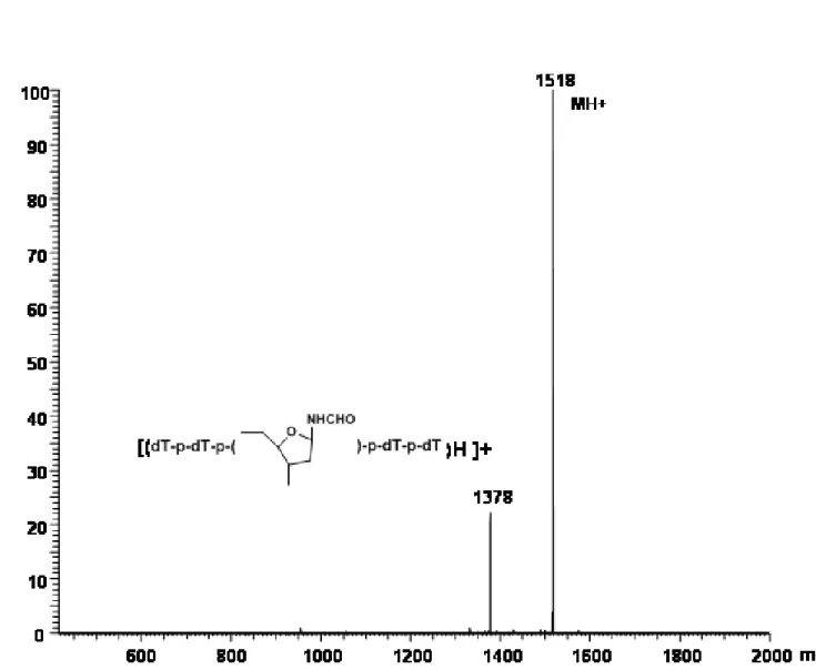
where the formyl H9 of 2-Ih is expected [Davies, J. R. et al, 1990]. The pattern observed in Figure 2.9 is consistent with formation of 2-Ih diastereomers in the oxidized 5-mer in a 1:4 ratio. Based on integrated signals from unreacted 5-mer and the 2-Ih diastereomers, > 95 % of the 5-mer was oxidized, with at least 40 % conversion to 2-Ih-containing product. The MALDI-TOF mass spectrum in the negative ion mode showed a strong ion at m/z 1516, corresponding to a gain of 34 mass units as expected for formation of the 2-Ih lesion, in addition to unmodified 5-mer (Figure 2.10). The negative ion ESI-MS acquired by loop injection (Figure 2.11) shows strong ions at m/z 1516 and 758 as expected for the [M – H] -and [M – H]2- ions, respectively, of the 5-mer containing the 2-Ih modification. In the ESI-MS, ions corresponding to unmodified 5-mer are not observed, consistent with the estimate from the NMR spectrum that only 5 % of the 5-mer was unreacted and that a substantial proportion of the 5-mer was converted to the 2-Ih-modified oligonucleotide. ESI-MS/MS of the ion at m/z 1516 (Figure 2.12) yielded a product ion at m/z 1376 corresponding to loss of a 2-iminoimidazole fragment (2-imino-5-oxo-2,5-dihydro-1H-4-imdazole-4-carboxamide) to yield a ribosyl formamide-containing 5-mer anion, a fragmentation pattern consistent with oxidation of the Gua to 2-Ih. The positive ion ESI-MS (Figure 2.13) by loop injection, was entirely in accord with this result, having ions at m/z 1518 ([MH]+) and 1540 ([MNa]+), along with product ions at m/z 1400 for loss of 2-iminoimidazole from (MNa)+, 729 [sodium
![Figure 2.1 Full scan ESI-MS + of NA-2-Ih: m/z 186 [MH] + , 208 [M + Na] + .](https://thumb-us.123doks.com/thumbv2/123dok_us/8297476.2197423/79.1188.242.958.139.721/figure-scan-esi-ms-na-ih-mh-na.webp)
![Figure 2.2 ESI-MS/MS + of NA-2-Ih: m/z 186 [MH] + , 169 [MH-NH 3 ] + , 158 [MH-CO] + , 141 [MH-formamide] +](https://thumb-us.123doks.com/thumbv2/123dok_us/8297476.2197423/80.1188.248.959.167.722/figure-esi-ms-ms-na-ih-mh-formamide.webp)