MINIREVIEW
Focal Adhesions: A Nexus for Intracellular Signaling
and Cytoskeletal Dynamics
Sarita K. Sastry1
and Keith Burridge
Department of Cell Biology and Anatomy and Lineberger Comprehensive Cancer Center, University of North Carolina, Chapel Hill, North Carolina 27599
INTRODUCTION
Focal adhesions (FAs) are specialized sites of cell attachment to the extracellular matrix (ECM) where integrin receptors link the ECM to the actin cytoskel-eton. Integrins cluster into supramolecular complexes with structural, cytoskeletal proteins like talin, vincu-lin, and␣-actinin, as well as numerous signaling mol-ecules, including c-Src, FAK, p130cas, and paxillin [1]. The composition and molecular architecture of FAs have been reviewed elsewhere [2– 4] and are beyond the scope of this brief review. FAs serve at least two significant cellular functions: to transmit force or ten-sion at adheten-sion sites to maintain strong attachments to the underlying ECM and to act as signaling centers from which numerous intracellular pathways emanate to regulate cell growth, survival, and gene expression [5, 6].
FAs are dynamic structures that assemble, disperse, and recycle (turnover) as cells migrate or enter into mitosis. Recent evidence reveals the complexity of these processes. Assembly/disassembly involves the co-ordinate regulation of Rho family GTPases through cross talk between integrins and numerous adhesion receptors (cadherins, cell adhesion molecules (CAMs), selectins, and syndecans), G-protein-coupled receptors (GPCRs), and receptor tyrosine kinases (RTKs), as well as the interplay between microtubules and actin. It is also apparent that FAs are themselves motile and het-erogeneous in composition. Finally, turnover of FAs entails communication with components of vesicle traf-ficking pathways and microtubules. This review high-lights recent findings relating to FA assembly, dynam-ics, and turnover.
FOCAL ADHESION ASSEMBLY
Contractility
FAs have long served as a model system for the study of cell–matrix interactions. These structures are prom-inent in many adherent cell types grown in culture, but are rarely observed in vivo. Several features of the tissue culture environment promote FA assembly [1]. FAs form during spreading or migration on flat, rigid substrates to which ECM components become ad-sorbed. The assembly of FAs in response to adhesion to the ECM is gradual, usually occurring within 1 to 2 h after cell attachment. Initially, nascent cell–matrix ad-hesions, or focal complexes, form at the cell periphery as a cell spreads or at the leading edge as a cell mi-grates. Focal complexes mature into FAs as cells be-come stably attached to their substrates and tension is exerted on these sites of adhesion. Actin filaments are indirectly tethered to integrins at FAs [4, 7]. In migrat-ing cells, FAs can provide traction on the substrate over which cells crawl, although some cells can migrate without FAs and large FAs retard motility due to ex-cessive adhesion [8].
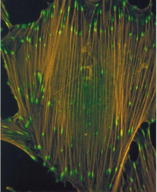
Additionally, cells in culture are grown under condi-tions that mimic a wound environment, frequently in the presence of serum factors such as lysophosphatidic acid (LPA). Normally, LPA is secreted into a wound to stimulate cell contraction, contributing to wound clo-sure. However, in tissue culture, contraction is opposed by adhesion to a rigid substrate. This generates iso-metric tension between sites of strongest adhesion. In turn, the isometric tension results in alignment of bun-dles of actin filaments (stress fibers) and the clustering of integrins, giving rise to FAs (Fig. 1). An experimen-tal system often utilized to study FA formation uses nonmigratory, adherent fibroblasts that have become quiescent as a result of being serum-starved. Under this condition, FAs and stress fibers are disassembled, despite contact with the underlying ECM. Quiescent
1
To whom reprint requests should be addressed. Fax: (919) 966-1856. E-mail: sksastry@med.unc.edu.
0014-4827/00 $35.00
25
fibroblasts respond rapidly to LPA stimulation, with FA assembly occurring within a few minutes [9].
The pioneering work of Ridley and Hall established the small GTP-binding protein, RhoA, as a cornerstone for FA assembly [10]. Activation of RhoA is essential for FA assembly in response to both integrin-mediated adhesion [11–13] and LPA stimulation [9]. The mech-anism by which RhoA drives FA assembly has recently been elucidated. RhoA stimulates actin–myosin con-tractility [14] via a kinase cascade leading to the phos-phorylation of the regulatory light chain of myosin II [15]. A downstream effector of RhoA, Rho kinase, can directly phosphorylate myosin light chain [16] and can also inhibit myosin phosphatase [17], both of which
result in enhanced light chain phosphorylation and hence increased contractility. Increased actin–myosin contractility results in bundling of actin filaments to generate stress fibers and clustering of integrins and associated proteins to form FAs [14, 18].
inhibit cell contractility as evidenced by a decrease in wrinkling of silicone rubber substrates upon which cells had been cultured [19]. In parallel, there was a decrease in the size and number of FAs. Finally, caldesmon overexpression resulted in increased cell spreading and membrane extensions, also a sign of decreased tension. Caldesmon acts downstream of RhoA, since it blocked FA assembly when coexpressed with activated forms of RhoA. It will be interesting to determine the physiological conditions under which caldesmon functions to affect FA assembly.
Activation of RhoA
Progress has also been made regarding the activa-tion of RhoA. Early work suggested that a tyrosine kinase acted upstream of RhoA and was essential for FA assembly [20]. The identity of this kinase remains elusive. It is clear, however, that many factors can regulate RhoA activity, including integrin signaling, other adhesion receptors, soluble factors like LPA, re-ceptor tyrosine kinase signaling, and components of the microtubule cytoskeleton (Fig. 2) [10, 21]. Using an affinity precipitation assay to directly measure RhoA activity, Ren et al. observed that integrin-mediated adhesion leads to a biphasic response in RhoA activity [22]. Attachment to ECM initially suppresses RhoA activity and this is followed by a modest activation phase. This activation is enhanced significantly in the presence of LPA. Thus LPA is a more potent stimulator of RhoA than is integrin-mediated adhesion to the ECM. This study also showed that adhesion is required to attenuate LPA-induced RhoA activity, since in sus-pended cells, LPA stimulation led to sustained RhoA activity.
Microtubule depolymerization was shown many years ago to stimulate cell contractility and the assem-bly of stress fibers [23]. Not surprisingly, subsequent work demonstrated that microtubule depolymerization also resulted in the assembly of FAs [24 –28]. Microtu-bule depolymerization was shown to elevate the level of myosin light chain phosphorylation [29], whereas the formation of stress fibers and FAs was blocked by the inhibitor of RhoA, Botulinum C3 exoenzyme [26, 28]. These findings suggested that microtubule depolymer-ization stimulates increased actin–myosin contractility by activating RhoA. This was confirmed by direct mea-surement of RhoA activity [22].
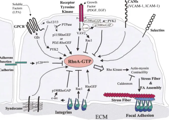
The signal transduction pathways leading to RhoA activation by LPA, integrins, RTKs, or microtubule depolymerization are intense areas of investigation. A schematic diagram of proposed pathways regulating RhoA activation is shown in Fig. 2. The immediate upstream activators of RhoA are guanine nucleotide
exchange factors (GEFs), which promote the exchange of GDP for GTP, thus inducing an active conformation of RhoA (or other family members) [30, 31]. The path-way by which LPA elevates RhoA–GTP levels may be the best characterized at present. LPA binds to a hep-tahelical G-protein-coupled receptor and activates G␣12/13 or G␥ subunits, which then associate with GEFs for RhoA. Introduction of G␣12/13 or G␥ sub-units into quiescent fibroblasts on their own stimulates FA assembly [32, 33]. In the case of G␣12/13 subunits, p115 RhoGEF [34] or PDZ RhoGEF [35] appear to be direct targets. It has also been suggested that the ty-rosine kinase PYK2 (CAK, CADTK, RAFTK) may play a role in GPCR signaling to RhoA [36].
The regulation of RhoA activity by integrins is com-plex. Integrins can either stimulate or inhibit RhoA activity depending on the cell type, engagement of spe-cific integrins, and time course of engagement [22, 37, 38]. This duality may reflect the role of integrin-medi-ated signals in promoting membrane extensions, a con-dition in which low RhoA activity is desirable, versus the role of integrins in establishing strong attachments across which tension is transmitted, for which higher RhoA activity is needed. As discussed above, in fibro-blasts, integrin engagement initially inhibits RhoA ac-tivity but later activates it, correlating with the com-pletion of cell spreading [22], during which time integrin-mediated activation of Rac1 is high [39]. Barry and colleagues found that addition of RGD pep-tides to quiescent fibroblasts stimulated FA and stress fiber assembly [11]. In adenocarcinoma cells, which are epithelial in origin, crosslinking of␣64 integrin stim-ulates RhoA activity, whereas crosslinking of1 inte-grins inhibits RhoA [37]. Arthur and colleagues iden-tified a pathway by which integrin engagement results initially in a decrease in RhoA activity [38]. It was found that incubation of fibroblasts with integrin li-gands, such as RGD peptides, caused a rapid drop in RhoA activity, but that this did not occur in cells defi-cient in the Src family tyrosine kinases (Src, Fyn, and Yes). This integrin-mediated drop in RhoA activity was restored if c-Src was reexpressed in these cells. Down-stream from c-Src, p190RhoGAP was identified as a target that is phosphorylated and activated, in re-sponse to integrin engagement. Interestingly, v-Src ex-pression in fibroblasts has long been known to disrupt FAs and stress fibers (see below).
discov-ered [30, 31, 40, 41] but little information exists con-cerning how these GEFs become activated by integrin signaling. One exception is Vav1, a hematopoietic GEF for Rho family GTPases. Vav1 is tyrosine phosphory-lated and activated in response to integrin engagement or clustering [42– 45].
Similar to integrins, receptor tyrosine kinases trans-mit both positive and negative signals to RhoA. Initial stimulation (within minutes) with growth factors such as PDGF or EGF promotes the formation of membrane extensions through activation of Rac1 while suppress-ing RhoA [46]. RhoA is likely inhibited via Src-depen-dent activation of p190 RhoGAP [47]. Furthermore, recruitment of p190 RhoGAP to RTKs may be regu-lated by PYK2 [48]. Prolonged addition of PDGF (more than 2 h) results in FA assembly, suggesting that growth factors can activate RhoA. Recently, a GEF that may mediate growth factor-dependent regulation of RhoA has been identified. Vav2, a widely expressed relative of Vav1, is tyrosine phosphorylated and acti-vated in response to EGF or PDGF stimulation [49 – 51]. Vav2 activates several Rho family members, in-cluding Rac1 and RhoA [49 –53].
Finally, how might depolymerization of microtubules lead to enhanced RhoA activity? It has been suggested that intact microtubules may sequester GEFs for RhoA that are released upon microtubule depolymerization [26]. In support of this idea, several GEFs have been found to bind tubulin or microtubules. For example, in hematopoietic cells the exchange factor Vav1 binds tubulin, although whether this affects GEF activity has not been determined [54]. The association of more widely expressed Vav family members, Vav2 or Vav3, with microtubules has not been fully investigated. GEFH1, which is specific for RhoA, has recently been shown to bind microtubules [55]. The existence of mul-tiple GEFs for RhoA reflects either a functional redun-dancy or that specific GEFs act on RhoA in response to distinct stimuli.
Although integrins and GPCRs are a major focus in upstream regulation of RhoA, other adhesion receptors also promote FA assembly, likely via the activation of RhoA. Recently, a role for syndecans in FA assembly has been demonstrated. Syndecan-4 is a transmem-brane member of the heparan sulfate proteoglycan family that localizes in FAs [56]. Its overexpression in cultured fibroblasts increases FAs and stress fibers [57, 58], whereas syndecan-4 null cells have impaired FAs [59]. Additionally, FA assembly on the cell binding domain of fibronectin is promoted by antibody ligation of syndecan-4 in a RhoA-dependent manner [60]. A particularly intriguing system in which multiple adhe-sion receptors, including selectins, integrins, and other CAMs, cooperate to potentially regulate RhoA is the interaction of leukocytes with endothelial cells during
inflammation. Adhesion of monocytes to endothelial cells induces the assembly of stress fibers and presum-ably FAs in the endothelial cells. Crosslinking of the endothelial cell adhesion molecules VCAM-1, ICAM-1, or E-selectin, but not ICAM-2 or ICAM-3, also stimu-lates stress fiber formation [61]. It will be important to determine whether these effects are due to increased RhoA activity. In contrast to the above-mentioned cell adhesion molecules, the crosslinking of which pro-motes FA assembly, the formation of adherens junc-tions of the cadherin type tends to inhibit FA assembly. The mechanism of this inhibition has not been deter-mined. Recent work, however, has identified a cad-herin-binding protein, p120 catenin, as a regulator of Rho family GTPases [62, 63]. Overexpression of p120 catenin disrupts focal adhesions and stress fibers and decreases RhoA activity in cells. In addition, p120 cate-nin elevates the activity of Rac1 and Cdc42 and binds to Vav2, a Rho family GEF [62].
FOCAL ADHESION DISASSEMBLY
FAs disassemble or disperse under a number of physiological situations. For example, adhesions to the ECM are released at the rear of a migrating cell and this is accompanied by a disruption of FAs. Another instance of FA disassembly occurs during mitosis, during which cells lose their attachments to the ECM and adopt a round morphology. Finally, in oncogenically transformed fibroblasts, FA integrity is often compromised [64]. Since FA assembly in-volves both the activation of RhoA and the stimula-tion of contractility, loss of FAs would ostensibly involve mechanisms that counteract these pathways. The role of integrin signaling in FA disassembly has recently been reviewed [65]. Here we highlight some novel and significant observations.
GAPs
Interplay of Rho and Ras Family GTPases
Our understanding of FA disassembly is further complicated by recent evidence that additional Rho family GTPases can affect FA organization. It has long been noted phenomenologically that the actions of Rac and RhoA are functionally antagonistic. Rac1 promotes membrane extension, whereas in many situations RhoA induces membrane retraction. Such a reciprocal relationship is most clearly seen in migrating fibro-blasts or in growth cones of neurons. In epithelial cells, activation of Rac promotes the assembly of cell– cell junctions and blocks FA formation, while activation of RhoA promotes a fibroblastic phenotype [46, 69 –71]. Rottner et al. have recently shown that expression of a dominant negative Rac1 mutant in quiescent fibro-blasts induces FA assembly [72]. This effect could be attributed to regulation of contractility by Rac1 effec-tors downstream of RhoA [73]. However, using affinity precipitation assays to measure RhoA activity, Sander et al. have shown that activation of Rac1 can itself suppress RhoA activity [46]. This was shown either by expression of TIAM-1, an exchange factor for Rac1, or by expression of activated mutants of Rac1 in NIH3T3 fibroblasts. Cdc42 expression also antagonized RhoA activity. The mechanism by which Rac1 (or Cdc42) inhibits RhoA is currently unknown. A signaling com-plex that links Rac1 effectors to GAPs for RhoA would be one potential mechanism.
A newly identified Rho family member, RhoE/rnd, also disrupts FAs and stress fibers [74, 75]. RhoE/rnd was identified as a binding partner for p190RhoGAP [76]. Unlike RhoA, which cycles between GDP- and GTP-bound states, RhoE is constitutively active in the GTP-bound state and is insensitive to GAPs. It is un-clear how RhoE functions. One possibility is that it titrates away downstream effectors from RhoA. It is also possible that RhoE can inhibit RhoA activity, al-though this has not been directly demonstrated. The function of RhoE within cells remains undetermined.
Finally, the Ras pathway is suggested to play a role in FA turnover. Active Ras is required for the turnover of FAs during cell migration [77]. Interestingly, the duration of Ras activation affects the activation state of RhoA. Transient expression of activated Ras in epithe-lial cells results in activation of Rac1 and inhibition of RhoA activity [71]. In contrast, sustained Ras activa-tion promotes enhanced RhoA activity [71] as well as a fibroblastic morphology [70, 71]. Knockout cell lines lacking p120 RasGAP, an upstream inhibitor of Ras, are unable to adopt a polarized morphology [78] or turn over FAs. This effect of p120 RasGAP is independent of Ras regulation. Instead, p120 RasGAP regulates cell polarity, and presumably FA turnover, via its interac-tion with p190 RhoGAP.
Tyrosine Phosphorylation
As mentioned earlier, signaling from a tyrosine ki-nase is necessary for FA assembly. It was thought that Src family kinases or perhaps FAK may be required for FAs to form. However, through the analysis of knock-out cell lines, it is apparent that Src family kinases play a role in FA disassembly or turnover [80]. As discussed above, c-Src is required for inhibition of RhoA by integrins [38]. Furthermore, overexpression of v-Src leads to FA disruption, while the expression of kinase-inactive v-Src in normal cells leads to formation of exaggerated FAs [81]. FAK is also likely to play a role in FA turnover or disassembly. FAK-null cells possess abnormally large FAs and are unable to mi-grate [82]. In contrast, overexpression of FAK stimu-lates motility [83]. Consistent with this finding is the observation that in permeabilized fibroblasts, an in-crease in tyrosine phosphorylation accompanies FA disruption in response to ATP [79].
Significant progress has also been made in identify-ing protein tyrosine phosphatases that may promote FA disassembly or turnover. Knockout cell lines of either SHP-2 or PTP-PEST exhibit enhanced FAs [84, 85]. The mechanism of this phenotype remains to be determined. However, one or more PTPases may act upstream of RhoA. Using calpeptin, an inhibitor orig-inally designed for the Ca2⫹-dependent protease
cal-pain, it was recently demonstrated that this inhibitor stimulates FA assembly in quiescent fibroblasts through inhibition of a PTPase [86]. Calpeptin-induced FA assembly was blocked by C3 exoenzyme, indicating that the PTPase acts upstream of RhoA. In addition two transmembrane PTPases, LAR and RPTP␣, have been shown to localize in FAs under restricted condi-tions [87, 88]. Finally, PTPases may play a major role in regulation of FA disassembly during mitosis. Com-parison of interphase versus mitotic cells shows that FAK, p130cas, and paxillin are dephosphorylated on tyrosine residues but phosphorylated on serine and threonine in mitosis [89]. These proteins are rapidly tyrosine phosphorylated in response to adhesion [3, 5, 18]. In mitotic extracts, FAK kinase activity is de-creased and its associations with p130cas and paxillin are disrupted. The disruption of this complex is thought to prevent integrin signaling until the comple-tion of cytokinesis. It will be interesting to determine which tyrosine phosphatases and which serine–threo-nine kinases act on these proteins in mitosis.
HETEROGENEITY OF CELL–MATRIX ADHESIONS
is noteworthy, however, that recent observations point to considerable molecular and structural diversity among FAs in a single cell and also within individual FAs. Using fluorescence ratio imaging, the distribution of several FA proteins was compared. This analysis has identified at least three structurally distinct types of adhesion sites whose molecular compositions differ. Classical FAs are large, spearheaded or ellipsoid in shape, and located at the cell periphery and contain vinculin, paxillin, phosphotyrosine, and␣v3 integrins [90]. In contrast, fibrillar adhesions are elongated, cen-trally located, and contain tensin,␣51 integrins, and fibronectin with little or no phosphotyrosine, vinculin, or paxillin. Finally, “mosaic” FAs are morphologically similar to “classical” FAs, but their content is variable. Interestingly, the assembly of these distinct adhesions depends on several critical factors. The first is contrac-tility. The assembly of classical FAs but not fibrillar adhesions was sensitive to contractility inhibitors [90]. The second is the physical state of the ECM. A nonim-mobilized fibronectin matrix that is adsorbed to the substrate promotes fibrillar adhesions, while an immo-bilized matrix, crosslinked to the substrate, leads to the formation of classical FAs [91]. The third factor is the type of integrin involved. Classical FAs typically contain ␣v3 integrins and fibrillar adhesions contain ␣51 [90]. Using GFP-tagged c-Src, Felsenfeld and co-workers noted that c-Src selectively localizes in phos-photyrosine-rich FAs formed by␣v3 integrins, but not those formed by␣51 integrins [92]. This type of anal-ysis has been performed only on some FA components. A more comprehensive survey will be informative and may reveal additional complexities.
TURNOVER AND DYNAMICS OF FOCAL ADHESIONS
Up to this point, much of this review has dealt with potential pathways leading to FA formation or disas-sembly. These are active, dynamic processes inti-mately associated with turnover or recycling of FAs. Information from knockout cell lines and biochemical screening has led to the identification of potential reg-ulators of FA turnover. Additionally, the use of elegant imaging techniques to visualize cell–substrate contact dynamics in live cells has contributed to our knowledge of how FAs undergo remodeling and turnover during cell spreading and motility.
Movement of Focal Adhesions
FAs that form at the front of a migrating cell gener-ally remain fixed relative to the substrate as the cell moves over them. FAs then disperse at the cell tail [93, 94]. Stationary FAs maintain stable attachments to the ECM to resist actin–myosin contraction that pro-pels the cell forward. However, in some situations FAs move relative to the substrate. A recent study found that static FAs occur primarily in motile cells. Using GFP-tagged1 integrin subunits, Smilenov et al. com-pared FA movement in stationary (nonmotile) and mi-grating fibroblasts [95]. Using time-lapse imaging of live cells and overlaying of sequential images, they found that FAs in nonmotile cells are not static. In-stead, FAs in stationary cells were observed to move linearly toward the cell center. This movement de-pended on actin–myosin contractility since FA move-ment was not observed in the presence of BDM, a myosin inhibitor. Microtubule disassembly, which
en-syndecan-4 stimulates focal adhesion and stress fiber assembly likely through activation of RhoA (dotted arrows). The formation of adherens junctions, containing cadherins, antagonizes focal adhesion and stress fiber formation, possibly via p120 catenin, whose overexpression inhibits RhoA activity. Finally, integrins transmit both positive and negative signals to RhoA. Initially, engagement of integrins with ECM inhibits RhoA. Integrins activate p190 RhoGAP through a c-Src-dependent mechanism. Integrins also activate Rac1 as a cell spreads, which can antagonize RhoA activity. As stable adhesions form, integrins activate RhoA, most likely through an integrin-dependent Rho GEF. Downstream of RhoA, actin–myosin contractility stimulates actin stress fiber formation and clustering of integrins and associated proteins to form focal adhesions. Actin–myosin contractility is positively regulated by RhoA effectors, like Rho kinase, and negatively regulated by caldesmon, an actin-binding protein.
hances RhoA activity [22] and contractility [23, 29], increased the rate of FA movement. Interestingly, al-though FA movement was typically not observed in migrating cells, distinct zones of FA behavior were discerned. At the leading edge of a migrating cell is a formation zone; between the leading edge and the nu-cleus, a persistence zone exists in which stable FAs continue to grow and mature; between the nucleus and the tail a culling zone exists, where FAs turn over; at the cell tail is a small motile zone [95]. The different behaviors of FAs in both nonmigratory fibroblasts and polarized, migrating fibroblasts are depicted in Fig. 3. These findings have led to a molecular clutch model in which FAs transition between a motile and a nonmotile state in a contractility-dependent manner, to balance adhesive forces and migratory cues. How universal this behavior of FAs is remains to be determined.
Cross Talk between Microtubules and Focal Adhesions
The state of the microtubule cytoskeleton can greatly influence the organization of FAs and actin stress fi-bers. An emerging view is that the relationship be-tween microtubules and FAs is reciprocal, in which the organization of one affects the dynamics of the other (Fig. 3). As mentioned earlier, disruption of microtu-bules activates RhoA, leading to increased actin–myo-sin contractility, FAs, and stress fibers [23–29]. Con-versely, elevated RhoA activity stabilizes microtubules [96]. In addition, Waterman-Storer and co-workers found that active microtubule polymerization was as-sociated with increased Rac1 activity and membrane protrusions [97]. Given the antagonism between Rac1 and RhoA, these findings suggest that sites of micro-tubule growth would be associated with locally high concentrations of active Rac1 and decreased RhoA ac-tivity. Consequently, microtubule growth would be ex-pected to promote focal adhesion disassembly. Indeed, Small and co-workers found that microtubule polymer-ization is associated with local destabilpolymer-ization of focal adhesions [98]. Small and his colleagues noted earlier that there is an association between the ends of micro-tubules and focal adhesions [98 –100] and they specu-lated that this association might stabilize the adhe-sions. More recent work from this group, however, has shown that this association is antagonistic: the target-ing of microtubules to focal adhesions causes the dis-assembly of these structures [101]. These investigators propose that growing microtubules negatively regulate focal adhesions by delivering a localized relaxing signal to this region. The evidence points to microtubule dy-namics regulating the activity of Rho family GTPases in very localized regions of the cytoplasm, but proof of
this awaits assays that will reveal the activity of these Rho family proteins at a subcellular level.
Delivery of Components to Focal Adhesions
An emerging concept is that some focal adhesion proteins are actively targeted to and from focal adhe-sions via a vesicle trafficking pathway. This also likely involves microtubules and may be another way in which microtubule behavior affects focal adhesion turnover. Several studies have implicated the ARF (ADP-ribosylation factor) family of GTPases in this targeting. ARFs have been shown to control intracel-lular membrane trafficking, including the delivery of membrane to sites of membrane protrusion [102]. They have also been shown to have a role in regulating cytoskeletal organization through an interplay with Rho family GTPases [103, 104]. ARF1 activity has been shown to be required for the recruitment of paxillin from the perinuclear compartment to focal adhesions [103]. Like Rho family GTPases, ARFs cycle between an inactive GDP-bound state and an active GTP-bound state. The mechanism by which paxillin is recruited to focal adhesions may lie in its interactions with ARF-GAPs. Paxillin has been shown to associate with a 95-kDa ARF-GAP, variously named PKL [105], PAG3/ PAP␣[106], or GIT [107]. This ARF-GAP acts on ARF6 [106] and is localized to focal adhesions [105]. Kondo et al. found that the activity of this ARF-GAP prevents paxillin recruitment to focal adhesions [106]. Another protein that is recruited to focal adhesions from the perinuclear regions is v-Src [108]. However, recruit-ment of v-Src to FAs does not require its kinase activity [81]. Whether this recruitment involves ARF activity has not yet been determined. An intriguing connection between ARFs and Src is suggested by the finding that ASAP-1, another ARF-GAP that localizes to FAs [109], is phosphorylated by Src family kinases [110]. Finally, some integrins associate with ARF-GEFs. For exam-ple, 2 integrin cytoplasmic domains associate with cytohesin-1, an ARF-GEF, and this interaction is im-plicated in the regulation of integrin affinity [111, 112]. The roles of ARFs and their regulatory GAPs and GEFs in focal adhesion turnover are only just begin-ning to be discovered. This promises to be an exciting area in the future.
CONCLUDING REMARKS
many components have yet to be compared. The role of ARFs, along with their regulatory GEFs and GAPs, is currently enigmatic. The evidence for cross talk be-tween ARFs and Rho family proteins is particularly intriguing. A great many factors influence the activity of Rho family proteins. In general, the effects of agents that stimulate or inhibit RhoA activity have been mea-sured on whole populations of cells. In many situa-tions, however, very localized, subcellular changes in RhoA activity are likely to be important. This is sug-gested by the studies on microtubule dynamics. The evidence indicates that the behavior of individual mi-crotubules regulates the local activity of RhoA or Rac1, thereby affecting FA assembly and turnover in specific regions of the cell. The development of methods to determine subcellular changes in activity of Rho family proteins should yield a greater understanding of how various factors control FA dynamics.
We thank Dr. Becky Worthylake, Bill Arthur, and Betty P. Liu for critical review of the manuscript and for helpful discussions. We apologize to those individuals whose work we were unable to cite due to space limitations. This work was supported by NIH Grants GM29860 and HL45100 (Keith Burridge).
REFERENCES
1. Burridge, K., and Chrzanowska-Wodnicka, M. (1996). Focal adhesions, contractility, and signaling. Annu. Rev. Cell Dev. Biol. 12, 463–519.
2. Jockusch, B., Bubeck, P., Giehl, K., Kroemker, M., Moschner, J., Rothkegel, M., Rudiger, M., Schluter, K., Stanke, G., and Winkler, J. (1995). The molecular architecture of focal adhe-sions. Annu. Rev. Cell Dev. Biol. 11, 379 – 416.
3. Yamada, K., and Geiger, B. (1997). Molecular interactions in cell adhesion complexes. Curr. Opin. Cell Biol. 9, 76 – 85. 4. Critchley, D. (2000). Focal adhesions—The cytoskeletal
con-nection. Curr. Opin. Cell Biol. 12, 133–139.
5. Schwartz, M., Schaller, M., and Ginsberg, M. (1995). Integrins: Emerging paradigms of signal transduction. Annu. Rev. Cell Biol. 11, 549 –599.
6. Howe, A., Aplin, A., Alahari, S., and Juliano, R. L. (1998). Integrin signaling and cell growth control. Curr. Opin. Cell Biol. 10, 220 –231.
7. Calderwood, D., Shattil, S., and Ginsberg, M. (2000). Integrins and actin filaments: Reciprocal regulation of cell adhesion and signaling. J. Biol. Chem. 275, 22607–22610.
8. Huttenlocher, A., Sandborg, R., and Horwitz, A. F. (1995). Adhesion in cell migration. Curr. Opin. Cell Biol. 7, 697–706. 9. Ridley, A., and Hall, A. (1992). The small GTP-binding protein Rho regulates the assembly of focal adhesions and actin stress fibers in response to growth factors. Cell 70, 389 –399. 10. Hall, A. (1998). Rho GTPases and the actin cytoskeleton.
Sci-ence 279, 509 –514.
11. Barry, S. T., Flinn, H. M., Humphries, M. J., Critchley, D. R., and Ridley, A. J. (1996). Requirement for Rho in integrin signalling. Cell Adhes. Commun. 4, 387–398.
12. Hotchin, N. A., and Hall, A. (1995). The assembly of integrin adhesion complexes requires both extracellular matrix and intracellular Rho/Rac GTPases. J. Cell Biol. 131, 1857–1865.
13. Clark, E., King, W., Brugge, J., Symons, M., and Hynes, R. (1998). Integrin-mediated signals regulated by members of the Rho family of GTPases. J. Cell Biol. 142, 573–586.
14. Chrzanowska-Wodnicka, M., and Burridge, K. (1996). Rho-stimulated contractility drives the formation of stress fibers and focal adhesions. J. Cell Biol. 133, 1403–1415.
15. Kaibuchi, K., Kuroda, S., and Amano, M. (1999). Regulation of the cytoskeleton and cell adhesion by the Rho family GTPases in mammalian cells. Annu. Rev. Biochem. 68, 459 – 486. 16. Amano, M., Ito, M., Kimura, K., Fukata, Y., Chihara, K.,
Nakano, T., Matsuura, Y., and Kaibuchi, K. (1996). Phosphor-ylation and activation of myosin by Rho-associated kinase (Rho-kinase). J. Biol. Chem. 271, 20246 –20249.
17. Kimura, K., Ito, M., Amano, M., Chihara, K., Fukata, Y., Nakafuku, M., Yamamori, B., Feng, J., Nakano, T., Okawa, K., Iwamatsu, A., and Kaibuchi, K. (1996). Regulation of myosin phosphatase by Rho and Rho-associated kinase (Rho-kinase). Science 273, 245–248.
18. Burridge, K., Chrzanowska-Wodnicka, M., and Zhong, C. (1997). Focal adhesion assembly. Trends Cell Biol. 7, 342–347. 19. Helfman, D., Levy, E., Berthier, C., Shtutman, M., Riveline, D., Grosheva, I., Lachish-Zalait, A., Elbaum, M., and Bershad-sky, A. (1999). Caldesmon inhibits nonmuscle cell contractility and interferes with the formation of focal adhesions. Mol. Biol. Cell 10, 3097–3112.
20. Ridley, A., and Hall, A. (1994). Signal transduction pathways regulating Rho-mediated stress fiber formation: Requirement for a tyrosine kinase. EMBO J. 13, 2600 –2610.
21. Schwartz, M., and Shattil, S. (2000). Signaling networks link-ing integrins and Rho family GTPases. Trends Biochem. Sci.
25, 388 –391.
22. Ren, X., Kiosses, W., and Schwartz, M. (1999). Regulation of the small GTP-binding protein Rho by cell adhesion and the cytoskeleton. EMBO J. 18, 578 –585.
23. Danowski, B. (1989). Fibroblast contractility and actin reorga-nization are stimulated by microtubule inhibitors. J. Cell Sci.
93, 255–266.
24. Bershadsky, A., Chausovsky, A., Becker, E., Lyubimova, A., and Geiger, B. (1996). Involvement of microtubules in the control of adhesion-dependent signal transduction. Curr. Biol.
6, 1279 –1389.
25. Enomoto, T. (1996). Microtubule disruption induces the forma-tion of actin stress fibers and focal adhesions in cultured cells: Possible involvement of the Rho signal cascade. Cell Struct. Funct. 2, 317–326.
26. Liu, B. P., Chrzanowska-Wodnicka, M., and Burridge, K. (1998). Microtubule depolymerization induces stress fibers, focal adhesions, and DNA synthesis via the GTP-binding pro-tein Rho. Cell Adhes. Commun. 5, 249 –255.
27. Pletjushkina, O., Belkin, A., Ivanova, O., Oliver, T., Jacobson, K., and Vasiliev, J. (1998). Maturation of cell–substratum focal adhesions induced by depolymerization of microtubules is induced by increased cortical tension. Cell Adhes. Commun.
5, 121–135.
28. Zhang, Q., Magnusson, M., and Mosher, D. (1997). Lysophos-phatidic acid and microtubule-destabilizing agents stimulate fibronectin matrix assembly through Rho-dependent actin stress fiber formation and cell contraction. Mol. Biol. Cell 8, 1415–1425.
30. Van Aelst, L., and D’Sousa-Schorey, C. (1997). Rho GTPases and signaling networks. Genes Dev. 11, 2295–2322.
31. Whitehead, I., Campbell, S., Rossman, K., and Der, C. (1997). Dbl family proteins. Biochim. Biophys. Acta 1332, F1–F23.
32. Buhl, A., Johnson, N., Dhanasekaran, N., and Johnson, G. (1995). G␣12 and G␣13 stimulate Rho-dependent stress fiber formation and focal adhesion assembly. J. Biol. Chem. 270, 24631–24634.
33. Ueda, H., Itoh, H., Yamauchi, J., Morishita, R., Kaziro, Y., Kato, K., and Asano, T. (2000). G protein␥subunits induce stress fiber formation and focal adhesion assembly in a Rho-dependent manner in HeLa cells. J. Biol. Chem. 275, 2098 – 2102.
34. Hart, M., Jiang, X., Kozasa, T., Roscoe, W., Singer, W., Gil-man, A., Sternweis, P., and Bollag, G. (1998). Direct stimula-tion of the guanine nucleotide exchange activity of p115 Rho-GEF by G␣13. Science 280, 2112–2114.
35. Fukuhara, S., Murga, C., Zohar, M., Igishi, T., and Gutkind, J. (1999). A novel PDZ domain containing guanine nucleotide exchange factor links heterotrimeric G proteins to Rho. J. Biol. Chem. 274, 5868 –5879.
36. Shi, C., Sinnarajah, S., Cho, H., Kozasa, T., and Kehrl, J. (2000). G␣13-mediated PYK2 activation: PYK2 is a mediator of G13␣-induced serum response element dependent tran-scription. J. Biol. Chem. 275, 24470 –24476.
37. O’Connor, K., Ngyuen, B., and Mercurio, A. (2000). RhoA func-tion in lamellae formafunc-tion and migrafunc-tion is regulated by the
␣64 integrin and cAMP metabolism. J. Cell Biol. 148, 253– 258.
38. Arthur, W. T., Petch, L., and Burridge, K. (2000). Integrin engagement suppresses RhoA activity via a c-Src-dependent mechanism. Curr. Biol. 10, 719 –722.
39. del Pozo, M., Price, L., Alderson, N., Ren, X., and Schwartz, M. (2000). Adhesion to the extracellular matrix regulates the coupling of the small GTPase Rac to its effector PAK. EMBO J.
19, 2008 –2014.
40. Tatsumoto, T., Xie, X., Blumenthal, R., Okamoto, I., and Miki, T. (1999). Human ECT2 is an exchange factor for Rho GTPases, phosphorylated in G2/M phases, and involved in cytokinesis. J. Cell Biol. 147, 921–928.
41. Rumenapp, U., Blomquist, A., Schworer, G., Schablowski, H., Psoma, A., and Jakobs, K. (1999). Rho-specific binding and guanine nucleotide exchange catalysis by KIAA0380, a Dbl family member. FEBS Lett. 459, 313–318.
42. Zheng, L., Sjolander, A., Eckerdal, J., and Andersson, T. (1996). Antibody-induced engagement of beta 2 integrins on adherent human neutrophils triggers activation of p21ras through tyrosine phosphorylation of the protooncogene prod-uct Vav. Proc. Natl. Acad. Sci. USA 93, 8431– 8436.
43. Yron, I., Deckert, M., Reff, M., Munshi, A., Schwartz, M., and Altman, A. (1999). Integrin-dependent tyrosine phosphoryla-tion and growth regulaphosphoryla-tion by Vav. Cell Adhes. Commun. 7, 1–11.
44. Gotoh, A., Takahira, H., Geahlen, R., and Broxmeyer, H. (1997). Cross-linking of integrins induces tyrosine phosphory-lation of the proto-oncogene product Vav and the protein ty-rosine kinase Syk in human factor-dependent myeloid cells. Cell Growth Differ. 8, 721–729.
45. Miranti, C. K., Leng, L., Maschberger, P., Brugge, J. S., and Shattil, S. J. (1998). Identification of a novel integrin signaling pathway involving the kinase Syk and the guanine nucleotide exchange factor Vav1. Curr. Biol. 8, 1289 –1299.
46. Sander, E., ten Klooster, J., van Delft, S., van der Kammen, R., and Collard, J. (1999). Rac downregulates Rho activity: Recip-rocal balance between both GTPases determines cellular mor-phology and migratory behavior. J. Cell Biol. 147, 1009 –1021.
47. Chang, J., Gill, S., Settleman, J., and Parsons, S. (1995). c-Src regulates the simultaneous rearrangement of actin cytoskele-ton, p190RhoGAP, and p120RasGAP following epidermal growth factor stimulation. J. Cell Biol. 130, 355–368. 48. Zrihan-Licht, S., Fu, Y., Settleman, J., Schinkmann, K.,
Shaw, L., Keydar, I., Avraham, S., and Avraham, H. (2000). RAFTK/PYK2 tyrosine kinase mediates the association of p190RhoGAP with RasGAP and is involved in breast cancer cell invasion. Oncogene 19, 1318 –1328.
49. Liu, B. P., and Burridge, K. (2000). Vav2 activates Rac1, Cdc42 and RhoA downstream from growth factor receptors but not1 integrins. Mol. Cell. Biol. 20, 7160 –7169.
50. Moores, S., Selfors, L., Fredericks, J., Breit, T., Fujikawa, K., Alt, F., Brugge, J., and Swat, W. (2000). Vav family proteins couple to diverse cell surface receptors. Mol. Cell. Biol. 20, 6364 – 6373.
51. Pandey, A., Podtelejnikov, A., Blagoev, B., Bustelo, X., Mann, M., and Lodish, H. (2000). Analysis of receptor signaling path-ways by mass spectrometry: Identification of Vav-2 as a sub-strate of the epidermal and platelet-derived growth factor receptors. Proc. Natl. Acad. Sci. USA 97, 179 –184.
52. Schuebel, K., Movilla, N., Rosa, J., and Bustelo, X. (1998). Phosphorylation-dependent and constitutive activation of Rho proteins by wild-type and oncogenic Vav-2. EMBO J. 16, 6608 – 6621.
53. Abe, K., Rossman, K., Liu, B. P., Ritola, K., Chiang, D., Camp-bell, S., Burridge, K., and Der, C. (2000). Vav2 is an activator of Cdc42, Rac1, and RhoA. J. Biol. Chem. 275, 10141–10149.
54. Fernandez, J., Keshvara, L., Peters, J., furlong, M., Harrison, M., and Geahlen, R. (1999). Phosphorylation- and activation-independent association of the tyrosine kinase substrates Cbl and Vav with tubulin in B-cells. J. Biol. Chem. 274, 1401– 1406.
55. Ren, Y., Li, R., Zheng, Y., and Busch, H. (1998). Cloning and characterization of GEF-H1, a microtubule-associated guanine nucleotide exchange factor for Rac and Rho GTPases. J. Biol. Chem. 273, 34954 –34960.
56. Couchman, J., and Woods, A. (1999). Syndecan-4 and inte-grins: Combinatorial signaling in cell adhesion. J. Cell Sci.
112, 3415–3420.
57. Echtermeyer, F., Baciu, P., Saoncella, S., Ge, Y., and Goetinck, P. (1999). Syndecan-4 core protein is sufficient for the assem-bly of focal adhesions and actin stress fibers. J. Cell Sci. 112, 3433–3441.
58. Longley, R., Woods, A., Fleetwood, A., Cowling, G., Gallagher, J., and Couchman, J. (1999). Control of morphology, cytoskel-eton, and migration by syndecan-4. J. Cell Sci. 112, 3421– 3431.
59. Ishiguro, K., Kadomatsu, K., Kojima, T., Muramatsu, H., Tsu-zuki, S., Nakamura, E., Kusugami, K., Saito, H., and Mura-matsu, T. (2000). Syndecan-4 deficiency impairs focal adhesion formation only under restricted conditions. J. Biol. Chem. 275, 5249 –5252.
61. Wojciak-Stothard, B., Williams, L., and Ridley, A. (1999). Monocyte adhesion and spreading on human endothelial cells is dependent on Rho-regulated receptor clustering. J. Cell Biol. 145, 1293–1307.
62. Noren, N., Liu, B., Burridge, K., and Kreft, B. (2000). p120 catenin regulates the actin cytoskeleton via Rho family GTPases. J. Cell Biol. 150, 567–580.
63. Anastasiadis, P., Moon, S., Thoreson, M., Crawford, H., Zheng, Y., and Reynolds, A. (2000). Inhibition of RhoA by p120 cate-nin. Nature Cell Biol. 2, 637– 644.
64. Hynes, R. (1992). Integrins: Versatility, modulation, and sig-naling in cell adhesion. Cell 69, 11–25.
65. Schoenwaelder, S. M., and Burridge, K. (1999). Bidirectional signaling between the cytoskeleton and integrins. Curr. Opin. Cell Biol. 11, 274 –286.
66. Taylor, J., Macklem, M., and Parsons, J. T. (1999). Cytoskel-etal changes induced by GRAF, the GTPase regulator associ-ated with focal adhesion kinase, are mediassoci-ated by Rho. J. Cell Sci. 112, 231–242.
67. Sekimata, M., Kabuyama, Y., Emori, Y., and Homma, Y. (1999). Morphological changes and detachment of adherent cells induced by p122, a GTPase-activating protein for Rho
274, 17757–17762.
68. Jantsch-Plunger, V., Gonczy, P., Romano, A., Schnabel, H., Hamill, D., Schnabel, R., Hyman, A., and Glotzer, M. (2000). CYK-4: A Rho family GTPase activating protein (GAP) re-quired for central spindle formation and cytokinesis. J. Cell Biol. 149, 1391–1404.
69. Braga, V., Machesky, L., Hall, A., and Hotchin, N. (1997). The small GTPases Rho and Rac are required for the establish-ment of cadherin-dependent cell– cell contacts. J. Cell Biol.
137, 1421–1431.
70. Zhong, C., Kinch, S., and Burridge, K. (1997). Rho-stimulated contractility contributes to the fibroblastic phenotype of Ras-transformed epithelial cells. Mol. Biol. Cell 8, 2329 –2344. 71. Zondag, G., Evers, E., ten Klooster, J., Janssen, L., van der
Kammen, R., and Collard, J. (2000). Oncogenic Ras downregu-lates Rac activity, which leads to increased Rho activity and epithelial–mesenchymal transition. J. Cell Biol. 149, 775–781. 72. Rottner, K., Hall, A., and Small, J. (1999). Interplay between Rac and Rho in the control of substrate contact dynamics. Curr. Biol. 9, 640 – 648.
73. Sanders, L., Matsumura, F., Bokoch, G., and de Lanerolle, P. (1999). Inhibition of myosin light chain kinase by p21-acti-vated kinase (PAK). Science 283, 2083–2085.
74. Nobes, C., Lauritzen, I., Mattei, M., Paris, S., Hall, A., and Chardin, P. (1998). A new member of the Rho family, Rnd1, promotes disassembly of actin filament structures and loss of cell adhesion. J. Cell Biol. 141, 187–197.
75. Guasch, R., Scambler, P., Jones, G., and Ridley, A. (1998). RhoE regulates actin cytoskeleton organization and cell mi-gration. Mol. Cell. Biol. 18, 4761– 4771.
76. Foster, R., Hu, K., Lu, Y., Nolan, K., Thissen, J., and Settle-man, J. (1996). Identification of a novel human Rho protein with unusual properties: GTPase deficiency and in vivo farne-sylation. Mol. Cell. Biol. 16, 2689 –2699.
77. Nobes, C., and Hall, A. (1999). Rho GTPases control polarity, protrusion, and adhesion during cell movement. J. Cell Biol.
144, 1235–1244.
78. Kulkarni, S., Gish, G., van der Geer, P., Henkemeyer, M., and Pawson, T. (2000). Role of p120 Ras-GAP in directed cell movement. J. Cell Biol. 149, 457– 470.
79. Crowley, E., and Horwitz, A. F. (1995). Tyrosine phosphoryla-tion and cytoskeletal tension regulate the release of fibroblast adhesions. J. Cell Biol. 131, 525–537.
80. Klinghoffer, R., Sachsenmaier, C., Cooper, J., and Soriano, P. (1999). Src family kinases are required for integrin but not PDGFR signal transduction. EMBO J. 18, 2459 –2471.
81. Fincham, V., and Frame, M. (1998). The catalytic activity of Src is dispensable for translocation to focal adhesions but controls the turnover of these structures during cell motility. EMBO J. 17, 81–92.
82. Ilic, D., Furuta, Y., Kanazawa, S., Takeda, N., Sobue, K., Nakatsuji, N., Nomura, S., Fujimoto, J., Okada, M., and Yamamoto, T. (1995). Reduced cell motility and enhanced focal adhesion contact formation in cells from FAK-deficient mice. Nature 377, 539 –544.
83. Cary, L., Chang, J., and Guan, J. (1996). Stimulation of cell migration by overexpression of focal adhesion kinase and its association with src and fyn. J. Cell Sci. 109, 1787–1794.
84. Yu, D., Qu, C., Henegariu, O., Lu, X., and Feng, G. (1998). Protein-tyrosine phosphatase Shp-2 regulates cell spreading, migration, and focal adhesion. J. Biol. Chem. 273, 21125– 21131.
85. Angers-Lousteau, A., Cote, J., Charest, A., Dowbenko, D., Spencer, S., Lasky, L., and Tremblay, M. (1999). Protein ty-rosine phosphatase-PEST regulates focal adhesion disassem-bly, migration, and cytokinesis in fibroblasts. J. Cell Biol. 144, 1019 –1031.
86. Schoenwaelder, S. M., and Burridge, K. (1999). Evidence for a calpeptin-sensitive protein-tyrosine phosphatase upstream of the small GTPase Rho. J. Biol. Chem. 20, 14359 –14367.
87. Serra-Pages, C., Kedersha, N., Fazikas, L., Medley, Q., De-bant, A., and Streuli, M. (1995). The LAR transmembrane protein tyrosine phosphatase and a coiled-coil LAR-interacting protein co-localize at focal adhesions. EMBO J. 14, 2827–2838.
88. Lammer, R., Lerch, M., and Ullrich, A. (2000). The carboxyl-terminal tyrosine residue of protein-tyrosine phosphatase␣ mediates association with focal adhesion plaques. J. Biol. Chem. 275, 3391–3396.
89. Yamakita, Y., Totsukawa, G., Yamashiro, S., Fry, D., Zhang, X., Hanks, S., and Matsumura, F. (1999). Dissociation of FAK/ p130cas/c-Src complex during mitosis: Role of mitosis-specific serine phosphorylation of FAK. J. Cell Biol. 144, 315–324. 90. Zamir, E., Katz, B., Aota, S., Yamada, K., Geiger, B., and Kam,
Z. (1999). Molecular diversity of cell–matrix adhesions. J. Cell Sci. 112, 1655–1669.
91. Katz, B., Zamir, E., Bershadsky, A., Kam, Z., Yamada, K., and Geiger, B. (2000). Physical state of the extracellular matrix regulates the structure and molecular composition of cell– matrix adhesions. Mol. Biol. Cell 11, 1047–1060.
92. Felsenfeld, D., Schwartzberg, P., Venegas, A., Tse, R., and Sheetz, M. (1999). Selective regulation of integrin– cytoskel-etal interactions by the tyrosine kinase Src. Nat. Cell Biol. 1, 200 –206.
93. Regen, C., and Horwitz, A. F. (1992). Dynamics of beta1 inte-grin-mediated adhesive contacts in motile fibroblasts. J. Cell Biol. 119, 1347–1359.
94. Palecek, S., Schmidt, C., Lauffenburger, D., and Horwitz, A. F. (1996). Integrin dynamics on the tail region of migrating fi-broblasts. J. Cell Sci. 109, 941–952.
96. Cook, T., Nagasaki, T., and Gundersen, G. (1998). Rho guanosine triphosphatase mediates the selective stabilization of microtubules induced by lysophosphatidic acid. J. Cell Biol.
141, 175–185.
97. Waterman-Storer, C., Worthylake, R., Liu, B., Burridge, K., and Salmon, E. (1999). Microtubule growth activates Rac1 to promote lamellipodial protrusion in fibroblasts. Nat. Cell Biol.
1, 45–50.
98. Kaverina, I., Krylyshkina, O., and Small, J. V. (1999). Micro-tubule targeting of substrate contacts promotes their relax-ation and dissocirelax-ation. J. Cell Biol. 146, 1033–1043.
99. Small, J. V., and Rinnerthaler, G. (1985). Cytostructural dy-namics of contact formation during fibroblast locomotion in vitro. Exp. Biol. Med. 10, 54 – 68.
100. Rinnerthaler, G., Geiger, B., and Small, J. V. (1988). Contact formation during fibroblast locomotion: Involvement of mem-brane ruffles and microtubules. J. Cell Biol. 106, 747–760. 101. Kaverina, I., Rottner, K., and Small, J. V. (1998). Targeting,
capture, and stabilization of microtubules at early focal adhe-sions. J. Cell Biol. 142, 181–190.
102. Radhakrishna, H., and Donaldson, J. (1997). ADP-ribosylation factor 6 regulates a novel plasma membrane recycling path-way. J. Cell Biol. 139, 49 – 61.
103. Norman, J., Barry, S., Holt, M., Cockcroft, A., and Critchley, D. (1998). ARF1 mediates paxillin recruitment to focal adhe-sions and potentiates Rho-stimulated stress fiber formation in intact and permeabilized Swiss 3T3 fibroblasts. J. Cell Biol.
143, 1981–1995.
104. Boshans, R., Szanto, S., van Aelst, L., and D’Souza-Schorey, C. (2000). ADP-ribosylation factor 6 regulates actin cytoskeleton remodeling in coordination with Rac1 and RhoA. Mol. Cell. Biol. 20, 3685–3694.
105. Turner, C., Brown, M., Perrotta, J., Riedy, M., Nikolopoulos, S., McDonald, A., Bagrodia, S., Thomas, S., and Leventhal, P. (1999). Paxillin LD4 motif binds PAK and PIX through a novel
95-kD ankyrin repeat, ARF-GAP protein: A role in cytoskel-etal remodeling. J. Cell Biol. 145, 851– 863.
106. Kondo, A., Hashimoto, S., Yano, H., Nagayama, K., Mazaki, Y., and Sabe, H. (2000). A new paxillin-binding protein, PAG3/ Pap␣/KIAA0400, bearing an ADP-ribosylation factor GTPase-activating protein activity, is involved in paxillin recruitment to focal adhesions and cell migration. Mol. Biol. Cell 11, 1315– 1327.
107. Vitale, N., Patton, W., Moss, J., Vaughan, M., Lefkowitz, R., and Premont, R. (2000). GIT proteins, a novel family of phos-phatidylinositol 3,4,5-trisphosphate-stimulated GTPase-acti-vating proteins for ARF6. J. Biol. Chem. 275, 13901–13906. 108. Fincham, V., Unlu, M., Brunton, V., Pitts, J., Wyke, J., and
Frame, M. (1996). Translocation of Src kinase to the cell pe-riphery is mediated by the actin cytoskeleton under control of the Rho family of small G proteins. J. Cell Biol. 135, 1551– 1564.
109. Randazzo, P., Andrade, J., Miura, K., Brown, M., Long, Y.-Q., Stauffer, S., Roller, P., and Cooper, J. (2000). The Arf GTPase-activating protein ASAP1 regulates the actin cytoskeleton. Proc. Natl. Acad. Sci. USA 97, 4011– 4016.
110. Brown, M., Andrade, J., Radhakrishna, H., Donaldson, J., Cooper, J., and Randazzo, P. (1998). ASAP1, a phospholipid-dependent ARF GTPase-activating protein that associates with and is phosphorylated by Src. Mol. Cell. Biol. 18, 7038 – 7051.
111. Kolanus, W., Nagel, W., Schiller, B., Zeitlman, L., Godar, S., Stockinger, H., and Seed, B. (1996). AlphaL beta2 integrin/ LFA-1 binding to ICAM-1 induced by cytohesin-1, a cytoplas-mic regulatory molecule. Cell 86, 233–242.
112. Nagel, W., Zeitlmann, L., Schilcher, P., Geiger, C., Kolanus, J., and Kolanus, W. (1998). Phosphoinositide 3-OH kinase acti-vates the beta2 integrin adhesion pathway and induces mem-brane recruitment of cytohesin-1. J. Biol. Chem. 273, 14853– 14861.