EVALUATING THE UTILITY OF PORTABLE X-RAY FLUORESCENCE TO THE DISCRIMINATION OF HUMAN REMAINS FROM COMMINGLED CONTEXTS
By
Abigail S. Gancz
Senior Honors Thesis Curriculum in Anthropology University of North Carolina at Chapel Hill
Spring 2019
Approved:
________________________________ Dr. Dale L. Hutchinson, Thesis Advisor
Dr. R.P. Stephen Davis Jr, Reader
Table of Contents
LIST OF FIGURES 3
LIST OF TABLES 4
INTRODUCTION 6
CHAPTER I: STANDARD & EMERGING COMMINGLING TECHNIQUES: 9
Definition of Commingling: 9
Approaches to Commingled Collections: 11
Analytical Methods Without Resolution: 12
Analytical Methods for Resolution: 14
Qualitative Techniques: 15
Quantitative Techniques: 15
Geometric morphometrics: 16
DNA Sequencing: 17
Archaeometric Techniques: 17
CHAPTER II: REVIEW OF PORTABLE X-RAY FLUORESCENCE (PXRF): 19
History and Development: 19
Underlying Physical Theory: 22
Instrumentation: 24
Applications: 24
Data Analysis: 25
Limitations: 26
CHAPTER III: STRUCTURE & ELEMENTAL COMPOSITION OF HUMAN BONE: 28
Skeletal Composition: 28
Bone Growth and Remodeling: 29
Elemental Incorporation Strategies: 30
Developmental and Environmental Effects: 31
Pathological Effects: 32
Nutritional Effects: 32
Diagenetic Effects: 33
Processing Effects: 34
History of Elemental Human Bone Studies: 34
CHAPTER IV: REVIEW OF APPLICATIONS OF PXRF TO BIOLOGICAL ANTHROPOLOGY: 37
Detection Studies: 37
Diet Studies: 38
Commingling Studies: 39
Methodological Studies: 41
Summary: 42
Comparisons and Contrasts of pXRF Studies: 43
Discussion: 47
CONCLUSIONS: 49
APPENDIX 1: PERIODIC TABLE 50
APPENDIX 2: HUMAN SKELETAL ELEMENTS 51
GLOSSARY: 52
LIST OF FIGURES
1. Types of Commingled and Fragmentary Assemblages 10
2. Hand mit Ringen (Hand with Rings) 20
LIST OF TABLES
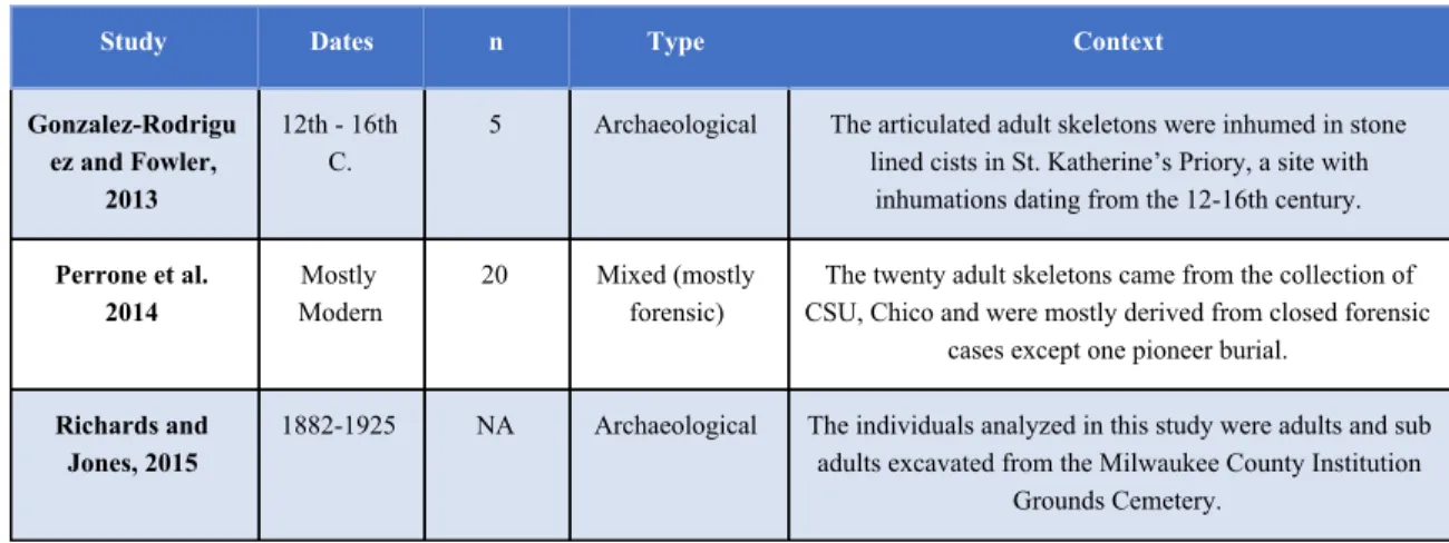
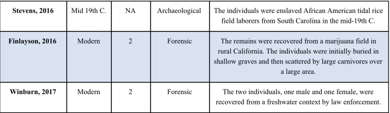
1. Dates, Contexts, and Sample Sizes of Commingling pXRF Studies 43
2. Chemical Elements Utilized by Commingling pXRF Studies 44
3. Chemical Elements and Ratios Considered Effective by Study 46
“It is dangerous, that power. It is most perilous. It must follow knowledge, and serve need. To light a candle is to cast a shadow.”
INTRODUCTION
This thesis is a response to the rapidly expanding number of biological anthropology and forensic publications (ex. Gonzalez-Rodriguez, 2013; Perrone, 2014; Richards and Jones, 2015; Stevens, 2016; Finlayson, 2016; Winburn, 2017) which attempt to utilize portable X-Ray Fluorescence (pXRF, also known as p-ED-XRF, handheld XRF (HH-XRF), and field-portable XRF (FP-XRF)) spectrometry for the purpose of segregating and analyzing commingled human remains. The impetus for this trend is a
byproduct of the rising number of handheld spectrometers owned by museums, universities, and private institutions across the globe. Museums and archaeological sites that lack the expensive high-end
spectrometry facilities formerly relegated to top research institutions have been keen to embrace the rapid, non-invasive, and non-destructive technique. The resulting publications and papers have sought to apply this methodology to a variety of materials including ceramics, lithics, metals, paintings, sculptures, soil samples, and organic remains. Within the last decade there has been a magnitudinal increase in the number of Society for American Archaeology Annual Meeting abstracts describing research related to pXRF (Abstract Archives, 2019).
Due to its enhanced accessibility and visibility, pXRF has also recently permeated the field of biological anthropology. The emerging applications have ranged from the detection of heavy metals like Mercury (Hg), Lead (Pb), and Iron (Fe) (Zuckerman, 2010; Little et al. 2014; Killoran, 2017), to the analysis of paleodiets (Gilbertson, 2015; Bergmann, 2018), to the comparisons of trace elements for the purpose of resolving commingling (Gonzalez-Rodriguez, 2013; Perrone, 2014; Richards and Jones, 2015; Stevens, 2016; Finlayson, 2016; Winburn, 2017). While many of these studies have returned findings strongly suggesting that pXRF is a valid and useful technique for resolving anthropological questions, there appears to be a general uncertainty regarding the underlying practical and theoretical limitations and capabilities of pXRF and the ways in which data collection can be optimized so that researchers can attain interpretable and quantitative results (Shugar and Mass, 2013, pg. 20). Indeed, several researchers have been exceptionally vocal about about the misuse and misinterpretation of pXRF data (ex. Shackley, 2012; Speakman and Shackley, 2013).
summarize and evaluate the past applications of spectrometry to the study of human remains, and (3) outline a collection of successful approaches for future projects as well as research frontiers which are yet unexplored.
In order to achieve these objectives, the thesis will be divided into the following chapters: Chapter I will introduce the concept of commingled remains and describe both the processes of their formation and their use. It will also discuss past, current, and developing methods of re-association such as quantitative, qualitative, and archaeometric techniques. Chapter II will provide a general introduction to pXRF and outline its history, development, theoretical models, and current instrumentation. This chapter will also address common concerns regarding the problematic use of this technique. Chapter III will outline the anatomy and elemental composition of human bone, including the molecular structure, chemical incorporation strategies, and non-pXRF investigations of trace elements in osseous tissues. Chapter IV will describe and analyze previous attempts to resolve commingling with respect to their stated goals, instrumental analysis procedures, statistical procedures, and findings. Chapter V will outline successful techniques and describe directions for future studies to explore.
Throughout the following discussion, the recurring question will be whether pXRF spectrometry is a valid approach to the resolution of commingled human remains. An ostensible affirmative response is complicated by the fact that, just like isotopic analysis, molecular techniques, and advanced quantitative methodologies, pXRF is far from ‘point and shoot’ or ‘plug and chug’. In addition to understanding the underlying principles of X-ray spectrometry, a successful investigator must possess a thorough
understanding the trace element composition of the human bone as well as an ability to combine disjoint streams of data into an interpretable form. Therefore, in order to validate the utility of pXRF as a useful technique for individuating (resolving) commingling, the following must be appropriately demonstrated:
1. There exists less intraskeletal variation than interskeletal variation for some set of chemical elements. Such variation can result from any of a number of processes, but should be predicted and explained by the researcher.
CHAPTER I: STANDARD & EMERGING COMMINGLING TECHNIQUES:
Commingling of human remains in forensic and bioarchaeological contexts poses significant barriers to the goals of anthropological investigators and is intrinsic to the collection on hand. Because of the diverse deposition, excavation, laboratory, and curational processes through which commingling occurs, the homo- or heterogeneity of individuals in the collection, and the questions asked by
investigators, every commingled scenario exhibits unique challenges. Sorting out individual remains to address commingling requires a considerable amount of time, resources, skill, and effort. Thus,
researchers must decide whether resolution is necessary to achieving their research goals, and if so, which of several individuation methodologies to follow. In this chapter, commingling and the processes leading to commingling will be defined, motivations for resolutions will be described, and existing and emerging methods of investigating commingled collections will be explored.
Definition of Commingling:
Commingling can be defined as the mixing of skeletal elements belonging to different individuals and is a frequent feature of both archaeological and forensic contexts. This process can occur through human processes (ossuary burials, lack of excavation records, subpar laboratory techniques) and natural processes (mechanical mixing through hydrological or geological agitations, mass disasters, etc).
Fig. 1. Types of commingled and fragmentary assemblages (from Osterholtz, 2014, pg. 2)
Long term usage assemblages occur when a depositional location such as a tomb has been utilized over a long period of time, frequently by specific communities for multi-generational deposition. Due to the temporal and spatial nature of such interment patterns, these types of collections frequently exhibit a higher degree of commingling and fragmentation since earlier individuals are frequently disturbed to make space for new depositions. Depending on whether secondary burial is a feature of the site, there may be a lower representation of smaller and more fragile skeletal elements. Because such assemblages are attritional, “demography will reflect mortuary programs” and thus the skeletal element composition of such assemblages will likely be different in age and sex ratios than others (Osterholtz, 2014, pg. 2).
Episodic usage assemblages, are, by contrast, typically formed in far shorter periods of time (single generation, often under a year). Reasons for their formation are often extraordinary events such as warfare, plague or religious activities. Episodic assemblages tend to exhibit a lesser degree of
skeletal elements, such as crania, occurred at the time of death (Osterholtz, 2014, pg. 2).
Demographically, these types of collections will not reflect natural populational attrition but might feature subpopulations such as young males (if warfare), the very old, infirm, and young (if plague), or social subgroups (if genocide).
Investigator-commingled assemblages are the third type of commingling and the only completely avoidable type. While modern commingling is generally unintentional and largely occurs due to subpar laboratory practices (lack of labeling of skeletal elements, unorganized notes, collection issues) many 19th and early 20th century legacy collections are commingled because they were assembled when certain skeletal elements (i.e., crania, spines) were valued above others for research purposes. Many long-term museum collections are also commingled due to accession and deaccession, cataloging changes, and administrative agendas (Osterholtz, 2014, pg. 173). Once commingling occurs in a laboratory or museum setting, it is unlikely to ever be resolved with absolute certainty and thus loses value to both modern and future investigators.
In addition to the three commingled assemblage patterns identified by Osterholtz, also worth mention are those that can be attributed to extreme natural and human processes such as construction and development, disturbance of burials by natural processes such as wind or water erosion (gradually or by natural disasters), and irresponsible excavation or collection practices by untrained personnel (Stevens, 2016, pg. 3).
Approaches to Commingled Collections:
Depending on the goals and research questions of the investigators, it may be neither necessary nor useful to resolve commingling in order to analyze a collection. However, in most paleopathological and forensic commingling scenarios, reassociating skeletal elements to form distinct individuals must precede further analysis. This is because estimations of age, sex, stature, health, lifestyle, and experiences generally require skeletal remains to be as complete as possible. However, due to the challenge of
For paleopathological studies, resolving commingling is critical. This is because many infections and nutrient deficiencies will systematically affect multiple skeletal elements. For example, consider a micronutrient deficiency such as scurvy or a condition such as syphilis, both of which lead to diagnostic osseous lesions on multiple skeletal elements. Without examining the morphology and pathology of different osseous tissues belonging to the same individual, the certain presence of a specific condition may be mistaken for a population containing several unhealthy individuals. In addition to gaining the ability to pinpoint nutritional and pathological conditions, resolving commingling is necessary for paleopathological investigation because epidemiological tools such as life-tables are complicated to construct for commingled assemblages, particularly when considering the prerequisites of much-improved emerging aging techniques. While techniques have been suggested to extrapolate information from single skeletal elements to the entire assemblage (Osterholt, 2014, pg. 38), neither precision or accuracy can be reliably achieved.
Forensic workers, driven by the need for positive identification and rapid return of remains to loved ones, value commingling resolution as much as if not more so than archaeological investigators. (Broehl, 2018, pg. 2). Many forensic assemblages caused by natural disasters, terrorist attacks, or
every-day violence need to be quickly resolved due to legal, economic, and political pressures. Because of cases such as these, forensic anthropologists require accurate ways of resolving commingling in order to provide accurate demographic data and ultimately return individuals to their families. A major difference between archaeological and forensic applications, however, is that forensic investigators have access to funds that enable them to perform DNA work and permission for destructive analysis. These resources, despite decreasing costs, still remain out of reach for many biological archaeologists.
Analytical Methods Without Resolution:
The MNI began being used in the mid 20th century and returns an estimate of the minimum number of individuals necessary to form an assemblage. It is typically determined by using the discrete skeletal element (i.e. proximal tibia) with the highest count with respect to characteristics such as age, sex, pair-matching, and pathologies. While it is extremely simple to calculate, the drawback of MNI is that it has a high probability of returning an underestimate of the original number of individuals. According to Adams and Konigsberg (2004), there are three major ways of calculating MNI where L signifies the number of left skeletal elements, R the number of right skeletal elements, and P the number of known pairs:
1. Max (L +R) 2. (L+2R)
3. L + − R P
As can be demonstrated using simple models (Adams and Konigsberg, 2004, pg. 139), all three of these methods return gross underestimations of commingled assemblages, (1) and (2) by approximately 30% and (3) by 9% when the probability of recovery is fairly high (p = 0.7). However, because the probability of recovery is nearly always unknowable, it is difficult to attain accurate estimations using MNI.
Like MNI, the Lincoln (also called the Peterson) Index was developed for use on living population of animals based on capture-recapture techniques (Adams and Konigsberg, 2004, pg. 139). The advantage of LI over MNI is that it can be used to estimate population sizes even if taphonomic biasing has occurred because it does not assume that all members of a population can or will be observed. Crucial to this calculation is extensive pair-matching, which can be achieved through osteological sorting or other techniques described later in this chapter. Preferably, the skeletal elements used for the P value ought to be those more likely to preserve, to be distinct, and have definitive features (i.e. femur, tibia, humerus, innominates). Analogously to living animal populations, one side of the skeleton (i.e., L) is treated as the initial catch (those animals which are first captured and tagged) while the other (i.e., R) is treated as the re-capture (those which are caught for a second time during an additional round of sampling). A general form of the LI can be calculated as:
Finally, MLNI was also adapted from ecological and zooarchaeological models, specifically from capture-recapture studies estimating faunal populations. When the expected sample size is small, this measure outperforms the LI (Adams and Konigsberg, 2004, pg. 141), although it can have a substantial negative bias if the number of recovered pairs is low (Broehl, 2018, pg. 8).
5. MLNI = INT[(L+1)((P+1)R+1)− 1 ]
While LI and MLNI are certainly an improvement over MNI, it is unlikely that MNI will ever be completely replaced due to its simplicity and the fact that calculations for LI and MLNI should be approached somewhat more carefully, with considerations for rates of pair matching and expected population sizes. However, these two measurements are advantageous because they are considerably less prone to bias and allow for consideration of taphonomic processes which are natural to commingled collections.
Analytical Methods for Resolution:
The best way to control commingling is to prevent it in the first place. While the first two commingling processes discussed earlier in this chapter (long term, episodic) are largely unavoidable, field, laboratory, and curational commingling can easily be prevented by keeping extensive and detailed excavation and shipping photos and records (both digitally and in print), labeling and tagging every element as soon as possible and with carefully considered identification numbers, bagging carefully and distinctly, keeping laboratories and analysis tables carefully segregated and organized, and limiting analysis to one assemblage and individual at a time. While these steps may seem arduous at the time, once commingling occurs, valuable information is irrevocably lost.
In the case where commingling is unavoidable, resolution of commingling must begin as early as possible, even during excavation. As soon as commingled skeletal elements are discovered, contextual information such as provenience, photographs, and observations ought to be recorded, preferably through precise georeferencing combined with high-quality 3D models achieved with scanners or
laboratory or in the field. Once skeletal elements arrive in the lab, more serious re-association can begin through qualitative factors, quantitative methodologies, DNA analysis, and a variety of other techniques.
Qualitative Techniques:
Qualitative techniques are typically the first step towards resolving commingling because they require little to no equipment, are relatively quick, and are generally reliable when performed by
experienced osteologists. All begin with an inventory of the remains where information is collected on the identification of each element, its siding, visible pathologies and taphanomies, sutures, and other
distinguishing characteristics. Once that information is collected and skeletal elements can be examined in logical groups (i.e., articulating skeletal elements, matching pairs, etc), pair-matching and joint-matching can be conducted.
In pair-matching, all left and right skeletal elements of a certain type are considered as possible pairs from a single individual. These determinations can be made based on a combination of morphology, trauma, taphonomy, and other characteristics. Although highly reliable when conducted by the
experienced practitioner, visual pair-matching can be difficult for inexperienced osteologists. Studies have shown that the success of this technique can range between 75.6% to over 90% based on skeletal element types (Broehl, 2018, pg. 10-11). Aside from experience, other factors influencing the utility of this technique are the size of the commingled sample and the general degree of preservation of the skeletal elements. The method can also be more successful if individuals of different sexes are being compared and if the skeletal elements being considered are sexually dimorphic (in that case, both long bones and innominates could be more easily distinguished).
Articulation, or joint, matching, is frequently used in conjunction with pair-matching and can be used to associate mandibles with crania, femurs with innominates, and so on. Through this process, individuals can be gradually re-assembled, although some skeletal elements such as phalanges, carpals, and ribs are resistant to this approach.
Quantitative Techniques:
assess the articulation and association of the hip joint (femur and acetabulum) (Stevens, 1015, 103-104). However, as both statistical and computational techniques improved, increasingly complex and
multivariate methods for addressing commingling emerged.
Of these, osteometric sorting is the most common. This technique relies on the assumption that the human skeleton varies in predictable and population-specific ways which allow distinct skeletal elements to have predictable relationships with each other (Stevens, 2016, pg. 104-105). Osteometric sorting utilizes confidence intervals derived from known reference populations to evaluate whether two skeletal elements originate from the same individual. Statistical methods to evaluate similarities and differences include the use of confidence intervals as well as t-tests (Broehl 2018, pg. 15). Unlike qualitative methods, osteometric sorting can be used to consider skeletal elements from different areas of the skeleton. On the other hand, it provides less definitive guarantees of matches and should therefore be used in combination with other methods (Broehl 2018, pg. 11). Due to a combination of research interests and practical limitations of osteological sorting, most studies have focused on long bones, innominates, and sacrums (Broehl, 2018, pg. 3). This technique is most useful when individuals are extremely different (in terms of age, sex, physique, etc) and when there are few individuals. When individuals are similar or numerous, overlapping measurements can complicate the results.
Geometric morphometrics:
DNA Sequencing:
By far the most accurate method to resolve commingling, DNA analysis has become widely available over the last decade for anyone with the financial resources to send out samples. Particularly in forensic contexts, DNA analysis is the principal method applied to the resolution of commingling.
However, this method is constrained by the large cost of processing samples, the degree of preservation of the bones, and whether legal, ethical, and political contexts permit destructive analysis
(Gonzalez-Rodriguez, 2013, pg. 1). This technique is most frequently employed in modern mass disaster events (wars, plane crashes, natural disasters, terrorist attacks) and is used to attain the highest degree of confidence in an identification. It can also be used when deceased individuals need to be associated with living relatives (ex. Cakar, 2018). Typically, this technique requires DNA extraction (and therefore destructive analysis), amplification, and extraction of profiles (usually based off of a discrete number of specified loci). Aside from the necessary destruction of minute portions of skeletal elements, further drawbacks of this commingling resolution technique are the preservation state of the DNA since time, erosion, and extreme climatic conditions such as heat, cold, and moisture can cause degradation. However, the clear advantage is that skeletal elements do not need to be identified in order to be reassociated.
Archaeometric Techniques:
Thus, when pXRF (portable X-Ray spectrometry) was developed, it was considered in the context of an extremely well established XRF method which had been in use for anthropology since the 1960’s, when Edward Olson utilized XRF to evaluate selenium (Se) and Hg in organic compounds (Olson, 1960). Unlike XRF, however, pXRF had the advantage of being nondestructive, portable, and (seemingly) simple to operate and interpret, in part due to the ways in which companies marketed the device to academic consumers. The portability of the devices was especially desirable for archaeologists because it allowed for in situ analysis of monumental buildings, artifacts, and fragile objects (Shugar and Mass, 2013, pg. 17). Finally, the cost of the devices was negligible in comparison to the cost of setting up a full-scale spectrometry or DNA laboratory, and instruments could often be acquired by anthropologists in cooperation with geology and chemistry departments.
The temptation to seize upon pXRF as a means of resolving commingling has thus been
CHAPTER II: REVIEW OF PORTABLE X-RAY FLUORESCENCE (PXRF):
X-Ray Fluorescence (XRF) analysis has evolved over the last century into a generally accepted and extremely versatile technique with direct and meaningful applications to disciplines as diverse as archaeology, environmental science, museum studies, conservation, and geochemistry. Advances in physical theory, software capability, and instrumentation have led to direct benefits in the form of a new generation of portable instruments that can perform relatively high-resolution multi-elemental analysis. Currently, of the various analytic techniques for the determination of a material’s elemental composition (XRF, ICP-MS, etc), pXRF holds major advantages as an effective, quick, and non-destructive
methodology. However, the same features which make pXRF such an enticing tool for researchers also make it problematic because if the underlying theories of the technique are not properly understood and accounted for by investigators pXRF will return useless or misleading data. Therefore, the following sections in this chapter will describe the history and development of modern instruments, the underlying physical theories of the method, differences between portable and stationary instruments, the applications of pXRF, approaches to data analysis, and concerns raised by pXRF experts regarding inappropriate use of the technique.
History and Development:
On November 8th, 1895, the German physicist Wilhelm Conrad Röntgen discovered the form of radiation which came to be known as X-rays (Panchbhai, 2015). Prior to this date, X-rays were simply counted among other types of unidentified radiation which emanated from gas-filled discharge tubes. Not unlike the fortuitous discovery of penicillin by Alexander Fleming, the story goes that Röntgen happened to place an electric tube on top of a book resting upon a photographic plate. Upon removing the book, Röntgen was amazed to discover the outline of a key which was hidden within the book’s pages. Clearly, “the strange light from the glass tube had penetrated the pages of the book”, and thus X-rays were
Johann Wilhelm Hittorf, Heinrich Hertz, Eugene Goldstein, Wilhelm Conrad Guericke, and William Crookes (Panchbhai, 2015).
In the months subsequent to his discovery, Röntgen continued to test his new form of radiation and by December 22, 1895, had photographed his spouse’s hand using X-rays, thus creating the first radiograph of a human being (fig. 2) (Panchbhai, 2015) . Röntgen also named the new rays as ‘X-rays’ 1 because of the variable ‘x’s mathematical attribute of denoting an unknown quality, although in many non-English languages they are known as Röntgen rays, after their discoverer. Soon after the first publications, the uses of the new imaging technology became starkly evident and were embraced by diverse medical professionals, especially dentists (Panchbhai, 2015 . Indeed, in modern times, dentistry is 2 one of the foremost applications of the technology.
Fig. 2. Hand mit Ringen (Hand with Rings). This is a 1896 print of Wilhelm Röntgen’s first radiograph, depicting his wife’s hand (Jaya, 2009).
Following the initial excitement about the utility of Röntgen’s rays, the next major step was taken in 1904 by Sir John Fleming, an English physicist and electrical engineer. By inventing the thermionic
1 Upon seeing her skeleton fingers on a radiograph, Bertha, Röntgen’s wife, is rumored to have exclaimed, “I have seen my
death!” (Panchbhai, 2015, pg. 92).
2 Ironically, many of these early adopters were also the first to suffer from the medical consequences of overexposure to X-rays,
diode, a vacuum tube which utilized a hot cathode by running a second electric current through it and thus emitting a significantly higher number of electrons, Fleming allowed the replacement of problematic cold-cathode tubes in X-ray work. Another advancement took place in 1906 when Charles Barkla, a Nobel-winning British physicist, discovered that each element had a characteristic X-ray spectrum when scattered by gases. Specifically, by observing the secondary X-rays which were emitted from a sample, Barkla found that “the polarization of X-rays, the gaps in atomic absorption, and the distinction between continuous and characteristic X-rays, which consisted of several series of X-rays, named K, L, M series” (Beckhoff, 2006). In other words, he discovered that the pattern of X-rays was related to the electron patterning of an atom. Advancements continued on through work by researchers such as Max von Laue, who discovered the diffraction of X-rays by Crystals, Sir William Bragg, who established Bragg’s law of X-ray diffraction, and William Coolidge, who invented the Coolidge X-ray tube still in use today. Eventually, the working principle of modern XRF emerged: it is possible to measure the energy of characteristic photons emitted by a sample and thus determine which chemical elements the sample is composed of, and even their concentration.
During World War I, X-rays were turned to military applications by researchers such as Marie Curie, who developed mobile X-ray units used for imaging soldiers in the field (Jorgensen, 2017). Concurrently, X-rays became ever-more integrated into medical, chemical, and physical research and practice. Perhaps the most well-known application of X-rays was the X-ray diffraction pattern captured by Rosalind Franklin in 1952 at King’s College in London. Using that tool, the three dimensional structure of DNA as a double helix composed of paired nucleotide bases was discovered.
As can be expected, World War II resulted in even further developments in X-ray technologies. Specifically, great strides were taken in the construction of x-ray fluorescence spectrometers. In 1948, the first successful commercial X-ray fluorescence spectrometer was constructed (Beckhoff, 2006). In 1958, an X-ray spectrometer was developed specifically for industrial applications. Once XRF was made available for public and commercial consumption, there began a rapid development and miniaturization of the various components which resulted in the eventual creation of portable instruments such as are
available today.
To begin describing the theory underlying X-ray fluorescence, it is important to first define what an X-ray actually is: a form of electromagnetic radiation generated by electrons slowed down in the outer field of an atomic nucleus or by a change of bound states of electrons in an electronic shell of an atom with wavelengths ranging from .01 to 10 nanometers and energies ranging from 100 eV to 100 keV (Beckhoff, 2006). Meanwhile, electromagnetic radiation above one MeV generated by nuclear processes is usually called γ-radiation. The divisions between these two types of radiations are to “to some extent artificial and misleading” (Beckhoff, 2006).
The process of XRF spectrometry has three major operational stages: primary radiation, ionization, and detection. Primary radiation occurs in the unit, which emits photons. The process of ionization takes places when samples are excited by X-rays which cause the atoms to eject electrons. When an electron is removed, it makes the electronic structure of the atom unstable and causes other electrons still in orbit to fill the vacancy left by the excited electron. When the outer orbital electron shifts to the open lower position, energy is released as a photon of a predictable energy- in fact, the energy difference between the orbitals. As described by Charles Barkla when he won the Nobel prize,
characteristic X-rays are given off when outer orbital electrons drop into a vacancy in an inner shell of an atom and a photon with the energy difference of the two orbitals is emitted. For those who work with X-rays, there exists a traditional notation used to describe transitions between different electron orbitals. For example, a transition from the L shell to the K shell results in a Kα X-ray, while a transition from the M shell to the K shell results in a Kβ X-ray. Although there are other transitions, these are the two major ones used in XRF analysis. The basic function of an XRF is therefore to sort through the energies of the detected photons and to determine the relative quantity of each element in the material. The term fluorescence describes the emission of photons when a material absorbs radiation. The process of detection again occurs in the unit, and describes the conversion of characteristic radiation into electrical signals which can be converted to interpretable data.
Beginning with the process of primary radiation, in order to excite electrons in the sample material an original source of radiation is needed. Therefore, XRF spectrometers will have X-ray generators which can emit either gamma or X-rays, with gamma rays being preferred for portable applications due to their lower energy requirements. The first X-ray generator used by Röntgen was an ion tube, specifically a Hittorf-Crooks tube filled with air at a reduced temperature. In this set up,
had a relatively short lifetime, was unstable, and difficult to control (Beckhoff, 2006). Therefore, when Coolidge introduced the schema for the hot filament electron emitter in a high vacuum X-ray tube, the stability and efficiency allowed it to remain the dominant equipment choice into the modern era (Beckhoff, 2006). With a hot filament set up, the heated cathode is usually a tungsten (W) wire and the anode typically an element such as chromium (Cr), copper (Cu), molybdenum (Mo) or W held at many kilovolts of potential difference relative to the cathode (Guthrie, 2012). Some manufacturers will use rhodium (Rh) as the standard anode material because its energies are suitable for exciting chemical elements both light and heavy. Thermal electrons accelerate to the anode from the cathode, causing bremsstrahlung radiation to be emitted . This radiation then leaves the unit through a beryllium window in 3 the side of the tube.
When it leaves the tube, the bremsstrahlung radiation then excites the characteristic radiation of the chemical elements in the sample material. As discussed, each element emits photons of characteristic energies. One important aspect of the excitement stage is that the chemical elements which can be detected are limited by the maximum energy emitted from the tube. For example, if the maximum energy emitted is 20 keV, then it is impossible to excite chemical elements with an atomic number exceeding 43 as the K binding energy would surpass the maximum. Other factors of importance during this stage are the penetration of the radiation, matrix effects, thickness of the material, and scattering patterns.
As the energy leaves the sample material, some of it will return to the unit. For detection of incoming photons, proportional counters or other detectors are used. These components work because X-ray photons from the sample material will ionize with detector atoms in a manner such that the amount of charge created is proportional to the energy of the photon. These charges create pulses which are proportional to the energy of each incoming photon. By recording each of these pulses, an energy spectrum with frequencies can be mapped and counts of photons can be used to describe which chemical elements are present and, if the machine is calibrated, in what quantities. Common ways of analyzing these data are by employing computer software to create spectra overlay or to create elemental concentration charts which can be used for quantitative analysis (Perrone, 2014, pg. 146).
The final step of analysis is to account for the errors inherent in the analysis and with each specific machine. As an X-ray moves through a material, its energy is modified by the matrix effects,
3 Bremsstrahlung radiation is generated when electrons are accelerated by the anode and release X-rays, and results
secondary excitation, absorption of emitted X-rays, and interference with characteristic X-rays (Beckhoff, 2006). Some of these factors can be controlled for and predicted using derivations of mathematical correction formulae. The formulae allow the raw readings, or counts, to be converted to actual numeric quantities which allow direct comparisons of elemental compositions.
Instrumentation:
Since the early 2000s, advancement of X-ray generation and detection technology has allowed for the miniaturization of bulky laboratory-based units into portable and lightweight handheld pXRF devices which can be easily packed, transported, and utilized in non-traditional settings (Shugar and Mass, 2013, pg. 17). This has permitted studies involving the analysis of large, bulky, or fragile objects which cannot be investigated in laboratories. Furthermore, pXRF does not require the destructive preparation
procedures required for a stationary XRF unit such as powdering or cutting (Perrone, 2014, pg. 147). The drawback of pXRF is that it has a significantly more limited detection range than XRF and is typically (though not always) limited to the accurate detection of element numbers 22 (titanium, Ti) to 41 (niobium, Nb) whereas a desktop XRF can precisely capture element numbers 11 (sodium, Na) to 92 (uranium, U) (Shackley, 2012). If detection of lower atomic number chemical elements is desirable, a vacuum can be used with a pXRF to improve readings. Another major limitation of pXRF is that since it is nondestructive, the thickness and homogeneity of the material must be accounted for. In comparison, most XRF samples are powdered and can thus be reliably replicated.
Applications:
Historically, XRF has been used for a variety of purposes mainly related to measuring chemical elements found in natural or industrial settings. The prime consumers of pXRF units are steel,
manufacturers of commercially available equipment as they compose an extremely minute share of the market. Therefore, most of the calibrations and factory-settings are not optimized for the uses researchers envision.
In the last three decades, archaeologists and museum curators have made extensive use of the archaeometric applications of XRF for the analysis of materials such as metals, ceramics, lithics, and textiles. Many of these studies have been geared towards either sourcing materials or determining whether they contained harmful or dangerous substances. The tool has also been used by forensic investigators at crime scenes to detect gunshot residues and body fluids, and thus it was a natural transition for biological anthropologists to utilize pXRF for projects ranging from the detection of heavy metals like Hg, Pb, and Fe (Zuckerman, 2010; Little et al. 2014; Killoran, 2017) to analysis of paleodiets (Gilbertson, 2015; Bergmann, 2018) and comparisons of trace elements for the purpose of resolving commingling
(Gonzalez-Rodriguez, 2013; Perrone, 2014; Richards and Jones, 2015; Stevens, 2016; Finlayson, 2016; Winburn, 2017).
Data Analysis:
Analysis of pXRF data in archaeometry is generally considered to be a qualitative or, at best, a semiquantitative endeavour (Holmqvist, 2016). Data examined for presence/absence is generally straightforward, but determining real compositions is challenging because comparative studies of pXRF and higher-sensitivity techniques such as inductively coupled plasma mass spectrometry (ICP-MS) have demonstrated that elemental concentrations from pXRF are inaccurate (despite generally following real patterns and trends) (Holmqvist, 2016). Such results are subpar because they are hard to validate and less precise than destructive methods. Additionally, such semi-quantitative values are specific to each pXRF unit and cannot be compared between different machines. For example, the same material analyzed with two different handheld units might return statistically different results.
inter-elemental interactions, variations in absorption and emittance, and surface effects (i.e., the ‘shielding effect’) (Holmqvist, 2016). In addition, a vacuum can help mitigate some of the issues of matrix
heterogeneity, as can helium flow.
In addition to optimized sample detection, if a researcher wants to convert pXRF count data into mass units, calibrations must be used which match the matrix of the sample (Holmqvist, 2016). The general recommendation offered by Speakman et al. (2011) is that investigators produce specific calibrations for their materials since manufacturer settings will neither be accurate nor precise for archaeological materials (Speakman et al., 2011). As described by Drake (2018), there are multiple assumptions which must hold true for a calibration to function as intended. First, the matrix should be homogenous; second, the calibration reference should be of the same material as the sample; third, every element in the sample should be present in the reference; fourth, the reference should have both minimum and maximum values of each element; and fifth, readings should be taken with the same parameters (unit, energy, current, filter, vacuum, etc) (Drake, 2018). The process of creating a calibration generally
involves using both a pXRF unit and a trusted method of detection (ex. ICP-MS) to analyze a sample. Therefore, the process of creating an appropriate calibration for archaeological materials is a destructive one.
Limitations:
There are multiple commonly expressed concerns regarding the application of pXRF to
archaeological studies. These include issues originating from non-ideal surfaces and matrices. Bones are a fantastic example of non ideal pXRF samples because they have very few flat surfaces and exhibit
extreme elemental and structural variation from one location to another and by depth. Furthermore, many archaeological materials do not account for surface level treatments common to many museum
collections. As demonstrated by Shugar (2013), a treatment such as a slip on pottery can dramatically alter the peaks and concentrations of chemical elements detected by pXRF (Shugar, 2013). Therefore, investigators must be either willing to accept inaccurate readings or remove the outer surface of analytes.
and interpret photon interactions. Even in the most straightforward analysis, the photons used by pXRF will exhibit at least four different phenomena in addition to X-ray fluorescence: incident beam absorption: elastic scattering (Rayleigh), inelastic scattering (Compton), and diffraction (Bragg) (Shugar and Mass, 2013, pg. 18). When archaeological objects with complex morphologies and heterogeneous matrices are involved, the resulting data will be problematic and potentially meaningless.
Despite these challenges associated with the use of pXRF, it is certainly possible to utilize this technique to answer specific anthropological questions so long as the investigator approaches the design, execution, and interpretation of the project with appropriate care. In the following chapters, the
CHAPTER III: STRUCTURE & ELEMENTAL COMPOSITION OF HUMAN BONE:
In order for pXRF to be considered a useful technique for the resolution of commingled human remains, the first criterion that must be demonstrated, as described in the introduction, is that there exists a lower degree of intraskeletal variation within the skeletal elements of a single individual than
interskeletal variation within a commingled population with respect to a subset of chemical elements. The purpose of this chapter is to demonstrate multiple lines of evidence why this might hold true for some, but not all, skeletal populations. In the process of achieving that objective, the physical structure and
elemental composition of osseous tissue will be described, elemental incorporation pathways will be outlined, and the history of elemental human bone studies will be discussed.
Skeletal Composition:
Human bone, synonymously called osseous tissue, is a dynamic feature of the body which is responsible for several vital functional roles such as red and white blood cell production, service as a mineral depository, and support of other anatomical structures. An adult human being has approximately 206 bones (excluding sesamoid bones) [see Appendix 2 with image of skeleton] while juveniles boast substantially more centers of ossification until their skeletal elements fuse into final forms by early adulthood. Osseous tissue is composed of multiple types of cells organized in a matrix-like structure that allows bones to be both strong and lightweight. These cells include osteoblasts and osteocytes which form and mineralize bone and osteoclasts which reabsorb osseous materials throughout the course of the human life.
orientations (Clarke, 2008, pg. S132). Because of this, newly deposited bone is on the periosteal (external surface) while older bone will be underneath. This feature of lamellar bone is an important consideration for studies proposing to utilize pXRF. In contrast to lamellar bone is woven bone, which is comprised of a more haphazard structure of collagen.
On a molecular level, bone is a complex amalgam of both organic (30% dry weight) and inorganic components (70% dry weight) (Gilbertson, 2015, pg. 79). Its composition is mainly water, phosphate, calcium (Ca), and a variety of trace elements (Stevens, 2016, pg. 108). The organic component is made up of cells (osteoblasts, osteoclasts, and osteocytes) as well as the osteoid, the organic portion of bone matrix which consists of collagen, proteoglycans, and glycoproteins (Zimmerman, 2015, pg. 132). This organic portion of bone is what provides the tissue with the tensile strength and flexibility that allows it to undergo physical stress. The part of the bone that gives its structure is the inorganic bony matrix which is composed mainly of hydroxyapatites, or mineral calcium phosphates, which can be described with the formula Ca (P O ) 5 4 3OH(Zimmerman, 2015, pg. 132)
Bone Growth and Remodeling:
Although bone growth is rapid during childhood, all skeletal elements continue to grow, develop, model, and remodel throughout the lifetime. That early childhood bone growth is referred to as
longitudinal and radial, whereas remodeling is a more gradual process by which the skeleton adjusts to physiological conditions, mechanical stress, and environmental factors. During the process of remodeling, human bone composition continually shifts based on nutrition, environment, and metabolic demand. Although bone is actively remodeled in humans of all ages, the rates are not constant, nor are the rates constant across different skeletal elements. There are several sequential phases to the remodeling process: (1) resorption, when osteoclasts digest old bone; (2) reversal, when cells appear on the bone surface; and (3) formation, when osteoblasts deposit new bone until replacement or growth occur. While the process nominally maintains existing structure, remodeling also serves to help the skeletal system adapt to new mechanical needs and to repair damage caused by stress. During these steps, the basic components of bone are removed and added on both a cellular level and molecular level. These processes are controlled by complex signaling pathways, especially during early life when bones are still undergoing development.
mass each year (Kini and Nandeesh, 2012). During this turnover, the chemical composition of the bone is transformed based on accessibility of different skeletal elements within the body. Therefore, different skeletal elements are informative of different time periods in an individual's life (e.g. teeth are useful indicators of childhood because they form during early life and are not remodeled, while skeletal elements such as ribs reflect more recent chemical environments).
Elemental Incorporation Strategies:
The major chemical elements found in bone are Ca and phosphorus (P) in addition to minor chemical elements such as magnesium (Mg), sulphur (S), iron (Fe), Zn, manganese (Mn) and cadmium (Cd). While all human bone includes a similar basic structure, variation between individuals of bone composition can be caused by a variety of factors including: diverse developments, exposure to dissimilar environments (i.e., heavy metals contaminants), different pathological experiences, consumption of chemically dissimilar food or water, effects of postmortem contamination (diagenesis), and human processing (post mortem). The chemical elements which are commonly detected using XRF include potassium (K), Ca, Fe, Mg, P, Zn, strontium (Sr), and Pb (Perrone, 2014, pg. 148). Both the distribution and the concentration of these chemical elements can vary considerably within the skeleton based on the type of bone, the speed of its turnover, and the chemical environment of the body at the time at which the tissue was formed. In the following section, the mechanisms by which these chemical elements are incorporated into the human body will be discussed in more depth.
Excluding diagenesis and processing, the other factors leading to elemental variation occur during life, typically during the process of deposition. During that time, the composition of the inorganic portion of bone can change due to ionic substitution of bone which causes the calcium phosphate matrix to be replaced by trace elements (Zimmerman, 2015, pg. 132). For example, the Ca ions can be replaced by a variety of chemical elements (Zimmerman, 2015, pg. 132). Meanwhile, the phosphate group (PO 4) can be exchanged for citrate, phosphate esters, diphosphates, pyrophosphates, and amino acids (Zimmerman, 2015, pg. 132). Lastly, the hydroxyl group (OH) can be switched for fluorine (F) or chlorine (Cl) (Zimmerman, 2015, pg. 132).
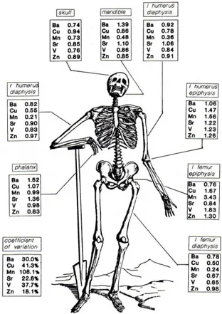
these chemical elements within individuals and even skeletal elements. For example, the variation in elemental concentrations by skeletal element can be seen through the fact that Zn, vanadium (V), nickel (Ni), Cr, Pb, Mn, and Co are more concentrated in epiphyseal bone regions while Ca, Sr, Na, and K tend to collect near the diaphysis (Zimmerman, 2015, pg. 132). Indeed, knowledge of the intra-skeletal variability in elemental signatures has been established for several decades and is a recurring issue investigators seeking to analyze elemental data (Stevens, 2016, pg. 11). Therefore, investigators should carefully consider the known distributions of the chemical elements relevant to their study and choose skeletal elements in ways that suit their research questions.
Developmental and Environmental Effects:
The chemical environment of both children and adults has a significant effect on which chemical elements are incorporated into bone. Thus, the elemental composition of bone contains information relevant to geographical origin and residence, diet, and environmental exposures (Byrnes, 2016, pg. 1042). The trend to use skeletal elemental composition to answer questions about origin is most evident in isotopic studies which frequently utilize oxygen and Sr comparisons between teeth and bone to determine whether an individual migrated from one area to another. This technique is effective because oxygen and Sr isotopic ratios vary based on the region where an individual is living (Gilbertson, 2015, pg. 88-99).
Pathological Effects:
The pathology of an individual can also influence which chemical elements make up osseous tissue. This is both because the pathogenesis of certain conditions can influence the chemical balance of the body and because certain cultures attempt to treat illness with chemical elements that are then incorporated into the matrix. The first mechanism is the relationship between elemental composition and pathology, a frequent subject of research that has been used to understand the physiological response to disease (ex. Karaaslan, 2014). Other examples include studies utilizing micro-XRF to investigate the distribution of trace elements within specific bone histological structures (e.g.. Pemmer et al. 2013).
The second mechanism through which the elemental composition of bone can be informative about pathology is through evidence of treatment. The best examples of such cases are those involving the use of Hg to treat syphilis in the 17th-19th centuries . For example, Zuckerman (2010) utilized pXRF on 4 English skeletal samples in order to explore the utilization of Hg for this medicinal purpose. Aside from syphilis, a high Hg concentration in an individual could also be associated with leprosy. The mechanism through which Hg is incorporated into osseous tissue is through the substitution of Hg for Ca in bone carbonate (Rasmussen, 2008).
Nutritional Effects:
Perhaps the most impactful factor influencing the incorporation of chemical elements into osseous tissue is the diet of an individual. Of course, diet is closely related both to pathological lesions as well as to the chemical environment during life (i.e., environmental contaminants such as Pb) and growth since differing physiological requirements will impact which chemical elements are kept in the body’s reserves. Some of the chemical elements most commonly associated with diet are Zn, Cu, Mo, Se, Sr, Mg, Mn, cobalt (Co), and Ni. Of these, the first four are predominantly associated with animal proteins, while the latter are linked to plant resources (Stevens, 2016, pg. 109). The composition of human bone,
Chemical elements that recur frequently in investigations of ancient diets are barium (Ba), Ca, and Sr, all alkaline earth metals which are incorporated into human tissue through the consumption of plants and animals (ex. Gilbert, 1994; Bergmann, 2018). The utility of these chemical elements to investigations of nutrition is derived from the fact that there is a reduction in an organism’s Ba/Ca and Sr/Ca ratios in relation to the diet that it consumes (Burton, 199, pg. 609). So, for example, a herbivore would have lower ratios than plants and carnivores would have lower ratios than omnivores. Therefore, reductions in Ba/Ca and Sr/Ca relate to ascensions in trophic level. While there are several issues associated with matching these ratios directly to plant versus meat diets (for example, unusually high Ca content in plants can skew ratios and thus diets with the same amount of meat can appear identical), “experimental results overwhelmingly demonstrate that bone Ba/Ca and Sr/Ca faithfully track dietary Ba/Ca and Sr/Ca” (Burton, 1999, pg. 609).
Diagenetic Effects:
Of all of the elemental incorporation strategies, diagenetic effects are considered the most problematic because they cause the elemental composition of retrieved osseous tissues to reflect not only the composition of the bones during life, but the chemical composition of a depositional environment following death. Diagenesis is the post-mortem alteration in the makeup of bone caused by contamination by surrounding soils (Price, 1992). This is a frequent issue in archaeological investigations seeking to study the composition of ancient remains (Burton, 1999, pg. 609). Although the general effects of diagenesis are known, there is so single universal trajectory for the process. Rather, a complex array of factors specific to the depositional environment and ranging from soil composition, breakdown and leaching of collagen, microbial attacks, hydrological and geological events, physical erosion, acidity, and more interact to alter the composition of bone (Stevens, 2016, pg. 112-113).
While diagenesis is largely unavoidable, there are several possible indicators that allow
the first component of osseous tissue to be altered, while bone hydroxyapatite is typically altered at slower rates (Gilbertson, 2015, pg. 1).
When diagenetic effects are suspected, the prudent anthropologist should collect samples of soil from the area of deposition in order to compare the elemental composition with recovered skeletal elements. Another good practice is to note the positioning of skeletal elements during their recovery since the positioning of a bone can influence the concentration of chemical elements (Lambert et al. 1985, pg. 480).
Processing Effects:
The final mechanism that can influence the elemental composition of skeletal elements is post-recovery processing. Admittedly, this mechanism is less relevant to forensic applications but can be extremely important in the analysis of museum collections recovered prior to the development of high tech analytical methodologies. Materials that were historically used to preserve and treat bones include paraffin wax, animal glue shellac, polyvinyl acetate emulsions, acrylic emulsions, and acrylic resins in solvent (Wills, 2014). While these may be visible in some scenarios as shiny or discoloured patches on the bone, at times it is difficult to determine if processing has occurred. However, application of foreign materials to skeletal materials is important to establish prior to analysis so that, if necessary, the external surface of skeletal elements can be removed and uncontaminated tissue can be analyzed.
History of Elemental Human Bone Studies:
As is clearly evident by the chemical element incorporation mechanisms described above, the composition of osseous tissue has the potential to be extremely informative both about the lives of deceased individuals as well as what happened to their remains after death. While it can be difficult to disentangle the various factors which influence the presences, absences, concentrations, and ratios of various chemical elements and form concrete conclusions about deceased individuals, many investigators have made the effort using various techniques ranging from mass spectrometers to pXRF units.
Among the earliest individuals to attempt such analysis was Robert Gilbert, who in 1975
and migrations (Stevens, 2016, pg. 108-109). Many of these researchers struggled against the same issues posed by anthropologists interested in the resolution of commingling: interskeletal vs intra skeletal variation in elemental incorporation and diagenesis.
Probably among the most versatile, accurate, and precise tool for such investigations has been the inductively coupled plasma mass spectrometer, or ICP-MS. While more time-intensive, expensive, and destructive than XRF, ICP-MS offers an extremely successful and reliable method to analyze the elemental composition of most substances. Indeed, prior to the development of pXRF, ICP-MS was a popular methodology for the attempts at using chemical elements to resolve skeletal commingling.
In 1986, Fulton et al. was published. This study utilized trace metal ratios to attempt to resolve commingling among a small number of individuals from an air crash scenario using ICP. Their results indicated that Mg/Zn ratios were the most reliable and that Zn/Na, Mg/Na, and Cr/Na could be used as supplements. However, they also found that single chemical elements (excluding arsenic) were not useful in distinguishing between individuals.
In 1988, Grupe set out to compare trace element content in bone specimens from different anatomical sites. Using individuals excavated in northern Germany from the 11-12th century CE, the researcher utilized neutron activation analysis and plasma emission spectrometry to investigate Ca, P, Sr, Mg, Ba, Zn, Fe, Co, Cr, Se, Sc, Antimony (Sb), Cs, and silver (Ag). What Grupe found was that there were trace element differences between trabecular and cortical bone and that trabecular bone was more susceptible to diagenetic impacts.
Fig. 3. Ratios of the trace element content in different bones in individual E64 (Francalacci, 1990, pg. 228).
More recent studies, such as Castro et al. (2010), have continued to explore possibilities for resolving commingling using trace element analysis. In their investigation, the researchers conducted elemental analysis of bone samples via ICP-MS from 12 individuals and determined that although full discrimination was not achieved, elemental analysis was a promising tool for separation of commingled remains.
CHAPTER IV: REVIEW OF APPLICATIONS OF PXRF TO BIOLOGICAL ANTHROPOLOGY:
This chapter will provide synopses of recent publications characterized by the application of pXRF to biological anthropology and specifically human bone. These studies are roughly sorted into four categories according to the focus of their research questions, as detection, diet, commingling, and
methodology. Detection studies are those which address questions pertaining to the presence or absence of particular chemical elements such as Pb or Hg. Diet studies are those which attempt to associate spectrometry data to the nutritional patterns of ancient populations. Commingling studies are those which seek to resolve mixed skeletal elements into discrete individuals. Finally, methodology studies are those which attempt to delimit the applications and interpretations of pXRF through formal experimentation on specific aspects of the technique.
Although determining the effectiveness of pXRF to the resolution of commingling is the primary objective of this discussion, studies within the other three categories have been included in this chapter because of their relevance to the topic at hand. Both detection and diet studies attempt to determine information about skeletal remains that could be used to differentiate individuals in certain commingling scenarios (i.e., social differentiations in diet, workplace heavy metal exposure, etc.) Similarly, the methodology studies have been incorporated because they are necessary to determining the applicability, precision, and accuracy of the technique.
Detection Studies:
In 2010, Molly Zuckerman applied pXRF to her research on syphilis in late medieval and early modern England (Zuckerman, 2010). Employing trace element analysis via pXRF, Zuckerman
ascertained differences in Hg levels in skeletal remains from several archaeological sites (Zuckerman, 2010, pg. 14). Multiple skeletal elements and locations were systematically analyzed and comparable Hg values were obtained. Overall, despite the heterogeneity of intraskeletal Hg distribution, Zuckerman found pXRF to be a valid way to study Hg exposure in archaeological populations.
Like Zuckerman, in 2018 Peter Killoran utilized pXRF to detect the heavy metals Arsenic, Lead, Cr, Cd, Ba, and Cs in 107 individuals from Kentucky in an effort to isolate individuals who received heavy-metal laden medications. Unlike previous investigations, Killoran found that the pXRF output was not sensitive enough to unequivocally determine the presence of mercury.
Diet Studies:
Dietary studies seeking a nondestructive substitution to ICP-MS and isotopic analysis techniques have appeared more recently than detection studies. An early iteration was the doctoral dissertation composed by Theresa Gilbertson from the University of South Florida in 2015. Focusing on ancient Peruvian coastal diets, Gilbertson sought to compare pXRF and stable isotope analysis. To do so, she tested 209 skulls via pXRF and compared those results to a subsampling of individuals for whom she used isotope ratio mass spectrometry (IRMS) on bone collagen and apatite (Gilbertson, 2015, pg. ix). The bivariate correlations and linear regressions that resulted indicated that pXRF was a valid way to quantify Sr, barium, and Fe for the purpose of dietary analysis (Gilbertson, 2015, pg. ix). Overall, Gilbertson concluded that pXRF could not yet replace destructive analysis in dietary studies.
Commingling Studies:
As discussed in chapter III, the use of trace element analysis to distinguish between commingled human remains is a tradition dating back to the mid 20th century. Therefore, several attempts have been made to utilize pXRF to discern differences between skeletons. One of the earliest and therefore
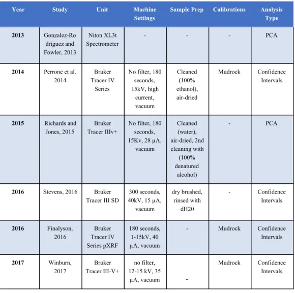
most-cited of such investigations was that by J. Gonzalez-Rodriguez and G. Fowler in 2013. Using a set of 5 mostly complete archaeological skeletons (four males, one female) excavated from the city of Lincoln (United Kingdom), Gonzalez-Rodriguez and Fowler tested trace element concentrations of Pb, Sr, Zn, Fe, Ca, and K in 23 skeletal elements ranging from cranium to long bones (Gonzalez-Rodriguez, 2013, pg. 5). Using principal component analysis (PCA), the researchers found that skeletal elements could be separated with a high degree of accuracy when two to four individuals were examined, although accuracy dropped when the number of individuals increased (Gonzalez-Rodriguez, 2013, pg. 1). The chemical elements found to be the most important for separation were Pb, Sr, Fe, Ca, and K and the ratios Pb/Ca, Zn/Fe, and Sr/Pb were found to be the most influential (Gonzalez-Rodriguez, 2013, pg. 5).
Overall, the findings of this research indicated that pXRF seemed to be effective but quickly lost strength in large contexts due to overlapping chemical element readings.
The Gonzalez-Rodriguez and Fowler study was followed by one by Alexandra Perrone et al. in 2014 which likewise investigated the utility of pXRF for the resolution of commingled human remains. The researchers focused on measuring intra- and inter- skeletal chemical element variation by using pXRF to analyze 20 compete adult skeletons, almost all from forensic anthropology cases. Using
ANOVA tests, the researchers found minimal variation between skeletal elements (intraskeletal variation) except for Mg. To simulate commingling, the researchers used 95% confidence intervals to determine whether individuals could be distinguished. The chemical elements which Perrone et al. found to be most useful for the resolution of commingling were Si, P, K, Ca, Mn, Fe, and Co. Like Gonzalez-Rodriguez and Fowler, Perrone et al. found that segregation rates dropped with increasing number of individuals. However, unlike Gonzalez-Rodriguez and Fowler, the pXRF used in this study was unable to detect Sr and Pb.
In 2015, John D. Richards and Catherine R. Jones from the University of Wisconsin-Milwaukee utilized pXRF both for detection and commingling and presented their work in a Society for American Archaeology (SAA) symposium. In part, this study was a response to the drawbacks of the
diagenesis. The Milwaukee County Institution Grounds Cemetery focused on in this study was suspected to have significant diagenetic alterations due to groundwater infiltration, the deposition of industrial and medical wastes, and other natural and anthropogenic activities (Richards and Jones, 2015, pg. 5). The investigators focused on several commingled burial lots and investigated cranial, scapular, humeral, ulnar, innominate, femural, and tubular elements. The chemical elements detected at ‘useful’ levels were
arsenic, Ca, copper (Cu), Fe, Mg, Mn, Ni, P, Rh, Sn, Sr, Zn, and zirconium (Zr). In this study, the pXRF elemental readings were not especially effective at resolving commingling, although Sr/Ca and Zn/Fe ratios did lead to some separation. Ultimately, the investigators concluded that pXRF might prove to be useful in combination with archaeological and osteological analyses, but that more careful care should be taken with how measurements are taken and that results should be calibrated to specific reference
materials (Richards and Jones, 2015, pg. 10).
Following the Richards and Jones study, in 2016 William D. Stevens of the University of South Carolina utilized pXRF to address commingling as part of his doctoral dissertation. In addition to utilizing classic approaches to commingling, Stevens also tested the efficacy of pXRF in separating skeletal remains of enslaved African American tidal rice field laborers from South Carolina from the 19th century. One interesting finding of Steven’s study was that differences existed in the distribution of chemical elements between upper and lower limbs (Stevens, 2016, pg. 158). However, pXRF generally worked well in conjunction with other approaches to commingling to create possible associations and support pair-matches (Stevens, 2016, pg. 159). As in earlier studies, difficulties arose as larger sets of
commingled remains were analyzed. Overall, Sr and Pb were found to be the best for sorting remains. Stevens conjectured that this was because these chemical elements reflect individual dietary differences and the possible origins of the enslaved (Stevens, 2016, pg. 245). Metals such as Cu and Fe, however, displayed high degrees of both variation and overlap, probably due to burial practices, and were thus not useful for the resolution of commingling (Stevens, 2016, pg. 245).
results indicated that P had the greatest potential for resolving commingling and that pXRF could be used to segregate skeletal elements when other techniques are ineffective (Finlayson et al. 2016, pg. 497).
In 2017, Winburn et al. published a similar investigation which utilized numerous commingling techniques to attempt to segregate two sets of commingled remains. Unlike the Finlayson 2016 study, the skeletal remains from this investigation originated from a freshwater context and, likely due to the resulting diagenetic alteration, pXRF was unable to resolve commingling (Winburn, 2017, pg. 24).
Methodological Studies:
The final category of study related to this discussion are methodological studies seeking to test the practical and theoretical limitations of the technique. An early example of this is the 2014 study conducted by Nicole C. Little and her colleagues. The goal of the investigation was to compare data collected from an ICP-MS to that derived from a pXRF with respect to Pb content in bone for 25
individuals. However, the researchers found that there were extreme differences between pXRF readings taken for unburred and burred bones (Little et al. 2014, pg. 21). This was attributed not only to differing Pb concentrations within and between skeletal elements but also to the differences in what ICP-MS and pXRF are measuring- average bulk composition versus analytical surfaces. The take away from this investigation is that researchers should consider what, if any, treatments skeletal elements should endure prior to being analyzed.
In 2016, another methodological study was published by Jennifer F. Byrnes and Peter J. Bush that considered the practical limitations of bone surface and analysis depth for accurate trace element
detection. The researchers focused on Sr, Cu, Pb, Zr, and Sn (Byrnes, 2016, pg. 1042). The study concluded that diagenesis was a vital factor for pXRF studies because surface effects created detectable differences in readings. Moreover, bone density and thickness must be considered in order to account for the expected penetration distances of specific chemical elements.
Summary:
As demonstrated by this chapter, over the last decade pXRF has been utilized frequently and enthusiastically by biological anthropologists seeking to add another technique to their commingling toolbox. While many of these researchers were aware of the studies that preceded their own, there have been limited systematic efforts to define and refine the methodology despite the promise and potential it seems to hold. Instead, there has been significant variation in sample preparation, analytical procedures, and adherence to accepted pXRF protocols as have been established for ceramic and lithic studies.