ENDO-EXOCYTIC TRAFFICKING IN REGULATION OF CDC42 POLARITY
Leah Joy Watson
A dissertation submitted to the faculty at the University of North Carolina at Chapel Hill in partial fulfillment of the requirements for the degree of Doctor of Philosophy in
the Department of Cell Biology and Physiology in the School of Medicine.
Chapel Hill 2014
iii ABSTRACT
Leah Joy Watson: Endo-exocytic Trafficking in Regulation of Cdc42 Polarity (Under the direction of Patrick Brennwald)
The precise subcellular localization of the Rho GTPase Cdc42 is essential for its spatial and temporal control of polarized growth and division. In budding yeast, the activation and clustering of Cdc42 on the cell surface designates the site of emergence for the daughter bud and it is towards this site that the actin cytoskeleton and exocytic pathways orient to promote bud formation. In turn, exocytic delivery of Cdc42 along actin cables has been suggested as a mechanism to reinforce Cdc42’s own polarized localization at the bud tip. Recycling via endocytosis and
GDI-dependent mechanisms are posited to contribute to Cdc42’s polarized localization by offsetting lateral membrane diffusion of Cdc42 molecules from the concentrated pool. The intimate relationship between Cdc42’s function in cell polarity and the maintenance of its own localization by the pathways it regulates has been extensively studied, however, the molecular mechanisms involved in the determination and maintenance of Cdc42 polarity remain unclear.
Using a novel in vivo assay developed in the lab, we found that disrupting distinct stages of endocytosis severely disrupted the ability of Cdc42 to associate with secretory vesicles. This implicates the possible involvement of multiple
iv
into sites of endocytosis. We also demonstrate that GFP-tagged Cdc42 is highly defective in its ability to associate with vesicles. Although GFP-Cdc42 has been a valuable tool for understanding mechanisms involved in Cdc42 polarity, our findings demonstrate differences in the itineraries of the tagged and untagged Cdc42
v
vi
ACKNOWLEDGEMENTS
First, I must thank God. Throughout graduate school, I have had to face many deaths in my family, a failed thesis project, many personal losses and frustrations. However, my faith has been essential for me to overcome every challenge,
appreciate every lesson and delight in my successes no matter the scale.
Thank you to my advisor, Dr. Patrick Brennwald. You taught me to extract lessons from every failure and success, to exploit imperfections in the attempt for perfection, and to question everything. I am forever grateful for your lessons, both intentional and unintentional.
Thank you to my committee members: Drs. Jim Bear, Keith Burridge, Adrienne Cox and Henrik Dohlman. Your advice and guidance during each of our meetings have been important to my development as a scientist.
vii
“little sister”, Kelly Watson, for critical readings of my manuscript, for giving me someone to laugh at (with), and for your support.
To my coffee buddies: Drs. Michelle Itano, Meghan Morgan-Smith and Katie Wolfe. For your friendship, for your laughter, for being there…Thank you.
Thank you to all of my UNC network, friends, and family for their support and encouragement throughout my time at UNC, especially: Kyle McKenna, Ashalla Freeman, Pat Phelps, Michael Johnson, Sabrice Guerrier, the
IMSD/STAD/TIBBs/BBSP office, Alan Anderson, Marva Taylor, Monique Sprueill, and MoniQue Honablew.
viii
TABLE OF CONTENTS
LIST OF FIGURES AND TABLES ... x
LIST OF ABBREVIATIONS ...xi
CHAPTER 1: Introduction and Background ... 1
1.1 Overview... 1
1.2 Establishment of cell polarity in budding yeast ... 2
1.3 Identification and characterization of Cdc42 as the “master regulator of polarity” ... 4
1.4 Cdc42 localization and polarity ... 7
1.5 Gaps in current understanding ... 9
1.6 Figures... 11
CHAPTER 2: Quantitative Analysis of Membrane Trafficking in Regulation of Cdc42 Polarity ... 14
2.1 Overview... 14
2.2 Introduction ... 15
2.3 Results... 16
2.3.1 A quantitative assay for Cdc42-vesicle association ... 16
2.3.2 Endocytosis is required for Cdc42 post-Golgi vesicle association ... 18
2.3.3 GFP-Cdc42 shows impaired association with post-Golgi vesicles ... 20
2.3.4 Quantification of Cdc42 density on vesicles and the plasma membrane polarity cap ... 22
2.4 Discussion ... 26
ix
2.6 Figures... 38
2.7 Supplementary Information ... 45
CHAPTER 3: Concluding Remarks and Future Studies ... 49
x
LIST OF FIGURES AND TABLES
Figure 1.1 Hierarchal model for polarity establishment in budding and
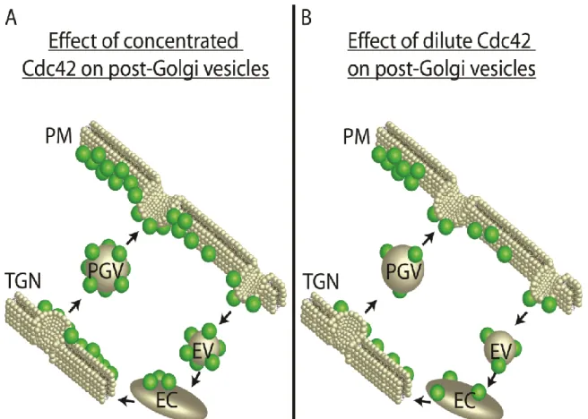
mating cells. ... 11 Figure 1.2 Rho GTPase localization and polarized growth. ... 12 Figure 1.3 Schematic representation of potential outcomes of the delivery
of post-Golgi secretory vesicles (A) sufficiently and (B)
insufficiently loaded with Cdc42. ... 13 Figure 2.1 An in vivo assay demonstrates the association of Cdc42 with
post-Golgi vesicles. ... 38 Figure 2.2 Endocytosis, but not Rho GDI, is required for Cdc42
association with post-Golgi vesicles. ... 39 Figure 2.3 Endosomal sorting mutants show defects in Cdc42 recycling
onto post-Golgi vesicles. ... 41 Figure 2.4 GFP-tagged Cdc42 has impaired ability to associate with
post-Golgi vesicles and exhibits synthetic growth defects with rdi1∆. ... 42 Figure 2.5 Quantitative analysis of Cdc42 density on post-Golgi vesicles
and the plasma membrane polarity cap. ... 43 Figure 2.S1 Comparisons of strains containing untagged and GFP-tagged
Cdc42. ... 45 Table 2.S1: Yeast strains used in this study ... 47 Figure 3.1 Schematic representation of the consequence of exocytic
xi
LIST OF ABBREVIATIONS
RHO Ras HOmolog
CDC42 Cell Division Cycle 42 RSR1 RaS-Related 1
GEF Guanine nucleotide Exchange Factor GAP GTPase-Activating Protein
GDI GDP Dissociation Inhibitor CDC24 Cell Division Cycle 24
RGA1/2 Rho GTPase Activating Protein BEM1/2/3 Bud EMergence
RDI1 Rho GDP Dissociation Inhibitor
FRAP Fluorescence Recovery After Photobleaching LatA/B LATrunculin
TGN Trans Golgi Network ROI Region Of Interest
1
CHAPTER 1: Introduction and Background 1.1 Overview
Cell polarity is defined as the partitioning of cellular materials into spatially distinct domains in response to external and/or internal stimuli. Virtually all cells polarize at some point during their lifetime. Whether to grow and divide or to perform highly specialized cellular functions such as axonal migration or activation of the immune response, the process of cell polarization is a critical component of eukaryotic cell biology [1-4].
Much of the current understanding of polarity and the identification of many key regulators can be attributed to studies using the budding yeast Saccharomyces cerevisiae. Polarity in budding yeast essentially is compartmentalizing the cell into a distinct cell “front” and “back” during either the production of a daughter bud or a mating projection. In particular for budding, the proper establishment of a cell “front” ensures that only one bud is constructed per cell cycle [2, 5]. Besides the
pronounced polarization state during most of their life cycle, budding yeast are a genetically tractable system with many conserved key components of polarity. As such, budding yeast are an excellent model system for dissecting the mechanisms underlying cell polarity.
2
evolutionarily conserved role in defining the cell “front” and “back”. They are important for several physiological processes—including axonal migration, membrane trafficking, actin organization, and morphogenesis—and pathological processes that include metastasis, cell survival and infinite proliferative potential [11, 12]. The Rho GTPase, Cdc42, is essential for polarity establishment in yeast [9, 10, 12, 13]. Its local activation at the plasma membrane designates the cell “front” or the site where the daughter bud will form [14, 15]. This chapter will discuss the
establishment of polarity in yeast, the identification and characterization of Cdc42 as the “master regulator of polarity”, the link between the localization of Cdc42 and bud emergence, and gaps in the current knowledge of Cdc42 polarity wherein my work will endeavor to fill.
1.2 Establishment of cell polarity in budding yeast
Polarity establishment generally involves 1) a cellular response to
intrinsic/extrinsic stimuli, 2) determination of a single, defined polarity axis direction and 3) construction of the axis via positioning of polarity factors and pathways
towards a spatial landmark [12]. The commitment to exit isotropic growth and trigger asymmetry in yeast results in either the formation of a bud or shmoo/mating
3
Budding yeast display two spatial patterns for placement of the nascent bud and subsequent separation from the mother. The bud of haploid yeast cells forms adjacent to the previous division site (axial budding pattern), while the bud forms at the polar opposite end of the previous division site (bipolar budding pattern) in diploid cells. Axial and bipolar budding patterns are dictated by distinct “landmark” proteins which are passed along from mother to nascent bud. Axial budding requires the gene products of BUD3, BUD4, AXL1, and AXL2/BUD10, whereas the gene products of BUD7, BUD8, BUD9, RAX1,and RAX2 are specific for the bipolar budding pattern [13, 15, 17-19]. The gene products of RSR1/BUD1, BUD2, and BUD5 comprise a general bud selection machinery that is involved in both axial and bipolar budding [13, 15, 17, 20]. After the initiation of the cell-cycle program, the procession of polarity establishment follows: 1) the general selection machinery (RSR1/BUD2/BUD5) interprets the axial and bipolar signal for bud placement 2) transmits these spatial coordinates to the polarity establishment machinery then 3) the polarity establishment machinery organizes the actin cytoskeleton and delivery of protein and vesicles towards this site on the cell surface to promote bud
emergence.
4
cells are inherently capable of switching from symmetrical to asymmetrical growth— albeit in a randomized orientation. Further examination of the establishment of polarity in yeast led to the characterization of the polarity establishment machinery which is comprised of the Cdc42 GTPase and its regulators (i.e. Cdc24, Rga1, Rga2, Bem2, and Bem3). The consensus of these studies has led to the
classification of Cdc42 as the central or “master” regulator of polarity [22], and as such, massive efforts remain focused on dissecting its function in polarized growth. 1.3 Identification and characterization of Cdc42 as the “master regulator of polarity”
The Rho GTPase CDC42 was originally identified from an extensive screen for mutants with growth arrest phenotypes resembling previously characterized mutations in the gene CDC24 [9, 10, 23, 24]. The cdc42-1 temperature sensitive mutant isolated from this screen displayed defects in actin cable distribution despite normal isotropic growth at the restrictive temperature of 37oC. This mutant also failed
5
The Cdc42 GTPase, like other GTPases, cycles between active, guanine triphosphate (GTP)-bound and inactive, guanine diphosphate (GDP)-bound states. GTPases are activated by guanine nucleotide exchange factors (GEFs) which stimulate the release of GDP and loading of GTP. Follow-up studies to the
aforementioned screen revealed Cdc24 as the sole GEF for yeast Cdc42 [12, 28]. The GTPase activating proteins (GAPs)—Bem2, Bem3, Rga1 and Rga2—inactivate Cdc42 by catalyzing its intrinsic ability to hydrolyze GTP [28]. Studies using
nucleotide-locked forms of Cdc42 reveal the requirement for its GTPase cycle in cell viability, the establishment of a single “front” or bud per cell cycle, and its ability to localize properly [29-32].
6
studies reveal that Rdi1-mediated rapid recycling of Cdc42 from the plasma
membrane is involved in maintaining the plasma membrane localization of Cdc42. Furthermore, the lethality imparted by RDI1 overexpression [34] strongly suggests Rdi1 could negatively impact Cdc42 function via unrestricted extraction of the GTPase from membranes [35, 36].
In the hierarchal order of polarity, the regulation of Cdc42 activity and membrane attachment by its GEF, GAPs and GDI are integral components of
7 1.4 Cdc42 localization and polarity
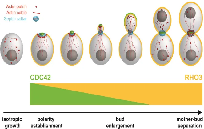
Wu et al. [37] demonstrated that the distinct localizations of Cdc42 and another Rho protein, Rho3, are integrated into their function such that their
localizations reflect the stages at which each GTPase regulates polarized growth. As the determinant of bud emergence and formation, Cdc42 localizes as a concentrated cap on the plasma membrane at the presumptive bud site of unbudded cells and the bud tip of small budded cells. Cdc42 disperses around the cell periphery as the bud enlarges, only to re-cluster at the mother-bud neck prior to its regulation of
cytokinesis (Figure 1.2) [12, 37]. Prenylation of the carboxyl-terminal CAAX moiety (A is aliphatic amino acid; X is any amino acid) of Cdc42 is required for its peripheral attachment to membranes. The 188Cys residue in the CAAX domain is prenylated via
the addition of a C20 geranylgeranyl isoprene group and mutational analysis of this
domain revealed the requirement of prenylation for Cdc42 activity and function [31]. The Rho3 GTPase, on the other hand, is prenylated by the addition of a C15
8
The isotropic-asymmetric switch in Cdc42 plasma membrane localization is thought to involve two distinct positive feedback loops: the adaptor-based signaling and the actomyosin-based transport systems. Both systems are thought to generate and maintain robust Cdc42 polarity by amplifying a spontaneously occurring cluster of GTP-Cdc42 on the cell surface [38-41]. For example, active Cdc42 binds to a signaling complex consisting of the adaptor, scaffold protein Bem1 (Bud EMergence 1), the GEF Cdc24 and a p21-activated kinase (PAK)-family kinase. As a result of its interaction with the GEF-Bem1-PAK complex, neighboring GDP-bound Cdc42 molecules become activated and thus ensues the perpetual recruitment of GEF-Bem1-PAK complexes and local activation of Cdc42 at the presumptive bud site.
In the case of actomyosin-based transport, a stochastically-generated cluster of active Cdc42 orients actin cable nucleation via localized formin activation [42, 43]. Actin cables serve as tracks for myosin-mediated transport of Cdc42-laden post-Golgi vesicles. The delivery and fusion of secretory vesicles carrying Cdc42 with the active pool at the plasma membrane reinforces further local activation of GDP-Cdc42 on the cell surface and drives membrane expansion.
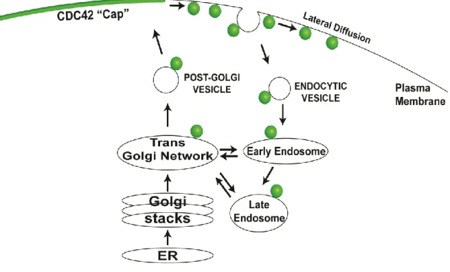
Previous studies using fluorescence recovery after photobleaching (FRAP) revealed the rapid cycling of GFP-Cdc42 between the plasma membrane and internal membrane compartments [30, 44]. These data indicate that the pool of activated Cdc42—or the Cdc42 polarity cap—is dynamically maintained [30, 44, 45]. Indeed, lateral diffusion of Cdc42 within the lipid bilayer can easily dilute the
9
cap-cytosol exchange have been proposed. First, endocytic recycling of Cdc42 from the plasma membrane polarity cap counterbalances its delivery on post-Golgi
secretory vesicles. Irazoqui et al. found that the partial depolymerization of F-actin structures using Latrunculin B (Lat B) results in the dispersal of Cdc42 from the plasma membrane polarity cap [45]. Furthermore, disruption of endocytosis, not RDI1, prevented Lat B-induced Cdc42 dispersal. Co-fractionation studies reveal the association of Cdc42 with both classes of secretory vesicles, Bgl2 and invertase, of which invertase-containing vesicles are known to initially sort through endosomes before entering another round of exocytosis [46]. Second, Rdi1 binds to and extracts prenylated GDP-Cdc42 from membrane compartments into the cytosol. Deletion of RDI1 significantly depletes the cell of cytosolic Cdc42 [47], whereas, overexpression of RDI1 dramatically increases the levels of cytosolic Cdc42 resulting in cell lethality [48].
1.5 Gaps in current understanding
Notwithstanding the breadth of research dedicated to elucidating mechanisms involved in Cdc42 polarization, gaps in our current understanding still remain. In the actin-based transport model, it is the delivery and fusion of secretory vesicles
10
cap is unknown, assessing the contribution of membrane trafficking has been challenging. Although recycling of Cdc42 by Rdi1 and endocytosis are presumably partially redundant pathways, neither endocytic uptake of GDP-Cdc42 nor the accessibility of the vesicular pool of Cdc42 for Rdi1 extraction have been
demonstrated. Furthermore, the difference between bulk and selective incorporation of Cdc42 into the endocytic pathway is still unclear. In light of these and other key concerns, the direct assessment of the contribution of membrane trafficking pathway to Cdc42 polarity will aid in further understanding Cdc42’s control of polarized
11 1.6 Figures
12
Figure 1.2 Rho GTPase localization and polarized growth. The re-localization of Cdc42 and Rho3 throughout the cell cycle reflects the stages at which they control polarized growth. The organization of the actin cytoskeleton and membrane
13
14
CHAPTER 2: Quantitative Analysis of Membrane Trafficking in Regulation of Cdc42 Polarity1
2.1 Overview
Vesicle delivery of Cdc42 has been proposed as an important mechanism for generating and maintaining Cdc42 polarity at the plasma membrane. This
mechanism requires the density of Cdc42 on secretory vesicles to be equal to or higher than the plasma membrane polarity cap. Using a novel method to estimate Cdc42 levels on post-Golgi secretory vesicles in intact yeast cells, we: 1) determined that endocytosis plays an important role in Cdc42’s association with secretory
vesicles 2) found that a GFP-tag placed on the N-terminus of Cdc42 negatively impacts this vesicle association and 3) quantified the surface densities of Cdc42 on post-Golgi vesicles which revealed that the vesicle density of Cdc42 is three times more dilute than that at the polarity cap. This work suggests that the immediate consequence of secretory vesicle fusion with the plasma membrane polarity cap is to dilute the local Cdc42 surface density. This provides strong support for the model in which vesicle trafficking acts to negatively regulate Cdc42 polarity on the cell surface while also providing a means to recycle Cdc42 between the cell surface and internal membrane locations.
1 Reproduced with permission from: Watson, L.J., Rossi, G., and Brennwald, P. (2014). Quantitative
15 2.2 Introduction
Growth along a defined axis is important for many biological processes. The subcellular localizations of key regulators and effectors of polarity are intricately linked with their control of the establishment and maintenance of the polarized axis [14, 37, 49, 53]. In budding yeast, the switch from isotropic to asymmetric growth is preceded by the accumulation of activated (GTP)-Cdc42—a conserved Rho
GTPase—at the presumptive bud site [12, 15]. The Cdc42 polarity cap is required to orient the actin and secretory pathways toward the nascent bud site and Cdc42 polarization is necessary and sufficient for determining the site of bud emergence [14, 49].
Generation and maintenance of robust Cdc42 polarity promotes membrane expansion during bud formation. Studies reveal that Cdc42 is dynamically
16
subject cells to lysis conditions or require fluorescently tagged protein—both of which may impede direct quantitative assessment of the membrane association of the native protein.
In this study, we make use of a novel assay to quantitatively assess the contribution of the recycling pathways to the polarity of endogenous Cdc42 and obtain estimations of the relative and absolute concentrations of Cdc42 on post-Golgi vesicles and the plasma membrane polarity cap. While our results implicate endocytic and exocytic trafficking in recycling of Cdc42, they also demonstrate that the density of Cdc42 proteinon exocytic vesicles is significantly lower than at the plasma membrane polarity cap. We discuss the implications of these findings on current models for Cdc42 polarization.
2.3 Results
2.3.1 A quantitative assay for Cdc42-vesicle association
17
post-Golgi vesicles observed by thin section electron microscopy [59-61] (Figure 2.1A). Consistent with results from biochemical studies, double-labeled
immunofluorescence staining with antibodies directed at Cdc42 and Sec4 revealed a striking re-localization of Cdc42 from the bud-tip to the Sec4-positive vesicle clusters in response to the Sec15 or Sro7 induction (Figure 2.1A). In both Sec15-and Sro7-induced cells we find that all the cytoplasmic clusters that are positive for Cdc42 are also positive for Sec4 and that greater than 70% of Sec4-positive clusters were positive for Cdc42 (Figure 2.1B). This is the similar to the level of co-localization observed at the plasma membrane polarity cap in uninduced cells (GAL-vector, Figure 2.1B).
As a first step in the quantification of Cdc42 levels found on specific
membrane compartments, we measured the ratio ofCdc42 fluorescence associated with Sec4-positive vesicle clusters or the plasma membrane polarity cap to an equivalent-sizedregion in the cytoplasm. The relative Cdc42 fluorescence
18
2.3.2 Endocytosis is required for Cdc42 post-Golgi vesicle association
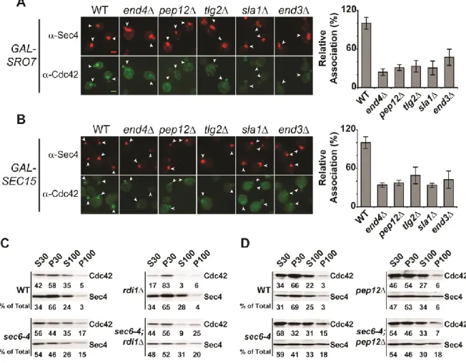
Recycling of Cdc42 to and from the plasma membrane polarity cap is thought to be critical to its ability to act in cell polarization. Two mechanisms for Cdc42 recycling have been proposed. One mechanism involves the Rho GDI protein which selectively extractsthe GDP-bound form of geranylgeranylated Cdc42 from the plasma membrane by providing a pocket for the hydrophobic prenyl group—similar to the role for Rab GDI in Rab GTPase recycling [62]. The second mechanism involves endocytic recycling ofCdc42 from the plasma membrane in a pathway that may function in parallel to its recycling by Rho GDI [44, 45, 63]. We made use of the vesicle clustering assay described above to examine the requirement of GDI or endocytosis in the association of Cdc42 with post-Golgi vesicles. To examine the endocytic requirement, we disrupted endocytosis using a deletion in END4 (also known as SLA2) which regulates the interaction between endocytic vesicles and the actin cytoskeleton during vesicle internalization [64]. As expected (Figure 2.2A), Cdc42 localization at the plasma membrane polarity cap is stable in cells lacking RDI1 or END4 [44-47]. However, induction of vesicle clusters in an end4
background resulted in >60% reduction in the relative concentration of Cdc42
19
cells had increased levels of Cdc42 associated with vesicles—which is consistent with the documented depletion of cytosolic Cdc42 in rdi1 cells [44, 46, 47]. Therefore, while endocytosis is important for Cdc42 association with post-Golgi vesicles, Rho GDI is completely dispensable for this association.
We next examined the association of Cdc42 with vesicle clusters in mutants known to have defects at distinct points in endocytic trafficking from the plasma membrane to endosomes and the Trans Golgi Network (TGN). Sla1, like End4, functions at the plasma membrane during endocytic vesicle formation, while Tlg2 and Pep12 are important for transport between the early endosome to the TGN and between the late endosome (or PreVacuolar Compartment) and the TGN,
respectively [65]. We found that defects in any of these gene products results in a significant and highly penetrantdefect in Cdc42 association with vesicleclusters, suggesting that Cdc42 recycling onto post-Golgi vesicles is likely to involve trafficking through multiple endocytic compartments (Figure 2.3A, B).
Cdc42 has previously been shown to associate with post-Golgi vesicles that accumulate in response to a sec6-4 mutation when analyzed by differential
centrifugation [57]. To examine the role of endocytic and GDI-mediated recycling on the association of Cdc42 with post-Golgi vesicles by differential centrifugation, we constructed double mutants of rdi1 or several endocytic mutants with sec6-4. In response to a sec6-4 mutation, cells shifted to 37oC accumulate post-Golgi secretory
20
Cdc42 association with the P100 fraction also occurs in response to the secretory vesicle accumulation (Figure 2.3C, D). We also observed that when rdi1, sec6-4 cells were examined, elevated levels of Cdc42 were maintained in the P100 fraction. In contrast, the P100 fraction of pep12, sec6-4 mutants—despite having normal accumulation of Sec4—was depleted of Cdc42 compared to sec6-4 cells (Figure 2.3C, D). The accumulation of secretory vesicles in theend4, sec6-4 double mutant was problematic and, unfortunately, this mutant could not be utilized for analysis by fractionation. Nonetheless, the results of fractionation clearly confirm both a role for endocytic recycling and the lack of a requirement for GDI function in the association of Cdc42 with exocytic vesicles.
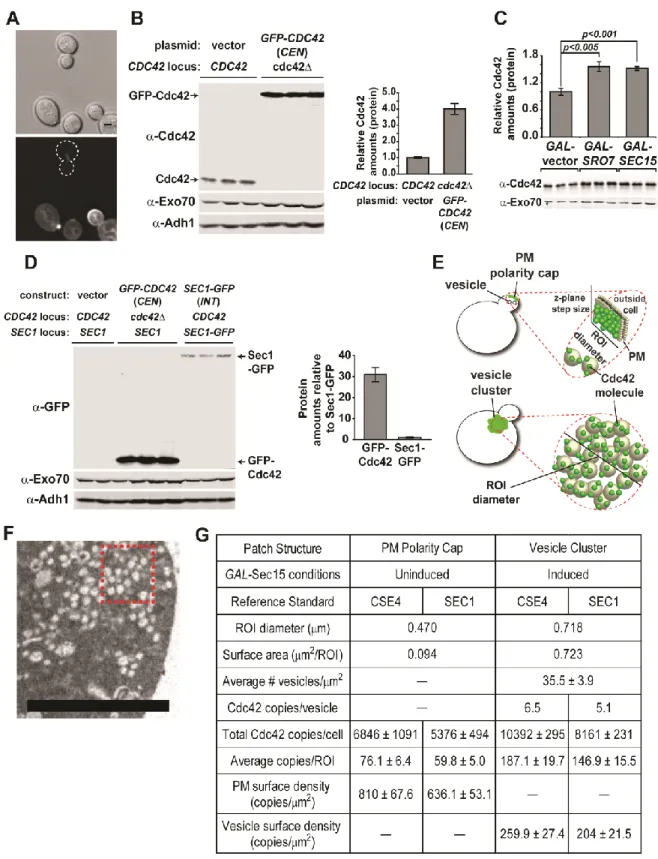
2.3.3 GFP-Cdc42 shows impaired association with post-Golgi vesicles Previous work from our lab has demonstrated an important role for the N-terminus of Rho family GTPases in determining their patterns of subcellular localization [37]. Perhaps not surprisingly, several groups have demonstrated significant growth defects associated with N-terminal GFP-tagged forms of Cdc42 expressed as the sole source of Cdc42 in the cell [55, 66, 67]. Since the work described above relied exclusively on untagged Cdc42 expressed from its
endogenous chromosomal locus, we examined the effect of a GFP-tagged form of Cdc42 on its association with post-Golgi vesicles in our vesicle clustering assay. We generated strains containingGAL-SEC15 with either GFP-tagged or untagged
CDC42 expressed behind their native promoter on a CEN/LEU2 plasmid as the sole source of CDC42. Expression from the CEN plasmids results in a slight, but
21
constructs show normal growth and polarization at 25oC (Figure 2.4A, B), the
GFP-tagged form of Cdc42 resulted in lethality at 37oC as previously reported (Figure
2.S1A, 2.4D) [55, 66, 67]. When induced with galactose, we saw the expected staining of untagged Cdc42 on the large cytoplasmic puncta co-stained by Sec4. However, the strain containing the GFP-tagged form of Cdc42 demonstrated very weak staining of these puncta that was only slightly above the levels of the
surrounding cytoplasm (Figure 2.4A, C). Thesedata indicate that the presence of a GFP tag on the N-terminus of Cdc42 results in a dramatic loss of post-Golgi vesicle association in this assay.
As mentioned above, Slaughter et al. [44] have proposed two parallel
mechanisms for recycling of Cdc42: 1) Rdi1-mediated membrane extraction and re-delivery through the cytosol and 2) endocytic uptake and rere-delivery on exocytic vesicles. A clear prediction of this model is that loss of the sole Rho GDI in yeast should demonstrate synthetic growth defects when combined with a form of Cdc42, in this case GFP-Cdc42, which disrupts its association with exocytic vesicles. We therefore utilized a plasmid shuffle assay to examine the effect of an rdi1 on the ability of GFP-Cdc42 to function as the sole source of Cdc42 in the cell. Previous reports have demonstrated that GFP-Cdc42 is unable to support growth at high temperatures [55, 66, 67]. We find that the temperature-sensitive nature of GFP-Cdc42 is accentuated by the presence of rdi1. In particular, the synthetic effect of the GFP tag and loss of Rho GDI is most apparent at 34.5oC, a temperature at
22
at ambient temperatures and the sustained polarized localization also suggests that there may be a third mechanism for recycling Cdc42 that is independent of both RDI1 and endocytic/exocytic recycling.
2.3.4 Quantification of Cdc42 density on vesicles and the plasma membrane polarity cap
While previous studies using subcellular fractionation as well as the immunofluorescence studies described above have established that Cdc42 is associated with post-Golgi vesicles in significant amounts, the precise density of Cdc42 molecules on the surface of these vesicles has not been determined. The vesicle clustering procedure described above presented a unique opportunity to address this question in vivo, without the numerous difficulties—such as degradation and membrane disassociation—that are often associated with biochemical
fractionation. Therefore, we set out to determine the absolute density of Cdc42 molecules on both post-Golgi vesicles (within the clusters) as well as at the plasma membrane polarity cap. This required having reliable estimates of: 1) the total number of molecules of Cdc42 in the cell 2) the membrane surface area associated with the vesicle clusters or the plasma membrane polarity cap and 3) the fractional amount of Cdc42 associated with each of these two regions.
Surprisingly, we were unable to find a direct estimate of Cdc42 copies per cell in whole proteome-tagging studies [68] or other published work. We therefore
23
number per sister kinetochore cluster is well established [69-71]. To compare the relative fluorescence levels of GFP-Cdc42 in the total cell and Cse4-GFP in the sister kinetochore clusters, the two strains were mixed and imaged by fluorescence microscopy (Figure 2.5A). With each sister kinetochore containing 80 copies of Cse4, the resulting comparison yielded an estimate of roughly 27,400 copies of GFP-Cdc42 per cell. We then compared the amount of plasmid-derived GFP-Cdc42 to that of endogenous Cdc42 in wild type cells by quantitative Western blot analysis (Figure 2.5B). From this analysis we estimate that wild type cells contain
approximately 6,800 copies of Cdc42 per cell.
Since the growth conditions for the vesicle clustering assays differed from the above conditions, we compared the effects of carbon source and vesicle clustering on Cdc42 amounts per cell. While we found there was little effect of carbon source (glucose vs. raffinose; data not shown) on the levels of Cdc42, there was a
significant (>50%) increase in Cdc42 amounts in strains when vesicle clusters were induced (Figure 2.5C). This is presumably due to the enlargement of cells and the inhibition of cell division during vesicle cluster formation. Based on this comparison, we estimate approximately 10,400 copies of Cdc42 per cell following the 8 hour galactose induction of Sec15—identical to the conditions used for fluorescence imaging.
24
approximately 35.5 ± 3.9 post-Golgi vesicles (90nm average diameter) per square micron for each thin section. Thus, the 0.718 micron diameter regions of interest (ROI) used for fluorescence imaging (0.2µm thick optical sections) of clusters
corresponded to approximately 71 vesicles or a total vesicle membrane surface area of 0.723 square microns per ROI. Fluorescence microscopy was used to determine the fractional amount of Cdc42 (% of total) associated with each ROI. Considering the total copy number of Cdc42 in the cell, our assessment yielded ~187 copies of Cdc42 in the vesicle cluster ROI. This corresponds to approximately 6.5 copies of Cdc42 per vesicle or a density of ~260 copies of Cdc42 per square micron of vesicle surface (Figure 2.5E, G).
To assess Cdc42 surface density at the plasma membrane polarity cap, we imaged cells using the same growth conditions as above but without Sec15
induction (GAL-vector). This imaging revealed a fractional fluorescence of
approximately 93 copies of Cdc42 per ROI. Since thin section electron micrographs of small budded yeast show an average of 1.3 ± 0.23 secretory vesicle in close proximity to the bud tip per section (per bud) or approximately 2.6 vesicles per 0.2 micron optical section, it was important to account for this contribution in our estimates. To accomplish this we subtracted the contribution of the 2.6 docked vesicles (~17 copies of Cdc42) from the total Cdc42 present in the ROI (93 copies) and divided the remaining amount (76 copies) by the plasma membrane surface area present in the ROI (0.094 µm2). As shown in Figure 2.5G, this equates to a
25
To corroborate our findings described above, we also generated an estimate of the Cdc42 copy number by quantitative immunoblot analysis using an alternative reference standard, Sec1—a protein which the total copies per cell (determined by immunoblot) has previously been determined [68]. We generated a strain in which Sec1-GFP was integrated at the SEC1 chromosomal locus such that it is expressed behind the native promoter and is the sole source Sec1 in the cell (see materials methods). Whole cell lysates of Sec1-GFP and GFP-Cdc42 expressing cells alongside an isogenic control strain were prepared, loaded by equivalent cell number and analyzed by Western blotting (Figure 2.5D). We applied the reported total copies of Sec1 (639 copies/cell) to the ratio of total GFP-Cdc42:Sec1-GFP and found the total copies of GFP-Cdc42 to be roughly 21,500. Using this copy number and the aforementioned comparative analyses of native Cdc42 protein levels under various conditions (see Figures 2.5B, C), we estimate ~5,400 copies of Cdc42 in polarized (uninduced) cells and ~8,200 copies of Cdc42 in cluster-forming (induced) cells. The compilation of the described data using both reference standards is
reported in Figure 2.5G. Similar to our results using the Cse4 standard, we found the plasma membrane cap density of 636 Cdc42 copies per square micron to also be roughly three times the vesicle membrane density (204 copies/µm2). Together these
26 2.4 Discussion
Delivery of Cdc42 by vesicle-mediated exocytic transport has been proposed to be an important mechanism by which Cdc42 polarity on the plasma membrane is both generated and maintained [57, 72]. In these models, Cdc42 associated with post-Golgi vesicles is delivered along actin cables to sites of polarized growth. The subsequent fusion of Cdc42-laden vesicles with the plasma membrane at these sites would promote Cdc42 polar cap formation. This would lead to a positive feedback loop by reinforcing the organization of actin cables oriented toward such sites, which in turn would bring more vesicles to this site [49, 57]. Another positive feedback loop could result from Cdc42’sdirect activation of the Exocyst tethering complex to promote its own polarizationby increasing the rates of vesicle docking and fusion at specific sites of the plasma membrane –in a manner that is independent of actin [72, 73]. A critical assumption in both of these models is that the surface density of Cdc42 on post-Golgi vesicles must exceed the surface density at the plasma membrane polarity cap for polarity to be generated and/or maintained [50].
Mathematical modeling studies by Savage et al. [74], examined the theoretical effect of exocytic fusion of vesicles depleted of Cdc42 on the plasma membrane polarity cap. In their model such a situation perturbed local polarity in a manner that could be overcome in the presence of an active GDI recycling mechanism. The
experimental data presented here demonstrate that such an effect is more than theoretical since the surface density we observe for Cdc42 on post Golgi vesicles is, in fact, roughly 3-fold more dilute than the density of Cdc42 we observe at the
27
vesicle fusion is to dilute the Cdc42 present at this site on the plasma membrane rather than to concentrate it, which is inconsistent with the actin-mediated positive feedback model [44, 57].
Since both the actin cytoskeleton and the vesicle docking/fusion apparatus are thought to direct traffic to sites on the plasma membrane with the most
concentrated Cdc42, the local effect of exocytic transport would be to antagonize or destabilize Cdc42 polarization on the plasma membrane [56, 75]. Local negative regulation may play an important role in building spatial flexibility into this system. This is similar to a model recently proposed by Dyer et al. [76] in which dilution of the scaffolding protein Bem1 by vesicle fusion would lead to wandering of the polarity cap. Our data suggest that in addition to dilution of polarity factors such as Bem1, vesicle fusion would result in dilution of Cdc42 itself which would contribute directly to the destabilizing effect of exocytic transport on polarity.
If the immediate effect of exocytic traffic is to antagonize Cdc42 polarity on the plasma membrane, then why is Cdc42 associated with post-Golgi vesicles at all? While the work presented here does not directly address this important question, we can speculate on possible roles for exocytic delivery of Cdc42. First, exocytic
delivery may represent a mechanism for recycling Cdc42 that has been removed by endocytosis [44, 45, 63]. Such a delivery system would fit with the notion that trafficking acts as a parallel pathway to the Rho GDI recycling mechanism as
proposed by Slaughter et al. [44]. All of our data is in complete agreement with such a recycling function for vesicular Cdc42. Another function for this recycling
28
the negative regulatory effects of exocytic transport on Cdc42 polarity a possibility explored through modeling in the Savage et al. study [74].
A recent study examined the apparent surface densities of GFP-Cdc42 within the cell by fluorescence correlative spectroscopy [77]. In this paper, Slaughter et al. concluded that the surface density of GFP-Cdc42 on vesicles (49 molecules/µm2)
was similar to that on the plasma membrane (46 molecules/µm2). The remarkably
low absolute density reported at the polarity cap was particularly surprising given that such a density would involve a polarity cap (0.1µm2) with only 4.3 GFP-Cdc42
molecules out of roughly 12,000 molecules per cell (based on a conservative estimate of the GFP-Cdc42/native Cdc42 expression levels for the GFP-Cdc42 constructs used) or less than 0.04% of the total cellular GFP-Cdc42. In contrast, our estimates of native Cdc42 densities on post-Golgi vesicles are almost 5-fold higher (260 molecules/µm2 or 6.5 copies per vesicle) than Slaughter et al. (2013) and our
estimates of densities on the plasma membrane (810 molecules/µm2) are 15-fold
higher. For comparison, our results indicate that a polarity cap (0.1µm2) contains 76
molecules of native Cdc42 or roughly 1.1% of the total cellular Cdc42. Given the low densities of GFP-Cdc42 and the inferred low plasma membrane polarity reported by Slaughter et al. (2013) it is difficult to reconcile their findings with our work except to note that widely different approaches were used to estimate densities.
29
Thus, the inability to gather information on the native protein may have introduced gaps in our understanding that were not previously apparent. Both our data and Slaughter et al. [44] support the notion that Rdi1 and vesicle traffic act as parallel routes for Cdc42 cycling to and from the plasma membrane, which is somewhat surprising given the apparent recycling defect of GFP-Cdc42 compared to the untagged form [55, 66, 67]. Furthermore, it is interesting that GFP-Cdc42 supports growth of rdi1cdc42 cells at temperatures below 30oC (Figure 2.4D). This may
indicate the existence of an additional or third recycling pathway that allows prenylated Cdc42 to be recycled between internal membranes and the plasma membrane polarity cap. Altogether, the data presented in this paper supports the importance of continued studies of the native protein alongside tagged forms of Cdc42 to improving our understanding of cell polarity.
2.5 Materials and Methods Yeast strains, reagents and media
30
raffinose followed by 8-10 hour inductions with 1% galactose (US Biological,
Swampscott, MA). Deletion mutants were generated by PCR amplification of either a KanMX or NatMX cassette using oligos designed against the –MX cassette and flanking DNA sequences of respective genes (e.g. END4, PEP12, etc.). The genomic DNA used as PCR template was extracted from deletion strains— developed by the Saccharomyces Genome Deletion Project— using standard
protocol for genomic DNA extraction [78]. Yeast transformations of the –MX cassette into GAL-vector, -SRO7 and –SEC15 strains were performed using lithium acetate method [79]. G418 sulfate was obtained from US Biological. clonNAT
(nourseothricin) was obtained from WERNER BioAgents (Jena, Germany). YIpLac211-GFP-linker-CDC42 plasmid was received as a gift from the Lew laboratory (Duke University, Durham, NC). GFP-linker-CDC42 (behind CDC42 promoter) was subcloned into a LEU2, CEN vector (pRS315) and introduced into wild-type and the CDC42-plasmid shuffle strain. Zymolyase 100T, ampicillin, Hepes (free acid), and 5-fluoroorotic acid (5-FOA) were obtained from US Biological
(Swampscott, MA). Sorbitol, β-mercaptoethanol, phenol, sodium azide, sodium fluoride, dithiothreitol (DTT), were obtained from Sigma Aldrich (St. Louis, MO). Chloroform, Terrific Broth, and dextrose were from Fisher Scientific (Pittsburgh, PA). Subcellular fractionation
Wild-type and sec6-4ts- cells with or without PEP12 or RDI1 disruptions were grown in rich (YPD) media overnight at 25oC to mid-log phase and shifted to 37oC
for 2hr to accumulate secretory vesicles. Approximately 200 OD599 units of cells
31
NaF) buffer. Cells were spheroplasted in 7.2ml of (100 mM Tris pH 7.5; 10 mM NaN3; 1.2 M Sorbitol; 21 mM β-mercaptoethanol; 0.05mg/ml Zymolyase 100T) buffer
for 30 min at 37oC and lysed in 6ml of ice-cold (10 mM triethanolamine, pH 7.2; 0.8M
sorbitol; protease inhibitor cocktail: 2 µg/ml each of leupeptin, aprotinin, antipain; 20 µM pepstatin A; 2 mM 4-(2-aminoethyl)benzenesulfonyl fluoride) buffer. Lysed cells were centrifuged, cold at 450 x g for 4 min to remove unbroken cells. Cleared lysates were centrifuged in a Sorvall centrifuge (30,000 x g for 15 min at 4oC) to
separate pellet and supernatant fractions. Supernatants were then centrifuged at 100,000 x g for 1hr at 4oC. Pellets were resuspended in lysis buffer at volumes equal
to the supernatant fractions. Equal volumes of supernatant, pellet and total lysate fractions were boiled in SDS sample buffer and separated on a 12.5% SDS-polyacrylamide gel. Western blotting was performed using polyclonal α-Sso1/2 (1:2000), polyclonal α-Sec4 (1:1000) or monoclonal α-Cdc42 (1:200) antibodies. Quantitative Western analysis was performed with the Odyssey Infrared Imaging System (LI-COR Biosciences, Lincoln NE).
Plasmid shuffle assay
32
temperatures shown in Figure 2.4 represent phenotypic separation between the RDI1 and rdi1 strains.
Immunofluorescence and fluorescence microscopy
Cells were grown to mid-log phase in 2% glucose media and shifted into 3% raffinose for 2/+ doublings. GAL-SRO7 and -SEC15 were induced by adding 1% galactose for 8-10 hours. Cells were fixed and processed for immunofluorescent staining as described previously [72, 80]. Double-labeled immunofluorescent staining of the plasma membrane polarity cap and post-Golgi vesicles was
performed using ammonium sulfate precipitated, monoclonal mouse α-Sec4 (1:200) and affinity-purified, polyclonal rabbit α-Cdc42 (1:75) antibodies. For background correction, control staining was performed using rabbit and mouse IgG antibodies that lacked reactivity to any yeast protein. Secondary antibodies were Rhodamine Red-X-conjugated AffiniPure Goat Anti-Mouse IgG and Fluorescein Isothiocyanate (FITC)-conjugated AffiniPure Goat Anti-Rabbit IgG (Jackson ImmunoResearch Laboratories Inc., West Grove, PA), respectively. Secondary antibodies were used at 1:100-1:200 dilution. Single-plane immunofluorescent, GFP-fluorescent, and differential interference contrast (DIC) images were acquired using Nikon model E600 and 2D-deconvolved using MetaMorph software (Molecular Devices). Figures were prepared from deconvolved images using Adobe Photoshop and Illustrator (CS5.1).
Quantitative analysis of polarized and vesicle clustering cells
33
regions of interest (ROIs) drawn within the centroid, or peak intensity, of either the plasma membrane polarity cap or the post-Golgi vesicle cluster and throughout the cytoplasm of the respective cell. Increases in fluorescence intensities of Cdc42 associated with the polarity cap versus the cluster relative to the cytoplasm were calculated in Microsoft Excel. Data was presented as either 1) relative fluorescence: the ratio of the average fluorescence intensities of the Cdc42-positive compartment relative to the cytosol, 2) relative association: the Cdc42 compartment:cytosol ratio expressed as percent association relative to 100% wild-type, 3) phenotype
penetrance: box and whisker plot of the Cdc42 compartment:cytosol ratio. Figures were prepared using Adobe Photoshop and Illustrator (CS5.1).
Ratio-metric analysis of GFP-Cdc42 using the Cse4 reference standard
Cells expressing either GFP-Cdc42 or Cse4-GFP were cultured at 25oC in
minimal media to mid-log phase. Equivalent OD599 of both strains were mixed and
34
ROIs of controls cells that were imaged in mixture with GFP-Cdc42 expressing cells. Total copies of GFP-Cdc42 in the cell were calculated by: (average GFP-Cdc42 fluorescence intensity ÷ average Cse4-GFP fluorescence intensity) x 80 Cse4-GFP copies/cell (yielded ~27,400 copies/cell). Approximately 40 cells for each strain were measured.
Comparative analysis of Cdc42 protein
To compare the amounts of endogenous Cdc42 to the plasmid-derived tagged and untagged forms, quantitative Western analysis was performed using the Odyssey Infrared Imaging System (LI-COR Biosciences, Lincoln NE). Strains were grown overnight (25oC) in minimal media to mid-log phase. Cells were then
transferred to rich media for 2 doublings prior to harvesting 7 OD599 units for glass
bead lysis. An aliquot of each of these cultures were diluted to 0.2-0.4 OD599 units
and counted using a hemocytometer. Cells were then subjected to glass bead lysis and lysates boiled in SDS sample buffer. Lysates were normalized based on
equivalent cell number, separated on an 11.5% polyacrylamide gel and analyzed by quantitative western blotting. The ratio of protein amounts of GFP-tagged to
endogenous Cdc42 were applied to the total cellular copies of GFP-Cdc42 to determine the total cellular copies of the native, untagged protein (yielded ~6,800 copies/cell).
35
equivalent cell number. The ratio of protein amounts of wild-type and vesicle clustering strains yielded ~10,400 copies of Cdc42 in GAL-SEC15 induced cells. Ratio-metric analysis of GFP-Cdc42 using the Sec1 reference standard
To generate the reference standard strain, an EcoR1 linearized plasmid (pB1114) containing a N-terminal deletion of SEC1 tagged with GFP was integrated into wild-type cells at the SEC1 locus using standard yeast transformation method [79]. The resulting strain expresses SEC1-GFP as the sole copy in the cell. Cells expressing either GFP-Cdc42 or Sec1-GFP, as the sole source, were cultured and prepared for quantitative Western blotting as described for comparative analysis of Cdc42 protein (above). Western blotting was performed using monoclonal mouse α-GFP from Roche Diagnostics (Indianapolis, IN), polyclonal rabbit α-Adh1 (1:2000), or affinity purified polyclonal α-Exo70 (1:100) antibodies. The total copies of Sec1 per cell [68] was applied to the ratio of GFP-Cdc42:Sec1-GFP to obtain total cellular copies of GFP-Cdc42 (~21,500 copies/cell). The comparative analysis of plasmid-borne to native Cdc42, induced to uninduced cells was applied to determine the total native Cdc42 copies per cell in uninduced and induced cells, ~5,400 and 8,200 respectively.
Quantification of Cdc42 vesicle and polarity cap surface densities
36
integrated fluorescence of the entire cell (as described for GFP-Cdc42). Also from these deconvolved z-series, ROI scans for the polarity cap and vesicle cluster were performed on the z-plane with the peak Cdc42 integrated fluorescence signal. ROI diameters used for polarity cap (0.47µm) and vesicle cluster (0.72µm) were chosen based on size that consistently fit regions with homogeneous staining. The fractional amount of the total integrated fluorescence intensity for the polarity cap and cluster was determined and converted to copies per ROI using this work’s estimates of the total copies of the native protein per cell. Approximately 50 cells per strain were used for this analysis.
Morphometric analysis of thin-section micrographs was performed to obtain surface densities of the polarity cap and vesicle cluster. Vesicle cluster analysis: vesicles were counted in six regions of known size in several thin-section
micrographs yielding an average cluster packing density of 35.5 ± 3.9 vesicles/µm2.
A 0.2µm-thick z-plane can accommodate two vesicles with a 90nm diameter. With this two-vesicle maximum per optical section and an ROI area of 0.407µm2, we
estimate that each ROI used for immunofluorescence contains an average of 71 vesicles/ROI and a total vesicle membrane surface area of 0.72µm2. Polarity cap
analysis: small-budded wild-type cells from six independent EM fields were examined for the absence/presence of vesicles within the bud that were either associated with or adjacent to the plasma membrane. This analysis yielded an average of 1.3 ± 0.23 vesicles/µm2 per thin-section or 2.6 vesicles/µm2 per IF optical
37
38 2.6 Figures
Figure 2.1 An in vivo assay demonstrates the association of Cdc42 with post-Golgi vesicles. A) Cdc42 localizes to Sec4+ post-post-Golgi vesicle clusters following GAL-overexpression of Sro7 and Sec15. Induced and uninduced cells were subjected to fixation and double-label immunofluorescence using monoclonal α-mouse Sec4 (red) and polyclonal α-rabbit Cdc42 (green) antibodies. Single-plane, 2D deconvolved images are shown. Cdc42/Sec4 co-staining for the plasma
membrane polarity cap and post-Golgi vesicle clusters is denoted by arrowheads. Scale bar = 2µm. B & C) Quantitative analyses of Cdc42 association with Sec4+ compartments. B) Approximately 40 cells were selected based on Sec4+ staining and scored for Cdc42 co-localization. The bar graph compares the percentage of puncta showing co-localization in polarized and cluster-forming cells. Error bars represent standard deviation. C) The average ratio of Cdc42 at the polarity cap or vesicle clusters over the cytoplasm was measured (see Materials and Methods) in cells acquired from three independent experiments (approx. 140 cells). Error bars represent the standard deviation. Two-tailed Student t test was performed
39
Figure 2.2 Endocytosis, but not Rho GDI, is required for Cdc42 association with post-Golgi vesicles. A) Cdc42 associates with the plasma membrane polarity cap in RDI1- and END4 (SLA2)-depleted cells. Deletions in RDI1 and END4 were introduced into the GAL-SRO7 vesicle clustering strain (see Materials and Methods). Cells were grown in raffinose media (25oC) and subjected to IF as in Figure 2.1. The
40
41
Figure 2.3 Endosomal sorting mutants show defects in Cdc42 recycling onto post-Golgi vesicles. A & B) Deletions in general endocytic regulators were
introduced into the GAL-SRO7 and –SEC15 strains (see Materials and Methods). Cells were induced, fixed and subjected to IF as in Figure 2.2. Effect on Cdc42 vesicle cluster association is denoted by arrowheads. Data is presented as percent association relative to wild-type.Approximately 40 cells per strain were scored. Student t test was performed on all endocytic mutants compared to wild-type: A & B): all comparisons yielded p<0.0001. Scale bar = 2µm. C & D) Cdc42 associates with sec6-4 derived post-Golgi vesicles in an endocytic-dependent, RDI1
-independent manner. PEP12 and RDI1 disruptions were introduced into the late-secretory mutant sec6-4 and an isogenic wild-type strain. Strains were grown in rich media overnight at 25oC to mid-log phase and shifted to 37oC for 2h to accumulate
secretory vesicles. Cells were lysed and differential centrifugation was performed. Samples of low- and high-speed supernatants (S30 and S100) and pellets (P30 and P100) fractionswere subjected to SDS-PAGE and Western analyses using
42
Figure 2.4 GFP-tagged Cdc42 has impaired ability to associate with post-Golgi vesicles and exhibits synthetic growth defects with rdi1∆. A) GFP-Cdc42 does not accumulate under conditions that form Sec4+ post-Golgi vesicle clusters. Vesicles clusters were induced in cells that complement a cdc42∆ with either untagged or GFP-tagged CDC42 expressed behind the CDC42 promoter on a LEU2/CEN plasmid. Cdc42 vesicle and PM association was visualized by double-labeled IF of Sec4 and Cdc42. Live cell imaging of GFP-Cdc42 was performed before and after GAL-induction. B & C) Quantitative analysis of average relative GFP-Cdc42 fluorescence intensity at the PM (B, live-cell versus IF) and the cluster (C, IF only). Polarity cap (B): n = 25. Vesicle cluster (C): n = 50. Data comparing GFP-Cdc42 expressing cells to untagged Cdc42 (CEN) was analyzed by Student t test. (B) Comparison of Live, GFP-Cdc42 to native Cdc42 by IF: p=0.0235 Scale bar = 4µm D) GFP-CDC42 is synthetically sick with rdi1∆. Untagged or GFP-tagged CDC42 were introduced into CDC42-plasmid shuffle strains that contained either RDI1 or rdi1∆.After selection on 5-FOA, growth on YPD was assessed at 25οC,
43
44
differential interference contrast (top). Scale bar = 2µm. B) Whole cell lysate
comparison of cells expressing GFP-Cdc42 (CEN) or the chromosomal (untagged) Cdc42 in rich media. Protein samples were normalized by cell equivalents as
described in Materials and Methods. Antibodies against Exo70 and Adh1 were used as loading controls. Quantification of the relative Cdc42 protein levels in GFP-Cdc42 expressing cells compared to cells expressing chromosomal Cdc42—when
normalized by equivalent cell number. C) Whole celll lysate comparison of Cdc42 protein levels in vector, GAL-Sro7 and Sec15 after 8h induction in 1% galactose. Protein samples were loaded in duplicate based on equivalent cellular number. Exo70 was used as a loading control. Student t test was performed to compare relative Cdc42 amounts in clustering (GAL-Sro7 or –Sec15) versus non-clustering wild-type cells (vector). D) Whole cell lysate comparison of GFP-tagged Cdc42 (CEN) and Sec1-GFP (INT) in rich media. Protein samples were loaded in triplicate based on equivalent cell number. Western blotting was performed using monoclonal α-GFP and polyclonal α-Exo70 and α-Adh1 antibodies. Quantification of amounts of GFP-tagged protein relative to Sec1-GFP are shown. E) Schematic representation of measured surface densities associated with the polarity cap and vesicle cluster. F) Representative thin-section electron micrograph of vesicle cluster packing
45 2.7 Supplementary Information
1. Figure 2.S1 2. Table 2.S1
Figure 2.S1 Comparisons of strains containing untagged and GFP-tagged Cdc42. A) Expression of GFP-CDC42 results in temperature sensitivity at 37oC.
Plasmid-borne (CEN) GFP-tagged and untagged CDC42 were introduced as the only source in the cell (see Experimental Procedures). These strains confirmed reported effects on growth at 37oC [55, 66, 67]. Four independent transformants for
each strain were picked and transferred to minimal media at permissive (25oC) and
non-permissive (37oC) temperatures. B) Effects of plasmid-borne (CEN) CDC42
on OD599 equivalents. Cells with either native, untagged or GFP-tagged CDC42 as
the sole copy of Cdc42 were grown in either 2% glucose or 3% raffinose media to mid-log phase (2/+ doublings) at 25oC. The same number of OD599 were
harvested, diluted to 0.2-0.7 OD599 and counted using a hemocytometer. Bar
graphs show the average number of cells per OD599 Unit for each strain grown in
46
plasmid-derived CDC42 show an increased cell diameter. Cells were grown as described in (B) and single plane, differential interference contrast (DIC) images were acquired using a Nikon model E600 microscope. Diameters of the mothers of budded cells were measured using ImageJ [81]. The average mother diameters of cells grown in glucose and raffinose are shown. Error bars represent the standard deviation; data were analyzed by two-tailed Student t test as compared to their respective wild-type control: all comparisons to control yielded p<0.01. D) Whole cell lysate comparison of native, untagged and GFP-tagged Cdc42 in 3% raffinose (minimal media). Left blot: samples were prepared and normalized to cell
47 Table 2.S1: Yeast strains used in this study
Strain MAT Genotype Reference
BY1807 a cdc42∆::HIS3; pRS316-CDC42; ura3-52; leu2-3,112 PB
Collection
BY2368 α GAL+; LEU2::GAL-SRO7; ura3-52; leu2-3, 112;
his3-∆200 [60]
BY2369 α GAL+; LEU2::GAL-SEC15; ura3-52; leu2-3, 112;
his3-∆200 [60]
BY2375 α GAL+; LEU2::GAL-vector; ura3-52; leu2-3, 112;
his3-∆200 [60]
BY2478 a ura3-52; leu2-3, 112 PB
Collection
BY2479 α rdi1∆::KANr; ura3-52; leu2-3, 112 PB
Collection
BY2480 a sec6-4ts-; ura3-52; leu2-3, 112 PB
Collection
BY2481 a sec6-4ts-; rdi1∆::KANr; ura3-52; leu2-3, 112 PB
Collection
BY2666 α GAL+; LEU2::GAL-SRO7; rdi1∆::NATr; ura3-52;
leu2-3, 112; his3-∆200 This study
BY2667 α GAL+; LEU2::GAL-SEC15; rdi1∆::NATr; ura3-52;
leu2-3, 112; his3-∆200 This study
BY2668 α GAL+; LEU2::GAL-vector; rdi1∆::NATr; ura3-52;
leu2-3, 112; his3-∆200 This study
BY2669 α GAL+; LEU2::GAL-SRO7; sla2∆::KANr; ura3-52;
leu2-3, 112; his3-∆200 This study
BY2670 α GAL+; LEU2::GAL-SEC15; sla2∆::KANr; ura3-52;
leu2-3, 112; his3-∆200 This study
BY2671 α GAL+; LEU2::GAL-vector; sla2∆::KANr; ura3-52;
leu2-3, 112; his3-∆200 This study
BY2685 α GAL+; LEU2::GAL-SRO7; pep12∆::KANr; ura3-52;
leu2-3, 112; his3-∆200 This study
BY2686 α GAL+; LEU2::GAL-SEC15; pep12∆::KANr; ura3-52;
48
BY2687 α GAL+; LEU2::GAL-vector; pep12∆::KANleu2-3, 112; his3-∆200 r; ura3-52; This study
BY2688 a pep12∆::KANr; ura3-52; leu2-3, 112 This study
BY2689 a sec6-4ts-; pep12∆::KANr; ura3-52; leu2-3, 112 This study
BY2707 α GAL+; LEU2::GAL-SRO7; tlg2∆::KANleu2-3, 112; his3-∆200 r; ura3-52; This study
BY2708 α GAL+; LEU2::GAL-SEC15; tlg2∆::KANleu2-3, 112; his3-∆200 r; ura3-52; This study
BY2709 α GAL+; LEU2::GAL-vector; tlg2∆::KANleu2-3, 112; his3-∆200 r; ura3-52; This study
BY2710 α GAL+; LEU2::GAL-SRO7; sla1∆::KANleu2-3, 112; his3-∆200 r; ura3-52; This study
BY2711 α GAL+; LEU2::GAL-SEC15; sla1∆::KANleu2-3, 112; his3-∆200 r; ura3-52; This study
BY2712 α GAL+; LEU2::GAL-vector; sla1∆::KANleu2-3, 112; his3-∆200 r; ura3-52; This study
BY2719 α GAL+; LEU2::GAL-SRO7; end3∆::KANleu2-3, 112; his3-∆200 r; ura3-52; This study
BY2720 α GAL+; LEU2::GAL-SEC15; end3∆::KANleu2-3, 112; his3-∆200 r; ura3-52; This study
BY2721 α GAL+; LEU2::GAL-vector; end3∆::KANleu2-3, 112; his3-∆200 r; ura3-52; This study
BY3036 a cdc42∆::HIS3; pRS315-CDC42; URA3::GAL-SEC15 This study
BY3037 a cdc42∆::HIS3; pRS315-GFP-CDC42; URA3::GAL-SEC15 This study
BY3050 a cdc42∆::HIS3; pRS316-CDC42; rdi1∆::KAN3,112 r; leu2- This study
BY3152 α GAL+; SEC1-GFP::URA3; ura3-52; his3-∆200 This study
49
CHAPTER 3: Concluding Remarks and Future Studies
Maintenance of the polarized distribution of Cdc42 on the cell surface is dynamically achieved through the constant exchange of Cdc42 molecules between the polarity cap and internal pools [30, 44, 45]. Both the targeted delivery of proteins and lipids to the bud tip via exocytic transport and the diffusion of Cdc42 molecules away from the polarity cap can threaten the stability of the polarity cap. Recycling of Cdc42 from the plasma membrane presumably stabilizes the polarity cap by
circumventing lateral membrane diffusion. Endocytic uptake and GDI-mediated retrieval of Cdc42 from the plasma membrane are considered parallel mechanisms for maintaining Cdc42’s localization in this manner. Irazoqui et al. [45] demonstrated that endocytic recycling of Cdc42 counterbalances the targeted exocytic delivery of Cdc42-laden vesicles to the polarity cap suggesting that membrane trafficking
serves to maintain the polarized localization of Cdc42. However, the feasibility of this concept of endo-exocytic trafficking of Cdc42 as a polarizing event is still quite
controversial as it requires vesicles to deliver a significant amount of Cdc42 relative to the site on the plasma membrane to which the vesicles will ultimately fuse (Figure 1.3). As the amounts of native, untagged Cdc42 on vesicles compared to the polarity cap has yet to be determined, direct assessment of the vesicular contribution to Cdc42 polarity has been challenging. Therefore, the objective of my thesis has been to address this and other concerns of the role of endo-exocytic trafficking in
50
First, using a novel phenotype that results in the accumulation of a pure population of Golgi-derived vesicles in the cytosol, we directly showed the
51
parallel to regulate Cdc42’s localization. Therefore, our findings also distinguished the requirements for vesicle recruitment from that involved in Cdc42’s recruitment to the polarity cap.
Second, we provided the first direct estimation of the molecular distribution of native Cdc42 on post-Golgi vesicles compared to the polarity cap. Previous analyses of the contribution of membrane trafficking to Cdc42 polarity have involved
mathematical modeling or fluorescence correlative spectroscopy of GFP-tagged Cdc42 [50, 77]. However, direct validation (or invalidation) of membrane trafficking as a polarizing agent required having precise estimations of the concentration of native Cdc42 on post-Golgi vesicles and the plasma membrane polarity cap. Using two independent methods, we found that while the amounts of native Cdc42 on vesicles is substantial in comparison to the surrounding cytosol, the concentration of Cdc42 at the polarity cap exceeded the vesicular concentration by threefold. Recent mathematical modeling studies suggests that fusion of secretory vesicles with the plasma membrane could result in dispersal of the polarity cap [50]. We
demonstrated that the concentration of Cdc42 on post-Golgi vesicles is not sufficient to reinforce the polarized localization of Cdc42, and thus we provided the first
experimental evidence for a negative role for membrane trafficking on Cdc42 polarity.
52
proper localization and function of Cdc42 and another Rho GTPase Rho3.
Exchanging the N-termini of these two proteins resulted in loss of Rho3 localization and function, while the chimeric Cdc42 protein adopted a “Rho3-like” localization and ability to function as the sole source of Rho3 in the cell. The impaired
association of the N-terminally tagged GFP-Cdc42 with post-Golgi vesicles presented in this thesis further demonstrated the significance of the extreme N-terminus in Cdc42’s localization. GFP-tagged forms of CDC42 have been shown to have a growth defect at higher temperatures when serving as the only copy of CDC42 in the cell (Figure S2.1) [55, 66, 67]. We found that the additional loss of RDI1 function further diminishes the ability of GFP-CDC42 to function as the sole source of CDC42 in the cell, which suggests GFP-Cdc42 has a recycling defect. While the worsening of the temperature sensitivity of GFP-CDC42 in RDI1-depleted cells was consistent with the parallel function of endocytosis and Rdi1, the ability of GFP-CDC42 to support growth at ambient temperatures suggested the possibility of a third independent mechanism for recycling Cdc42 from the polarity cap.
Altogether, these findings suggested that although studies using GFP-Cdc42 have contributed extensively to our understanding the polarization of Cdc42, tagging Cdc42 at the N-terminus could obscure the detection of additional mechanisms involved in Cdc42’s localization and its function in polarized growth.
53
antagonize rather than promote Cdc42 polarity (Figure 3.1). The antagonistic effect is consistent with the role of negative feedback systems in supporting spatial and temporal flexibility in responses to external/internal stimuli—such as that proposed for patch wandering during chemical gradient detection or in patch competition during the establishment of a single cell “front” [67, 76]. However, the direct
trafficking of Cdc42-laden vesicles to the polarity cap—which is the site on the cell surface with the most concentrated Cdc42 distribution—could promote septin ring formation which in turn would serve as a barrier to restrict Cdc42 molecules to the polarity cap thereby preventing lateral membrane diffusion of Cdc42 [75].
3.1 Future Studies
The work presented in this thesis has provided several insights into the role of membrane trafficking in Cdc42 polarity. However, several unanswered questions remain. We found that Cdc42’s association with post-Golgi secretory vesicles heavily depended on a functional endocytic pathway. Selective endocytic uptake of cell surface proteins has been shown to be involved in the stabilization of plasma membrane localization [85-89]. This may involve clathrin-mediated enrichment of cell surface proteins at sites of endocytosis followed by trafficking through endosomal and Golgi compartments to be redelivered to the plasma membrane as post-Golgi vesicle cargo [85]. As the disruption of endocytic genes known to regulate
54
uptake. There are many functional redundancies within the yeast genome that could obscure the identification of such an adaptor. Therefore, the identification of a
Cdc42-specific adaptor may require large screens of potential candidates and/or the development of high-throughput assays.
Exchanging the N-terminal sequences of Rho3 and Cdc42 was previously shown by our lab to cause Cdc42 to adopt a dispersed “Rho3-like” localization pattern and function [37]. Work in this thesis revealed a significant reduction in Cdc42’s ability to be recycled onto post-Golgi secretory vesicles when a GFP-tag was placed on its N-terminus (Figure 2.4). Together with the requirement for endocytic recycling in Cdc42’s association with secretory vesicles, these
observations evoke the possibility of a recycling element within the N-terminus of Cdc42 and the importance of this region in Cdc42’s recruitment to sites of
endocytosis prior to internalization. The endocytic uptake of the alpha receptor Ste2 depends on the Sla1/End4/End3 pathway and an internalization signal located within its cytoplasmic tail [64, 90, 91]. The involvement of the Sla1/End4/End3 pathway in Cdc42’s recycling onto secretory vesicles suggests the possibility of an
internalization signal. However, as the N-terminus contains both GTP-binding and effector-binding domains, it is also possible that this region could facilitate