PRMT5-Selective Inhibitors Suppress Inflammatory T Cell
Responses and Experimental Autoimmune Encephalomyelitis
Lindsay M. Webb,*
,†Stephanie A. Amici,* Kyle A. Jablonski,* Himanshu Savardekar,*
Amanda R. Panfil,
‡Linsen Li,
xWei Zhou,
xKevin Peine,
{Vrajesh Karkhanis,
‖Eric M. Bachelder,
{Kristy M. Ainslie,
{Patrick L. Green,
‡Chenglong Li,
xRobert A. Baiocchi,
‖and Mireia Guerau-de-Arellano*
,#,**
,††In the autoimmune disease multiple sclerosis and its animal model, experimental autoimmune encephalomyelitis (EAE), ex-pansion of pathogenic, myelin-specific Th1 cell populations drives active disease; selectively targeting this process may be the basis for a new therapeutic approach. Previous studies have hinted at a role for protein arginine methylation in immune responses, in-cluding T cell–mediated autoimmunity and EAE. However, a conclusive role for the protein arginine methyltransferase (PRMT) enzymes that catalyze these reactions has been lacking. PRMT5 is the main PRMT responsible for symmetric dimethylation of arginine residues of histones and other proteins. PRMT5 drives embryonic development and cancer, but its role in T cells, if any, has not been investigated. In this article, we show that PRMT5 is an important modulator of CD4+T cell expansion. PRMT5 was transiently upregulated during maximal proliferation of mouse and human memory Th cells. PRMT5 expression was regulated upstream by the NF-kB pathway, and it promoted IL-2 production and proliferation. Blocking PRMT5 with novel, highly selective small molecule PRMT5 inhibitors severely blunted memory Th expansion, with preferential suppression of Th1 cells over Th2 cells. In vivo, PRMT5 blockade efficiently suppressed recall T cell responses and reduced inflammation in delayed-type hypersensitivity and clinical disease in EAE mouse models. These data implicate PRMT5 in the regulation of adaptive memory Th cell responses and suggest that PRMT5 inhibitors may be a novel therapeutic approach for T cell–mediated inflammatory disease. The Journal of Immunology, 2017, 198: 1439–1451.
M
ultiple sclerosis (MS) is a chronic inflammatory disease of the CNS that affects 2 million young adults worldwide (1). MS is driven by myelreactive in-flammatory T cells, resulting in axonal demyelination and dis-ability (2). The reactivation and expansion of myelin-specific inflammatory T cells is associated with active MS disease, in-cluding relapses (3–8). Therefore, drugs that suppress these processes may prevent or curtail the spread of this devastating disease.MS is associated with increased Th1 and Th17 inflammatory responses (9) and deficient Th2 and regulatory T cell responses (10). In particular, an imbalance between reciprocal Th1 and Th2 responses was reported to be an important etiologic factor in MS.
Several studies showed that T cells from MS patients favor the proinflammatory Th1 phenotype as opposed to a Th2 phenotype (11–13). Furthermore, although myelin-reactive T cells are pre-sent in healthy individuals, MS patients have increased frequen-cies of myelin-specific T cells with an activated memory phenotype (14–16).
Upon re-exposure to Ag, memory T cells multiply quickly, providing a large army of responding T cells. TCR stimulation results in the activation of several signaling pathways, including the Notch, c-Myc, NFAT, ERK, JNK, NF-kB, and mTOR pathways (17). Nuclear translocation of NFAT, Oct, NF-kB and AP-1 transcription factors activate transcription of the pro-proliferative cytokine IL-2 (18). In addition, Notch and c-Myc induce T cell
*Division of Medical Laboratory Science, School of Health and Rehabilitation Sci-ences, College of Medicine, The Ohio State University, Columbus, OH 43210; †Biomedical Sciences Graduate Program, College of Medicine, The Ohio State Uni-versity, Columbus, OH 43210;‡College of Veterinary Medicine, The Ohio State University, Columbus, OH 43210;xDivision of Medicinal Chemistry and Pharma-cology, College of Pharmacy, The Ohio State University, Columbus OH 43210; {Division of Molecular Pharmaceutics, Eshelman School of Pharmacy, University
of North Carolina, Chapel Hill, NC 27599;‖Division of Hematology, Department of Internal Medicine, College of Medicine, The Ohio State University Wexner Medical Center, Columbus, OH 43210;#Institute of Behavioral Medicine Research, College of Medicine, The Ohio State University, Columbus, OH 43210; **Department of Microbial Infection and Immunity, College of Medicine, The Ohio State University, Columbus, OH 43210; and††Department of Neuroscience, College of Medicine, The Ohio State University, Columbus, OH 43210
ORCIDs:0000-0002-3204-568X(L.M.W.);0000-0003-3895-1429(W.Z.); 0000-0003-2293-4499(V.K.);0000-0001-9460-3168(C.L.);0000-0002-0600-4593(M.G.-d.-A.).
Received for publication October 3, 2016. Accepted for publication December 15, 2016.
This work was supported by funds from the Drug Development Institute at The Ohio State University Comprehensive Cancer Center (to M.G.-d.-A., R.A.B., and C.L.), National Institutes of Health National Institute of Allergy and Infectious Diseases Grants R01AI121405 and 1R21AI127354 (both to M.G.-d.-A.), the Leukemia
Lymphoma Society Translational Research Program (to R.A.B. and C.L.), The Ohio State University School of Health and Rehabilitation Sciences start-up funds (to M.G.-d.-A.), National Institutes of Health Grant IHAI111125CA100730 (to P.L.G.), and the Comprehensive Cancer Center Medicinal Chemistry Shared Resource (Core Cancer Center Support Grant P30CA016058).
Address correspondence and reprint requests to Dr. Mireia Guerau-de-Arellano, Di-vision of Medical Laboratory Science, The Ohio State University School of Health and Rehabilitation Sciences, Atwell Hall 535, 453 West 10th Avenue, Columbus, OH 43210. E-mail address: [email protected]
The online version of this article contains supplemental material.
Abbreviations used in this article: ADA, adenosine deaminase; AUC, area under the curve; Bay11, Bay 11-7082; CMP5, compound 5; DTH, delayed-type hypersensitiv-ity; EAE, experimental autoimmune encephalomyelitis; MBP, myelin basic protein; MOG, myelin oligodendrocyte glycoprotein; MS, multiple sclerosis; PRMT, protein ar-ginine methyltransferase; SAM,S-adenosyl methionine; SDM, symmetric dimethylation; shRNA, short hairpin RNA; siRNA, small interfering RNA; Tg, transgenic.
This article is distributed under The American Association of Immunologists, Inc., Reuse Terms and Conditions for Author Choice articles.
CopyrightÓ2017 by The American Association of Immunologists, Inc. 0022-1767/17/$30.00
proliferation (19), whereas the mTOR pathway is essential for glucose metabolism in proliferating T cells (20). Memory T cells transition quickly from a nonproliferative resting state to maximal proliferation 2–4 d after Ag exposure. This is followed by a return to a resting state 7–10 d later (21). Although this process is es-sential in the immune response against bacterial and other infec-tions, memory T cell expansion in response to self-antigens can be harmful, resulting in excessive inflammation and autoimmunity.
The role, if any, that arginine methylation plays in this process remains vastly unexplored. However, previous studies provide some clues for further investigation. A role for methylation in physiologic immune responses was first suggested by the clinical signs of a debilitating immunodeficiency observed in adenosine deaminase (ADA)-deficient patients (22, 23). In ADA-deficient cells, the accumulation of adenosine and deoxyadenosine inhibits
S-adenosyl methionine (SAM)-dependent methylation reactions (23, 24). In particular, TCR/CD28-mediated proliferation and cytokine production are inhibited in ADA-deficient patients (22, 23). Similarly, global methyltransferase inhibitors were shown to have strong immunosuppressive properties and abrogate T cell– mediated autoimmunity (25–28). Methylthioadenosine, a naturally occurring metabolite of SAM, can act as a methylation inhibitor when present at high concentrations (27). It was recently shown that alterations in the tumor environment result in high tumoral methylthioadenosine levels that inhibit protein arginine methyl-ation and suppress anticancer human T cell responses. Therefore, accumulating evidence hints that the T cell effects of global methyltransferase inhibition are due to inhibition of protein argi-nine methyltransferases (PRMTs) (29, 30). However, conclusive evidence demonstrating that PRMTs are responsible for T cell suppression, as well as a specific role for the main symmetric dimethylation (SDM) enzyme PRMT5, has been lacking.
PRMTs are a family of enzymes that catalyze arginine methylation of nucleosomal histones on chromatin and other proteins. Among PRMTs, type I PRMTs (PRMT1–4, PRMT6, and PRMT8) catalyze asymmetric dimethylation, whereas type II PRMTs (PRMT5 and PRMT9) catalyze SDM at thev-NH2 of arginine (31). Although PRMT9 can catalyze SDM of certain substrates, most SDM reactions of histones are catalyzed by PRMT5 (32). Until recently, PRMT7 was considered a type II PRMT, but it was reclassified as a monomethylating type III PRMT (33). The modifications catalyzed by PRMTs play a crucial role in a variety of cellular processes, from differenti-ation to signaling and proliferdifferenti-ation (34). Among PRMTs, PRMT5 appears to play a particularly relevant role in the reg-ulation of cell death and malignant transformation processes in mouse and humans (35–38). Indeed, PRMT5 is upregulated in various human lymphoid malignancies (39–41) and solid tu-mors (42–48), and it promotes cancer cell proliferation and survival (38, 46, 49, 50). This led to the development of se-lective PRMT5-inhibiting drugs, such as compound 5 (CMP5) and EPZ015666, as potential new therapies in cancer (43, 51, 52). Although PRMT5 is clearly involved in tumor growth and survival, its role in T cell responses and its impact on auto-immunity are unknown.
In this study, we set out to investigate the role of PRMT5 activity in the expansion of pathogenic Th1 cells that leads to MS. We found that transient PRMT5 upregulation in response to TCR engagement in vitro is conserved in mouse and human memory Th1 and Th2 CD4+T cells. PRMT5 upregulation in Th1 cells required NF-kB signaling, and inhibition of PRMT5 activity with PRMT5-selective inhibitors blunted IL-2 secretion and proliferative re-sponses of memory Th cells. Interestingly, pathogenic Th1 cells were more sensitive to PRMT5 inhibition than were benign Th2 or
naive T cells, a desirable immunological profile for MS drugs. In vivo, treatment with the novel PRMT5 inhibitor HLCL65 suppressed Ag-specific T cell responses and inflammation in the delayed-type hypersensitivity (DTH) model of inflammation and the experimental autoimmune encephalomyelitis (EAE) model of MS. This is the first evidence, to our knowledge, that PRMT5 plays an essential role in pathogenic Th1 cell responses. Further, we describe novel potent PRMT5-selective inhibitors that may provide a novel therapeutic strategy for Th1-mediated inflamma-tory autoimmune disease.
Materials and Methods
Mice
B10.PL (Jackson Laboratory) and myelin basic protein (MBP)Ac1–11– specific TCR-transgenic (Tg) mice [described by Goverman et al. (53)] were bred in specific pathogen–free conditions at The Ohio State Uni-versity Laboratory Animal Resources. C57BL/6 and BALB/c mice were purchased from Taconic and the Jackson Laboratory, respectively. All animal procedures were approved under electronic Institutional Animal Care and Use Committee protocol number 2013A00000151.
Reagents
The PRMT5 inhibitor CMP5 was designed and synthesized at The Ohio State University as previously described (51). Briefly, the compound was designed to fit into the PRMT5 enzyme crystal structure, partially covering the binding pockets for both the methyl group donor, SAM, and the ac-ceptor protein arginine group. Stock CMP5 was dissolved in DMSO ve-hicle at a concentration of 100 mM and further diluted to 25mM in DMSO for in vitro assays. PRMT5 inhibitor HLCL65 was dissolved in DMSO vehicle at a concentration of 50 mM and was further diluted for in vitro assays. Stock Bay 11-7082 (Bay11) was dissolved in DMSO vehicle at a concentration of 10 mM and was further diluted$1:1000 for in vitro assays.
HLCL65-binding interaction prediction
The crystal structure of human PRMT5:MEP50 complex (PDB ID: 4GQB) was used to predict the binding interaction of HLCL65 within the PRMT5 active site. The cocrystallized SAM analog (A9145C) and the histone H4– derived substrate peptide were deleted from the binding site. The small molecule ligand HLCL65 was prepared by Maestro (Schro¨dinger). Mo-lecular docking was accomplished by AutoDock 4. The binding energy of the protein–ligand interaction in the shown binding mode was 213.14 kcal/mol.
Histone methyltransferase assays
Histone methylation was performed using 2mg of HeLa S3 core histones in the presence or absence of 15ml of affinity-purified human SWItch/sucrose nonfermentable–associated Flag-tagged PRMT5 or Flag-tagged PRMT7, as described previously (54). Reaction mixtures were spotted on Whatman P-81 filter paper and washed five times with 10 ml of 0.1 mm sodium carbonate buffer (pH 9) to remove unincorporated [3H]SAM, and meth-ylated peptides were detected by scintillation counting.
Cells
CD28 (1mg/ml for Th1, 2mg/ml for Th2) for further experiments. Naive CD4+T cells were isolated with the mouse naive CD4+T Cell Isolation Kit (Miltenyi Biotec) and activated with 5mg/ml coated anti-CD3 and 2mg/ml soluble anti-CD28. CCMCL1 cells are a mantle cell lymphoma cell line that was described previously (55).
Western blot
Cells were lysed in Passive Lysis Buffer (Promega) or RIPA buffer (10 mM Tris, 150 mM NaCl, 1% Triton X-100, 0.1% SDS, 1% deoxycholate) containing protease inhibitors and phosphatase inhibitors (Thermo Fisher). Protein concentrations were determined using a NanoDrop 2000 or BCA assay (Thermo Fisher). Equal quantities of protein (5–10mg) were sepa-rated on 10–14% SDS-PAGE gels and transferred to nitrocellulose or polyvinylidene difluoride membranes (162-0177; Bio-Rad). Membranes were incubated with rabbit anti-PRMT5 Ab (1:500, ab31751; Abcam), anti-PRMT1 Ab (CST 2449S; Cell Signaling Technology), anti-PRMT7 Ab (ab22110; Abcam), or mouse anti-b-actin Ab (1:20,000, A1978; Sigma) overnight at 4˚C or for 3 h at room temperature. After incubation with HRP-conjugated anti-rabbit Ab (1:15,000, A0545) or anti-mouse Ab (1:20,000, A9044; both from Sigma), Western blots were developed with SuperSignal West Pico Chemiluminescent Substrate (Thermo Fisher), and the luminescent signal was captured on film and developed on a Konica-Minolta SRX-101A or digitally on a Fuji LAS-4000 Imaging System. After incubation with fluorescently labeled anti-rabbit or anti-mouse sec-ondary Abs (Odyssey LI-COR), Western blots were imaged with Odyssey CLx. The Western blotting bands were analyzed by ImageJ (Bio-Arts, Fukuoka, Japan) or Image Studio software.
RNA isolation
Total RNA was isolated with a mirVana kit (Life Technologies), according to the manufacturer’s instructions, and stored at 280˚C until analysis. RNA concentration and quality were determined using a NanoDrop 2000.
Real-time PCR
RNA (300–1000 ng) from profiled samples were cDNA transcribed using random primers and Superscript II (Applied Biosystems); TaqMan quantitative real-time PCR was performed using mTbx21(Mm00450960_m1) and mHPRT (Mm0044968_m1) primer sets (Life Technologies), according to the manu-facturer’s instructions. Samples were cDNA transcribed using random primers and Superscript III (Applied Biosystems of similar amplification efficiency for test and control genes). An initial denaturation step at 95˚C for 10 min was followed by 40 cycles of denaturation at 95˚C for 15 s and primer annealing/ extension at 60˚C for 60 s. Results were analyzed using the comparative Ct method.
Cytokine ELISA
Cytokines were detected in supernatants at various points poststimulation using a sandwich ELISA. Mouse IL-2 reagents were from BD, mouse IL-17 reagents were purchased from eBioscience (Capture: 14-7175-85, Detec-tion: 13-7177-85), human IL-2 reagents were purchased from BioLegend (Capture: 500302, Detection: 517605), and recombinant human IL-2 was purchased from Miltenyi Biotec. ELISA was performed as previously described (12).
[3H]thymidine proliferation assay
Th1 and Th2 cell lines were plated on anti-CD3/CD28–coated wells (100,000–125,000 cells per well) and treated with CMP5 inhibitor, or vehicle control (DMSO), and/or increasing concentrations of IL-2. Two days after treatment, cells were pulsed with 1mCi of tritiated thymidine ([3H]thymidine). After 18 h, cells were harvested on a Filtermate 196 harvester, and the amount of [3H]thymidine incorporated into the DNA was measured using a TopCount microplate scintillation and lumines-cence counter (both from Packard/Perkin-Elmer, Waltham, MA).
Intracellular flow cytometry
On collection day, cells were treated with PMA/ionomycin and GolgiStop (BD Biosciences) for 4–6 h and washed with FACS buffer prior to Fc region blockade and surface Ab staining (10 min, 4˚C). Samples were fixed with Fixation/Permeabilization buffer and washed with Perm/Wash Buffer (554715; BD Biosciences). The intracellular proteins T-bet (644807; BioLegend), IL-17 (506916; BioLegend), and RORgt (12-698880; eBio-science) were stained with the corresponding Abs (T-bet clone: 4B10, IL-17 clone: TC11-18H10.1, and RORgt clone: AFKJS-9) for 30 min at 4˚C. T-bet+RORgt+, IL-17+RORgt+, and T-bet+IL-17+ double-positive pop-ulations were analyzed within the CD4+CD44+T cell gate.
Short hairpin RNA lentivirus transfection and transduction
Lentiviral vectors expressing five PRMT5-targeted short hairpin RNAs (shRNAs; target set RHS4533-EG10419) and the universal negative control, pLKO.1 (RHS4080), were acquired from Open Biosystems. HEK293T cells (Takara Clontech) were transfected with lentiviral vectors plus DNA vectors encoding HIV Gag/Pol and VSV-G in 10-cm dishes with Lipofectamine 2000 (Life Technologies), according to the manufacturer’s instructions. Lentiviral particle–containing supernatant was collected after 72 h, filtered through 0.45-mm filters, and concentrated using ultracentrifugation in a Sorvall SW 41 swinging bucket rotor. Human Th1 cells were prepared by resuspending 500,000 cells in 50ml of concentrated lentivirus plus 8mg/ml Polybrene. Human Th1 cells were transduced by spinoculation at 20003g for 2 h at room temperature and incubated for 1 h at 37˚C. Virus was washed out, and cells were plated on anti-CD3/CD28–coated plates for proliferation or protein.
Small interfering RNA transfection with Neon electroporation
To knockdown PRMT5, we selected three small interfering RNAs (siRNAs) targeting different areas in the PRMT5 gene. Two siRNAs (si#1: 59-AAT TCC AAG GTG CAA TAG CGG CCT GTC TC-39and si#2: 59-ACA CUU CAU AUG UCU GAG A-39) were synthesized in-house with the Silencer siRNA Construction Kit (AM1620M; Thermo Fisher). The third siRNA (si#3) was purchased from Ambion (cat. no. s77695). For siRNA trans-fection, the Neon Transfection System (Invitrogen) was used, following the manufacturer’s instructions and adapting them as indicated below. Human Th cells were prepared by washing twice with PBS, removing all of the supernatant after the last wash. Five million primary T cells were resus-pended in 100ml of T buffer containing 1.5mg of siRNA and electro-porated (one pulse at 2100 V for 20 ms) using a Neon 100-ml transfection pipette. Cells were mixed 1:1 with 100ml of 2% Viability Buffer (a gift from Dr. R. Han, The Ohio State University) to promote cell viability after electroporation. Finally, cells were plated into media lacking penicillin/ streptomycin. Proliferation and protein expression were monitored as in-dicated.
OVA-induced DTH
CFA (Difco) and OVA emulsion was prepared at a 1:1 v/v ratio for a final concentration of 1500mg of OVA/1 ml of PBS. BALB/c mice were in-jected with 100ml of emulsion in the dorsal proximal scruff and the base of the tail (150mg of OVA per mouse). Control groups included non-immunized mice and non-immunized mice that were not subsequently chal-lenged with OVA. One week after immunization, aggregated OVA was prepared by suspending in PBS at a concentration of 10 mg/ml in a 15-ml tube. Solution was heated in an 80˚C water bath for 60 min. Mice were challenged with 300mg of aggregated OVA by injecting 30ml of solution into the left footpad of immunized mice. After an additional week, mice were rechallenged in the same manner (nonimmunized mice were also challenged at this step). Twenty-four hours after the second challenge, mice were euthanized by CO2asphyxiation and cervical dislocation. Each footpad was measured using calipers for swelling (pre-euthanasia) and weighed for changes in mass. Additionally, spleens were removed and processed for in vitro studies.
Experimental autoimmune encephalomyelitis
For induced EAE, commercial Hooke Reagent or myelin oligodendrocyte glycoprotein (MOG; CS Bio) and CFA (Difco) emulsion were used. CFA/MOG emulsion was prepared in a 1:1 v/v ratio for a final con-centration of 1000 mg MOG/1 ml of PBS. C57/B6 mice (Taconic) re-ceived 100ml of emulsion s.c. in the dorsal proximal scruff and the base of the tail. About 2 h after immunization, mice were injected i.p. with 100 ml of 2 ng/ml pertussis toxin. Twenty-four hours later, mice were injected again with 100ml of 2 ng/ml pertussis toxin. Mice were moni-tored for disease every day and treated with 25 mg/kg HLCL65 or DMSO vehicle control. At the indicated time points, mice were eutha-nized by injection with 20 mg/ml ketamine and 4 mg/ml xylazine (120ml/20 g mouse) and perfused with PBS. Spleens, brains, and spinal cords were collected from representative mice and processed for in vitro studies. To isolate brain and spinal cord mononuclear cells, brains and spinal cords were processed through a 70-mm strainer and separated by a 70–30% isotonic Percoll gradient.
Results
PRMT5 protein is upregulated upon memory T cell reactivation
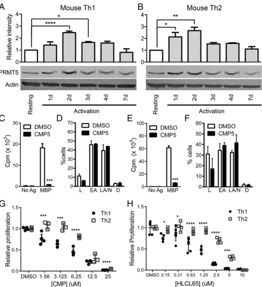
PRMT5 is overexpressed in several lymphoid malignancies, where it promotes uncontrolled cell growth and survival of transformed cells (35). However, its role in nonmalignant memory T cell proliferative responses is unknown. After exposure to their cog-nate Ag, previously sensitized T cells activate a signaling cascade that enhances metabolic activity and drives maximum prolifera-tion at 2–3 d postactivaprolifera-tion. Subsequently, the proliferative rate of T cells gradually decreases, and cells that survive the contraction period return to a nonproliferative resting state 7 d after activation (21). To determine whether PRMT5 plays a role in this process, PRMT5 expression was analyzed by Western blotting at various time points after MBPAc1–11TCR-Tg mouse memory Th1 or Th2
cells (characterized in Supplemental Fig. 1) were restimulated with immobilized anti-CD3/CD28. Compared with resting mem-ory T cells, PRMT5 was upregulated 2.5-fold in Th1 cells and 2.4-fold in Th2 cells at the 48-h time point, the peak of PRMT5 expression (Fig. 1A, 1B). PRMT5 was subsequently downregu-lated at day 4, reaching baseline levels by day 7. These results led us to hypothesize that PRMT5 promotes proliferation during the normal cycle of T cell activation.
Selective PRMT5 inhibition blunts TCR-mediated memory T cell expansion
To determine whether PRMT5 activity is required for memory T cell proliferation, resting memory Th1 and Th2 T cells were activated in vitro, and the extent of T cell expansion was measured by [3H]thymidine-incorporation assay in the presence of the
FIGURE 1. Proliferating murine Th1 and Th2 T cells express increased PRMT5 protein levels. MBP-specific TCR-Tg memory Th1 (A) and Th2 (B) cells were lysed at resting or at 1, 2, 3, 4, or 7 d after anti-CD3/CD28 stimulation and analyzed for PRMT5 protein expression by Western blotting. Resting cells were nonproliferating T cells and were collected 7–10 d after activation of the Th cell line with MBPAc1–11and irradiated splenocytes.b-actin was used as a loading control. Relative intensity quantification data are shown above a representative blot and were determined by normalizing PRMT5 expression to b-actin expression using Image Studio. Data are representative of three or four independent experiments (n= 5 for experiment shown). *p,0.05, **p, 0.01, ****p,0.001, one-way ANOVA, followed by the Sidak multiple-comparison adjustedttest. MBP TCR-Tg memory Th1 (CandD) and Th2 (Eand
previously described PRMT5 inhibitor, CMP5 (51), or DMSO vehicle control. CMP5 was designed to selectively and reversibly bind within the PRMT5 active site to prevent transfer of the methyl group from the donor, SAM, to the arginine
substrate-binding pocket. CMP5 selectively inhibits PRMT5-mediated SDM but not other PRMTs (51). Overall, these data indicate that CMP5 is a selective PRMT5 inhibitor. CMP5 treatment of mouse Th1 and Th2 cells strongly inhibited T cell proliferation (Fig. 1C, 1E). A detailed analysis of apoptosis status via annexin V/propidium iodide staining revealed that there were no signifi-cant differences in apoptotic or dead cells in cells treated with PRMT5 inhibitor (Fig. 1D, 1F), indicating that the reduced pro-liferation could not be explained by cell death. Interestingly, we noticed a small, but significant, difference in Th1 versus Th2 suppression with CMP5. Th1 cell proliferation was more sensitive to PRMT5 inhibitors than was Th2 cell proliferation (95.4 versus 90.5% inhibition, respectively, p, 0.005,ttest). To further ex-plore this phenomenon, we analyzed the IC50 values of the
PRMT5 inhibitor CMP5 for both cell types. Indeed, we confirmed that CMP5 inhibited Th1 cell proliferation (IC50= 3.7mM) more
potently than Th2 cell proliferation (IC50 = 9.2 mM) (Fig. 1G,
Table I. IC50values for PRMT5 inhibitors CMP5 and HLCL65 in mouse and human Th1 and Th2 cells
Compound Cell Type IC50(mM) Hill Slope R2
CMP5 mTh1 3.7 20.8334 0.96 mTh2 9.2 23.174 0.98 hTh1 26.9 23.454 0.94 hTh2 31.6 26.276 0.96 HLCL65 mTh1 1.1 21.392 0.91 mTh2 4 22.321 0.97 hTh1 5.7 23.291 0.98 hTh2 14.3 24.15 0.97
h, human; m, mouse.
Table I). To determine whether this phenomenon was isolated or could be replicated with CMP5 derivatives, we took advantage of a second-generation bioavailable PRMT5-selective inhibitor, HLCL65 (Supplemental Fig. 2A, 2B). HLCL65 selectively inhibited PRMT5-mediated SDM (Supplemental Fig. 2C, 2D). HLCL65 inhibited T cell proliferation more potently than CMP5, and it suppressed Th1 cells (IC50= 1.1mM) more effectively than
Th2 cells (IC50= 4.0mM) (Fig. 1H, Table I). Overall, these data
indicate that PRMT5 promotes murine memory T cell expansion and that inflammatory memory Th1 cells are more sensitive to targeting with PRMT5 inhibitors than are Th2 cells. To determine whether the proliferation or differentiation of newly activated naive T cells was similarly dependent on PRMT5 activity, we treated freshly isolated naive CD4+T cells with PRMT5 inhibi-tors. CMP5 and HLCL65 suppressed T cell proliferation in a dose-dependent manner (Supplemental Fig. 3A, 3B). Interestingly, and reminiscent of Th2 cells’ behavior, naive CD4+T cells were more resistant than memory Th1 cells to PRMT5 inhibitors (Supplemental Fig. 3A, 3B). We also observed decreased IFN-gin supernatants from naive T cell cultures differentiated with CMP5 and HLCL65 in the absence of exogenous polarizing signals (Supplemental Fig. 3C, 3D). Overall, these results show
prefer-ential suppressive effects of PRMT5 inhibitors on memory Th1 responses that drive inflammatory autoimmune diseases, such as MS.
PRMT5 is essential for human T cell activation and expansion
Targeting of pathogenic Th cell expansion may be beneficial as a therapy in human Th1–mediated autoimmune diseases. To deter-mine whether PRMT5 plays a similar role in human T cells, we restimulated previously differentiated human memory Th1- and Th2-enriched cells from healthy donors (characterized in Supplemental Fig. 4) and analyzed PRMT5 expression by Western blot from 0 to 7 d. We found that, similar to mouse T cells, PRMT5 was upregulated 2.1-fold in Th1 cells and 1.9-fold in Th2 cells by 48 h postactivation (Fig. 2A, 2B), and was subsequently downregulated to resting levels by 7 d postactivation. Importantly, PRMT5 was also essential for the expansion of human T cells. The PRMT5-selective inhibitor CMP5 preferentially suppressed the proliferation of human Th1 cells over Th2 cells (43 versus 9% inhibition, respectively,p,0.05) (Fig. 2C, 2E), but it had min-imal effects on cell death (Fig. 2D, 2F). To further evaluate the increased sensitivity of Th1 cells over Th2 cells to PRMT5 inhi-bition, we calculated the IC50of human T cells when treated with
CMP5 (Th1 IC50 = 26.9 mM versus Th2 IC50 = 31.6 mM) or
HLCL65 (Th1 IC50= 5.7mM versus Th2 IC50= 14.3mM) (Fig.
2G, 2H, Table I). To genetically validate a role for PRMT5 in
proliferation, we knocked down PRMT5 expression via shRNA lentiviral transduction in human Th1 cells. PRMT5 shRNA par-tially reduced PRMT5 protein levels (45%) but not other PRMTs (PRMT1 and PRMT7), and it significantly decreased Th1 cell proliferation (Fig. 3A). Although significant, the mild suppression of proliferation could be explained by the low lentiviral trans-duction efficiency expected in primary T cells, coupled with the proliferative advantage of untransduced (i.e., PRMT5-sufficient) cells. To increase efficiency, we used the Neon Transfection System, which provided 70–90% transfection efficiency in our primary human Th1/2 cell lines (Fig. 3B). Electroporation with any of three PRMT5-specific siRNAs efficiently suppressed hu-man Th1 and Th2 cell proliferation to a degree that correlated with PRMT5 knockdown (Fig. 3C, 3D and a positive Pearson corre-lation analysis of relative proliferation versus PRMT5 expression in Th1 and Th2 cells,r= +0.7481,p,0.0001, data not shown). These results validate a role for PRMT5 in T cell–proliferative responses. Interestingly, we observed that PRMT5 siRNA trans-fection was accompanied by PRMT1 protein suppression, partic-ularly in Th2 cells (Fig. 3C, 3D). Because the three siRNAs target different areas in PRMT5 (59untranslated region, exon 3, or exon 15), and each one was confirmed to lack complementarity with PRMT1, it is unlikely that this is a direct effect of the siRNAs on PRMT1 but rather is an indirect effect via PRMT5. Further studies should aim to elucidate the significance of this finding. Overall, these data indicate that PRMT5 is functionally conserved in mouse and human T cells and plays a critical role in memory Th cell reactivation and expansion.
PRMT5 expression is dependent upon TCR-induced NF-kB signaling
The NF-kB pathway was associated with PRMT5 in hematologic malignancies (56), but the pathways involved in PRMT5 expres-sion in T cells are unknown. T cell reactivation activates several signaling pathways, including the NF-kB pathway, leading to IL-2 production and proliferation. NF-kB transcription factors are kept inactive in the cytoplasm through binding to the inhibitory Ik-B subunits. Upon T cell activation, the Ik-B subunit is phosphory-lated by IKKa and proteasomally degraded, allowing nuclear FIGURE 4. T cell activation drives PRMT5
expres-sion in an NF-kB–dependent manner. Differentiated human Th1 (A) or Th2 (B) cells were reactivated on anti-CD3/CD28 and treated with DMSO vehicle or increasing amounts of the NF-kB pathway inhibitor Bay11 for 8 h. Cells were lysed for Western blot analysis of PRMT5 expression 48 h after initial acti-vation.b-actin was used as a loading control. Relative intensity quantification data are shown above a repre-sentative blot quantified with Image Studio. Data are representative of three independent experiments (n= 3 for experiment shown). Differentiated human Th1 (C) and Th2 (D) cells were treated as described in (A) and (B), and viability was monitored by trypan blue clusion. Data were pooled from three independent ex-periments (n= 6). (E) Differentiated human Th1 cells were treated as described in (A), and supernatants were collected to analyze the concentration of IL-2 produc-tion by ELISA. Data are representative of three inde-pendent experiments (n= 2). Plot error bars show6 SD. *p ,0.05, **p, 0.01, one-way ANOVA, fol-lowed by the Sidak multiple-comparison adjustedttest. n.d., not detected; n.s., not significant.
translocation of NF-kB and activation of transcription (57). To test whether NF-kB played a role in the upregulation of PRMT5 ex-pression after TCR/CD28 costimulation, we treated human Th cells with the IKK-ainhibitor Bay11 during the first 8 h of acti-vation. After 48 h of TCR/CD28 stimulation, we observed a de-crease in PRMT5 protein levels in healthy human donor Th1 cells treated with Bay11 (Fig. 4A). In contrast, PRMT5 expression in Th2 cells was less dependent on NF-kB signaling than was that in Th1 cells, as evidenced by stable levels of PRMT5 expression in Th2 cells treated with Bay11 (Fig. 4B). There were no differ-ences in cell death with Bay11 treatment (Fig. 4C, 4D), indicating that the changes in protein expression observed could not be explained by cell death. As expected (58), NF-kB inhibition also resulted in a 65% reduction in downstream cytokine IL-2 levels in Th1 cells (Fig. 4E). These data are consistent with NF-kB sig-naling promoting PRMT5 expression and IL-2 secretion during TCR-mediated activation of Th1 and Th2 cells.
PRMT5 inhibition suppresses IL-2 secretion, and IL-2 restoration rescues T cell expansion
TCR-mediated activation of the NF-kB pathway promotes IL-2, an important pro-proliferative T cell cytokine. We observed reduced IL-2 with Bay11-mediated suppression of PRMT5, and PRMT5 was linked to IL-2 production in Jurkat cancer T cells (59). Therefore, we explored whether PRMT5 inhibition af-fected IL-2 secretion in reactivated mouse or human Th1 and Th2 memory T cells. Vehicle-treated mouse (Fig. 5A) and human (Fig. 5B) Th1 cells secreted high levels of IL-2 at 24 and 48 h postactivation. In contrast, and as described previ-ously for Th2 cells (60), no IL-2 was detected in Th2 cell
su-pernatants (data not shown). PRMT5 inhibition resulted in a reduction in IL-2 secretion that ranged from 50 to 75% for mouse Th1 cells and from 30 to 80% for human Th1 cells (Fig. 5A, 5B). These data suggested that loss of IL-2 secretion may contribute to the suppression of proliferation observed in Th1 cells treated with PRMT5 inhibitors. To test whether the blunted T cell proliferation observed after PRMT5 inhibition could be rescued with IL-2 supplementation, memory Th1 cells were activated with anti-CD3/CD28 in the presence or absence of PRMT5 inhibitor and with increasing amounts of exogenous IL-2. Doses from 1 to 20 ng/ml were chosen because it was calculated that$10 ng/ml would be needed to restore the su-pernatant IL-2 levels observed in the vehicle Th1 condition (as in Fig. 5A). T cell proliferation was evaluated using a tritiated thymidine–incorporation assay. Treatment with 25 mM CMP5 inhibited mouse Th1 cell proliferation by 91% (p , 0.001,
ttest), and addition of IL-2 enhanced proliferation in the ve-hicle condition, reaching a peak at 5 ng/ml (Fig. 5C). Addition of IL-2 in the presence of PRMT5 inhibitor increased prolif-eration in a dose-dependent manner, reaching 100% of the control values at 10 ng/ml (Fig. 5C). Similarly, treatment with 25mM CMP5 suppressed human Th1 cell proliferation by 50%, and addition of exogenous IL-2 rescued proliferation (Fig. 5D). The recovery of Th1 T cell proliferation with exogenous IL-2 indicates that IL-2 pathways are active downstream of IL-2R and supports the notion that inhibition of IL-2 secretion by PRMT5 inhibitors contributes to the observed reduction in Th1 T cell proliferation. However, additional mechanisms may play a role in PRMT5 inhibitor–mediated suppression of prolifera-tion, particularly in Th2 cells.
PRMT5 inhibition suppresses in vivo OVA-induced DTH inflammatory responses
The effectiveness of PRMT5 inhibitors at suppressing inflamma-tory memory T cell responses suggested that they may be beneficial in inflammatory or autoimmune disease. To test this, we used the OVA-induced DTH mouse model and HLCL65, a more potent and bioavailable derivative of CMP5 (Figs. 1H, 2H, Supplemental Fig. 2). First, we analyzed PRMT5 expression in the spleen of un-treated mice after OVA immunization with CFA. We observed that, at 10 d after immunization, PRMT5 expression was upreg-ulated significantly in the spleen (Fig. 6A), suggesting that PRMT5 expression is relevant to in vivo DTH immune responses. In the DTH model (outlined in Fig. 6B), OVA immunization with CFA induces an OVA-specific T cell response that causes footpad inflammation in mice upon subsequent exposure to adjuvant-free OVA and memory CD4+ T cell expansion. HLCL65 treatment during the rechallenge period reduced footpad swelling, a measure of inflammation, by 40% (p,0.05, Fig. 6C). In addition, com-pared with vehicle, HLCL65 treatment reduced OVA-specific T cell proliferation by 36% (Fig. 6D) and IFN-g production by 70% (Fig. 6E). These data indicate that our novel PRMT5 hibitor HLCL65 suppresses T cell–mediated responses and in-flammation in vivo.
Prophylactic or therapeutic treatment with HLCL65 ameliorates EAE
The ability of PRMT5 inhibitors to suppress in vivo inflammatory T cell responses could be beneficial in the autoimmune disease MS. We observed that PRMT5 was upregulated in the spleen at 5 and 10 d after immunization with CFA/MOG (Fig. 7A), suggesting that PRMT5 plays an important role in the immune response against myelin Ags in vivo. In the MOG-induced murine EAE model, T cell responses against this myelin Ag resulted in ascending paralysis. We first tested whether short-term prophylactic HLCL65 treatment (5 d, 25 mg/kg every other day starting at immunization) could prevent EAE. Indeed, HLCL65 treatment resulted in delayed disease onset (16.9 d for HLCL65-treated mice versus 13.4 d for vehicle-treated mice) and a 33% reduction in disease incidence compared with vehicle-treated mice (Table II). Importantly, HLCL65-treated mice presented reduced EAE dis-ease burden, as measured by the area under the curve (AUC), compared with vehicle control (AUC HLCL65 versus vehicle: 2.1 versus 8.2,p= 0.016) (Fig. 7B, Table II). These clinical effects were associated with reduced MOG-specific T cell–proliferative responses (Fig. 7C), reduced CNS IL-17 production (Fig. 7D), and a trend toward suppressed Tbet mRNA expression in the CNS (Fig. 7E). With these promising results, we tested HLCL65 during a therapeutically relevant window for MS patients (i.e., after clinical signs had developed). HLCL65 treatment, beginning at 14 d after immunization (average score at treatment initiation = 2.7 out of 5), suppressed existing clinical signs of EAE, as mea-sured by the total disease burden (AUC HLCL65 versus vehicle: 19.161.6 versus 27.316 3.1, Fig. 7F, Table III). This disease suppression correlated with a reduction in MOG-specific T cell proliferation in HLCL65-treated mice (Fig. 7G). Additionally, inflammatory Th1 and Th17 responses were diminished in
HLCL65-treated mice (Fig. 7H, 7I). These data further support that PRMT5 activity may be essential for T cell function and that our novel PRMT5-selective inhibitors effectively suppress T cell– mediated inflammation. Next, to test whether PRMT5 inhibitors could suppress preformed encephalitogenic Th17 cells, spleno-cytes from MBP TCR-Tg mice that had spontaneously developed EAE (average score = 1.7) were activated with MBPAc1–11in the
presence or absence of CMP5 and HLCL65. CMP5 treatment significantly suppressed the frequency of pathogenic T-bet+ RORgt+(61) (Fig. 7J) and classic IL-17+RORgt+(Fig. 7K) Th17 cells in a dose-dependent manner. A similar dose-dependent de-crease in pathogenic T-bet+IL-17+Th17 cells (62) was apparent, but it did not reach statistical significance (Fig. 7L). Similar re-sults were observed with HLCL65 treatment (data not shown). Taken together, these results indicate that PRMT5 promotes pathogenic Th1 and Th17 cell responses that lead to inflammation and autoimmunity.
Discussion
Memory T cell reactivation after Ag exposure rapidly induces T cell proliferation and effector function. This process can be beneficial, as in vaccination immunity, or deleterious, as in perpetuation of pathogenic responses in autoimmunity. In this article, we show by expression knockdown and pharmacologic means that PRMT5, a methyltransferase that catalyzes SDM of arginine residues in histones and other proteins, promotes the activation and expansion of memory Th lymphocytes following Ag re-exposure.
The first indications of a key role for arginine methylation in lymphocyte activation originated from conditions and treatments that inhibit all SAM-dependent methylation reactions (25, 26). PRMTs were proposed to mediate some of these effects (29), but the role of individual PRMTs in these processes remained unre-solved. We found that Ag re-exposure in memory T cells upreg-ulates PRMT5 expression as T cells proliferate and expand, followed by a contraction phase in which PRMT5 expression is progressively lost. The temporal link between PRMT5 expression and proliferation, together with the observed inhibition of prolif-eration upon selective PRMT5 inhibition, indicates that PRMT5 activity is necessary for TCR engagement–induced memory T cell expansion. Th2 cell expansion was less dependent on PRMT5 activity than was that of Th1 cells. This difference was reproduced in mouse and human Th cells, indicating that this is a conserved difference that may impact human disease. However, differential sensitivity to PRMT5 inhibition did not appear to stem from dif-ferences in PRMT5 expression, which was equivalent in Th1 and Th2 cells. It is possible that PRMT5 activity is lower in Th2 cells than in Th1 cells as a result of the expression of type I methyl-transferases, which compete with PRMT5 for substrates (63). This difference offers the intriguing possibility that targeting PRMT5 may modulate the Th1/Th2 balance defect observed in autoimmune/inflammatory diseases, such as MS (12, 13).
The exact chain of events that leads to PRMT5 upregulation in T cells is unclear. A link between the NF-kB pathway leading to activation of the repressive p65/HDAC/Sp1 complex and loss of PRMT5 targeting microRNA was reported in mantle cell
10 d after immunization and activated in the presence or absence of MOG. MOG-specific proliferation was monitored via [3H]thymidine incorporation (G), IFN-g+– secreting cells were quantified by flow cytometry (gated on CD4+, CD44+cells) (H), and IL-17 production was measured by ELISA (I). *p,0.05, ***p,0.001, ****p,0.0001, one-way ANOVA, followed by the Sidak multiple-comparison adjustedttest. (J–L) Splenocytes were isolated from MBP TCR-Tg mice with spontaneous EAE and activated with MBPAc1–11in the presence of the indicated concentrations of the PRMT5 inhibitors CMP5 and HLCL65 or DMSO vehicle control for 48 h. Frequencies of RORgt+
lymphoma (64). We also found previously that NF-kB inhibition suppresses PRMT5 expression in EBV-transformed cells (51). Because TCR engagement activates the NF-kB pathway in T cells, a similar mechanism may regulate PRMT5 expression in T cells. Indeed, blocking NF-kB signaling attenuated, but did not com-pletely eliminate, PRMT5 expression in human Th1 cells. This indicates that, although NF-kB is an important driver of PRMT5 expression in Th1 cells, other TCR-induced pathways play a more significant role in regulating PRMT5 expression, especially in Th2 cells. TCR signaling cascades include the NFAT, ERK1/2, p38, and JNK MAPK pathways. Interestingly, inhibitors of the p38 and JNK MAPK pathways, but not the ERK1/2 pathway, were shown to inhibit hypoxia-induced upregulation of PRMT5 in lung epi-thelial cells (65). Although future studies are required to clarify the extent to which these pathways affect PRMT5 upregulation in T cells, NF-kB appears to play a major role in TCR-induced PRMT5 expression in human Th1, but not Th2, cells. Addition-ally, several studies showed that PRMT5 activates NF-kB sig-naling through arginine methylation of p65 (56, 66–68), suggesting that the NF-kB–PRMT5 signaling axis could involve a positive-feedback loop. Additional studies are required to validate this feedback loop and evaluate its role in T cells.
Several pathways downstream of TCR activation converge upon activation of the IL-2 promoter to induce T cell proliferation (18, 19, 69). In this study, we found that IL-2 secretion is dependent on PRMT5 activity and that addition of exogenous IL-2 to PRMT5 inhibitor–treated cells restored proliferation in Th1 cells. A role for PRMT5 in IL-2 production is consistent with the observations of Richard et al. (59); they reported that PRMT5 siRNA sup-presses IL-2 secretion in the Jurkat cancer T cell line. This effect is thought to be mediated by PRMT5-catalyzed arginine methyl-ation on histones. In support of this hypothesis, symmetrically dimethylated proteins associate with the IL-2 promoter after T cell activation. In contrast, PRMT5 did not associate directly with the IL-2 promoter. These data are consistent with PRMT5 indirectly regulating IL-2 expression via SDM of target proteins. Although the specific proteins that are methylated and bind to the IL-2
promoter remain to be defined, two proteins that form an IL-2 promoter–binding complex, NF-45 and NF-90, were proposed as candidate targets (59). Another candidate is the TCR signaling protein Vav-1, whose SDM was reported to promote IL-2 ex-pression (70). Overall, our data point to IL-2 as one of the mechanisms by which PRMT5 regulates proliferation in Th1 cells. However, because we observed only a 60% reduction in IL-2 production, yet T cell proliferation is reduced by 90–95% when treated with CMP5, it is likely that PRMT5 regulates proliferation by several mechanisms. Because Th2 cells do not secrete large amounts of IL-2, further studies are required to determine the mechanism by which PRMT5 promotes Th2 cell proliferation.
Memory T cell responses play a critical role in chronic T cell– mediated diseases, such as autoimmunity and allergy (71, 72). For example, increased memory T cells were found in MS patients with active disease, and they increased further during disease flare (4), whereas the memory/naive T cell ratio diminishes in patients responding to therapy (73). Importantly, inhibition of methyl-transferases successfully suppresses T cell activation and estab-lished clinical EAE and other inflammatory/autoimmune diseases (25–28), but the lack of selectivity has prevented the development of these treatments as therapy. Our data indicate that selective PRMT5 inhibition reproduces the suppression of memory T cell expansion observed with pan-methyltransferase inhibitors and may be similarly effective in autoimmunity. Indeed, in vivo treatment with PRMT5 inhibitors suppressed two models of inflammatory/autoimmune disease: DTH footpad inflammation and EAE CNS inflammation. Importantly, clinically established EAE disease was responsive to PRMT5 inhibitor treatment. Our data are consistent with T cells being a major target of PRMT5 inhibitors in EAE, although we cannot rule out a clinical contri-bution of PRMT5 inhibition in non-T cells (CNS cells or APCs). Effects on APCs could result in reduced TCR engagement and T cell responses. However, in vitro experiments showed similar suppressive effects when T cells are activated by anti-CD3/CD28 (Fig. 1E) or Ag-loaded APCs (Fig. 1C). In addition, HLCL65 treatment of EAE suppressed previously generated memory T cell Table II. Prophylactic treatment with HLCL65 ameliorates EAE
Disease Parameter DMSO HLCL65 Change from DMSO pValue
EAE incidence (%) 90 57 233 n/a
Disease onset (d; mean6SD) 13.4460.53 16.8660.96 +3.42 d 0.0053
AUC (mean6SD) 8.21461.58 2.14361.41 26.071 0.0159
Score on day 17 (mean6SD) 2.360.37 1.060.48 21.3 0.048 Maximum score (mean6SD) 2.4560.37 1.060.48 21.45 0.0285
n 10 7 n/a n/a
n/a, not applicable.
Table III. Therapeutic treatment with HLCL65 ameliorates EAE
Disease Parameter DMSO HLCL65 Change from DMSO pValue
EAE incidence (%): pretreatment 100 100 0 n/a
Disease onset (d): pretreatment (mean6SD) 11.6760.49 12.4360.37 +0.76 NS
Score on day 14 (mean6SD)a 2.760.40 2.760.40 0 NS
AUC: pretreatment (mean6SD) 4.6061.15 3.9160.91 20.69 NS AUC: posttreatment (mean6SD) 27.3163.13 19.1161.61 28.2 0.0329 Maximum score: pretreatment (mean6SD) 2.760.40 2.760.40 0 NS Maximum score: posttreatment (mean6SD) 3.8860.38 2.9360.44 20.95 0.0535 Score on day 22 (mean6SD) 3.560.51 2.1460.19 21.36 0.0228
n 6 7 n/a n/a
aTreatment initiation occurred on day 14.
responses, which are less dependent on APC costimulation. The similarity in the suppression of proliferation and inflammatory cytokines from in vitro and in vivo DTH/EAE studies is also con-sistent with T cells being a major target. To investigate relevance to human disease, we analyzed genome-wide association studies from the International Multiple Sclerosis Genetics Consortium and the Wellcome Trust Case Control Consortium. Interestingly, rs4410871 was identified as a high-frequency single-nucleotide polymorphism in theMYC locus in MS patients (74). MYC was shown to be upregulated after T cell activation (75) and to promote PRMT5 expression (37, 38). Taken together, these data suggest that PRMT5 could play a significant role in human disease.
In summary, this is the first report, to our knowledge, of the role of PRMT5 expression in in vitro and in vivo nonmalignant T cell responses. Our work identifies PRMT5 as an epigenetic modifier enzyme that promotes memory Th cell expansion. Memory T cell expansion of inflammatory Th1 cells and, to a lesser extent, Th2 cells, was dependent on PRMT5 activity. Finally, PRMT5 inhib-itors suppressed T cell–mediated inflammatory and autoimmune disease, suggesting that PRMT5 may be a promising therapeutic target for autoimmune and other T cell–mediated diseases.
Acknowledgments
We thank Chad Bennett, Erandi De Silva, Larry Schaaf, and Bence Boelscewski of the Drug Development Institute team for their insightful discussions.
Disclosures
R.A.B. and C.L. have a patent on PRMT5 inhibitors. M.G.-d.-A. has a patent pending. The other authors have no financial conflicts of interest.
References
1. World Health Organization; Multiple Sclerosis International Federation. 2008. Atlas: multiple sclerosis resources in the world 2008. Available at: http://www. who.int/mental_health/neurology/Atlas_MS_WEB.pdf. Accessed: December 30, 2016.
2. Frohman, E. M., M. K. Racke, and C. S. Raine. 2006. Multiple sclerosis—the plaque and its pathogenesis.N. Engl. J. Med.354: 942–955.
3. Crucian, B., P. Dunne, H. Friedman, R. Ragsdale, S. Pross, and R. Widen. 1995. Alterations in peripheral blood mononuclear cell cytokine production in re-sponse to phytohemagglutinin in multiple sclerosis patients.Clin. Diagn. Lab. Immunol.2: 766–769.
4. Okuda, Y., M. Okuda, B. R. Apatoff, and D. N. Posnett. 2005. The activation of memory CD4(+) T cells and CD8(+) T cells in patients with multiple sclerosis. J. Neurol. Sci.235: 11–17.
5. Khoury, S. J., C. R. Guttmann, E. J. Orav, R. Kikinis, F. A. Jolesz, and H. L. Weiner. 2000. Changes in activated T cells in the blood correlate with disease activity in multiple sclerosis.Arch. Neurol.57: 1183–1189. 6. Putheti, P., M. Morris, L. Stawiarz, N. Teleshova, P. Kivisa¨kk, M. Pashenkov,
M. Kouwenhoven, M. K. Wiberg, L. Bronge, Y.-M. Huang, et al. 2003. Multiple sclerosis: a study of chemokine receptors and regulatory T cells in relation to MRI variables.Eur. J. Neurol.10: 529–535.
7. Jensen, J., A. R. Langkilde, C. Fenst, M. S. Nicolaisen, H. G. Roed, M. Christiansen, and F. Sellebjerg. 2004. CD4 T cell activation and disease activity at onset of multiple sclerosis.J. Neuroimmunol.149: 202–209. 8. Scolozzi, R., A. Boccafogli, M. R. Tola, L. Vicentini, A. Camerani, D. Degani,
E. Granieri, L. Caniatti, and E. Paolino. 1992. T-cell phenotypic profiles in the cerebrospinal fluid and peripheral blood of multiple sclerosis patients.J. Neurol. Sci.108: 93–98.
9. Kebir, H., K. Kreymborg, I. Ifergan, A. Dodelet-Devillers, R. Cayrol, M. Bernard, F. Giuliani, N. Arbour, B. Becher, and A. Prat. 2007. Human TH17 lymphocytes promote blood-brain barrier disruption and central nervous system inflammation.Nat. Med.13: 1173–1175.
10. Venken, K., N. Hellings, M. Thewissen, V. Somers, K. Hensen, J.-L. Rummens, R. Medaer, R. Hupperts, and P. Stinissen. 2008. Compromised CD4+ CD25 (high) regulatory T-cell function in patients with relapsing-remitting multiple sclerosis is correlated with a reduced frequency of FOXP3-positive cells and reduced FOXP3 expression at the single-cell level.Immunology123: 79–89. 11. Windhagen, A., D. E. Anderson, A. Carrizosa, K. Balashov, H. L. Weiner, and
D. A. Hafler. 1998. Cytokine secretion of myelin basic protein reactive T cells in patients with multiple sclerosis.J. Neuroimmunol.91: 1–9.
12. Guerau-de-Arellano, M., K. M. Smith, J. Godlewski, Y. Liu, R. Winger, S. E. Lawler, C. C. Whitacre, M. K. Racke, and A. E. Lovett-Racke. 2011. Micro-RNA dysregulation in multiple sclerosis favours pro-inflammatory T-cell-mediated autoimmunity.Brain134: 3578–3589.
13. Couturier, N., F. Bucciarelli, R. N. Nurtdinov, M. Debouverie, C. Lebrun-Frenay, G. Defer, T. Moreau, C. Confavreux, S. Vukusic, I. Cournu-Rebeix, et al. 2011. Tyrosine kinase 2 variant influences T lymphocyte polarization and multiple sclerosis susceptibility.Brain134: 693–703.
14. Raddassi, K., S. C. Kent, J. Yang, K. Bourcier, E. M. Bradshaw, V. Seyfert-Margolis, G. T. Nepom, W. W. Kwok, and D. A. Hafler. 2011. Increased fre-quencies of myelin oligodendrocyte glycoprotein/MHC class II-binding CD4 cells in patients with multiple sclerosis.J. Immunol.187: 1039–1046. 15. Lovett-Racke, A. E., J. L. Trotter, J. Lauber, P. J. Perrin, C. H. June, and
M. K. Racke. 1998. Decreased dependence of myelin basic protein-reactive T cells on CD28-mediated costimulation in multiple sclerosis patients. A marker of activated/memory T cells.J. Clin. Invest.101: 725–730.
16. Cao, Y., B. A. Goods, K. Raddassi, G. T. Nepom, W. W. Kwok, J. C. Love, and D. A. Hafler. 2015. Functional inflammatory profiles distinguish myelin-reactive T cells from patients with multiple sclerosis.Sci. Transl. Med.7: 287ra74. 17. Nakayama, T., and M. Yamashita. 2010. The TCR-mediated signaling pathways
that control the direction of helper T cell differentiation.Semin. Immunol.22: 303–309.
18. Jain, J., C. Loh, and A. Rao. 1995. Transcriptional regulation of the IL-2 gene. Curr. Opin. Immunol.7: 333–342.
19. Guy, C. S., K. M. Vignali, J. Temirov, M. L. Bettini, A. E. Overacre, M. Smeltzer, H. Zhang, J. B. Huppa, Y.-H. Tsai, C. Lobry, et al. 2013. Distinct TCR signaling pathways drive proliferation and cytokine production in T cells. Nat. Immunol.14: 262–270.
20. Huang, W., and A. August. 2015. The signaling symphony: T cell receptor tunes cytokine-mediated T cell differentiation.J. Leukoc. Biol.97: 477–485. 21. Corradin, G., H. M. Etlinger, and J. M. Chiller. 1977. Lymphocyte specificity to
protein antigens. I. Characterization of the antigen-induced in vitro T cell-dependent proliferative response with lymph node cells from primed mice. J. Immunol.119: 1048–1053.
22. Cassani, B., M. Mirolo, F. Cattaneo, U. Benninghoff, M. Hershfield, F. Carlucci, A. Tabucchi, C. Bordignon, M. G. Roncarolo, and A. Aiuti. 2008. Altered in-tracellular and exin-tracellular signaling leads to impaired T-cell functions in ADA-SCID patients.Blood111: 4209–4219.
23. Blaese, R. M., K. W. Culver, A. D. Miller, C. S. Carter, T. Fleisher, M. Clerici, G. Shearer, L. Chang, Y. Chiang, P. Tolstoshev, et al. 1995. T lymphocyte-directed gene therapy for ADA- SCID: initial trial results after 4 years. Sci-ence270: 475–480.
24. German, D. C., C. A. Bloch, and N. M. Kredich. 1983. Measurements of S-adenosylmethionine and L-homocysteine metabolism in cultured human lymphoid cells.J. Biol. Chem.258: 10997–11003.
25. Yang, M.-L., A. J. Gee, R. J. Gee, C. I. Zurita-Lopez, S. Khare, S. G. Clarke, and M. J. Mamula. 2013. Lupus autoimmunity altered by cellular methylation metabolism.Autoimmunity46: 21–31.
26. Saso, Y., E. M. Conner, B. R. Teegarden, and C. S. Yuan. 2001. S-Adenosyl-L-homocysteine hydrolase inhibitor mediates immunosuppressive effects in vivo: suppression of delayed type hypersensitivity ear swelling and peptidoglycan polysaccharide-induced arthritis.J. Pharmacol. Exp. Ther.296: 106–112. 27. Moreno, B., H. Hevia, M. Santamaria, J. Sepulcre, J. Mun˜oz, E. R.
Garcı´a-Trevijano, C. Berasain, F. J. Corrales, M. A. Avila, and P. Villoslada. 2006. Methylthioadenosine reverses brain autoimmune disease.Ann. Neurol.60: 323– 334.
28. Moreno, B., B. Fernandez-Diez, A. Di Penta, and P. Villoslada. 2010. Preclinical studies of methylthioadenosine for the treatment of multiple sclerosis.Mult. Scler.16: 1102–1108.
29. Parry, R. V., and S. G. Ward. 2010. Protein arginine methylation: a new handle on T lymphocytes?Trends Immunol.31: 164–169.
30. Henrich, F. C., K. Singer, K. Poller, L. Bernhardt, C. D. Strobl, K. Limm, A. P. Ritter, E. Gottfried, S. Vo¨lkl, B. Jacobs, et al. 2016. Suppressive effects of tumor cell-derived 59-deoxy-59-methylthioadenosine on human T cells. OncoImmunology5: e1184802.
31. Gary, J. D., and S. Clarke. 1998. RNA and protein interactions modulated by protein arginine methylation.Prog. Nucleic Acid Res. Mol. Biol.61: 65–131. 32. Yang, Y., A. Hadjikyriacou, Z. Xia, S. Gayatri, D. Kim, C. Zurita-Lopez,
R. Kelly, A. Guo, W. Li, S. G. Clarke, and M. T. Bedford. 2015. PRMT9 is a type II methyltransferase that methylates the splicing factor SAP145.Nat. Commun. 6: 6428.
33. Zurita-Lopez, C. I., T. Sandberg, R. Kelly, and S. G. Clarke. 2012. Human protein arginine methyltransferase 7 (PRMT7) is a type III enzyme forming
v-NG-monomethylated arginine residues.J. Biol. Chem.287: 7859–7870. 34. Bedford, M. T. 2007. Arginine methylation at a glance.J. Cell Sci.120: 4243–
4246.
35. Stopa, N., J. E. Krebs, and D. Shechter. 2015. The PRMT5 arginine methyl-transferase: many roles in development, cancer and beyond.Cell. Mol. Life Sci. 72: 2041–2059.
36. Karkhanis, V., Y.-J. Hu, R. A. Baiocchi, A. N. Imbalzano, and S. Sif. 2011. Versatility of PRMT5-induced methylation in growth control and development. Trends Biochem. Sci.36: 633–641.
37. Koh, C. M., M. Bezzi, D. H. P. Low, W. X. Ang, S. X. Teo, F. P. H. Gay, M. Al-Haddawi, S. Y. Tan, M. Osato, A. Sabo`, et al. 2015. MYC regulates the core pre-mRNA splicing machinery as an essential step in lymphomagenesis.Nature523: 96–100.
39. Pal, S., R. A. Baiocchi, J. C. Byrd, M. R. Grever, S. T. Jacob, and S. Sif. 2007. Low levels of miR-92b/96 induce PRMT5 translation and H3R8/H4R3 methylation in mantle cell lymphoma.EMBO J.26: 3558–3569.
40. Wang, L., S. Pal, and S. Sif. 2008. Protein arginine methyltransferase 5 sup-presses the transcription of the RB family of tumor suppressors in leukemia and lymphoma cells.Mol. Cell. Biol.28: 6262–6277.
41. Chung, J., V. Karkhanis, S. Tae, F. Yan, P. Smith, L. W. Ayers, C. Agostinelli, S. Pileri, G. V. Denis, R. A. Baiocchi, and S. Sif. 2013. Protein arginine meth-yltransferase 5 (PRMT5) inhibition induces lymphoma cell death through reactivation of the retinoblastoma tumor suppressor pathway and polycomb re-pressor complex 2 (PRC2) silencing.J. Biol. Chem.288: 35534–35547. 42. Shilo, K., X. Wu, S. Sharma, M. Welliver, W. Duan, M. Villalona-Calero,
J. Fukuoka, S. Sif, R. Baiocchi, C. L. Hitchcock, et al. 2013. Cellular localization of protein arginine methyltransferase-5 correlates with grade of lung tumors. Diagn. Pathol.8: 201.
43. Yan, F., L. Alinari, M. E. Lustberg, L. K. Martin, H. M. Cordero-Nieves, Y. Banasavadi-Siddegowda, S. Virk, J. Barnholtz-Sloan, E. H. Bell, J. Wojton, et al. 2014. Genetic validation of the protein arginine methyltransferase PRMT5 as a candidate therapeutic target in glioblastoma.Cancer Res.74: 1752–1765. 44. Han, X., R. Li, W. Zhang, X. Yang, C. G. Wheeler, G. K. Friedman, P. Province,
Q. Ding, Z. You, H. M. Fathallah-Shaykh, et al. 2014. Expression of PRMT5 correlates with malignant grade in gliomas and plays a pivotal role in tumor growth in vitro.J. Neurooncol.118: 61–72.
45. Powers, M. A., M. M. Fay, R. E. Factor, A. L. Welm, and K. S. Ullman. 2011. Protein arginine methyltransferase 5 accelerates tumor growth by arginine methylation of the tumor suppressor programmed cell death 4.Cancer Res.71: 5579–5587.
46. Gu, Z., S. Gao, F. Zhang, Z. Wang, W. Ma, R. E. Davis, and Z. Wang. 2012. Protein arginine methyltransferase 5 is essential for growth of lung cancer cells. Biochem. J.446: 235–241.
47. Bao, X., S. Zhao, T. Liu, Y. Liu, Y. Liu, and X. Yang. 2013. Overexpression of PRMT5 promotes tumor cell growth and is associated with poor disease prog-nosis in epithelial ovarian cancer.J. Histochem. Cytochem.61: 206–217. 48. Kim, J.-M., H. Y. Sohn, S. Y. Yoon, J. H. Oh, J. O. Yang, J. H. Kim, K. S. Song,
S. M. Rho, H. S. Yoo, Y. S. Kim, et al. 2005. Identification of gastric cancer-related genes using a cDNA microarray containing novel expressed sequence tags expressed in gastric cancer cells.Clin. Cancer Res.11: 473–482. 49. Cho, E. C., S. Zheng, S. Munro, G. Liu, S. M. Carr, J. Moehlenbrink, Y. C. Lu,
L. Stimson, O. Khan, R. Konietzny, et al. 2012. Arginine methylation controls growth regulation by E2F-1.EMBO J.31: 1785–1797.
50. Panfil, A. R., J. Al-Saleem, C. M. Howard, J. M. Mates, J. J. Kwiek, R. A. Baiocchi, and P. L. Green. 2015. PRMT5 is upregulated in HTLV-1-mediated T-cell transformation and selective inhibition alters viral gene ex-pression and infected cell survival.Viruses8: 7.
51. Alinari, L., K. V. Mahasenan, F. Yan, V. Karkhanis, J. H. Chung, E. M. Smith, C. Quinion, P. L. Smith, L. Kim, J. T. Patton, et al. 2015. Selective inhibition of protein arginine methyltransferase 5 blocks initiation and maintenance of B-cell transformation.Blood125: 2530–2543.
52. Chan-Penebre, E., K. G. Kuplast, C. R. Majer, P. A. Boriack-Sjodin, T. J. Wigle, L. D. Johnston, N. Rioux, M. J. Munchhof, L. Jin, S. L. Jacques, et al. 2015. A selective inhibitor of PRMT5 with in vivo and in vitro potency in MCL models. Nat. Chem. Biol.11: 432–437.
53. Goverman, J., A. Woods, L. Larson, L. P. Weiner, L. Hood, and D. M. Zaller. 1993. Transgenic mice that express a myelin basic protein-specific T cell re-ceptor develop spontaneous autoimmunity.Cell72: 551–560.
54. Pal, S., S. N. Vishwanath, H. Erdjument-Bromage, P. Tempst, and S. Sif. 2004. Human SWI/SNF-associated PRMT5 methylates histone H3 arginine 8 and negatively regulates expression of ST7 and NM23 tumor suppressor genes.Mol. Cell. Biol.24: 9630–9645.
55. Zhao, X., S. Chen-Kiang, S. Shetty, M. Di Liberto, J. Bodo, L. Durkin, K. Eng, O. Elemento, M. R. Smith, and E. D. Hsi. 2015. CCMCL1: a new model of aggressive mantle cell lymphoma.Blood125: 2730–2732.
56. Tanaka, H., Y. Hoshikawa, T. Oh-hara, S. Koike, M. Naito, T. Noda, H. Arai, T. Tsuruo, and N. Fujita. 2009. PRMT5, a novel TRAIL receptor-binding
pro-tein, inhibits TRAIL-induced apoptosis via nuclear factor-kappaB activation. Mol. Cancer Res.7: 557–569.
57. Nomura, F., T. Kawai, K. Nakanishi, and S. Akira. 2000. NF-kappaB activation through IKK-i-dependent I-TRAF/TANK phosphorylation.Genes Cells5: 191– 202.
58. Sriskantharajah, S., M. P. Belich, S. Papoutsopoulou, J. Janzen, V. Tybulewicz, B. Seddon, and S. C. Ley. 2009. Proteolysis of NF-kappaB1 p105 is essential for T cell antigen receptor-induced proliferation.Nat. Immunol.10: 38–47. 59. Richard, S., M. Morel, and P. Cle´roux. 2005. Arginine methylation regulates
IL-2 gene expression: a role for protein arginine methyltransferase 5 (PRMT5). Biochem. J.388: 379–386.
60. Mosmann, T. R., and R. L. Coffman. 1989. TH1 and TH2 cells: different patterns of lymphokine secretion lead to different functional properties. Annu. Rev. Immunol.7: 145–173.
61. Ghoreschi, K., A. Laurence, X.-P. Yang, C. M. Tato, M. J. McGeachy, J. E. Konkel, H. L. Ramos, L. Wei, T. S. Davidson, N. Bouladoux, et al. 2010. Generation of pathogenic T(H)17 cells in the absence of TGF-b signalling. Nature467: 967–971.
62. Yang, Y., J. Weiner, Y. Liu, A. J. Smith, D. J. Huss, R. Winger, H. Peng, P. D. Cravens, M. K. Racke, and A. E. Lovett-Racke. 2009. T-bet is essential for encephalitogenicity of both Th1 and Th17 cells.J. Exp. Med.206: 1549–1564. 63. Bonham, K., S. Hemmers, Y.-H. Lim, D. M. Hill, M. G. Finn, and K. A. Mowen. 2010. Effects of a novel arginine methyltransferase inhibitor on T-helper cell cytokine production.FEBS J.277: 2096–2108.
64. Liu, S., L.-C. Wu, J. Pang, R. Santhanam, S. Schwind, Y.-Z. Wu, C. J. Hickey, J. Yu, H. Becker, K. Maharry, et al. 2010. Sp1/NFkappaB/HDAC/miR-29b regulatory network in KIT-driven myeloid leukemia.Cancer Cell17: 333–347. 65. Lim, S. K., Y. W. Jeong, D. I. Kim, M. J. Park, J. H. Choi, S. U. Kim, S. S. Kang, H. J. Han, and S. H. Park. 2013. Activation of PRMT1 and PRMT5 mediates hypoxia- and ischemia-induced apoptosis in human lung epithelial cells and the lung of miniature pigs: the role of p38 and JNK mitogen-activated protein ki-nases.Biochem. Biophys. Res. Commun.440: 707–713.
66. Lu, T., and G. R. Stark. 2015. NF-kB: regulation by methylation.Cancer Res. 75: 3692–3695.
67. Wei, H., B. Wang, M. Miyagi, Y. She, B. Gopalan, D.-B. Huang, G. Ghosh, G. R. Stark, and T. Lu. 2013. PRMT5 dimethylates R30 of the p65 subunit to activate NF-kB.Proc. Natl. Acad. Sci. USA110: 13516–13521.
68. Harris, D. P., S. Bandyopadhyay, T. J. Maxwell, B. Willard, and P. E. DiCorleto. 2014. Tumor necrosis factor (TNF)-ainduction of CXCL10 in endothelial cells requires protein arginine methyltransferase 5 (PRMT5)-mediated nuclear factor (NF)-kB p65 methylation.J. Biol. Chem.289: 15328–15339.
69. Kemp, M. L., L. Wille, C. L. Lewis, L. B. Nicholson, and D. A. Lauffenburger. 2007. Quantitative network signal combinations downstream of TCR activation can predict IL-2 production response.J. Immunol.178: 4984–4992. 70. Blanchet, F., A. Cardona, F. A. Letimier, M. S. Hershfield, and O. Acuto. 2005.
CD28 costimulatory signal induces protein arginine methylation in T cells. J. Exp. Med.202: 371–377.
71. Devarajan, P., and Z. Chen. 2013. Autoimmune effector memory T cells: the bad and the good.Immunol. Res.57: 12–22.
72. Endo, Y., K. Hirahara, R. Yagi, D. J. Tumes, and T. Nakayama. 2014. Pathogenic memory type Th2 cells in allergic inflammation.Trends Immunol.35: 69–78. 73. Blanco, Y., E. A. Moral, M. Costa, M. Go´mez-Choco, J. F. Torres-Peraza,
L. Alonso-Magdalena, J. Alberch, D. Jaraquemada, T. Arbizu, F. Graus, and A. Saiz. 2006. Effect of glatiramer acetate (Copaxone) on the immunopheno-typic and cytokine profile and BDNF production in multiple sclerosis: a longi-tudinal study.Neurosci. Lett.406: 270–275.
74. International Multiple Sclerosis Genetics Consortium, Wellcome Trust Case Control Consortium 2, S. Sawcer, G. Hellenthal, M. Pirinen, C. C. Spencer, N. A. Patsopoulos, L. Moutsianas, A. Dilthey, Z. Su, C. Freeman, S. E. Hunt, et al. 2011. Genetic risk and a primary role for cell-mediated immune mecha-nisms in multiple sclerosis.Nature476: 214–219.