Received 26 Jan 2016
|
Accepted 5 May 2016
|
Published 13 Jun 2016
A splicing isoform of TEAD4 attenuates the
Hippo–YAP signalling to inhibit tumour proliferation
Yangfan Qi
1,
*, Jing Yu
2,
*, Wei Han
1,
*, Xiaojuan Fan
3
, Haili Qian
4
, Huanhuan Wei
3
, Yi-hsuan S. Tsai
5
, Jinyao Zhao
1
,
Wenjing Zhang
1
, Quentin Liu
1
, Songshu Meng
1
, Yang Wang
1
& Zefeng Wang
3
Aberrant splicing is frequently found in cancer, yet the biological consequences of such
alterations are mostly undefined. Here we report that the Hippo–YAP signalling, a key
pathway that regulates cell proliferation and organ size, is under control of a splicing switch.
We show that TEAD4, the transcription factor that mediates Hippo–YAP signalling,
under-goes alternative splicing facilitated by the tumour suppressor RBM4, producing a truncated
isoform, TEAD4-S, which lacks an N-terminal DNA-binding domain, but maintains YAP
interaction domain. TEAD4-S is located in both the nucleus and cytoplasm, acting as a
dominant negative isoform to YAP activity. Consistently, TEAD4-S is reduced in cancer cells,
and its re-expression suppresses cancer cell proliferation and migration, inhibiting tumour
growth in xenograft mouse models. Furthermore, TEAD4-S is reduced in human cancers, and
patients with elevated TEAD4-S levels have improved survival. Altogether, these data reveal a
splicing switch that serves to fine tune the Hippo–YAP pathway.
DOI: 10.1038/ncomms11840
OPEN
1Institute of Cancer Stem Cell, Second Affiliated Hospital Collaborative Innovation Center of Oncology, Dalian Medical University, Dalian 116044, China. 2Department of Biostatistics, University of North Carolina at Chapel Hill, Chapel Hill, North Carolina 27599, USA.3Key Laboratory of Computational Biology,
CAS-MPG Partner Institute for Computational Biology, Shanghai Institutes for Biological Sciences, Chinese Academy of Sciences, Shanghai 200031, China. 4State Key Lab of Molecular Oncology, Peking Union Medical College and Chinese Academy of Medical Sciences, Beijing 100021, China.5Lineberger
H
ippo–YAP signalling is a key pathway that regulates cell
proliferation, cell contact inhibition and organ size
1–3.
The transcriptional output of this pathway is mediated by
TEAD proteins that partner with YAP to activate genes that
stimulate cell proliferation
4,5. As a key effector of the Hippo
pathway, YAP lacks DNA-binding motif, and thus recognizes its
targets through interacting with TEAD proteins
6. Under normal
condition, YAP is translocated into the nucleus to promote
cell growth; however, the activation of Hippo causes YAP
phosphorylation,
leading
to
cytoplasmic
retention
and
degradation of YAP
7,8. Thus, defects of the Hippo signalling
cause overgrowth phenotypes due to deregulation of proliferation
and apoptotic defects
9,10. Hippo–YAP pathway is directly
involved in cancer development
11–13, and inhibition of the YAP
activity provides a valuable route for cancer prevention and
treatment
14–16. In current model, Hippo signalling is mainly
regulated via protein phosphorylation and degradation
10,17.
Intriguingly, some key components of the Hippo–YAP pathway
undergo extensive regulation in RNA level through alternative
splicing (AS), a major mechanism to expand coding capacity of
human genome. For example, MST1 has multiple isoforms with
C-terminal truncations, and YAP has eight splicing isoforms with
different internal sequences
18. However, the biological functions
of these isoforms are unclear.
AS elicits control over the major hallmarks of cancer
19–21,
including apoptosis
22, angiogenesis
23and
epithelial–mesen-chymal transition (EMT)
24. However, the functional
conse-quences of most cancer-related splicing alterations are undefined.
AS is generally regulated by splicing factors that specifically bind
cis
-elements in pre-messenger RNA (mRNA) to affect the
splicing efficiency
25–27. The alteration of splicing factor levels
and activities is a main cause of splicing dysregulation in
cancer
28–30. For example, a general splicing factor, RBM4,
functions as a potent tumour suppressor by controlling AS
events critical to cell proliferation, migration and apoptosis
31,32.
Expression of RBM4 is significantly reduced in lung cancer and
strongly correlated with patient survival, making it a valuable
prognostic predictor.
Here we report that the Hippo pathway is under control of a
new splicing switch in TEAD4, producing a truncated isoform,
TEAD4-S, which lacks an N-terminal DNA-binding domain, but
maintains YAP interaction domain. TEAD4-S is located in both
the nucleus and cytoplasm, acting as a dominant negative isoform
to YAP activity. Splicing of TEAD4-S is facilitated by the tumour
suppressor RBM4. Consistently, TEAD4-S is reduced in cancer
cells, and its re-expression suppresses cancer cell proliferation and
migration, inhibiting tumour growth in xenograft mouse model.
Furthermore, TEAD4-S is reduced in human cancers, and
patients with elevated TEAD4-S levels have improved survival.
Altogether, these data reveal a splicing switch that serves to fine
tune the Hippo–YAP pathway. To our knowledge, this is the first
report that the Hippo–YAP pathway is regulated through RNA
splicing, which probably exemplify a new general regulatory
mode of cell proliferation in RNA level. We expect that splicing
misregulations of other genes in the Hippo–YAP pathway also
play critical roles in cancer development and thus should be
explored as new route of potential cancer therapy.
Results
Identification of an AS isoform of TEAD4
. The transcription
factor-mediating YAP-dependent gene expression, TEAD4,
contains two functional domains: an N-terminal domain
speci-fically recognizing the enhancer of targeted genes through direct
binding of DNA and a C-terminal motif that interacts with
transcription co-activator YAP to promote transcription of target
genes
33. On the basis of the annotation of the RNA sequencing
(RNA-seq) data, we discovered that the exon 3 of human TEAD4
can be skipped to generate a new isoform, producing a truncated
TEAD4 protein with an alternative start site at exon 6. This short
isoform of TEAD4 (TEAD4-S) lacks an N-terminal DNA-binding
domain, but still contains an intact C-terminal YAP-binding
motif (Fig. 1a; Supplementary Fig. 1a,b).
To validate the presence of this TEAD4-S isoform, we
measured the levels of TEAD4-S mRNA and protein in different
human tissues. While the full-length TEAD4 (TEAD4-FL)
represents the major form in all tested tissues, the TEAD4-S
mRNA is ubiquitously spliced with small tissue-specific variations
(Supplementary Fig. 1c). Consistently, using an antibody against
the common sequence of both TEAD4 isoforms (amino acids
151–261), we detected TEAD4-S protein in all tissues with
abundance comparable to a canonical isoform in some tissues
(for example, spleen and skeleton muscle; Supplementary
Fig. 1d). These two TEAD4 isoforms have distinct subcellular
localizations: while canonical TEAD4-FL is primarily located in
the nucleus, TEAD4-S is found in both the nucleus and cytoplasm
with no obvious preference (Fig. 1b). Importantly, both TEAD4
isoforms were found to bind YAP protein in a
co-immunopre-cipitation assay (Fig. 1c), supporting the finding that TEAD4-S
still contains an intact C-terminal YAP-binding motif.
Splicing of TEAD4-S is regulated by RBM4
. To identify the
possible splicing factors that control the AS of TEAD4, we
analysed the sequence of exon 3 in TEAD4 and discovered a
putative RNA motif resembling a known binding site of RBM4
(CGGCCGG)
34. RBM4 is a general splicing factor that functions
as a tumour suppressor in a number of human cancers
31.
This observation raises the possibility that RBM4 may directly
control TEAD4 splicing. To test this possibility, we generated 293
cells that stably express RBM4 on tetracycline induction,
and found that RBM4 promotes the splicing of TEAD4-S in a
time-dependent manner (Fig. 1d; Supplementary Fig. 1e).
Such regulation is consistent across various cell types, as
overexpression of RBM4 increased the TEAD4-S mRNA and
protein levels in various cultured cancer cells from pancreatic,
lung, liver and breast cancers (Fig. 1e; Supplementary Fig. 1f).
Next, we generated a minigene reporter containing the exon
2–4 of TEAD4 (Fig. 1f, upper panel). Consistent with the previous
results in Fig. 1d,e, RBM4 expression indeed caused skipping of
exon 3 in this artificial reporter (Fig. 1f, lower-left panel).
Remarkably, a mutant splicing reporter destroying the putative
RBM4-binding site (mut 1, CTTATA) abolished the splicing
regulation of TEAD4 by RBM4, whereas another mutant reporter
with a replaced RBM4-binding motif (mut 2, GTAACG)
35restored the RBM4 regulation (Fig. 1f, lower-right panel).
We further confirmed that RBM4 directly bound to TEAD4
pre-mRNA using RNA-IP (Fig. 1g). Consistently, the mutant
RNA with the destroyed RBM4-binding site (mut 1) failed to
interact with RBM4, while the replacement of this site
with a different RBM4-binding sequence (mut 2) reinstated the
RNA–RBM4 interaction (Fig. 1h). Furthermore, we used
an antisense oligonucleotide (ASO3, TAATGGTGCCGGCCGT
GCCC) to block the RBM4-binding site in exon 3 of TEAD4. As
expected, RBM4 could no longer regulate TEAD4 splicing in the
presence of antisense oligos (Supplementary Fig. 1g). Collectively,
these data suggest that RBM4 indeed recognizes this putative
binding site to control TEAD4 splicing.
TEAD4-S antagonizes TEAD4-FL to repress YAP signalling
.
canonical TEAD4 with its YAP-binding domain. We directly test
this hypothesis with a reporter system containing a luciferase
gene driven by a TEAD-dependent promoter
36. We found that
expression of YAP alone or co-expression of YAP/TEAD4-FL
significantly increased luciferase activity, whereas co-expression
of TEAD4-S/YAP suppressed the gene activation in a
dose-dependent manner, suggesting that TEAD4-S has an opposite
activity to canonical TEAD4. Importantly, when increasing
amount of TEAD4-S was co-expressed with TEAD4-FL,
TEAD4-S can inhibit the transcriptional activity of TEAD4-FL
in a dose-dependent manner (Fig. 2a; Supplementary Fig. 2a). We
further demonstrated that TEAD4-S can directly disrupt the
binding of canonical TEAD4-FL to YAP as judged by reciprocal
co-immunoprecipitation assays (Fig. 2b,c; Supplementary Fig. 2b),
suggesting that TEAD4-S functions as a dominant negative
isoform via competing with TEAD4-FL. In addition, while
100
400 bp 200 bp 400 bp
200 bp
400 bp 200 bp
200 bp
200 bp
200 bp 200 bp
400 bp 200 bp
50 50
25
TEAD4-S% 0 0
WT
– + – + – + RBM4
RBM4 mut1
Exon 3
TEAD4-FL Tubulin
PANC-1
+ + – –
+ +
+ + – – –
– + + – – FLAG-control
FLAG-RBM4 FLAG IP
TEAD4 TEAD4 RT–PCR
RT–PCR
RT–PCR RT–PCR FLAG IP
TEAD4 TEAD4 FLAG-RBM4
WT Mut1 Mut2 INPUT
INPUT
MDA-MB-231
HepG2 Tubulin RBM4 TEAD4-S TEAD4-FL TEAD4-S TEAD4-FL
Control ControlRBM4 RBM4 Control RBM4
Control RBM4
RBM4 TEAD4-S
TEAD4-FL TEAD4-S RT–PCR TEAD4-FL
(h) 48 24 8 4 0 Tet
55 kD 35 kD 35 kD 55 kD
55 kD
70 kD 35 kD Total lysate
55 kD 70 kD Vector Exon 3 skipping
YAP-interacting motif YAP-interacting motif TEAD4-S
DNA-binding domain
TEAD4-FL TEAD4
pre-mRNA
434 434 105
38 12
11 10 9 8 7 6 5 4 3 2
a
b
c
1
130
Merge DAPI
Anti-Flag
Flag-TEAD4-FL
TEAD4-FL
TEAD4-S
Flag-TEAD4-S HA-YAP
HA-YAP1 +
+ – –
– – – – + + + +
FLAG-TEAD4-FL FLAG-TEAD4-S HA-YAP1
IP:
FLA
G
35 kD 35 kD 55 kD RT–PCR
Western
blot Western
blot
TEAD4-S
mut2 – –
+ + +
+ + – – Control TEAD4 reporter
Mut 1 Mut 2
d
e
f
g
h
TEAD4-FL is found to bind the promoter of its endogenous
target CTGF, TEAD4-S cannot bind the same DNA as judged by
chromatin immunoprecipitation, presumably due to lack of the
N-terminal DNA-binding domain (Fig. 2d; Supplementary
Fig. 2c). Together, these data suggest that TEAD4-S functions
as a dominant negative isoform.
In line with the finding that RBM4 promotes the splicing of
TEAD4-S, co-expression of RBM4 with YAP also reduced
TEAD4-mediated gene activation, whereas expression of RBM4
alone had no effect (Fig. 2a). This observation was further
supported by measuring the endogenous targets of YAP/TEAD4,
as co-expression of YAP/TEAD4-S diminished the
YAP-depen-dent activation of CTGF and ITGB in two different cell lines
(Fig. 2e,f; Supplementary Fig. 2d). Consistently, co-expression of
RBM4 with YAP can also reduced the activation of these two
genes by YAP (Fig. 2e,f), while expression of RBM4 alone had no
effect (Supplementary Fig. 2e), further confirming that
RBM4-mediated new TEAD4 splicing switch to control the YAP
signalling pathway.
To further assess the impact of the RBM4-mediated TEAD4
splicing switch on YAP signalling and cellular functions,
particularly cell proliferation, we conducted transcriptome
profiling by RNA-seq in cells transfected with YAP alone, YAP/
TEAD4-FL, YAP/TEAD4-S and YAP/RBM4 (Supplementary
Fig. 2f; Supplementary Data 1). As expected, cells with
co-expression of YAP with TEAD4-FL or TEAD4-S displayed
limited
overlapping
between
significantly
affected
genes
(Supplementary Fig. 2g). Consistent with the finding that
RBM4 increases splicing of TEAD-S, the genes that were up- or
downregulated on YAP/TEAD4-S expression significantly overlap
with those altered in the same directions by YAP/RBM4
expression, whereas the genes altered in different directions are
mutually exclusive (Supplementary Fig. 2h;
P
o
2.2
10
16by
w
2-test).
By comparing all samples with the vector-transfected control,
we identified a set of 429 YAP-activated genes that were enhanced
by co-expression of TEAD4-FL, but were repressed by
co-expression of TEAD4-S or RBM4 (Fig. 2g; Supplementary
Data 2). Amazingly, these genes form a densely connected
network as evaluated by the analysis of STRING database of gene
interaction (Fig. 2h). The largest cluster of genes contains many
key regulators of cell cycle, including transcription factor (for
example, E2F1 and Myc), some cyclins (for example, CCNB1 and
CCNE2) and genes required for chromatin alignment and
segregation (for example, NDC80, CDCA8, SPDL1 and CENPF).
In addition, the genes differentially regulated by two TEAD4
isoforms also include components in key signalling pathways that
affect cell proliferation (for example, CSNK2A1, PCNA and other
components of the Hippo pathway), as well as genes involved in
RNA splicing (for example, SMN2, SF3A2 and many splicing
factors) and translation (for example, eIF3C, eIF1B and ribosomal
proteins).
In addition, gene ontology analysis also suggested that the cell
cycle, cell proliferation and RNA processing are main functional
pathways that are activated by YAP YAP TEAD4, but
diminished
by
the
non-canonical
TEAD4
isoform
(Supplementary Fig. 2i). Taken together, these data indicate
that the short isoform of TEAD4 harbours functions to
antagonize conventional TEAD4, thus attenuating the Hippo–
YAP–TEAD signalling cascade.
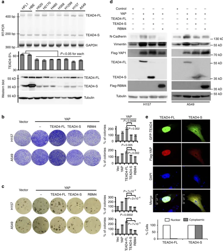
TEAD4-S represses cancer cell proliferation and EMT
. Hippo–
YAP pathway is known to control tumorigenesis
10. The negative
regulation of this pathway by the short isoform of TEAD4
suggests that TEAD4-S may repress cancer cell proliferation. To
test this possibility, we measured the relative levels of TEAD4 in a
panel of lung cancer cells. We found that, while TEAD4-S is the
major isoform in the normal lung fibroblast and bronchial
epithelial cells (Fig. 3a; HFL1 and HBE), all tested lung cancer
cells have reduced TEAD4-S level compared with the normal cells
(Fig. 3a). Furthermore, in lung cancer cells with stable
overexpression of YAP alone or co-expression of YAP with
TEAD4-FL, TEAD4-S or RBM4 (Supplementary Fig. 3a),
TEAD4-S inhibited cell proliferation as compared with vector
or TEAD4-FL (Fig. 3b,c). Such inhibition was comparable to that
of RBM4 in both anchorage-dependent and -independent cell
growth in two distinct cancer cell lines (Fig. 3b,c).
Hippo–YAP signalling has also been shown to regulate the
EMT of cancer cells
37,38. The inhibition of TEAD4-S to
YAP-activated cell proliferation makes us to speculate that it may also
repress the EMT. To test this, we examined whether the two
isoforms of TEAD4 have distinct impacts on the EMT. As shown
in Fig. 3d, co-expression of YAP/TEAD4-FL strongly induced
EMT in two primary tumour cell lines as judged by increased
levels of N-cadherin and Vimentin; however, the expression of
TEAD4-S or RBM4 suppressed the induction of EMT markers
(Fig. 3d). Interestingly, in H157 cells co-expressing YAP and
TEAD4-FL, the majority of YAP is retained in the nucleus as
pointed dots that are co-localized with TEAD4-FL (Fig. 3e;
Supplementary Fig. 3b). However, in cells expressing TEAD4-S,
the YAP is found in both nuclear and cytoplasmic compartments,
presumably through the interaction with TEAD4-S that is
diffused into the cytoplasm (Fig. 3e; Supplementary Fig. 3b).
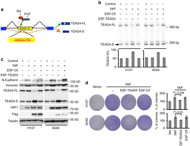
To further examine the functional role of TEAD4 splicing
switch, we applied a recently developed approach, engineered
splicing factors (ESFs)
39–42, to specifically manipulate TEAD4
splicing and to test whether the splicing changes of TEAD4 can
directly affect YAP-mediated EMT and tumour proliferation. We
designed and generated a PUF domain that can specifically bind
16
a
YAP, TEAD4, or RBM4
TEAD4 binding sites
F-Luci R-Luci
8
Relativ
e lucif
er
ase
activity
Relativ
e mRNA
Relativ
e mRNA
0 + CTGF-Luc
Vector Vector
Flag-TEAD4-FL Flag-TEAD4-FL
TEAD4-S TEAD4-S
HA-YAP HA-YAP Flag-TEAD4-S
Total lysate Total lysate
IP: FLAG IP: FLAG
70 kD 55 kD 55 kD 35 kD 70 kD 70 kD 55 kD 35 kD 70 kD
YAP TEAD4-FL (ng) TEAD4-S (ng) RBM4 (ng)
CTGF
TEAD4-S
H157 cell A549 cell TEAD4-FL
HA-YAP1 Flag-TEAD4-FL TEAD4-FL TEAD4-S HA-YAP1 HA-YAP1 FLAG-TEAD4-FL FLAG-TEAD4-S HA-YAP1
ltgb ltgb
Ctgf Ctgf
6 3 0
3.00
–3.00 2.00
–2.00 1.00
–1.00 0.00
Vec YAP
YAP/TEAD4-FL
YAP/TEAD-FL YAP/TEAD4-S
Vec YAP
YAP/TEAD4-FLYAP/TEAD4-S
YAP/TEAD-S YAP/RBM4
Vec YAP
YAP/TEAD4-FLYAP/TEAD4-S YAP/RBM4 YAP/RBM4
Vec YAP
YAP/TEAD4-FLYAP/TEAD4-S YAP/RBM4
YAP/RBM4 YAP
9 30 10
5 0 15
0
Mitochondrial function
Cell proliferation signals
Ribosome and translation
RNA splicing and processing Cell cycle
regulation 10
5 0
200 bp
0.1% input TEAD4 IP IgG IP 0.1% input TEAD4 IP IgG IP
+ + + + + + + + + + + + + + + +
+ – – – – 100 100 100 100 100 100 100 100
20 20 50 –
+ + +
+ + + + +
+ ++ + –
– – – – – –
– – – – – –
– – + +
+ +
+ + + + +
– –
– – – – – –
– –
–
– – – – – –
b
c
d
e
f
to an 8-nt RNA sequence (GAGCCTTG) in exon 3 of TEAD4,
and fused the designer PUF domain with a glycine-rich motif that
can inhibit exon inclusion on binding to pre-mRNA. The
resulting ESF, ESF-TEAD4, is designed to bind and inhibit the
inclusion of exon 3 in TEAD4, thus promoting the TEAD4-S
splicing (Fig. 4a). When transfecting ESF-TEAD4 in YAP stably
expressed lung cancer cells, H157 and A549, we found that the
ESF-TEAD4 specifically shifted TEAD4 splicing towards the
short isoform as compared with control ESF (Fig. 4b). As
expected, the splicing switch to TEAD4-S can indeed suppress the
YAP-mediated EMT (Fig. 4c) and the tumour cell proliferation
(Fig. 4d) in both lung cancer cell lines tested.
TEAD4-S inhibits tumour growth
in vivo
. To further assess
whether TEAD4-S affects cancer growth
in vivo
, we determined
whether expression of TEAD4-S could suppress tumour growth
in a xenograft mouse model. We generated H157 cells with stable
expression of YAP, YAP/TEAD4-FL, YAP/TEAD4-S or YAP/
RBM4, and injected these cells subcutaneously into the flanks of
nude mice to measure tumour growth. Consistent with our
in vitro
results, cells expressing TEAD4-S developed smaller
tumours as compared with cells with YAP alone, YAP/TEAD4-FL
or even the vector control (Fig. 5a), suggesting that TEAD4-S
functions as an inhibitor of tumour development. In addition, the
xenograft tumours with TEAD4-S developed much slower than
cells with YAP, YAP/TEAD4-FL or even vector control (Fig. 5b),
further supporting that TEAD4-S inhibits cancer progression
in vivo
. As a positive control, RBM4 also inhibited tumour
development
in vivo
, consistent with its role as antitumour
splicing factor to promote TEAD-S.
Next, we surgically collected paired non-small cell lung cancer
(NSCLC) samples and adjacent normal tissues from seven
patients, and measured the relative levels of two TEAD4 isoforms.
Compared with the paired normal tissues, the relative mRNA
levels of TEAD4-S were significantly reduced in six out of seven
primary NSCLC specimens (Fig. 5c), and the TEAD4-S protein
was substantially decreased in five out of seven specimens
(Fig. 5d), suggesting a general reduction of TEAD4-S expression
despite the obvious heterogeneity in different tumour specimens.
Intriguingly, in three of the NSCLC specimens (samples 4, 5 and
7), the truncated TEAD4-S protein was the predominant isoform
in normal tissues, but was almost completely eliminated in
tumours, implying that the AS switch in TEAD4 could play a
major role in the tumorigenesis of these patients.
To further evaluate the clinical relevance of TEAD4 splicing in
all cancers, we analysed the databases from TCGA consortium
that contains various large-scale RNA-seq results from thousands
of patients with various tumours
43(Supplementary Fig. 4a).
Strikingly, TEAD4-S levels were significantly altered in 7 out of 11
tumour types analysed with reduction in the majority of tumours
(6 out of 7; Fig. 5e), consistent with our results in the
anti-tumour activity of TEAD4-S. To further investigate the clinical
significance of TEAD4 splicing in cancers, we used a survival
analysis tool, Kaplan–Meier plotter, to analyse TCGA data sets
for the overall survival of various cancer patients with different
TEAD4-S levels. Strikingly, a higher level of TEAD4-S was
significantly associated with the improved overall survival in
patients with lung and colon cancers (Fig. 5f), and to a lesser
extent, in patients with liver and breast cancers (Supplementary
Fig.
4b,
positive
but
non-significant
association).
These
observations indicate that TEAD4-S could be recognized as an
independent prognostic factor for the survival of cancer patients.
Collectively, this finding validated the mechanistic link between
the TEAD4-S isoform and cancer progression, highlighting the
importance of this splicing switch of the Hippo–YAP–TEAD
pathway in regulating human cancer progression and patient
survival.
Discussion
Extensive splicing misregulation is one of molecular hallmarks of
cancer
44–47; however, the functional implication is far from clear.
Here we report a model in which the AS plays a key regulatory
role in mediating the activation of Hippo–YAP signals. In the
current model, activation of the Hippo pathway by multiple
extracellular cues is converged to its main effector YAP, whose
phosphorylation leads to cytoplasmic retention and protein
degradation. When unphosphorylated, YAP translocates into
the nucleus and interacts with transcription factors TEAD1–4 to
activate gene expression and promote cell proliferation
10. We
demonstrated that the AS imposes a new layer of regulation:
skipping of exon 3 in TEAD4 produces a short isoform that
interacts and neutralizes YAP in both the nucleus and cytoplasm,
leading to the attenuation of YAP signalling (Fig. 4g). The
splicing of TEAD4-S is controlled by RBM4 through direct
binding to its pre-mRNA. Consistently, RBM4 and TEAD4-S
inhibit tumour progression in cultured cells and xenograft
tumours. Altogether, these data represent a new mechanism on
how the AS affects tumorigenesis through mediating the key
signalling cascades such as the Hippo–YAP pathway.
Genomic analyses of TCGA data sets indicate that splicing of
TEAD4 is commonly altered in cancer patients to reduce
TEAD4-S. Strikingly, re-expression of TEAD4-S significantly
reduced tumour development in cultured cells and mouse
model. Since the Hippo–YAP pathway has a broad impact on
HFL1HBE H520H2170H460H358H1299H157 A549
400 bp
a
d
e
b
c
R
T
-PCR
TEAD4-S%
W
ester
n b
lot
A549
Ve
c
RBM4
TEAD4-S
TEAD4-FL
YA
P
Ve
c
RBM4
TEAD4-S
TEAD4-FL
YA
P
H157
% of colonies
Merge
0
µ
m
25
0
µ
m
25
D
API
Flag-Y
AP
GFP-TEAD4
% of colonies
% of colonies
% of colonies % Cells
300 bp
P< 0.05 for each
30
35 kD 55 kD 55 kD 60 200 bp
GAPDH TEAD4-S TEAD4-FL
55 kD
55 kD
55 kD A549
H157
TEAD4-S TEAD4-FL
35 kD 35 kD 70 kD 130 kD
Tubulin
Vector YAP
100 0 200
100 0 200 300
100 0 200 300
0
TEAD4-S TEAD4-FL
50 100 100
0 200
YAP
P= 0.0008
P= 2×10⁻⁶
P= 7×10⁻⁵
P= 7×10⁻⁶
P= 0.002
P= 0.002
P= 0.0002
Cytoplasmic Nuclear
P= 0.005
RBM4 TEAD4-S TEAD4-FL –
A549
H157
Vector YAP
RBM4 TEAD4-S TEAD4-FL –
Tubulin TEAD4-S
TEAD4-FL
RBM4 TEAD4-S TEAD4-FL YAP Control
Vimentin
TEAD4-FL
TEAD4-S Flag-YAP1
Flag-RBM4 N-Cadherin
– – – –
–
– –
– – –
– – –
– – – –
–
– –
– – –
– – – +
+ + +
+
+ +
+ +
+ +
+
+ +
tumorigenesis and cancer development, modulating TEAD
splicing may provide a new approach for potential therapeutic
interventions of cancer. In particular, TEAD4-S is controlled by
RBM4, a master regulator of many cancer-related splicing events.
Activation of RBM4 increases the production of TEAD4-S, which
in turn may potentially inhibit tumorigenesis through multiple
oncogenic pathways.
We also noticed that the mRNA and protein levels of two
TEAD4 isoforms are not always consistent across different cell
lines and tissues (Figs 3a; and 5c,d), suggesting that the two
isoforms may be differentially controlled in the levels of protein
translation and/or degradation. This observation adds additional
layer of complexity in controlling TEAD4 isoforms in addition to
splicing regulation at RNA level. Adding to the complexity is the
YAP paralogue TAZ that also binds TEAD4 with similar affinity
(reviewed in ref. 48). We speculate that the two TEAD4 isoforms
may also differentially regulate TAZ-mediated branch of the
Hippo pathway; however, an additional research is required to
reveal the detailed mechanisms.
Human genome has four TEAD paralogues, TEAD1–4, all of
which can interact with YAP to promote transcription. The
N-terminal DNA-binding domains of all TEAD proteins are
highly conserved. On the basis of the sequence annotation,
TEAD2 and TEAD3 also have putative splicing variants with a
truncated N-terminal domain that in principal act similarly to
TEAD4-S (Supplementary Fig. 5), although it is unclear whether
these splicing isoforms are indeed translated. Our observations
that TEAD4-S functions as a dominant negative isoform to
sequester/neutralize YAP imply that it may also antagonize all
other TEAD paralogues. It is possible that this finding presents a
general regulatory switch of all TEAD proteins, and future studies
are warranted to fully address the role of all noncanonical TEAD
isoforms.
Multiple AS isoforms can also be found in other main
components of the Hippo–YAP pathway to differentially affect
cellular signals. For example, by skipping the exon 4, YAP can
produce a splicing isoform containing single WW domain
(YAP1-1). In contrast to the canonical isoform YAP1-2 that
contains two WW domains, the YAP1-1 does not bind
angiomotin and thus is not sequestered by angiomotin in the
cytoplasm
49, In addition, YAP1-1 does not interact with p73 that
is the functional partner of YAP1-2 in response to the stress of
ultraviolet or serum depravation
50. Our finding provides a
mechanistic link between Hippo signalling and AS regulation,
which may be a tip of the iceberg for a general regulatory mode.
Previously, the Hippo–YAP pathway is well known to be
Gly PUF
Exon 3
Control
Control
YAP
YAP
ESF-Ctl
ESF-Ctl
ESF-TEAD4
ESF-TEAD4
N-Cadherin Vimentin TEAD4-FL
TEAD4-FL TEAD4-FL
TEAD4-S
TEAD4-S TEAD4-S
YAP1 Flag Tubulin
H157
H157 100
200 100 200 100 0 0
400 bp
200 bp
50 0
A549
A549 +
+ + + +
+ + + + + +
+ – – –
– –
– – –
– – – – – – – – – – – – – – –
130 kD 55 kD 55 kD 35 kD 70 kD 55 kD 55 kD
Vector
H157
TEAD4-S%
% of colonies
% of colonies
A549
YAP
Ve
c
YA
P
YAP
ESF-TEAD4
ESF-Ctl
P= 0.008
P= 0.03
– ESF-TEAD4 ESF-Ctl +
+ + + +
+ + + + + +
+ – – –
– –
– – –
– – – – – – – – – – – – – – –
a
b
c
d
controlled by a phosphorylation cascade that elicits rapid
regulation, the new control mode through splicing may present
a slower but more stable regulatory dynamics that is important
to cancer cell reprogramming. We speculate that splicing
misregulations of other components in the Hippo–YAP
pathway also play critical roles in cancer development,
and thus should be explored as a new route of potential cancer
therapy.
Vector Ctl YAP YAP/TEAD4-S
YAP/TEAD4-FL YAP/RBM4
800
600
400
200
0
7 9 11 13 15 17
T7
TEAD4-FL TEAD4-S Tubulin TEAD4-FL
TEAD4-S GAPDH
T6 T5 T4 T3 T2
T1 N2 N3 N4 N5 N6
N1 N7
Days after inoculation
55 kD 400 bp
200 bp 300 bp
1
0.5
1.0
1.0 0.8 0.6 0.4 0.2 0.0
0 1 2 3 4 5
Years of survival Years of survival
Low PSI (more TEAD4-S) High PSI (less TEAD4-S)
0.8 0.6 0.4
BLCA
**
***
***
**
N.S. N.S.*
N.S. N.S.***
**
BRCA COAD HNSC KIRC KIRP LIHC LUAD PRAD THCA UCEC
Cancer Normal Extracellular signals
Hippo
YAP P
TEAD4-FL
TEAD4-S
TEAD4-binding site
RBM4
Promote transcription
YAP/TEAD targets
35 kD 55 kD T7
T6 T5 T4 T3 T2
T1 N2 N3 N4 N5 N6
N1 N7
Volume of tumours (mm
3)
Relative level of TEAD4-S
PSI
Survival rate
Ctl
YAP
YAP+TEAD4-s
YAP+TEAD-L
YAP+RBM4
LUAD; P= 0.002 COAD; P= 0.04
0 1 2 3 4 1.0
0.8 0.6 0.4 0.2 0.0
a
b
d
c
e
f
g
Methods
Splicing reporter constructs
.
To construct the TEAD4 reporter, we used PCR reactions to amplify a fragment containing exon 2, part of intron 2, exon 3, part of intron 3 and exon 4 of TEAD4, and ligated this fragment to the pCDNA3-FLAG vector digested with NheI/NotI. To generate TEAD4 reporters with mutated RBM4-binding sites, Quikchange approach was applied with different paired primers. All primers used in this study were listed in Supplementary Table 1.ESF expression constructs
.
To express ESFs in cultured cells, we generated expression constructs using the pCI-neo vector (Promega). We started with an expression construct that encodes from N- to C terminal, FLAG epitope, Gly-rich domain of hnRNP A1 (residues 195–320 of NP_002127), and the MS2 coat protein (gift of Dr R. Breathnach form Institut de Biologie-CHR 1). The fragment encoding the MS2 coat protein fragment was removed using BamHI/SalI digestion and replaced with a fragment encoding a NLS (PPKKKRKV) and the PUF domain of human Pumilio1, which was amplified using primers Pum-F1 and Pum-R1 (Supplementary Table 1). The resulting construct expresses a Gly-PUF-type ESF under the control of a cytomegalovirus (CMV) promoter. To make ESF-TEAD4, we introduced the point mutations in consecutive steps in the PUF domain to make it recognize an 8-nt RNA sequence (GAGCCTTG) using a QuikChange Site-Directed Mutagenesis kit (Stratagene) following the manufacturer’s instructions.Human tissue total protein and cDNA panels
.
Human Tissue total protein and complementary DNA (cDNA) panels were purchased from Amsbio Company. Each panel contained 10 different human tissues, including the brain, colon, liver, kidney, heart, lung, skeletal muscles, pancreas, spleen and stomach.Cell culture and transfection
.
293 FlpIn/T-Rex cells were purchased from Invi-trogen. All other cell lines were purchased from American Type Culture Collection (Manassas, VA, USA). The HEK293T human embryonic kidney, HeLa cervical cancer, 293 FlpIn/T-Rex and PANC-1 human pancreatic carcinoma cell lines were cultured in DMEM (high glucose) medium containing 10% fetal bovine serum (FBS; Hyclone) and 1% penicillin/streptomycin (P/S). The A549 human lung carcinoma cell line was cultured in F-12 K medium containing 10% FBS and 1% P/S. The H157 human lung carcinoma cell line was cultured in RPMI-1640 medium containing 10% FBS and 1% P/S. The HT-1080 human fibrosarcoma and HepG2 human hepatocellular carcinoma cell lines were cultured in Eagle’s Minimum Essential Medium containing 10% FBS and 1% P/S. The MDA-MB-231 human breast cancer cell line was cultured in L-15 medium containing 10% FBS and 1% P/S. The cell lines were tested for mycoplasma contamination using the microbiological culture method, and all the lines were free of mycoplasma.To generate stable cell line expressing RBM4 on tetracycline induction, we used pCDNA5 FRT/TO vector and 293 FlpIn/T-Rex cells (Invitrogen). The FLAG-tagged full-length RBM4 was cloned into the vector, and transfected with pOG44 in 1:9 ratio. The stably integrated cells were selected with 100mg mllof hygromycin
at 2 days after transfection forB2 weeks to obtain individual colonies. One day before the induction, the cells were transferred to hygromycin-free medium. The inductions were carried out by adding tetracycline to a final concentration of 2mg ml1. The induced cells were collected at several time points after induction
to extract RNA and protein for further analysis.
To determine the localization of TEAD4-FL or TEAD4-S, HeLa cells were plated onto a coverslip in six-well plates 1 day before transfection. An amount of 1mg of GFP-tagged TEAD4-FL or TEAD4-S vectors was transfected using lipofectamine 2000 according to the manual. After 48 h, cells were fixed for further immunofluorescence analysis.
To stably express RBM4 (or other proteins) in PANC1 cells (or other cells), we used lentiviral vectors. We transfected 293 cells with flag-RBM4 or pCDH-flag-empty vectors as per the manufacture’s protocols. The supernatant media containing virus was collected by centrifugation to remove any cellular contaminant. The resulting viral particles were used to infect H157 cells, and stably integrated cells were selected by 5mg mllof puromycin for 1 week. The expression of transgenes was confirmed by western blots before further analysis.
To determine the effect of overexpression of RBM4 on TEAD4 splicing changes, 0.2mg of TEAD4 mini-gene reporters was co-tranfected with 0.4mg of RBM4, using lipofectamine 2000 according to the manual. After 48 h, cells were collected for further analysis of RNA and protein levels.
Assay of splicing with semi-quantitative RT–PCR
.
The total RNA was isolated from transfected cells with TRIzol reagent (Invitrogen) according to the manu-facturer’s instructions, followed by 1-h DNase I (Invitrogen) treatment at 37°C, and then heat inactivation of DNase I. Total RNA (2mg) was then reverse-tran-scribed with SuperScript III (Invitrogen) using poly T primer, and one-tenth of the room temperature product was used as the template for PCR amplification (25 cycles of amplification, with trace amount of Cy5-dCTP in addition to non-fluorescent dNTPs). Reverse transcription–PCR (RT–PCR) products were separated on 10% polyacrylamide gel electrophoresis (PAGE) gels, and scanned using a Typhoon 9400 scanner (Amersham Biosciences). The amount of each splicing isoform was measured using ImageQuant 5.2.Western blot
.
Cells were lysed in lysis buffer containing 50 mM HEPES, 150 mN NaCl (4.38 g), 1 mM EDTA, 1% (w/v) CHAPS and Sigma protease inhibitor cocktail. Subsequently, the cell lysates were boiled in 2SDS–PAGE loading buffer for 10 min, and then resolved by 10% SDS–PAGE and transferred to the nitrocellulose membrane. All primary antibodies were diluted 1,000 times for western blotting if not specified. The following antibodies were used in this study: TEAD4 (#ab58310) antibody, anti-Myc tag antibody (#ab9106) and anti-HA tag antibody (#ab9110) were purchased from Abcam; N-cadherin (#610921) antibody was purchase from BD; Vimentin (#5741) and YAP (#12395) antibodies were purchased from Cell Signaling Technology; and alpha-tubulin (#T5168, 1:5000 dilution) and FLAG M2 (#F1804) were purchased from Sigma-Aldrich. RBM4 antibody (#11614-1-AP) was purchased from Proteintech. Bound antibodies were visualized using the ECL kit (GE Healthcare).Clinical tissues samples collection
.
Fresh lung cancer tissues and normal adjacent tissues were collected from patients with pathologically and clinically confirmed lung carcinomas. All human tumour tissues were obtained with written informed consent from patients or their guardians before participation in the study. The Institutional Review Board of the Dalian Medical University approved use of the tumour specimens in this study. All of tissue specimens were kept in liquid nitrogen and sectioned for protein or mRNA extraction.High-throughput mRNA-sequence and data analysis
.
RNAs from H157 cells stably expressing YAP, YAP/TEAD4-FL, YAP/TEAD4-S, YAP/RBM4 and control vectors were purified using Trizol method, and subsequently cleaned using the RNAeasy Kit (Qiagen). The RNAs were digested in column with RNAse-free DNAse as per the manufacturer’s instruction. Total RNA not exceeding 3mg was further used to purify polyadenylated RNA using the Illumina TruSeq Total RNA Sample Prep kits. We used the Ribo-Zero Human to remove the cytoplasmic rRNA. The mRNA purified was further analysed using the Bioanalyzer (Agilent Technologies) before generation of cDNA library with bar-coded ends. RNA-seq libraries were robotically prepared using the Illumina TruSeq Total RNA Sample Prep kits according to the manufacturer’s protocol. The RNA-seq data set was deposited to the Gene Expression Omnibus with accession number GSE80372. To estimate the gene expression levels, we used RSEM package and bowtie2 (refs 51,52) to align all reads to human reference genome (UCSC hg19 version). Then, we provided a fragment length distribution with options of ‘–fragment-length-mean 75’ and ‘–fragment-length-sd 10’ to calculate transcript expression levels. Subsequently, we used EBSeq tool53to examine differential expression genes of pair-wise comparison based on empirical Bayesian methods.Heat map
.
We included genes that met the following criteria: (i) FPKM (fragments per kilobase of transcript per million mapped reads) values of a given gene are not equal in all samples; (ii) at least one of the FPKM values in all samples isZ3; and (iii) the ratio of the maximum FPKM value and the minimum FPKM value in all samples isZ2. Then, we used the log 2 ratio of FPKM values of the included genes, normalized by the FPKM value of control sample, as input of Cluster 3.0 (ref. 54). We clustered the data set using the hierarchical clustering method based on Pearson correlation with average linkage, and further viewed the results using Java TreeView.We selected the cluster in this pattern—upregulated by YAP–TEAD434, but downregulated by YAP–TEAD305 and YAP–RBM4—as our target data set. This data set includes the genes that are differentially regulated by TEAD4 isoforms. The heat map shown is ordered by the FPKM value of YAP.
The gene ontology analysis was performed using DAVID gene ontology analysis software to search for enriched pathways. The functional association of TEAD4 targets were analysed using the protein interaction data from STRING database, generating a set of functional interaction networks. The sub-network containing more than five nodes were demonstrated.
Soft agar assay
.
Equal volumes of 1.2% agar and 2DMEM (or RPMI-1640) mediums were mixed and placed onto six-well dishes to generate 0.6% base agar. A549 cells (or H157 cells) expressing YAP, YAP/TEAD4-FL, YAP/TEAD4-S, YAP/RBM4 and control vectors were seeded in 0.3% top agar (104cells per plate) and incubated at 37°C in humidified atmosphere for 3 weeks. Colonies were stained with crystal violet and counted.Colony formation assay
.
A549 cells (or H157 cells) expressing YAP, YAP/ TEAD4-FL, YAP/TEAD4-S, YAP/RBM4 and control vectors (5,000 cells per dish) were seeded in the 10-cm dishes and incubated at 37°C in humidified incubator for 2 weeks. Colonies were fixed and stained with crystal violet, and the number of colonies was counted.subcutaneously with 1106H157 cells expressing YAP, YAP/TEAD4-FL, YAP/ TEAD4-S, YAP/RBM4 and control. Nine mice were used for each group. Mice were raised in the following 3 weeks. The mice were then monitored for tumour volume and overall health. The size of the tumour was determined by caliper measurement of the subcutaneous tumour mass every 3 days. Tumour volume was calculated according to the formula (4/3)pr12r2, (r1or2). Each experimental group
contained nine mice. At the end of 17 days, all mice were killed, and tumours were removed for further analysis. For all data points, three independent measurements were performed and means were used for calculation.
RNA immunoprecipitation
.
293T cells (1106) expressing RBM4 or control vector are collected and washed twice with 10 ml of PBS, and then resuspended in 10 ml of PBS. Formaldehyde (37% stock) is added to the above solution to a final concentration of 1% and incubated at room temperature for 10 min with slow rotating. Crosslinking reactions are quenched by the addition of glycine solution (pH 7.0) to a final concentration of 0.25 M, followed by incubation at room temperature for 5 min. The cells are collected by centrifugation at 700gfor 4 min at 4°C, followed by two washes with ice-cold PBS. Fixed cells are resuspended in 2 ml of radioimmunoprecipitation assay (RIPA) buffer (50 mM Tris-Cl, pH 7.5, 1% NP-40, 0.5% sodium deoxycholate, 0.05% SDS, 1 mM EDTA, 150 mM NaCl) con-taining protease inhibitors. The cells are subsequently lysed by three rounds of sonication. Insoluble material is removed by microcentrifugation at 16,000gfor 10 min at 4°C. An aliquot of solubilized cell lysate is mixed with protein A– Sepharose beads along with nonspecific competitor tRNA. This mixture is rotated for 1 h at 4°C, followed by microcentrifugation at 1200gfor 5 min. The supernatant is removed and used for immunoprecipitation.Protein A or protein G–Sepharose beads are coated with the Flag antibody for 2 h at 4°C, followed by extensive washing with RIPA buffer containing protease inhibitors. Before immunoprecipitation, the beads are incubated for 10 min in RNasin. The precleared lysate is diluted with RIPA buffer, mixed with the antibody-coated beads and incubated with rotation for 60–90 min. The beads are collected using a minicentrifuge at 2800gfor 45 s. The beads are washed five or six times with 1 ml of highstringency RIPA buffer (50 mM Tris-Cl, pH 7.5, 1% NP-40, 1% sodium deoxycholate, 0.1% SDS, 1 mM EDTA, 1 M NaCl, 1 M urea, 0.2 mM phenylmethyl sulphonyl fluoride) by 10-min rotation at room temperature. The beads containing the immunoprecipitated samples are collected and resuspended in 100ml of 50 mM Tris-Cl, pH 7.0, 5 mM EDTA, 10 mM dithiothreitol and 1% SDS. Samples (resuspended beads) are incubated at 70°C for 45 min to reverse the crosslinks. The RNA is extracted from these samples using Trizol according to the manufacturer’s protocol, and reverse-transcribed into cDNA for PCR detection.
Immunofluorescence staining
.
To determine the localization of TEAD4-FL, TEAD4-S and YAP, we performed immunofluorescence assay. In brief, cells were plated on coverslips to appropriate density. Transfected cells were fixed on the coverslips with 4% paraformaldehyde in 1PBS for 15 min at room temperature and washed with 1PBS three times. Cells were then permeabilized with 0.2% Triton X-100 for 10 min. After blocking in 3% bovine serum albumin for 30 min, slides were incubated with indicated antibodies (Flag or Myc, 1:100 dilution) antibody diluted in 1% bovine serum albumin for 2 h. Subsequently, slides were washed with 1PBS for three times, and then incubated with fluorophore-conjugated secondary antibodies for 1 h. The coverslips were then washed and mounted with mounting medium (Vector shield’s mounting medium with 4,6-diamidino-2-phenylindole). Cells were visualized using an Olympus fluorescence microscope, and photographs were generated using a Kodak digital camera.Luciferase reporter assay
.
For the luciferase reporter assay, HEK293T cells were seeded in 24-well plates. Cells were co-transfected with 100 ng of CTGF promoter/firefly luciferase reporter plasmid and different amount of YAP (100 ng), TEAD4-FL (100 ng), TEAD4-S (20 or 100 ng), 100 ng TEAD4-FL with increasing amounts of TEAD4-S (20, 50 and 100 ng) and RBM4 (100 ng) plasmids and 5 ng of pRL-TK Renilla plasmids using lipofectamine 2000 (Invitrogen). After 48 h of transfection, cells were either lysed in protein lysis buffer for protein extraction or in passive lysis buffer (Promega) for luciferase assay measured with the Dual-Luciferase Reporter Assay System (Promega), using the TD-20/20 Luminometer (Turner Designs). The relative luciferase activities were determined by calculating the ratio of firefly luciferase activities over Renilla luciferase activities.Assay of CTGF and ITGB expression with real-time PCR
.
The real-time PCR was performed using the Maxima SYBR Green qPCR Master Mix (Thermo Scientific) and a 7500 real-time PCR system (Life Technologies) according to the manufacturer’s instructions. The expression level of CTGF and ITGB was normalized to the endogenous expression of GAPDH.Statistics
.
Statistical analyses of colony formation, soft agar and splicing changes were performed using Student’st-test.Data availability
.
RNA-seq data that support the findings of this study have been deposited in Gene Expression Omnibus of NCBI with the accession code GSE80372. The authors declare that all the data supporting the findings of this study are available within the article and its Supplementary Information files.References
1. Zhao, B., Lei, Q. Y. & Guan, K. L. The Hippo-YAP pathway: new connections between regulation of organ size and cancer.Curr. Opin. Cell Biol.20,638–646 (2008).
2. Zhao, B.et al.Inactivation of YAP oncoprotein by the Hippo pathway is involved in cell contact inhibition and tissue growth control.Genes Dev.21,
2747–2761 (2007).
3. Ota, M. & Sasaki, H. Mammalian Tead proteins regulate cell proliferation and contact inhibition as transcriptional mediators of Hippo signaling.Development
135,4059–4069 (2008).
4. Marti, P.et al.YAP promotes proliferation, chemoresistance, and angiogenesis in human cholangiocarcinoma through TEAD transcription factors. Hepatology62,1497–1510 (2015).
5. Hansen, C. G., Ng, Y. L., Lam, W. L., Plouffe, S. W. & Guan, K. L. The Hippo pathway effectors YAP and TAZ promote cell growth by modulating amino acid signaling to mTORC1.Cell Res.25,1299–1313 (2015).
6. Li, Z.et al.Structural insights into the YAP and TEAD complex.Genes Dev.24,
235–240 (2010).
7. Zhao, B., Li, L., Lei, Q. & Guan, K. L. The Hippo-YAP pathway in organ size control and tumorigenesis: an updated version.Genes Dev.24,862–874 (2010). 8. Meng, Z., Moroishi, T. & Guan, K. L. Mechanisms of Hippo pathway
regulation.Genes Dev.30,1–17 (2016).
9. Chan, S. W.et al.The Hippo pathway in biological control and cancer development.J. Cell. Physiol.226,928–939 (2011).
10. Yu, F. X., Zhao, B. & Guan, K. L. Hippo pathway in organ size control, tissue homeostasis, and cancer.Cell163,811–828 (2015).
11. Ehmer, U. & Sage, J. Control of proliferation and cancer growth by the Hippo signaling pathway.Mol. Cancer Res.14,127–140 (2016).
12. Moroishi, T., Hansen, C. G. & Guan, K. L. The emerging roles of YAP and TAZ in cancer.Nat. Rev. Cancer15,73–79 (2015).
13. Mo, J. S., Park, H. W. & Guan, K. L. The Hippo signaling pathway in stem cell biology and cancer.EMBO Rep.15,642–656 (2014).
14. Jiao, S.et al.A peptide mimicking VGLL4 function acts as a YAP antagonist therapy against gastric cancer.Cancer Cell25,166–180 (2014).
15. Johnson, R. & Halder, G. The two faces of Hippo: targeting the Hippo pathway for regenerative medicine and cancer treatment.Nat. Rev. Drug Discov.13,
63–79 (2014).
16. Liu-Chittenden, Y.et al.Genetic and pharmacological disruption of the TEAD-YAP complex suppresses the oncogenic activity of TEAD-YAP.Genes Dev.26,
1300–1305 (2012).
17. He, M.et al.New insights into posttranslational modifications of Hippo pathway in carcinogenesis and therapeutics.Cell Div.11,4 (2016). 18. Gaffney, C. J.et al.Identification, basic characterization and evolutionary
analysis of differentially spliced mRNA isoforms of human YAP1 gene.Gene
509,215–222 (2012).
19. Tsai, Y. S., Dominguez, D., Gomez, S. M. & Wang, Z. Transcriptome-wide identification and study of cancer-specific splicing events across multiple tumors.Oncotarget6,6825–6839 (2015).
20. David, C. J. & Manley, J. L. Alternative pre-mRNA splicing regulation in cancer: pathways and programs unhinged.Genes Dev.24,2343–2364 (2010). 21. Venables, J. P. Unbalanced alternative splicing and its significance in cancer.
Bioessays28,378–386 (2006).
22. Schwerk, C. & Schulze-Osthoff, K. Regulation of apoptosis by alternative pre-mRNA splicing.Mol. Cell19,1–13 (2005).
23. Harper, S. J. & Bates, D. O. VEGF-A splicing: the key to anti-angiogenic therapeutics?Nat. Rev. Cancer8,880–887 (2008).
24. Warzecha, C. C.et al.An ESRP-regulated splicing programme is abrogated during the epithelial-mesenchymal transition.EMBO. J.29,3286–3300 (2010). 25. Matlin, A. J., Clark, F. & Smith, C. W. Understanding alternative splicing:
towards a cellular code.Nat. Rev.6,386–398 (2005).
26. Wang, Z. & Burge, C. B. Splicing regulation: from a parts list of regulatory elements to an integrated splicing code.RNA.14,802–813 (2008).
27. Matera, A. G. & Wang, Z. A day in the life of the spliceosome.Nat. Rev. Mol. Cell Biol.15,108–121 (2014).
28. Sveen, A., Kilpinen, S., Ruusulehto, A., Lothe, R. A. & Skotheim, R. I. Aberrant RNA splicing in cancer; expression changes and driver mutations of splicing factor genes.Oncogene35,2413–2427 (2016).
29. Bechara, E. G., Sebestyen, E., Bernardis, I., Eyras, E. & RBM5, Valcarcel J. 6, and 10 differentially regulate NUMB alternative splicing to control cancer cell proliferation.Mol. Cell52,720–733 (2013).
31. Wang, Y.et al.The splicing factor RBM4 controls apoptosis, proliferation, and migration to suppress tumor progression.Cancer Cell26,374–389 (2014). 32. Liang, Y. C., Lin, W. C., Lin, Y. J. & Lin, J. C. The impact of RNA binding motif
protein 4-regulated splicing cascade on the progression and metabolism of colorectal cancer cells.Oncotarget6,38046–38060 (2015).
33. Chen, L.et al.Structural basis of YAP recognition by TEAD4 in the hippo pathway.Genes Dev.24,290–300 (2010).
34. Uniacke, J.et al.An oxygen-regulated switch in the protein synthesis machinery.Nature486,126–129 (2012).
35. Wang, Y., Ma, M., Xiao, X. & Wang, Z. Intronic splicing enhancers, cognate splicing factors and context-dependent regulation rules.Nat. Struct. Mol. Biol.
19,1044–1052 (2012).
36. Zhao, B.et al.TEAD mediates YAP-dependent gene induction and growth control.Genes Dev.22,1962–1971 (2008).
37. Zhang, H.et al.Yap1 is required for endothelial to mesenchymal transition of the atrioventricular cushion.J. Biol. Chem.289,18681–18692 (2014). 38. Shao, D. D.et al.KRAS and YAP1 converge to regulate EMT and tumor
survival.Cell158,171–184 (2014).
39. Wang, Y., Cheong, C. G., Hall, T. M. & Wang, Z. Engineering splicing factors with designed specificities.Nat. Methods6,825–830 (2009).
40. Wang, Y., Wang, Z. & Tanaka Hall, T. M. Engineered proteins with Pumilio/ fem-3 mRNA binding factor scaffold to manipulate RNA metabolism.FEBS J.
280,3755–3767 (2013).
41. Wang, Y. & Wang, Z. Design of RNA-binding proteins: manipulate alternative splicing in human cells with artificial splicing factors.Methods Mol. Biol.1421,
227–241 (2016).
42. Wei, H. & Wang, Z. Engineering RNA-binding proteins with diverse activities. Wiley Interdiscip. Rev. RNA6,597–613 (2015).
43. Weinstein, J. N.et al.The Cancer Genome Atlas Pan-Cancer analysis project. Nat. Genet.45,1113–1120 (2013).
44. Oltean, S. & Bates, D. O. Hallmarks of alternative splicing in cancer.Oncogene
33,5311–5318 (2013).
45. Chabot, B. & Shkreta, L. Defective control of pre-messenger RNA splicing in human disease.J. Cell Biol.212,13–27 (2016).
46. Scotti, M. M. & Swanson, M. S. RNA mis-splicing in disease.Nat. Rev.17,
19–32 (2016).
47. Daguenet, E., Dujardin, G. & Valcarcel, J. The pathogenicity of splicing defects: mechanistic insights into pre-mRNA processing inform novel therapeutic approaches.EMBO Rep.16,1640–1655 (2015).
48. Hau, J. C.et al.The TEAD4-YAP/TAZ protein-protein interaction: expected similarities and unexpected differences.Chembiochem14,1218–1225 (2013).
49. Oka, T., Schmitt, A. P. & Sudol, M. Opposing roles of angiomotin-like-1 and zona occludens-2 on pro-apoptotic function of YAP.Oncogene31,128–134 (2012).
50. Oka, T., Mazack, V. & Sudol, M. Mst2 and Lats kinases regulate apoptotic function of Yes kinase-associated protein (YAP).J. Biol. Chem.283,
27534–27546 (2008).
51. Li, B. & Dewey, C. N. RSEM: accurate transcript quantification from RNA-Seq data with or without a reference genome.BMC bioinformatics12,323 (2011).
52. Langmead, B., Salzberg, S. L. & Sudol, M. Fast gapped-read alignment with Bowtie 2. Nat Methods9,357–359 (2012).
53. Leng, N.et al.EBSeq: an empirical Bayes hierarchical model for inference in RNA-seq experiments.Bioinformatics29,1035–1043 (2013).
54. de Hoon, M. J.et al.Predicting gene regulation by sigma factors inBacillus subtilisfrom genome-wide data.Bioinformatics20,101–108 (2004).
Acknowledgements
We thank Drs Woan-Yuh Tarn and Kun-liang Guan for providing reagents. We thank Dr Xiaoling Li for critical reading of manuscript. This work is supported by the National Natural Science Foundation of China (31471235, 81422038 and 91540110 to Y.W.; 31570823 to Z.W.; 31400726 to W.Z.), the NIH grant R01CA158283 (to Z.W.), Young Thousand Talents Program of China (to Y.W.), the Education Department of Liaoning Province in China (the ‘Program for Pan-Deng Scholars’ to Y.W.), and program for Chang Jiang Scholars and Innovative Research Team in University IRT13049.
Author contributions
Y.W. and Z.W. designed the experiments, interpreted the results and wrote the manu-script. Y.Q., W.H., H.Q., J.Z., H.W. and W.Z. performed the experiments. Q.L. and S.M. help to interpret the data. J.Y., X.F. and Y.T. analysed the RNA-seq and TCGA data.
Additional information
Accession codes:RNA-seq data that support the findings of this study have been deposited in Gene Expression Omnibus of NCBI with the accession code GSE80372.
Supplementary Informationaccompanies this paper at http://www.nature.com/ naturecommunications
Competing financial interests:The authors declare no competing financial interests.
Reprints and permissioninformation is available online at http://npg.nature.com/ reprintsandpermissions/
How to cite this article:Qi, Y.et al.A splicing isoform of TEAD4 attenuates the
Hippo–YAP signalling to inhibit tumour proliferation.Nat. Commun.7:11840
doi: 10.1038/ncomms11840 (2016).

![Bis{μ 3,3′ (1,3,4 thiadiazole 2,5 diyldithio)bis[pentanedionato(1−)]}bis[diaquanickel(II)] dimethylformamide disolvate trihydrate](data:image/gif;base64,R0lGODlhAQABAIAAAP///wAAACH5BAEAAAAALAAAAAABAAEAAAICRAEAOw==)