Nonnative SOD1 trimer is toxic to motor neurons in a
model of amyotrophic lateral sclerosis
Elizabeth A. Proctor
a,b,c, Lanette Fee
c, Yazhong Tao
c, Rachel L. Redler
c, James M. Fay
c, Yuliang Zhang
d, Zhengjian Lv
d,
Ian P. Mercer
c, Mohanish Deshmukh
e,f, Yuri L. Lyubchenko
d, and Nikolay V. Dokholyan
a,b,c,1aCurriculum in Bioinformatics and Computational Biology, University of North Carolina at Chapel Hill, Chapel Hill, NC 27599;bProgram in Molecular and Cellular Biophysics, University of North Carolina at Chapel Hill, Chapel Hill, NC 27599;cDepartment of Biochemistry and Biophysics, University of North Carolina at Chapel Hill, Chapel Hill, NC 27599;dDepartment of Pharmaceutical Sciences, University of Nebraska Medical Center, Omaha, NE 68198; eNeuroscience Center, University of North Carolina at Chapel Hill, Chapel Hill, NC 27599; andfDepartment of Cell Biology and Physiology, University of North Carolina at Chapel Hill, Chapel Hill, NC 27599
Edited by Barry Honig, Howard Hughes Medical Institute, Columbia University, New York, NY, and approved December 2, 2015 (received for review August 21, 2015)
Since the linking of mutations in the Cu,Zn superoxide dismutase gene (sod1) to amyotrophic lateral sclerosis (ALS) in 1993, re-searchers have sought the connection between SOD1 and motor neuron death. Disease-linked mutations tend to destabilize the native dimeric structure of SOD1, and plaques containing mis-folded and aggregated SOD1 have been found in the motor neu-rons of patients with ALS. Despite advances in understanding of ALS disease progression and SOD1 folding and stability, cytotoxic species and mechanisms remain unknown, greatly impeding the search for and design of therapeutic interventions. Here, we de-finitively link cytotoxicity associated with SOD1 aggregation in ALS to a nonnative trimeric SOD1 species. We develop methodol-ogy for the incorporation of low-resolution experimental data into simulations toward the structural modeling of metastable, multi-domain aggregation intermediates. We apply this methodology to derive the structure of a SOD1 trimer, which we validate in vitro and in hybridized motor neurons. We show that SOD1 mutants designed to promote trimerization increase cell death. Further, we demonstrate that the cytotoxicity of the designed mutants corre-lates with trimer stability, providing a direct link between the presence of misfolded oligomers and neuron death. Identification of cytotoxic species is the first and critical step in elucidating the molecular etiology of ALS, and the ability to manipulate formation of these species will provide an avenue for the development of future therapeutic strategies.
neurodegeneration
|
protein aggregation|
protein misfolding|
ALS|
structural modelingP
rotein misfolding and aggregation are linked to cell death
and disease progression in neurodegenerative diseases, such
as Alzheimer’s disease, Parkinson’s disease, and amyotrophic
lateral sclerosis (ALS). In these diseases and others, the formation
of amyloid plaques, often observed post mortem, has long been
thought to play a role in neurodegeneration, but toxicity has never
been confirmed (1–3). Recent research has shown that small,
sol-uble oligomers, rather than insolsol-uble amyloids, are likely to be the
cytotoxic species causing neurodegeneration (4–14). These small,
soluble oligomers undergo aberrant interactions with cell
machin-ery and activate cell death pathways, but their exact stoichiometry is
not known and their properties have yet to be characterized.
Re-cently, metastable soluble Cu,Zn superoxide dismutase (SOD1)
oligomers have been identified that contain an epitope associated
with disease-linked species of SOD1, mutants of which are
impli-cated in a subset of ALS (15–18). Size exclusion chromatography
(SEC) of these oligomers revealed a size range of two to four
monomers, consistent with previous findings of potentially
cyto-toxic SOD1 oligomers (19–21).
Knowledge of the structures of these species would not only
allow for definitive testing of their toxicity but could potentially
lead to an understanding of disease mechanism and therapeutic
strategies against diseases for which no cure or effective treatment
exists (22). By their nature, however, these oligomers are only
metastable, and therefore difficult to isolate. More importantly, this
instability, and consequent transient nature, combined with the
proposed size range of the implicated oligomers, means that a
high-resolution structure cannot be achieved using traditional methods.
Although atomic structures have been explored for folding
inter-mediates of small protein domains (23) or stable oligomeric
struc-tures of disease-relevant peptide sequences (24), structural
characterization of metastable aggregation products of full-length
disease-linked proteins has remained elusive.
Here, we combined experimental and computational
ap-proaches to produce a molecular model of a toxic metastable
protein oligomer. Using state-of-the-art high-speed atomic force
microscopy (HS-AFM), we established the trimeric
stoichiome-try and highly dynamic nature of the isolated oligomers. We
applied limited proteolysis to the isolated SOD1 trimers,
allow-ing us to determine the solvent accessibility and rigidity of the
structure. Using this information as constraints in molecular
simulations, we evolved a structural model of the SOD1 trimer
that is consistent with our experimental data. Using our model,
we determined residues critical to trimer formation, and we used
rational mutagenesis to verify our model and manipulate trimer
formation in vitro and in living cells. We find that mutations that
stabilize the trimeric form of SOD1 result in increased cell death
over WT SOD1 or SOD1 mutants that inhibit trimer formation,
demonstrating the neurotoxicity of the SOD1 trimer and its potential
relevance to ALS etiology.
Significance
Protein aggregation is a hallmark of neurodegenerative dis-ease and is hypothesized to cause neuron death. Despite ex-tensive study of disease-associated aggregating proteins, mechanisms of neuron death remain a mystery, and no cures or effective treatments yet exist. Here, we demonstrate the tox-icity of a small aggregate of the Cu,Zn superoxide dismutase (SOD1) protein, associated with amyotrophic lateral sclerosis (ALS). We present an experimentally verified structural model of this toxic species and show that SOD1 mutants designed to promote formation of this aggregate increase cell death, pro-viding a direct link between aggregate presence and neuron death. Knowledge of toxic species and the ability to manipu-late their formation provides a valuable direction for pursuit of therapeutic strategies in ALS.
Author contributions: E.A.P. and N.V.D. designed research; E.A.P., L.F., Y.T., R.L.R., J.M.F., Y.Z., Z.L., and I.P.M. performed research; M.D. and Y.L.L. contributed new reagents/ana-lytic tools; E.A.P., L.F., Y.T., R.L.R., J.M.F., Y.Z., Z.L., M.D., Y.L.L., and N.V.D. analyzed data; and E.A.P., L.F., and N.V.D. wrote the paper.
The authors declare no conflict of interest. This article is a PNAS Direct Submission.
1To whom correspondence should be addressed. Email: [email protected].
Results
Determination of SOD1 Oligomer Stoichiometry.
To model
poten-tially toxic SOD1 oligomers accurately, we first determined the
number of SOD1 monomers participating in the oligomers using
definitive images obtained by both regular AFM and HS-AFM.
We find that the metastable SOD1 oligomers are stabilized at
pH 3.5, where the SOD1 aggregation pathway has been shown to
proceed more rapidly while retaining the same features and
characteristics as the physiological process. We have
demon-strated previously that although low pH accelerates progression
along the aggregation pathway by stabilizing species further
down that pathway, it does not affect the pathway itself (25). At
pH 3.5, where the SOD1 oligomers are stable, oligomer volume
is
∼
71.7 nm
3, threefold greater than oligomer volume of the
monomer (
∼
25.3 nm
3;
Fig. S1A
). These data are supported by
HS-AFM images resolving the three-partite structure of the
olig-omer (Fig. 1
A
). We conclude that the primary SOD1 oligomer
previously identified by Redler et al. (15) is a trimer. Upon
in-crease of pH to 7.4, where the SOD1 oligomer is less stable, the
majority of trimers dissociate into monomers, as confirmed by
volumetric AFM measurements (Fig. 1
B
and
Fig. S1A
). We find
that any larger aggregates encountered at low pH are mainly short
fibrillar structures (
Fig. S1B
).
SOD1 Trimer Features Nonnative Secondary, Tertiary, and Quaternary Structure.
After purification (26), aggregation, and isolation of the
SOD1 trimer (15), we conducted limited proteolysis, followed by
MS, to identify unstructured, solvent-accessible regions (27).
Sev-eral proteolytic cuts are located in regions that are buried in the
dimer interface or contain secondary structure elements in the
SOD1 dimer (Fig. 2
A
), which would make proteolysis impossible
or highly unlikely if native secondary and tertiary structure were
conserved. Limited proteolysis of SOD1 dimer and isolated SOD1
monomer demonstrate that the pattern of proteolytic cleavage
differs significantly (Fig. 2
B
). In addition to secondary and tertiary
structure differences, we conclude from proteolytic data that the
interface(s) between monomers in the SOD1 trimer must be
lo-cated in a different region of the monomer from the native SOD1
dimer interface (i.e., a significant change in quaternary structure).
The SOD1 trimer therefore is not simply the result of an addition
of a
“loose”
monomer to a preexisting native-like SOD1 dimer, but
features significant rearrangement from the native configuration
and fold that necessitates complete dissociation of the native dimer
and aggregation of partially unfolded monomers, in agreement
with previous findings (25, 28).
To characterize this rearrangement and obtain a structure of
the SOD1 trimer, we performed coarse-grained discrete
molec-ular dynamics (DMD) simulations of three unbound SOD1
monomers with a knowledge-based force field based on the
re-quirement for the experimentally obtained proteolytic cut sites to
be unstructured and solvent-exposed (
Fig. S2
). We designed our
force field such that native contacts are attractive to each other
and proteolytic cut sites are repulsive to all other residues,
ful-filling the requirements for cleavage and introducing competition
between the native and nonnative states (
Fig. S3
). After extensive
coarse-grained sampling, followed by equilibration in a
physics-based, all-atom force field (29), we obtained a model of the SOD1
trimer that agrees with experimental results (Fig. 2
C
and
Movie S1
):
The majority of identified proteolytic cut sites are surface-exposed
and reside in unstructured regions. Due to competition between the
native fold and the artificially imposed, experimentally derived bias,
not all proteolytic cut sites are surface-exposed in the set of lowest
energy structures, although they are present in the population as a
whole (
Dataset S1
). We find that, consistent with our conclusions
from limited proteolysis data, the individual monomers in the SOD1
trimer are severely misfolded and deviate significantly, with rmsds
from the native monomer of 12.5 Å, 19.3
Å, and 11.7 Å. More
importantly, as predicted from proteolysis data, the potentially
cy-totoxic SOD1 trimer does not make use of the same monomer–
monomer interface as the native SOD1 dimer. Instead, residues
comprising the SOD1 dimer interface are spread into discrete
re-gions on each monomer, with a significant fraction solvent-exposed
in the SOD1 trimer (
Movie S2
). Additionally, the recently
discov-ered epitope of the C4F6 Ab (18), which preferentially binds the
SOD1 trimer (
Fig. S4A
) as well as several other disease-linked
SOD1 species (15, 16) over the native dimer or monomer, is
completely exposed on the surface of the trimer structure (
Fig.
S4B
), further supporting our model and suggesting the potential
toxicity of the SOD1 trimer. The atomic coordinates of our SOD1
trimer model are available for download at
dokhlab.org
.
Mutations in Predicted Monomer–Monomer Interfaces Affect SOD1 Trimer Formation.
To verify our model of the SOD1 trimer, we
designed single-residue mutations to the predicted SOD1 trimer
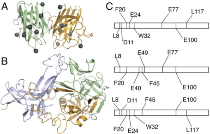
Fig. 1. SOD1 forms nonnative trimer. (A) HS-AFM analysis reveals that un-der stabilizing conditions (pH 3.5), SOD1 oligomer features a tripartite structure, in line with volumetric AFM measurements of dry samples, where oligomer volume is∼71.7 nm3and monomer volume is∼25.3 nm3(Fig. S1A). We observe three distinct monomers in HS-AFM recordings at a SOD1 concen-tration of 30 nM. The three subunits form a nonspherical compact particle, which is stable over long time periods. (Scale bar: 5 nm.) (B) In destabilizing conditions (pH 7.4), SOD1 trimers dissociate into three distinct monomers, even at the high end of physiological concentration (100 nM). (Scale bar: 5 nm.)
Fig. 2. Hybrid experimental/computational method leads to a model of metastable SOD1 trimer. (A) SOD1 trimer proteolytic cut sites (gray spheres, shown here on the native dimer structure) in the dimer interface and sec-ondary structural elements suggest significant structural differences be-tween native and trimeric structures. (B) Structural model of the metastable SOD1 trimer obtained using limited proteolysis data as constraints in several rounds of coarse-grained and atomistic DMD simulations (Fig. S2). (C) Represen-tation of the SOD1 linear sequence, residues 1–153. Proteolytic cut sites (vertical lines) determined in limited proteolysis experiments differ significantly in SOD1 monomer (Top), dimer (Middle), and trimer (Bottom), supporting structural rearrangement during the aggregation process and SOD1 trimer formation.
BIOPHYSICS
AND
COMPUTATION
AL
interfaces and tested the effects of these mutations on trimer
formation in vitro. Mutations that we predict to stabilize the
SOD1 trimer should result in an increased trimer population,
whereas mutations that we predict to destabilize the trimer
structure should result in a decreased or absent trimer
pop-ulation. Using Monte Carlo energy minimization, we predicted
the change in free energy (ΔΔG) of mutation for all interface
residues to all possible amino acids, and selected for
experi-mental evaluation those mutations having a significant effect
on SOD1 trimer stability (
j
ΔΔG
mutj>
3 kcal/mol). To ascertain
that the outcomes of any mutation observed in vitro are caused
by directly affecting SOD1 trimer formation and not due to
stabilization or destabilization of native SOD1 species, we
se-lected only those mutants that feature a negligible effect
(
j
ΔΔG
mutj<
1 kcal/mol) on the native SOD1 dimer and
monomer species (Protein Data Bank ID code 1SPD) (Fig. 3).
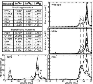
We observed the aggregation time course of our designed
SOD1 mutants using SEC. All seven of our designed
destabi-lizing mutations successfully decrease or eliminate the
pop-ulation of SOD1 trimer. Upon destabilization of the trimer
interfaces, we observe one of two outcomes: (
i
) inhibited trimer
formation with resulting increased populations of SOD1
mono-mer and dimono-mer species or (
ii
) inhibited trimer formation with a
resulting increase in large aggregates. Exhibiting the first
out-come, in four cases (P62Y-SOD1, G108H-SOD1, I99H-SOD1,
and N65V-SOD1), we observe the formation of a nonnative
extended dimer species, confirmed by AFM (Fig. 3 and
Figs. S5
and
S6
). In the second possible outcome of trimer-destabilizing
mutations, we observe the immediate formation of large
aggre-gates in D101I-SOD1, G147P-SOD1, and N53I-SOD1 (Fig. 3
and
Figs. S5
and
S6
). In these high-aggregation mutants, some
SOD1 trimer is formed in the early hours of the time course,
quickly followed by the appearance of large aggregates. None of
these three trimer-destabilizing mutations form SOD1 trimer,
instead quickly aggregating into large, potentially insoluble
ag-gregates, supporting the hypothesis that large, insoluble
aggre-gates, such as fibrils, lie on a different, competing pathway from
the formation of small, soluble aggregates.
Because the SOD1 trimer is a nonnative, metastable,
in-termediate aggregation state, stabilizing its formation is a much
more challenging task than destabilizing it. In stabilizing one
transient species, we may unknowingly stabilize, destabilize, or
even create another transient species. Although we can account
for the stabilization or destabilization of known structures, such
as the native SOD1 dimer and monomer, aggregation is a complex
process, and we cannot account for other metastable species that
may be perturbed during our mutation of trimer interface residues,
which may affect whether or not we observe increased levels of
SOD1 trimer. Despite these difficulties, we succeeded in predicting
and designing a SOD1 trimer-stabilizing mutant. The F20L-SOD1
mutant demonstrates a clear stabilization of the SOD1 trimer
species: monomer, dimer, and trimer species are all present at time
t
=
0, but the trimer species quickly dominates and stabilizes over
t
=
24 h, with no larger aggregates forming (Fig. 3). Five additional
mutations predicted to stabilize the SOD1 trimer, while exhibiting a
trimer population, result in stabilization of other nonnative species
ranging from misfolded monomer to protofibrils (
Fig. S5
), with
stoichiometry confirmed by AFM (
Fig. S6
). This finding suggests
that the nonnative monomer–monomer interfaces we identify in the
SOD1 trimer are also exploited by other potentially toxic species.
Trimer-Stabilizing SOD1 Mutants Promote Cell Death.
Because
mu-tations can cause conformational changes in proteins, especially
when located in a structurally important region like an interface,
SOD1 mutations clinically linked to ALS could cause an entirely
different trimeric species to form, which we would not capture in
our model. Computational testing of the stabilizing effect of each
mutation on all possible SOD1 trimer structures would be
pro-hibitive, and still may not yield an answer to the crucial question:
Do disease mutations cause an increase in the population of
trimeric SOD1? Instead, we address the question of SOD1
trimer disease relevance from another direction: by directly
testing the toxicity of SOD1 trimer in motor neuron-like cells
(NSC-34 cells). We transfected cells with expression constructs
for each of our designed SOD1 mutants, WT SOD1, and
A4V-SOD1, an aggressive ALS-linked mutation responsible for the
majority of familial ALS cases in North America (30, 31), which
we use as a positive control for ALS-relevant cell death. The
A4V mutation has previously been observed to promote the
formation nonnative SOD1 trimer (15). We observed
signifi-cantly increased cell death in NSC-34 hybrid motor neuron cells
expressing A4V-SOD1 (positive control) or our designed
stabilizing SOD1 mutants (Fig. 4). Conversely, our
trimer-destabilizing mutants do not cause cell death, with cell viability
comparable to cells expressing WT SOD1 (Fig. 4). Similarly,
expression of A4V-SOD1 or trimer-stabilizing SOD1 mutants,
but not trimer-destabilizing SOD1 mutants, resulted in elevated
levels of cleaved caspase-3 compared with WT SOD1 (Fig. 4).
Finally, we note that the amount of cell death present was
cor-related to the stability of the SOD1 mutant trimer (
Fig. S7
).
Discussion
We conclude that the SOD1 trimer causes cell death in motor
neuron-like cells, and that the nonnative trimeric interfaces
ap-pear to be shared by multiple cytotoxic SOD1 species. Studies
from many groups have suggested potential mechanisms of
SOD1 oligomer neurotoxicity but were unable to characterize
these oligomers fully. Excellent reviews of these mechanisms can
be found elsewhere (32–34). Many of these studies resulted in
such broad characterization that we may only conclude that the
toxic SOD1 species was soluble; however, in those studies that
determine a more limited size range, we note that the size of these
oligomers closely mirrors the size of the SOD1 trimer, and we
propose that the trimeric form of SOD1 that we describe in this
work is a likely candidate for the aberrant and detrimental
inter-actions with cellular machinery observed in these previous studies.
Given that SOD1 has been shown to undergo aberrant
ag-gregation in both familial and sporadic ALS (35–37), we propose
that formation of SOD1 trimer is a common pathogenic
mech-anism in ALS, causing the death of motor neurons and progression
of the disease. At the time of writing, 62% of residues known to
feature disease-relevant point mutations are located in the
pro-posed SOD1 trimer interfaces (
Table S1
and
Dataset S1
).
More-over, at least two-thirds of disease mutations located in the trimer
interfaces are overall stabilizing to trimer formation; the effect of
the remaining third cannot currently be established because of the
metastable nature of these oligomers. Due to metastability, the
SOD1 trimer exists as an ensemble of diverse structures (
Fig. S4C
and
Dataset S1
), and although the effect of a mutation on one
trimeric structure may be destabilizing, the same mutation could
promote formation of another trimeric form, resulting in a higher
fraction of disease mutants stabilizing a trimeric form of SOD1.
Recognizing and sequestering SOD1 trimer or preventing its
formation is therefore a potential strategy for preventing
neu-rodegeneration in ALS. Because of the high concentration of
SOD1 in motor neurons (30–100
μM), stoichiometric binding of
a drug compound to native SOD1 may not be a viable option to
abolish trimer formation completely. One option is to stabilize
larger, nontoxic aggregation products; this strategy has been
adopted for amyloid-β
aggregates in Alzheimer’s disease, to
positive effect in a cellular model (9). However, another strategy
would involve identifying and blocking toxic interactions of
SOD1 trimer in the cell. Alternatively, the structural knowledge
that we uncover in this work can be used for the design of
rec-ognition and sequestering strategies. The characterization of
structural features, such as the identification of nonnative
ag-gregation interfaces in this work, provides a key step in the
understanding of ALS disease etiology and development of
therapeutic or preventive strategies.
Materials and Methods
Purification of SOD1.For determination of surface-accessible sites in the SOD1 trimer in limited proteolysis studies, we isolated and purified SOD1 from human erythrocytes, as previously described (26). To generate SOD1 mutants for aggregation time courses using SEC, and WT SOD1 for comparison of comparable species, we performed cloning, expression, and purification of human recombinant SOD1 inSaccharomyces cerevisiaeas described elsewhere (15, 26, 38). We determined the concentration of purified SOD1 species by measuring theAat 280 nm with an extinction coefficient of 10,800 M−1·cm−1. We stored samples at−80 °C after flash-freezing in liquid nitrogen.
Formation and Isolation of SOD1 Species.We formed SOD1 trimer by in-cubating SOD1 dimer at a concentration of 100–120μM in 50 mM acetate, 150 mM NaCl, and 10 mM EDTA at pH 3.5 for 24 h. We then isolated trimer and monomer using an AKTA Purifier with a Superdex 200 10/300 GL column (GE Healthcare) equilibrated in either 50 mM phosphate and 150 mM NaCl at pH 3.5 (for use with V8 protease) or 50 mM acetate, 150 mM NaCl, and 10 mM EDTA at pH 3.5 (for all other proteases). We isolated monomer species fresh daily. We obtained trimer fractions from gel filtration chromatogra-phy, concentrated using an Amicon Ultra filter (Millipore) and used fresh or stored at 4 °C overnight and used the next day.
AFM.For AFM imaging of dried samples, we immersed freshly cleaved mica stripes (5.0×1.5 cm; Asheville Schoonmaker Mica Co.) in plastic cuvettes containing 167μM 1-(3-aminopropyl)silatrane (APS) solution for 30 min (39), followed by deionized water rinsing and drying in argon flow. We stored the APS mica in a vacuum chamber for use over the next few weeks (39). We cut the strips into∼0.75×0.75-cm pieces for sample preparation. We diluted the SOD1 samples to the appropriate concentrations (3.3–10 nM) with the corresponding buffers before deposition onto APS mica [50 mM sodium acetate, 150 mM NaCl, and 10 mM EDTA (pH 3.5) or 20 mM Tris·HCl and 150 mM NaCl (pH 7.4)]. We deposited 20 mL of diluted sample onto APS mica surfaces for 2 min, followed by thorough rinsing with deionized water and drying with argon flow. We kept the specimens in a vacuum chamber for at least 3 h, after which we mounted the specimens onto metal discs with double-sided tape (carbon-coated; Ted Pella, Inc.). We performed AFM im-aging on a MultiMode 8 atomic force microscope (Bruker NanoA, Bruker Co., Santa Barbara, CA) equipped with PeakForce modulus with microlever (MSNL; Bruker) probes. We acquired AFM images with 512×512 pixels at a scan rate of 4–6 Hz. We conducted volume and height analysis using Femtoscan (Advanced Technologies Center). We used“Enum features,”a functional tool provided by the software, to measure the heights and vol-umes of the protein in AFM images (40). We measured the background over the noncovered surface area. We performed Gaussian fitting of the volume and height values using Origin 6.0 software (Microcal Software, Inc.).
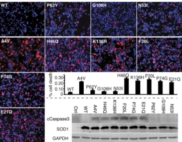
We acquired HS-AFM images using an HS-AFM instrument, operating in tapping mode in liquid (RIBM). We prepared samples as reported by Miyagi et al. (41). Briefly, we glued freshly cleaved 1.5-mm mica discs to the glass rods and modified with 167μM APS for 30 min. After rinsing with 20μL Milli-Q water (AquaMax-Ultra, LabWater.com, a division of APS water Services Corporation, Van Nuys, CA) and then three times with 20μL of pH 3.5 buffer (50 mM sodium acetate with an additional 150 mM NaCl and 10 mM EDTA), we deposited the SOD1 samples onto the APS mica in the same buffer. We per-formed measurements at two different concentrations of SOD1: 100 nM and 30 nM. After 5 min of incubation, we rinsed the surface with the same buffer used for imaging. To compare the effect of pH on sample dynamics, we removed the pH 3.5 buffer by pipette and injected the pH 7.4 buffer (20 mM Tris with 150 mM NaCl) to continue sample scanning. We used BL-AC10DS-A2 cantilevers (Olympus) with carbon tips obtained by electron beam deposition for im-aging (39). The spring constant of the AFM probes is between 0.1 and 0.2 N/m, with resonance frequency between 400 and 1,000 kHz in water. We performed continued scanning over the selected area (50 nm×50 nm) with a scan rate of approximately three frames per second. Images contain 128×128 pixels. We processed HS-AFM images using home-built software written by Atsushi Miyagi, Y.L.L. laboratory, University of Nebraska Medical Center, Omaha, NE. Fig. 4. SOD1 trimer is cytotoxic. Expression of A4V-SOD1 (positive control)
and trimer-stabilizing mutant SOD1s in NSC-34 motor neuron-like cells in-creases the incidence of cell death compared with WT SOD1 (negative con-trol), whereas trimer-destabilizing mutants have no effect on viability. Applied 3 d posttransfection, red stain [propidium iodide (PI)] identifies nuclei of dead cells, whereas blue stain (Hoechst) identifies nuclei of all cells. Incidence of cell death is measured as the percentage of total cells exhibiting PI staining (left to right fromUpper Left): WT, 4%; P62Y, 8%; G108H, 5%; N53I, 6%; A4V, 23%; H46Q, 29%; K136H, 25%; F20L, 27%; P74G, 24%; and E21Q, 22%. (Magnification: 20×.) Cell death correlates with trimer stability with aPvalue of 0.0476, or aPvalue of 0.000966 with exclusion of the outlier P62Y, which is extremely destabilizing (Fig. S7). Elevated levels of the apoptotic marker cleaved caspase 3 in NSC-34 motor neuron hybrid cells expressing A4V-SOD1 (positive control) and trimer-stabilizing SOD1 mutants (demon-strated by Western blot,Lower Right) confirm increased cytotoxicity of these mutants compared with WT SOD1 (negative control) and trimer-destabilizing SOD1 mutants.
BIOPHYSICS
AND
COMPUTATION
AL
Limited Proteolysis.We performed limited proteolysis at 25 °C in 100 mM buffer (acetate, phosphate, or Tris) at the appropriate pH for each enzyme: V8 protease reactions in 100 mM phosphate buffer (pH 4.0), chymotrypsin reactions in 100 mM Tris 50 mM CaCl2buffer (pH 7.8), and pepsin reactions in 100 mM acetate buffer (pH 3.5), quenched by addition of 1 M Tris buffer (pH 8.0) to a final concentration of 150 mM. We performed 1- to 50-min time course proteolysis reactions for trimer, allowing the reaction to proceed for a sufficient time to allow a single proteolytic cut and no more than four proteolytic cuts: 1 min for V8 and pepsin reactions and 2 min for chymo-trypsin reactions. This technique preserves the structural integrity of the trimer during the period of the limited proteolytic reaction, ensuring rele-vance of the obtained structural information. Time course reactions for monomer and dimer extended longer, as is necessary to obtain SOD1 cleavage. The proteolysis reaction for dimer contained an equimolar SOD1 concentration to the trimer reaction (7–15μM), but we performed reactions with SOD1 monomer at the concentration eluted from the FPLC (2.6–5μM), because we could not concentrate the monomer without significant ag-gregation. We determined protease/SOD1 ratios empirically. We quenched samples by the addition of 100 mM PMSF (1 mM final) and froze them until ready for further use. We desalted samples using Ziptip C18 pipette tips (Millipore) and eluted the peptides in 20μL of MS grade solvent [50% ace-tonitrile, 50% water, and 0.1% TFA (vol/vol); Fisher].
MS.We performed MS on an AB SCIEX 4800 PLUS MALDI-TOF-TOF instrument at the University of North Carolina Michael Hooker Proteomics Center using α-cyano-4-hydroxycinnamic acid as the matrix. Data output was analyzed in Data Explorer, with the signal-to-noise ratio set to 20–30, MS/MS tolerance set to±1 Da, and peptide charge set to 1+. We identified peptides using Mascot (Matrix Science) (42) with the UniProt database, with the search limited to the taxonomyHomo sapiens. We used the data from the earliest time points that resulted in cleavage (1–2 min) for constraints in modeling (Table S2).
Incorporation of Experimental Constraints.To incorporate information from limited proteolysis experiments into DMD simulations, we created an algo-rithm for converting knowledge of the sequence positions of proteolytic cleavage sites into pairwise simulation constraints (Fig. S2). The formation of trimeric SOD1 is made possible by a perturbation to the native state, which must occur as local unfolding in a background of native interactions. To account for this phenomenon, we first represent the native background as a G ¯o potential (43), which rewards contacts that are present in the native structure, assigning a pairwise attraction of 1 kcal/mol between pairs of residues with a distance between beta-carbons that is less than 7.5 Å. Next, we define a bias potential based on the location of proteolytic cleavage sites. Because each cleavage site must necessarily be (i) solvent-exposed and (ii) unstructured, we assign a repulsive interaction of energyE0between the cleavage sitejand every other residue in the system. Because residues near a solvent-exposed site are also likely to be solvent-exposed, and because an unstructured region of∼12 residues surrounding the cleavage site is necessary for the proteolytic enzyme to gain access to the site (27, 44), we apply a stepwise, decreasingly repulsive potential to the two residues on either side of each cleavage site, such that the pairwise repulsive interaction of the respective resi-due with each resiresi-due in the system has energy:
ErðiÞ=
X
j∈cleavage sites
E0expð−ji−jj=2Þ, whereji−jj≤2.
For each identified cleavage site, we thus generate 5npairwise constraints, wherenis the total number of residues in the system. All G ¯o and experi-mental constraint potentials are additive. Thus, the combined potential function is:
H=−X
i<j
Δij•δijE0+λ
X
i<j
δijððErðiÞ+ErðjÞÞ=2Þ,
whereΔijis the native contact matrix of the conformation,δijis the contact matrix of the current conformation, andλis a scaling parameter discussed in the next section.
Parameterization of Bias Potential.To scale the two terms of our bias po-tential, we tested the effects of various values ofλfrom 0.0 to 30 at intervals of 0.3 (from 0.0 to 3.0) or 3 (from 3 to 30) in coarse-grained (45) replica exchange simulations of apo-SOD1 monomer, which features a well-defined folding pathway. We examined the resulting energetic profiles and folding trajectories, and selected values ofλfor which folding intermediates are introduced by the competition between G ¯o and experimental energy terms but distinct folding transitions are evident. Atλ=0 (G ¯o potential only), we
find a single, sharp folding transition with no intermediates, as expected from experimental findings (46). Atλ≥1.5, we cannot resolve individual thermodynamic transitions, and protein unfolding occurs with increasing linearity asλincreases. We find that thatλvalues 0.33≤λ≤1.2 fit our cri-teria for selection, and that values in this range produce nearly identical final structures in our test apo-monomer system (Fig. S3). We therefore se-lectedλ=0.99 for use in modeling of trimeric SOD1.
Coarse-Grained DMD.To obtain initial trimeric SOD1 structures that agree with experimental data, we use DMD (47, 48) replica exchange (REX) (49) simulations. We apply our bias potential to a coarse-grained model of three natively folded SOD1 apo-monomers placed in proximity to each other but not initially bound. We use a four-bead protein model (45) for increased sampling and for simplified application of the bias potential to the Cβbead of each residue, with the exception of Ala, for which the bias potential is applied to the Cαbead. For each value ofλ, we determined the ideal number of replicas and spread of replica temperatures such that we sample energetic space spanning the entire melting transition and that exchange of replicas occurs with an acceptance rate between 0.2 and 0.7, with exchange attempted every 1,000 time steps. Forλ=0.99, we used 27 replicas, with temperatures of 0.435, 0.445, 0.455, 0.462, 0.470, 0.480, 0.490, 0.503, 0.513, 0.523, 0.538, 0.552, 0.566, 0.581, 0.595, 0.610, 0.625, 0.639, 0.654, 0.674, 0.694, 0.713, 0.733, 0.753, 0.768, 0.788, and 0.815 kcal·mol−1·kB−1. We per-formed simulations for 106time steps.
We evaluated the experimental agreement of each structure snapshot from simulation by calculating the average number of contacts made by each pro-teolytic cut site residue (Nc). Our rationale is that to demonstrate agreement with limited proteolysis results, each proteolytic cut site residue should make as few contacts with other residues as possible, because its ability to be cleaved by proteolytic enzymes denotes its solvent accessibility and lack of participation in secondary structure interactions. However, although the proteolytic cleavage sites should demonstrate a minimum of structural interaction, SEC results (15) indicate that the structure should be a compact, associated trimer. We there-fore use a combination of proteolytic cut site contacts and trimer radius of gyration (Rg) to select a pool of candidate structures from all REX simulation trajectories. When we examined these two criteria together, we found a cluster of structures having Nc<1.5 and Rg<30 Å. We clustered this pool of candidate structures by pairwise rmsd, and selected the centroid of the largest cluster for further simulation and structural refinement.
All-Atom DMD.We reconstructed the four-bead centroid structure obtained from the DMD REX simulations described above to an all-atom model (29) and performed structural minimization using Chiron (50) to remove clashes introduced by reconstruction. To equilibrate our structure in a physical force field, we first performed low-temperature (below the melting transition) all-atom REX simulations using 26 replicas with temperatures of 0.350, 0.360, 0.370, 0.380, 0.390, 0.400, 0.410, 0.420, 0.430, 0.440, 0.450, 0.460, 0.470, 0.480, 0.490, 0.500, 0.510, 0.520, 0.530, 0.540, 0.550 0.560, 0.570, 0.580, 0.590, and 0.600 kcal·mol−1·kB−1. We selected the ideal number of replicas and spread of replica temperatures such that exchange of replicas occurs with an acceptance rate between 0.2 and 0.7, with exchange attempted every 1,000 time steps. We performed simulations for 106time steps. We isolated the lowest energy structure from all simulation replicas, and con-tinued simulation of that structure at a temperature of 0.350 kcal·mol−1·k
B−1 for an additional 106 time steps. Finally, we selected the lowest energy structure from this single-temperature trajectory as our final model, and verified the quality of the model using Gaia (51).
Computational Mutagenesis.Using our final SOD1 trimer model, we per-formed computational mutagenesis of every residue residing in an interface between monomer chains. We designate a residue as residing in an interface if any atom of that residue is within 4.5 Å of any atom of a residue belonging to a different monomer chain. We performed computational mutagenesis of each interface residue to every possible residue and calculated the change in free energy upon mutation (ΔΔGmut) using Eris (52, 53). Due to complexity in their interactions, we did not perform mutations to Cys.
Measurement of Cytotoxicity of SOD1 Trimer.We maintained neuroblastoma spinal cord hybrid cell lines (NSC-34) in DMEM containing 10% (vol/vol) FBS. We plated∼5,000 NSC-34 cells in one well of a 24-well plate with differen-tiation medium [1% FBS, 1% nonessential amino acid supplement, (vol/vol) 10μM of RA]. After a 2-d differentiation period in DMEM, we transfected cells with 2μg of plasmid encoding WT SOD1 or SOD1 mutants (within the pCI-Neo vector) using Lipofectamine 3000 (Thermo Fisher Scientific) according to the manufacturer’s instructions. To verify that SOD1 had been properly trans-fected, we performed immunostaining. We fixed cells 24 h posttransfection using 4% (vol/vol) PFA for 10 min at room temperature. We blocked with 3% (vol/vol) goat serum, followed by Abs against SOD1 (Sigma) and Hoechst 33342 counterstain. We performed propidium iodide (red) and Hoechst (blue) staining in culture 3 d posttransfection without fixation. We performed a Western blot with∼20,000 cells per well, collecting cells 2 d posttransfection. We lysed cells in PBS with 0.5% Triton X-100, 0.5 mM EDTA, and EDTA-free protease inhibitor (Roche Life Sciences). We denatured cell lysates and
resolved using 18% (vol/vol) SDS/PAGE, and incubated with Abs against cleaved caspase-3 (Cell Signaling Technology), SOD1 (Calbiochem), and GAPDH (Chemcon), followed by secondary Ab incubation. We visualized protein bands using an Odyssey IR imaging system (LI-COR Biosciences). Cell viability was not assayed in all mutants because we were unable to verify SOD1 transfection in five of our predicted mutants, four of which are trimer-destabilizing muta-tions: D124Q-SOD1, G147P-SOD1, I99H-SOD1, D101I-SOD1, and N65V-SOD1.
ACKNOWLEDGMENTS.We thank Dr. Feng Ding and Dr. Michael Caplow for helpful discussions, and Dr. Joan S. Valentine for graciously providing the EG118 yeast strain and the yEP351:hwtSOD1 vector. MS data were collected at the University of North Carolina Michael Hooker Proteomics Center with help from Nedyalka Dicheva. This work was supported by NIH Grants R01GM080742 (to N.V.D.), R01GM096039 (to Y.L.L.), and R01GM078366 (to M.D.). E.A.P. was supported by a Ruth L. Kirschstein National Research Service Award (F31AG039266) from the National Institute on Aging.
1. Cleveland DW, Rothstein JD (2001) From Charcot to Lou Gehrig: Deciphering selective motor neuron death in ALS.Nat Rev Neurosci2(11):806–819.
2. Rothstein JD (2009) Current hypotheses for the underlying biology of amyotrophic lateral sclerosis.Ann Neurol65(Suppl 1):S3–S9.
3. Walker LC, LeVine H, 3rd (2012) Corruption and spread of pathogenic proteins in neurodegenerative diseases.J Biol Chem287(40):33109–33115.
4. Redler RL, Dokholyan NV (2012) The complex molecular biology of amyotrophic lat-eral sclerosis (ALS).Prog Mol Biol Transl Sci107:215–262.
5. Zetterström P, et al. (2007) Soluble misfolded subfractions of mutant superoxide dismutase-1s are enriched in spinal cords throughout life in murine ALS models.Proc Natl Acad Sci USA104(35):14157–14162.
6. Kirkitadze MD, Bitan G, Teplow DB (2002) Paradigm shifts in Alzheimer’s disease and other neurodegenerative disorders: The emerging role of oligomeric assemblies.
J Neurosci Res69(5):567–577.
7. Arrasate M, Mitra S, Schweitzer ES, Segal MR, Finkbeiner S (2004) Inclusion body formation reduces levels of mutant huntingtin and the risk of neuronal death.Nature
431(7010):805–810.
8. Caughey B, Lansbury PT (2003) Protofibrils, pores, fibrils, and neurodegeneration: Separating the responsible protein aggregates from the innocent bystanders.Annu Rev Neurosci26:267–298.
9. Bieschke J, et al. (2012) Small-molecule conversion of toxic oligomers to nontoxic
β-sheet-rich amyloid fibrils.Nat Chem Biol8(1):93–101.
10. Rakhit R, et al. (2007) An immunological epitope selective for pathological monomer-misfolded SOD1 in ALS.Nat Med13(6):754–759.
11. Jonsson PA, et al. (2004) Minute quantities of misfolded mutant superoxide dis-mutase-1 cause amyotrophic lateral sclerosis.Brain127(Pt 1):73–88.
12. Israelson A, et al. (2010) Misfolded mutant SOD1 directly inhibits VDAC1 conductance in a mouse model of inherited ALS.Neuron67(4):575–587.
13. Pedrini S, et al. (2010) ALS-linked mutant SOD1 damages mitochondria by promoting conformational changes in Bcl-2.Hum Mol Genet19(15):2974–2986.
14. Nishitoh H, et al. (2008) ALS-linked mutant SOD1 induces ER stress- and ASK1-dependent motor neuron death by targeting Derlin-1.Genes Dev22(11):1451–1464. 15. Redler RL, Fee L, Fay JM, Caplow M, Dokholyan NV (2014) Non-native soluble oligo-mers of Cu/Zn superoxide dismutase (SOD1) contain a conformational epitope linked to cytotoxicity in amyotrophic lateral sclerosis (ALS).Biochemistry53(14):2423–2432. 16. Bosco DA, et al. (2010) Wild-type and mutant SOD1 share an aberrant conformation
and a common pathogenic pathway in ALS.Nat Neurosci13(11):1396–1403. 17. Brotherton TE, et al. (2012) Localization of a toxic form of superoxide dismutase 1 protein
to pathologically affected tissues in familial ALS.Proc Natl Acad Sci USA109(14):5505–5510. 18. Rotunno MS, et al. (2014) Identification of a misfolded region in superoxide dismutase 1
that is exposed in amyotrophic lateral sclerosis.J Biol Chem289(41):28527–28538. 19. Banci L, et al. (2007) Metal-free superoxide dismutase forms soluble oligomers under
physiological conditions: A possible general mechanism for familial ALS.Proc Natl Acad Sci USA104(27):11263–11267.
20. Banci L, et al. (2008) SOD1 and amyotrophic lateral sclerosis: mutations and oligo-merization.PloS One3(2):e1677.
21. Luchinat E, et al. (2014) In-cell NMR reveals potential precursor of toxic species from SOD1 fALS mutants.Nat Commun5:5502.
22. Auclair JR, Boggio KJ, Petsko GA, Ringe D, Agar JN (2010) Strategies for stabilizing superoxide dismutase (SOD1), the protein destabilized in the most common form of familial amyotrophic lateral sclerosis.Proc Natl Acad Sci USA107(50):21394–21399. 23. Korzhnev DM, Religa TL, Banachewicz W, Fersht AR, Kay LE (2010) A transient and
low-populated protein-folding intermediate at atomic resolution.Science329(5997):1312–1316. 24. Laganowsky A, et al. (2012) Atomic view of a toxic amyloid small oligomer.Science
335(6073):1228–1231.
25. Khare SD, Caplow M, Dokholyan NV (2004) The rate and equilibrium constants for a multistep reaction sequence for the aggregation of superoxide dismutase in amyo-trophic lateral sclerosis.Proc Natl Acad Sci USA101(42):15094–15099.
26. Wilcox KC, et al. (2009) Modifications of superoxide dismutase (SOD1) in human eryth-rocytes: A possible role in amyotrophic lateral sclerosis.J Biol Chem284(20):13940–13947. 27. Hubbard SJ, Campbell SF, Thornton JM (1991) Molecular recognition. Conformational analysis of limited proteolytic sites and serine proteinase protein inhibitors.J Mol Biol
220(2):507–530.
28. Rakhit R, et al. (2004) Monomeric Cu,Zn-superoxide dismutase is a common mis-folding intermediate in the oxidation models of sporadic and familial amyotrophic lateral sclerosis.J Biol Chem279(15):15499–15504.
29. Ding F, Tsao D, Nie H, Dokholyan NV (2008) Ab initio folding of proteins with all-atom discrete molecular dynamics.Structure16(7):1010–1018.
30. Deng HX, et al. (1993) Amyotrophic lateral sclerosis and structural defects in Cu,Zn superoxide dismutase.Science261(5124):1047–1051.
31. Juneja T, Pericak-Vance MA, Laing NG, Dave S, Siddique T (1997) Prognosis in familial amyotrophic lateral sclerosis: Progression and survival in patients with glu100gly and ala4val mutations in Cu,Zn superoxide dismutase.Neurology48(1):55–57. 32. Bruijn LI, Miller TM, Cleveland DW (2004) Unraveling the mechanisms involved in
motor neuron degeneration in ALS.Annu Rev Neurosci27:723–749.
33. Joyce PI, Fratta P, Fisher EMC, Acevedo-Arozena A (2011) SOD1 and TDP-43 animal models of amyotrophic lateral sclerosis: Recent advances in understanding disease toward the development of clinical treatments.Mamm Genome22(7-8):420–448. 34. Rotunno MS, Bosco DA (2013) An emerging role for misfolded wild-type SOD1 in
sporadic ALS pathogenesis.Front Cell Neurosci7:253.
35. Shibata N, Asayama K, Hirano A, Kobayashi M (1996) Immunohistochemical study on superoxide dismutases in spinal cords from autopsied patients with amyotrophic lateral sclerosis.Dev Neurosci18(5-6):492–498.
36. Shibata N, et al. (1994) Cu/Zn superoxide dismutase-like immunoreactivity in Lewy body-like inclusions of sporadic amyotrophic lateral sclerosis.Neurosci Lett179(1-2): 149–152.
37. Gruzman A, et al. (2007) Common molecular signature in SOD1 for both sporadic and familial amyotrophic lateral sclerosis.Proc Natl Acad Sci USA104(30):12524–12529. 38. Goscin SA, Fridovich I (1972) The purification and properties of superoxide dismutase
from Saccharomyces cerevisiae.Biochim Biophys Acta289(2):276–283.
39. Shlyakhtenko LS, Gall AA, Lyubchenko YL (2013) Mica functionalization for imaging of DNA and protein-DNA complexes with atomic force microscopy.Methods Mol Biol
931:295–312.
40. Portillo AM, Krasnoslobodtsev AV, Lyubchenko YL (2012) Effect of electrostatics on aggregation of prion protein Sup35 peptide.J Phys Condens Matter24(16):164205. 41. Miyagi A, Ando T, Lyubchenko YL (2011) Dynamics of nucleosomes assessed with
time-lapse high-speed atomic force microscopy.Biochemistry50(37):7901–7908. 42. Perkins DN, Pappin DJ, Creasy DM, Cottrell JS (1999) Probability-based protein
iden-tification by searching sequence databases using mass spectrometry data.Electrophoresis
20(18):3551–3567.
43. Go N, Abe H (1981) Noninteracting local-structure model of folding and unfolding transition in globular proteins. I. Formulation.Biopolymers20(5):991–1011. 44. Hubbard SJ, Eisenmenger F, Thornton JM (1994) Modeling studies of the change in
conformation required for cleavage of limited proteolytic sites.Protein Sci3(5): 757–768.
45. Ding F, Borreguero JM, Buldyrey SV, Stanley HE, Dokholyan NV (2003) Mechanism for the alpha-helix to beta-hairpin transition.Proteins53(2):220–228.
46. Stroppolo ME, Malvezzi-Campeggi F, Mei G, Rosato N, Desideri A (2000) Role of the tertiary and quaternary structures in the stability of dimeric copper, zinc superoxide dismutases.Arch Biochem Biophys377(2):215–218.
47. Dokholyan NV, Buldyrev SV, Stanley HE, Shakhnovich EI (1998) Discrete molecular dynamics studies of the folding of a protein-like model.Fold Des3(6):577–587. 48. Zhou Y, Karplus M (1997) Folding thermodynamics of a model three-helix-bundle
protein.Proc Natl Acad Sci USA94(26):14429–14432.
49. Okamoto Y (2004) Generalized-ensemble algorithms: enhanced sampling techniques for Monte Carlo and molecular dynamics simulations.J Mol Graph Model22(5): 425–439.
50. Ramachandran S, Kota P, Ding F, Dokholyan NV (2011) Automated minimization of steric clashes in protein structures.Proteins79(1):261–270.
51. Kota P, Ding F, Ramachandran S, Dokholyan NV (2011) Gaia: Automated quality as-sessment of protein structure models.Bioinformatics27(16):2209–2215.
52. Yin S, Ding F, Dokholyan NV (2007) Modeling backbone flexibility improves protein stability estimation.Structure15(12):1567–1576.
53. Yin S, Ding F, Dokholyan NV (2007) Eris: An automated estimator of protein stability.
Nat Methods4(6):466–467.
BIOPHYSICS
AND
COMPUTATION
AL