GENETIC APPROACHES TO THE STUDY OF CORONAVIRUS REPLICATION AND PATHOGENESIS
Damon Joshua Deming
A dissertation submitted to the faculty of the University of North Carolina at Chapel Hill in partial fulfillment of the requirements for the degree of Doctor of Philosophy in the
Department of Microbiology and Immunology
Chapel Hill 2006
Abstract
DAMON J. DEMING: GENETIC APPROACHES TO THE STUDY OF CORONAVIRUS REPLICATION AND PATHOGENESIS
(Under the direction of Dr. Ralph S. Baric)
The recently developed coronavirus reverse genetic systems have been a tremendous asset for improving our understanding of the viruses’ complex replication strategy,
pathogenesis, mechanisms of host-range expansion, and in the development of anti-viral therapies. We completed two studies using coronavirus infectious clones. The first
evaluated a severe acute respiratory syndrome coronavirus (SARS-CoV) vaccine to protect against an antigenically divergent strain. The second study determined the requirement for proteolytic processing of a highly conserved region of the replicase polyprotein for efficient replication.
Ideally, a SARS-CoV vaccine should confer long-term protection, especially in vulnerable senescent populations, against both the 2003 epidemic strains and zoonotic strains that may yet emerge from animal reservoirs. Using Venezuelan equine encephalitis virus replicon particles (VRP) expressing the 2003 epidemic Urbani SARS-CoV strain spike (S) glycoprotein (VRP-S) or the nucleocapsid (N) protein from the same strain (VRP-N) as candidate vaccines, we tested their ability to protect young and senescent mice when
the epidemic strain. VRP-S, but not VRP-N, provided protection for both young and
senescent mice when challenged with the epidemic strain. When challenged with icGDO3-S, VRP-S protected young mice but only partially protected senescent animals. VRP-N
vaccinated mice demonstrated enhanced pulmonary inflammation, which included eosinophils among the cellular infiltrates, following SARS-CoV or icGDO3-S challenge.
The highly conserved region at the carboxy-terminus of the coronavirus replicase ORF1a polyprotein is processed by the main proteinase (Mpro) into mature products including nsp7, nsp8, nsp9 and nsp10, proteins with predicted or identified activities involved with RNA synthesis. Mpro continuous translation and processing of ORF1ab polyproteins is required for replication, but specific cleavage events may be dispensable. We determined the requirement for the nsp7-10 proteins and their proteolytic processing during the replication of murine hepatitis virus (MHV), which is phylogenetically grouped with the human
coronaviruses OC43 and SARS-CoV. Using the MHV reverse genetics system, in frame deletions of the coding sequences for nsp7, 8, 9, and 10 were either deleted, or the flanking cleavage sites ablated, and the effect upon replication determined. Viable viruses were characterized through analysis of Mpro processing, subgenomic RNA transcription, and in vitro growth fitness. Deletion of any of the four regions encoding nsp7 through 10 was lethal. Disruption of the cleavage sites flanking the protein domains were lethal with the exception of the nsp9/10 cleavage site, which resulted in a mutant virus with severely attenuated replication. In order to determine if a distinct function could be attributed to pre-processed forms of the replicase polyprotein including nsp7-10, the genes encoding nsp7 and nsp8 were rearranged. The mutant virus MHV8/7 was not viable, suggesting that the
TABLE OF CONTENTS
Page
LIST OF TABLES ……… vi
LIST OF FIGURES ………..……… vii
Chapter I Introduction .………...………... 1
ORF1 Domain Organization and Expression ………..………. 3
Organization of the Structural and Accessory protein genes ……...………. 6
Expression of ORF2+ Proteins via Subgenomic mRNAs …………..…….. 7
Nidoviruses as Emerging and Reemerging Infectious Agents ………..…... 8
Nidovirus Reverse Genetics Systems ………..……. 15
II Vaccine Efficacy in Senescent Mice Challenged with Recombinant SARS- CoV Bearing Epidemic and Zoonotic Spike Variants Abstract ………. 28
Introduction ………... 29
Methods ……… 31
Results ………... 39
III MHV-A59 ORF1A REPLICASE PROTEIN NSP7-NSP10 PROCESSING IN REPLICATION
Abstract ………. 73
Introduction ………... 74
Methods ……… 77
Results ………... 83
Discussion ………. 89
IV Summary ………... 105
SARS-CoV vaccines, senescent animal models, and heterologous challenge virus ………. 105
Future Directions ……….. 111
MHV replicase protein processing in replication ………. 112
Future Directions ……….. 116
LIST OF TABLES
Table Page
1 Summary of Vaccine Groups and Select Results for Mouse Experiments ………… 60 S1 Titers and PRNT80 Dilutions for Individual Senescent Mice ………. 61-2 2 Primers, template DNA, and restriction sites used in the generation of
deletion and cleavage mutants ………... 95 3 nsp7-10 cleavage sites of wild-type MHV and deletion mutants ……….. 96 4 Mutagenesis of the nsp7-10 cleavage sites ……… 96 5 Genomic variation of nsp9 between MHV, MHV9/10, and the passage
15 mutants ……….. 97
LIST OF FIGURES
Figure Page
1 Genome organization of MHV-A59 and SARS-CoV ………... 22
2 ORF1ab polyprotein: Proteolytic processing and conserved elements ……..……... 22
3 Expression of ORF2+ from subgenomic RNAs ……… 23
4 Generation of subgenomic RNA by discontinuous attenuated transcription ………. 24
5 Initial version of targeted recombination reverse genetics system for coronaviruses ………. 25
6 Improved targeted recombination system using cell specificity for screening ……… 25
7 Rearrangement of structural genes to limit the occurrence of multiple recombination events ………... 26
8 Multi-component reverse genetics system for SARS-CoV ………... 27
9 VRP Expression of SARS S and N and VRP-S Induction of Anti-SARS S Antibody ……….. 63
10 VRP-S Induces Short- and Long-Term Protection against icSARS-CoV Challenge ………. 64
11 Synthetic Reconstruction of icGD03-S ……….. 65
12 VRP-S Induces Short-Term Protection against icGDO3-S in Young and Partial Protection in Old Mice ………. 66
13 An 80% Plaque Reduction Neutralization Titers (PRNT80) for VRP-S and VRP-S+N Hyperimmune Serum ………... 67
14 ELISA Titers for Anti-S and Anti-HA IgG in Vaccinated Animals ……….. 68
15 Pathogenic Findings Following Homologous Challenge ……….. 69
16 Pathogenic Findings Following Heterologous Challenge ……….. 70
17 Kinetics of VRP-N–Associated Inflammation ………... 71
Figure Page 19 MHV genome organization, proteolytic processing of the
replicase polyproteins, and putative cleavage sites of nsp7-10 ………. 98
20 RTPCR verification of the replication deficiency of non syncitia forming cleavage mutant viruses ………... 99
21 Replication kinetics of the viable cleavage mutants and MHV9/10 revertants ……….. 100
22 Characterizing the nsp9 genetic components of MHVp15-1 and MHVp15-3 ………....………... 101
23 ORF1a Polyprotein Processing in Recombinant Viruses ………....…… 102
24 Recombinant and wildtype virus RNA Synthesis ……… 103
Chapter I
Introduction
The order Nidovirales includes a broad group of mammalian, avian and crustacean
viruses grouped among the families Coronaviridae, Arteriviridae, and Roniviridae. The
family Coronaviridae is further divided into the genera Coronaviruses and Toroviruses.
Although nidoviruses differ significantly in genome size, sequence, virion morphology, and host-range specificity, they are grouped within the same order due to several shared traits (reviewed in (66)). All Nidoviruses are positive-stranded RNA viruses with large replicase domains (designated ORF1a and ORF1b) which are functionally conserved and encoded at the 5’ end of the genome. Translation of the replicase polyprotein is regulated by a
ribosomal frame-shift event which directs the expression of either an ORF1a or an ORF1a/b full-length polyprotein, which is in turn proteolytically processed by virally encoded
proteases. Another characteristic unique to nidoviruses is that several structural and
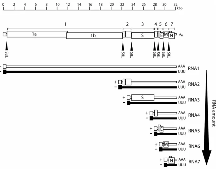
nonstructural ORFs are encoded downstream of the replicase polyprotein and expressed by a 3’ co-terminal nested set of subgenomic mRNAs (sg mRNAs) generated by a unique strategy of attenuated transcription. There are significant differences between the genome sizes of nidoviruses ranging from the smallest arterivirus genomes of ~13 kb to the largest
translated by host cell ribosomes. For many nidoviruses, including coronaviruses, a typically narrow host range is dictated by the highly specific interaction between the glycoprotein spike displayed on the virion and a particular receptor on permissive cells. However, characteristic high mutation and recombination rates allow these viruses to evolve to infect new cells and expand beyond their normal tissue tropisms and host range limitations.
Coronavirus reverse genetic systems were recently developed and have been a tremendous asset as new tools in facilitating our understanding of the viruses’ complex replication strategy, pathogenesis, and mechanisms of host-range expansion. Reverse genetics systems are also proving to be useful in the development of safe and effective anti-viral therapies. The small size of the arterivirus, the smallest of the nidoviruses with
genomes ranging in size from 13-16kb, facilitated the rapid development of molecular clones as early as 1997 (205). In contrast, the large size of the coronavirus genome and E. coli
associated toxicity of regions within the polymerase gene had delayed the construction of stable full-length cDNA templates until 2000 (2, 229). Fortunately for coronavirus research, several strategies were successfully employed to overcome these limitations and develop viable reverse genetics systems.
ORF1 Domain Organization and Expression
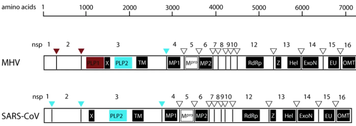
cysteine protease, PLP. In some coronaviruses, such as EAV and IBV, the first PLP domain is inactive, suggesting that although proteolytically inactive, they may serve another function in replication. SARS-CoV lacks the first PLP and relies upon the activity of its one PLP, denoted PLpro2 due to its position relative to that of other coronaviruses, for cleavage of the 5’ end of the replicase polyproteins. The three TMs (TM1, MP1, and MP3) likely anchor the replication complex to intracellular membranes. MP1 and MP2 flank another functionally conserved domain within the ORF1a polyprotein, a papain-like protease responsible for mediating cleavage of most of the replicase polyproteins into its constituent components. These proteases are often referred to as either 3CLpro, due to their structural and substrate similarity to the picornavirus 3C proteinase, or Mpro, the main protease. The Mpro designation refers to the fact that these proteases are responsible for processing the majority of the ORF1 polyprotein, including all components of the ORF1b, which encode the major replicative enzymes and most highly conserved domains. A narrowly conserved domain encoding an ADP-ribose 1’-phosphatase (X) was recently identified within the nsp3 of corona- (152, 153) and toroviruses (48).
constitutive proteolytic processing of the replicase polyprotein is required for replication and suggests that distinct roles may exist for both the fully processed and precursor forms of the polyproteins (13, 45, 103, 165, 168, 243). The necessity for the proteolytic processing of the polyprotein for efficient nidovirus replication has also been demonstrated in equine arteritis virus (EAV) and the mouse hepatitis coronavirus (MHV) by mutation of the cleavage sites recognized by Mpro or PLP (46, 206).
Several domains conserved across all nidoviruses fall within ORF1b (Fig. 2). In order from N to C-terminus, these elements are the RNA-dependent RNA polymerase (RdRp) (18, 47, 67), the putative Zinc-binding domain (ZBD; Z in Fig. 2) (171), a
superfamily-1 helicase (Hel) (92, 93, 171), and an endoribonuclease (EndoU; EU in Fig. 2) (7, 91). The orientation of the RdRp and Hel domains, with the RdRp N-terminal to that of the Hel, is unique to the nidoviruses among message-sense ssRNA viruses. Also, the ZBD and NendoU domains are unique to nidoviruses and as such are considered hallmark genetic markers for the order (66). Other ORF1 functional domains are conserved over many, but not all, nidoviruses. For example, exoribonuclease (ExoN) (137) and a putative ribose-2’-O-methyltransferase (O-MT; OMT in Fig. 2) are only conserved within the pp1ab of corona-, toro-, and roniviruses (39, 178, 212).
Many elements of the replicase gene have no known function. One such region of the coronavirus genome encodes four small proteins, denoted nsp7 to nsp10 in coronaviruses, are translated from 3’ end of ORF1a and are conserved among corona- and toroviruses (66). These proteins are processed by Mpro into mature products of 10, 22, 12, and 15 kDa,
respectively. The nsp7-10 co localize with the replication complex bound to double
Although the replicative function of these proteins are yet to be experimentally demonstrated, recent work has provided some insight into their purpose. Structure analyses of SARS-CoV nsp7 and 8 demonstrated that the two proteins form a hexadecameric supercomplex with electrostatic properties favorable for nucleic acid binding that may function as a processivity factor for the RdRp (234). A biochemical report described a primase function associated with the SARS-CoV nsp8 protein, implicating another important role for one of these small proteins in replication (32, 89). The SARS-CoV nsp9 crystal structure has also been resolved and shown to form homodimers possessing single-stranded RNA-binding properties, and it has been suggested that the protein may serve to stabilize nascent and template RNA during replication, transcription, and processing (22, 53). Temperature sensitive mutations localized in nsp10 suggest that the protein may be involved with negative strand synthesis (165). Recent reports describing the refinement of the nsp10 structure have revealed that the protein includes two Zn fingers, exhibits nucleic acid binding affinity (97), and that it may form a spherical dodecameric structure made up of 12 nsp10-11 fused subunits (187). Another recent structural study has described nsp10 crystals that formed monomers and homodimers and possessing nucleic acid binding affinity (97). Collectively, this data implies that the nsp7-nsp10 proteins are important – if not critical – to coronavirus, and likely torovirus, replication.
Organization of the Structural and Accessory protein genes
express a single nucleocapsid protein (N) as well as the proteins making up the enveloped virion (66). In coronaviruses, the structural genes maintain the same order: 5’- spike glycoprotein (S), envelope protein (E), membrane spanning integral protein (M), and
nucleocapsid (N) - 3’ (Fig. 1). Some group-2 coronaviruses, such as BCoV, mouse hepatitis virus (MHV), and HCoV-OC43 also include a gene encoding hemagluttinin esterase (HE). SARS-CoV is an exception. In addition to the major structural genes, “accessory” proteins are encoded by ORFs located among, or overlapping with, the structural genes. These proteins, of which coronaviruses typically have between 2 and 8, may or may not be integrated within the mature virion, are not ubiquitously present within viruses of a given group, and are often dispensable for in vitro replication (42, 57, 73, 74, 145, 169, 217, 227). Although not involved with efficient growth in tissue culture, deletion of some of these accessory ORFs have been reported to attenuate pathology in animal models (42, 73, 145).
Expression of ORF2+ Proteins via Subgenomic mRNAs
proximity. In the case of corona- and arteriviruses, their transcription strategy may more specifically be called discontinuous attenuated transcription. This refers to an additional recombination event that transfers the incomplete negative sense RNA strand to a TRS located near the 5’ end of the genomic template (Fig. 4, step 2). Transcription continues to include the complementary sequence of the 5’ UTR on the newly synthesized minus strand. In turn, the minus sense strand serves as the template for mRNA sense RNAs (Fig. 4, step 3). Typically, there is a decreasing chance that the polymerase will retain its association with the genomic template as it progresses past each TRS which leads to a nested set of mRNAs which decreases in relative amount the longer the mRNA.
Nidoviruses as Emerging and Reemerging Infectious Agents
It is estimated that 73% of human emerging and reemerging infectious diseases -- pathogens rapidly increasing in incidence, expanding in geographic range, or extending infection into new host species -- are zoonotic pathogens that have bridged the species barrier (108, 221). At 37% of all emerging and reemerging pathogens, RNA viruses are well
represented (221). In order for a virus to expand outside of its normal host range, the virus must evolve the capacity to interact with novel cellular factors and adapt to evade or usurp mechanisms which normally function to ablate virus entry, replication, or transmission in a new host species. An emerging virus requires both the opportunity to interact with a new prospective host as well as possess molecular mechanisms with which to adapt and replicate efficiently within the hostile cellular environment. With a very broad distribution among several different animal species, many of which maintain close contact to humans,
With replication characterized by high mutation and recombination rates, coronaviruses also possess the means to rapidly evolve to changing cellular environments and selective
pressures.
With established reservoirs in humans, wild and domesticated animals, coronaviruses have plenty of opportunity to make contact with species normally outside of their restricted host range. Many animals hosting coronaviruses are maintained close to other animals or humans, such as in the case of livestock and companion animals, such as equine, swine, bovine, canine, feline, and avian species. Several examples of emergent viruses have been found during the extensive studies of the coronaviruses’ ability to expand their host range. For instance, the porcine epidemic diarrhea virus (PEDV), an economically significant cause of severe swine gastroenteritis in Europe and Asia, is closely related to the human
coronavirus HCV-229E and is believed to be the result of transmission from humans to swine (15, 51, 148). BCoV is believed to have passed into several species of ruminants including elk (128), waterbuck, sambar deer, and white-tailed deer populations (201), dogs (54), and has been associated with at least one enteric infection in humans (76, 236). Close genetic and antigenic similarities between the group II coronaviruses BCoV, HCoV-OC43, and porcine hemagglutinating encephalomyelitits virus (PHEV) suggests that they may have only recently diverged from a common ancestor (211).
other species (72, 119, 180, 202). Similarly, sero-positive wet-market animal handlers who were asymptomatic for signs of SARS suggests that a SARS-like progenitor virus was also transmitted to humans during their contact with animals in the live-animal markets (28, 72, 150)}. Indeed, antibody detected in a low percentage (1.8%) of people in Hong Kong 2 years prior to the epidemic suggests that exposure to SARS-like viruses had infringed into human populations at least 2 years before the virus evolved the ability to efficiently replicate within a human host, cause disease, and spread from human-human (240).
Although nidoviruses have the opportunity to expand their host ranges, they must be able to exploit such opportunities by rapidly adapting to fit their new host. The potential for a virus to successfully adapt to a new host and cross the species barrier involves its ability to adapt to new or changing cellular environments and find new ecological niches via genetic variation (124). Nidoviruses can explore the range of viable genetic variation through two mechanisms, mutation and recombination. As viruses dependent upon a polymerase lacking a proof-reading mechanism, replication introduces approximately 1x10⁻³ - 1x10⁻⁵ errors per replication cycle (130), or up to 3 mutations per newly synthesized coronavirus genome. This high rate of error is common for RNA viruses and leads to the generation of genetically variable quasispecies and contributes to their genetic plasticity and ability to rapidly evolve to changes in selective pressure (8, 86, 210). Comparing the sequences of SARS-CoV isolated from live-market animals and early human cases to those of the late epidemic virus illustrates the rapid adaptation and high mutation rate, estimated at approximately
two-mutations per human passage (between 1.8x10⁻⁶ and 8.3x10⁻⁶ nucleotide substitutions per site
There is reason to believe that multiple strains of SARS-like CoVs emerged independently into the human population, although not as successful as the Urbani and related epidemic strains in adapting to human hosts. Between December 16, 2003 and January 8, 2004, four patients were independently hospitalized in Guangdong Province, China and confirmed as SARS cases. The patients did not have contact with each other or other SARS patients, and all four patients presented mild symptoms, and likely contacted the SARS-CoV through contact with infected animals from live-markets. Analysis of these isolates showed sequence similarity closer to zoonotic strains than that of the initial epidemic strain (98, 142, 180, 195). Given the facts that they were found relatively late in the
epidemic and their sequences did not appear to derive from the epidemic strain, these viruses likely represent an independent reemergence of a SARS-CoV into human populations whose success may have been limited due to the rapid response of the Chinese government to quarantine infected individuals and cull animals suspected of harboring the virus.
A second aspect of coronavirus biology that contributes to remarkably high
adaptability to new hosts is a high rate of recombination. In 1995, the high recombination rates of coronaviruses were recognized as an aspect of replication that would likely
contribute to these viruses becoming known as a significant threat as emerging pathogens (3). Coronaviruses, along with arteriviruses and toroviruses, rely on homologous recombination as part of their replication strategy to generate subgenomic RNAs from which to express their downstream genes. Using complementation of temperature sensitive mutants, the
(61, 62) and arteriviruses (138). The increasing occurrence of recombination moving from the 5’ to 3’ end of genome is likely a reflection of the increasing number of templates with which recombination can occur as a result of the co-terminal nested set of subgenomic RNA strands formed during replication (61, 129). Indeed, the highly efficient targeted RNA recombination system relies upon the frequent recombination between these RNA templates.
New strains of nidoviruses have been generated by recombination in the lab and in the wild. For example, recombination resulting in viable viruses has been illustrated in experimentally infected animals with murine coronaviruses (100) and in eggs infected with IBV (106). Evidence of homologous recombination between coronaviruses in the wild has also been found in novel strains of IBV (26, 94, 110, 114), including recombination
Nidoviruses have the opportunity and ability to quickly adapt to new cellular environments, and ongoing studies are devoted to understanding the molecular changes mapping to expanded tropisms. Coronavirus specificity is primarily mediated at binding and entry by the interaction of the spike glycoprotein with specific cellular receptors (23, 37, 44), as transfection of genomic RNA (117, 136) or expression of the appropriate receptor (44, 52, 199, 225) allows infection of otherwise non-permissive cells. Not surprisingly, many of the mutations critical for extended host ranges are the result of mutations within the spike glycoprotein.
account for the altered tropism of viruses such as FCoV. The lethal form of FCoV, feline infectious peritonitis virus (FPIV), likely arises from accumulated mutations within the S gene which alter the tropism of the persistent low virulence feline enteric coronavirus (FECV) from cells of the enteric tract to macrophages (160, 209).
A second in vitro model of coronavirus host range expansion uses persistently infected mixed cell cultures. A culture containing two cell lines, the MHV permissive DBT and the resistant Syrian baby hamster kidney (BHK) were infected with A59, MHV-JHM, or a combination of the two strains. Although both MHV strains are unable to infect BHK cells, MHV-JHM causes receptor independent fusion between DBT and BHK cells in vitro, and was included in the study for its potential to enhance virus evolution and
adaptation to the BHK cells (6, 63). The ratio of the permissive DBTs to resistant BHKs was changed over time with passage, with the relative amount of DBT cells being decreased. This pressure for the virus to adapt to infect the normally non-permissive cells produced an extended host range mutant in the case of the co-infected cultures. After 200 days of virus derived from the MHV-A59/JHM cultures were able to efficiently infect BHK cells. This virus also had adapted the ability to infect human, primate, as well as retaining efficient replication in murine cell lines, further emphasizing the plasticity of the coronavirus genome and demonstrating the ability of these viruses to rapidly evolve new tropisms.
express the viral receptor, human aminopeptidase N (APN) (199, 218). Although primary cell cultures established from the animals were permissive to infection, mice were resistant to direct infection (218). However, infection of double transgenic mice expressing APN and deficient in Stat1 were highly susceptible to HCoV-229E infection (112), indicating that the
virus was not able to replicate within an immunocompetent animal despite successful binding and entry.
Nidovirus Reverse Genetics Systems
Reverse genetics systems allow viral genomes to be directly manipulated and linked to a given phenotype. The development of reverse genetic systems sparked a revolution in Nidovirus research, significantly contributing to the understanding of gene function and factors that regulate transcription, replication, pathogenesis, assembly and release. The first Nidovirus reverse genetics system was developed by Paul Masters in 1992 based on a targeted recombination system that matured to allow for the ready manipulation of the 3’ most ~10Kb of the genome. However, this system didn’t allow modification to most of the replicase gene which makes up nearly two-thirds of the viral genome. Modification of the replicase genes required the development of a full length cDNA based infectious clone. An infectious clone provides a cDNA template which can be manipulated by standard molecular biological techniques to alter the viral genome sequence. Virus is generated from the full-length cDNA either by placing a polymerase II promoter or a T7 promoter upstream of the viral sequence. RNA is transcribed from the template, either directly from the DNA
replication complex that directs the transcription of subgenomic mRNAs and genome length RNA replication, resulting in the production of infectious progeny viruses.
Although an infectious DNA clone for the arterivirus equine arteritis virus (EAV) was reported in 1997, attempts to develop a system for coronaviruses were complicated by the large genome size and cDNA instability when amplified in bacterial vectors. Eventually, stable coronavirus infectious cDNA systems were developed by overcoming the
amplification difficulties by one of three different strategies. One strategy, and the first to report the successful generation of infectious virus from a full-length infectious clone, makes use of highly stable bacterial artificial chromosomes. A second strategy disrupts toxic regions encoded within the cDNA copy of the viral genome by separating the clone into contiguous fragments in multiple bacterial plasmids. The full-length cDNA clone is then reconstructed by excising the viral cDNA from the bacterial plasmids and ligating them together in vitro. A more recent approach is stably cloning the full length genome in poxvirus vectors. All three of these systems, targeted RNA recombination, full-length infectious cDNA expressed in stable amplification systems, and infectious clones amplified as multi-component cDNAs, are currently used in research and have relative strengths.
Targeted RNA Recombination
genome, followed by selection conditions which efficiently promote and amplify the
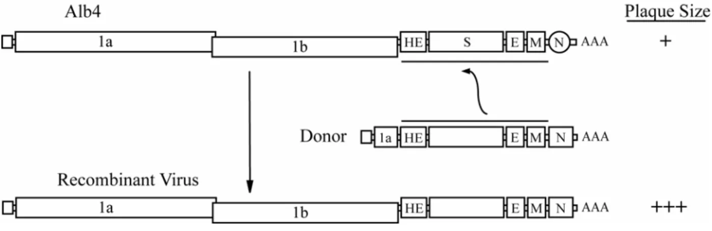
replication of recombinants over the parent genome. The earliest version of the system relied upon a temperature sensitive mutant genome recipient, Alb4, which contained a mutation in the nucleocapsid gene (N) that allowed normal replication at permissive temperatures (<34oC) but becomes severely attenuated at higher, or nonpermissive, temperatures (~39oC) (Fig. 5). Successful recombination transferred the donor RNA lacking the thermolabile mutation to the host genome and the resulting recombinants were capable of efficient growth and plaqued efficiently at the nonpermissive temperature. Recombinant viruses were
selected on the basis of plaque size when grown at the nonpermissive temperature. Several improvements have been made to the system since its original conception over a decade ago. The original iteration of the targeted RNA recombination system was limited to the production of robust viruses, since selection was made based on the
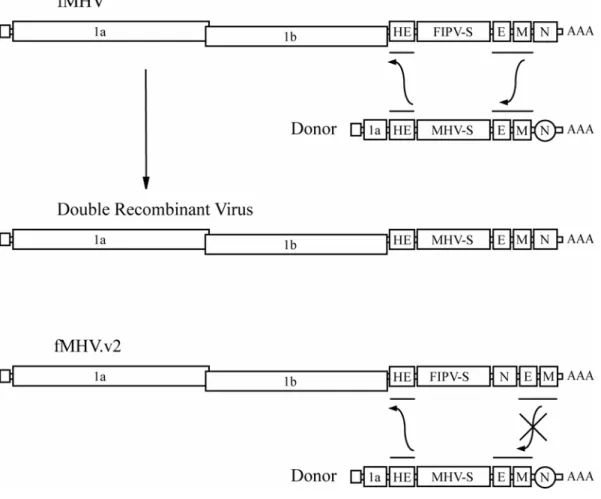
comparison of growth fitness at the nonpermissive temperature. In the case of genetic manipulations resulting in viruses attenuated at the nonpermissive temperature, whether due to their own temperature sensitivity or simply to a loss of growth fitness in general, selection was lost. The target size of the portion of the genome amenable to mutagenesis was mostly limited to the nucleocapsid gene located at the 3’ end of the genome. These limitations were overcome by taking advantage of the narrow host ranges of certain coronaviruses. A
previous data that argued that the S glycoprotein was a principle determinant of coronavirus host range. More importantly, fMHV was unable to infect cells derived from mice which are permissive to MHV. However, following successful recombination with a donor construct containing MHV S, the recombinant virus simultaneously lost the ability to infect feline cells and gained the ability to infect murine cells. This system provided a powerful positive selection step (i.e., growth on murine cells) which readily allowed for the isolation of
attenuated mutant viruses. Later improvements to the targeted recombinant system include a mechanism for limiting multiple recombination events, especially at the 3’ end of the
genome. Although successful recombination required that a functional copy of the MHV S gene be selected, there was the possibility of further recombination events occurring (Fig. 7). These are particularly likely when an introduced mutation at the 3’ end of the genome is debilitating. The likelihood of multiple recombination events occurring was reduced by moving the N gene of the recipient virus immediately downstream of the S gene. In the event of a second recombination event downstream of S, the resulting virus would lack the essential N gene and would be unviable. The targeted recombination system has been extended to MHV-JHM (144), TGEV, and FIPV and remains a powerful technology for altering genes and sequence motifs at the 3’-most 10 Kb of the genome (74).
Infectious cDNA clones
BAC System
The final missing fragment of the TGEV genome, which caused instability in standard bacterial vectors, was added in the last step and the full-length cDNA was transferred to a bacterial artificial chromosome (BAC), which attains a high degree of stability by
maintaining a very low plasmid copy number, no more than two copies per cell (2). The full-length genomic cDNA is transfected directly into cells and RNA is transcribed from within the nucleus of a cell from a CMV promoter located upstream of the viral sequence. Notably, the majority of viral RNA does not undergo deleterious mRNA splicing and is transferred from the nucleus of transfected cells into the cytoplasm where the virus replicates normally. After detecting that there was some level of instability in the TGEV BAC system with passage in E. coli, the system was further stabilized by inserting an intron into the region of
the ORF1 gene responsible for the toxicity in bacteria. Expression of full-length infectious cDNA clones of the human coronaviruses OC43 (186) and SARS-CoV (1) have also been reported using this approach. The full-length cDNA copies of these two clones, however, were not constructed by rebuilding an incomplete DI genome but were pieced together using unique restriction sites either present within the genome or added with the introduction of silent mutations.
Vaccinia Virus Expression System
After reporting difficulty in maintaining cDNA stability in bacterial systems, including BAC, Thiel and associates described a technique by which full-length cDNA of the human
coronavirus 229E was amplified in a vaccinia virus cloning vector (196) (reviewed in (197)). The cDNA was systematically assembled and cloned into a vaccinia virus vector.
cells and recombinant vaccinia viruses were rescued by co-infection with fowlpox virus. Recombinant vaccinia virus DNA was purified and used as template for the in vitro transcription of genomic RNA, which is then electroporated into cells. Avian infectious bronchitis virus (IBV) (24) was derived from a full-length cDNA by an alternative strategy using vaccinia virus vectors. Following transfection of the vaccinia virus DNA containing the IBV cDNA, cells were infected with fowlpox virus expressing TR RNA polymerase. A T7 promoter was engineered before the IBV cDNA, and viral RNA was transcribed within the cell initiating viral replication.
One of the novel aspects of propagating coronavirus cDNA within the vaccinia virus is the ability to mutate the clone by taking advantage of homologous recombination. There are two recombination steps in the procedure. A donor plasmid DNA containing the desired mutation and containing a dominant selectable marker gene recombines with the recipient cDNA by homologous recombination. Recombinant viruses containing the dominant marker are selected. Next, the selected virus is allowed to grow in the absence of selection. This results in two recombination events: the cDNA reverts to its original sequence or the selectable marker gene is excised and the clone successfully integrates the modification. Isolates containing the mutation are screened by PCR (17). A similar strategy was recently employed for creating and modifying an MHV-A59 infectious clone (36).
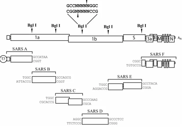
generated after the genome fragments are excised from the plasmid vectors and ligated. Synthetic RNA is produced in vitro through the use of a polymerase promoter at the 5’ end of the construct and electroporated into cells. This strategy was first applied to the development of a six-component TGEV infectious clone (229). Subclones were flanked by BglI or BstXI, restriction enzymes that recognize symmetric palindromes but cleave within asymmetric sites resulting in a 3- or 4- nucleotide complementary overhang. Use of these enzymes allows for a high degree of variability among different BglI digests, unique complementation, and high-specificity between contiguous fragments.
Figure 1. Genome organization of MHV-A59 and SARS-CoV. The 5’ two-thirds of the genome encode the polycistronic replicase protein in ORF1. Two polyproteins are expressed from ORF1, pp1a and 1ab. Pp1ab is a c-terminal extension of pp1a which is dependent upon a -1 ribosomal frame-shift event at a conserved pseudoknot structure. The primary structural genes, Spike glycoprotein (S), Envelope associated protein (E), Membrane protein (M), and Nucleocapsid (N), as well as a series of virus specific accessory proteins are encoded within the 3’ one-third of the genome from ORFs 2-7 for MHV-A59 and ORFs 2-9 in SARS-CoV.
Figure 2. ORF1ab polyprotein: Proteolytic processing and conserved elements.
Cleavage is mediated by three proteases in MHV or by two in SARS-CoV. The box color for each proteolytic domain and triangles at each nsp interface are color coordinated to the protease which cleaves at each position (red for PLpro1, blue for PLpro2, and white for Mpro).
MHV and SARS-CoV conserved domains are shown, including the accessory protease (PLP), ADP-ribose 1’-phosphatase (X), transmembrane domains (TM, MP1 and MP2), main
Figure 4. Generation of subgenomic RNA by discontinuous attenuated transcription. (1) The replication complex begins to transcribe the negative strand from the genomic RNA template. (2) The polymerase has a chance of disassociating from the template upon
encountering transcriptional regulatory sequences (TRS) located immediately upstream of each ORF and reannealing with the genomic template at the 5’-most TRS. (3) After
Figure 5. Initial version of targeted recombination reverse genetics system for coronaviruses. Alb4 produced a limited number of small plaques at the nonpermissive temperature. Following transfection of subgenomic mRNA 7 and infection of Alb4, RNA recombinants would be generated that resulted in wildtype plaque phenotypes, evidenced by large plaques that could be easily distinguished from Alb4.
Figure 8. Multi-component reverse genetics system for SARS-CoV. cDNA stability in E. coli DNA amplification vectors is maintained by separating the viral genome over several
CHAPTER II
Vaccine Efficacy in Senescent Mice Challenged with Recombinant SARS-CoV Bearing Epidemic and Zoonotic Spike Variants
Abstract
In 2003, severe acute respiratory syndrome coronavirus (SARS-CoV) was identified
as the etiological agent of severe acute respiratory syndrome, a disease characterized by
severe pneumonia that sometimes results in death. SARS-CoV is a zoonotic virus that
crossed the species barrier, most likely originating from bats or from other species including
civets, raccoon dogs, domestic cats, swine, and rodents. A SARS-CoV vaccine should confer
long-term protection, especially in vulnerable senescent populations, against both the 2003
epidemic strains and zoonotic strains that may yet emerge from animal reservoirs. We report
the comprehensive investigation of SARS vaccine efficacy in young and senescent mice
following homologous and heterologous challenge. Using Venezuelan equine encephalitis
virus replicon particles (VRP) expressing the 2003 epidemic Urbani SARS-CoV strain spike
(S) glycoprotein (VRP-S) or the nucleocapsid (N) protein from the same strain (VRP-N), we
demonstrate that VRP-S, but not VRP-N vaccines provide complete short- and long-term
protection against homologous strain challenge in young and senescent mice. To test VRP
vaccine efficacy against a heterologous SARS-CoV, we used phylogenetic analyses,
synthetic biology, and reverse genetics to construct a chimeric virus (icGDO3-S) encoding a
clusters among the zoonotic SARS-CoV. icGD03-S replicated efficiently in human airway
epithelial cells and in the lungs of young and senescent mice, and was highly resistant to
neutralization with antisera directed against the Urbani strain. This work tests the hypothesis
that vaccination of young and old mice with VRP-S and VRP-N provide protection from
replication by epidemic and heterologous strains of SARS-CoV. Although VRP-S vaccines
provided complete short-term protection against heterologous icGD03-S challenge in young
mice, only limited protection was seen in vaccinated senescent animals. VRP-N vaccines not
only failed to protect from homologous or heterologous challenge, but resulted in enhanced
immunopathology with eosinophilic infiltrates within the lungs of SARS-CoV–challenged
mice. VRP-N–induced pathology presented at day 4, peaked around day 7, and persisted
through day 14, and was likely mediated by cellular immune responses. This study identifies
gaps and challenges in vaccine design for controlling future SARS-CoV zoonosis, especially
in vulnerable elderly populations. The availability of a SARS-CoV virus bearing
heterologous S glycoproteins provides a robust challenge inoculum for evaluating vaccine
efficacy against zoonotic strains, the most likely source of future outbreaks.
Introduction
bats(113, 118, 149) or from other species including civets, raccoon dogs, domestic cats, swine, and rodents(72). New zoonotic variants may emerge as evidenced by sporadic cases of human disease in late 2003 and early 2004, which arose from strains distinct from that of the epidemic (195). In 2004, several laboratory-acquired infections were reported, including secondary spread resulting in fatal disease (141). Given the significant health and economic impact, the development of an effective vaccine strategy that is protective against both epidemic and zoonotic SARS-CoV strains is highly desirable.
Attenuated and killed SARS-CoV, DNA, and viral vectored vaccines are being evaluated in a number of animal models including mouse, ferret, hamster, and primate (9, 20, 21, 81, 90, 99, 170, 181, 183, 192, 215, 223, 232, 238), and have demonstrated that the SARS-CoV spike (S) glycoprotein is the principal component of protective immunity (20, 155, 161). Although strong immune responses are elicited against both S glycoprotein and nucleocapsid (N) protein (20, 154, 215, 241), passive transfer studies have illustrated that only anti-S antibody confers protection from SARS-CoV replication in the mouse model (9, 99, 188). Vaccine development faces a series of potential concerns including reversion or recombination repair of attenuated vaccine strains, induction of immune-mediated
enhancement of pathology, waning immune protection, lack of cross-protection for heterologous strains, and limited vaccine efficacy within senescent populations.
model, raising the specter of vaccine-mediated immune enhancement of disease following heterotypic challenge (224). Another potential problem is that SARS vaccines might fail to induce antibodies that protect from infection with divergent strains of SARS-CoV. The S glycoprotein of SARS-CoV contains about 2%–20% amino acid variation between zoonotic and the 2003 epidemic strains (195, 224), possibly limiting the effectiveness of monotypic SARS-S vaccines. Finally, studies measuring the duration of protective immunity or vaccine efficacy in animals greater than 4 mo post-boost have not yet been reported (99).
In this report, the efficacy of Venezuelan equine encephalitis virus replicon particle (VRP) vaccines expressing the Urbani SARS-CoV S glycoprotein (VRP-S) and N protein (VRP-N), either individually or in combination (VRP-S+N), are determined in young and senescent mouse models. We tested whether the senescent mouse model, which exhibits an age-related susceptibility to SARS-CoV similar to that seen in the human disease (158), will provide a sensitive measure of vaccine efficacy and reveal potential complications in SARS-CoV vaccine development for vulnerable elderly populations. We evaluated the duration of protective immunity following homologous and heterologous SARS virus challenge,
examining the impact of waning immunity on long-term protection. Through the use of publicly available SARS-CoV sequence databases, bioinformatics approaches, synthetic biology, and reverse genetics, we constructed a viable heterologous challenge virus to test the ability of current vaccine regimens to protect against zoonotic strains; the likely source of future epidemics (195).
Viruses and Cells. The Urbani, Tor-2, recombinant Urbani (icSARS), and a recombinant
chimeric virus encoding the S gene of GDO3 SARS-CoV (icGD03-S ), strains were propagated on VeroE6 cells in Eagle’s MEM supplemented with 10% fetal calf serum, kanamycin (0.25 μg/ml), and gentamycin (0.05 μg/ml) at 37 °C in a humidified CO2
incubator. For virus growth, cultures of VeroE6 cells were infected at a multiplicity of infection (MOI) of 1 for 1 h, the monolayer washed twice with 2 ml of PBS and overlaid with complete MEM. Virus samples were harvested at different times post-infection and titered by plaque assay. Plaques were visualized by neutral red staining and then counted.
Human nasal and tracheobronchial epithelial cells were obtained from airway specimens rejected from patients undergoing elective surgery under University of North Carolina (UNC) Institutional Review Board–approved protocols by the UNC Cystic Fibrosis (CF) Center Tissue Culture Core. Briefly, primary cells were expanded on plastic to generate passage 1 cells and plated at a density of 250,000 cells per well on permeable Transwell-Col (T-Col, 12-mm diameter; Corning [http://www.corning.com]) supports. Human airway epithelium (HAE) cultures were generated by provision of an air–liquid interface for 4–6 wk to form well-differentiated, polarized cultures that resemble in vivo pseudo-stratified
mucosciliary epithelium, and infected with wild-type or recombinant SARS-CoV as
previously described by our laboratory (174). All virus work was performed in a biological safety cabinet (BSC cabinet) in a biosafety level three (BSL3) laboratory containing
Construction and Isolation of the icGDO3-CoV Variant Virus. The GD03-S glycoprotein
sequence has been reported. A synthetic DNA containing the 5′-most GD03 mutations was purchased (Blue Heron Biotechnology [http//www.blueheronbio.com]) and inserted into the SARS-E fragment. The plasmid clone (SARS-E GD03) was fully sequenced and shown to contain all of the appropriate mutations. The remaining GDO3 mutation was incorporated by PCR mutagenesis (5′ amplicon A: 5′-CTGTTTTCCCTGGGATCGC-3′; 3′ amplicon A: 5′ -NNNNNNCACCTGCTTTTGGGCAACTCCAATGCC-3′; 5′ amplicon B: 5′
-NNNNNNCACCTGCAGTTGCCCAAAATGTTCTCTATGAGAAC-3′; 3′ amplicon B: 5′- CATAAATTGGATCCATTGCTGG), followed by seamless ligation of the amplicons as previously described (230) into the SARS-F subclone. The final construct (SARS-F GD03) was fully sequenced and found to contain the appropriate set of GD03-S glycoprotein alleles.
The icGDO3-S was generated as previously described. Infectious clone fragment plasmid DNA was prepared in Escherichia coli (TOP-10, Invitrogen
[http://www.invitrogen.com]), isolated, and purified (Qiagen [http://www.qiagen.com]). Infectious clone fragments B, C, D, and E were digested with BglI. Infectious clone fragments A and F were digested with EcoRI and NotI, respectively. Infectious clone fragments A and F were then dephosphorylated and then digested with BglI. Individual cDNA fragments were gel purified (Qiagen) and ligated (Roche [http://www.roche.com]) to form a full-length genomic cDNA and then chloroform extracted and EtOH precipitated. N cDNA and full-length viral genomic cDNA were then used as templates for in vitro
harvested 48 h post-electroporation. Virus was plaque purified and then passaged twice in Vero cells. The resultant stock was plaque titered and cyropreserved at −80 °C.
Western Blot Analysis. Twelve hours post-infection, Urbani–, icSARS-CoV–, SARS-CoV–,
Tor-2–, and icGD03-S–infected cells were washed in 1X PBS, lysed in buffer containing 20 mM Tris-HCL (pH 7.6), 150 mM NaCl, 0.5% deoxycholine, 1% nonidet-p-40, 0.1% sodium dodecyl sulphate (SDS), and post-nuclear supernatants added to an equal volume of 5 mM EDTA/0.9% SDS, resulting in a final SDS concentration of 0.5%. Samples were then heat inactivated for 30 min at 90 °C in the BL3 prior to removal. At BL2, samples were again heat inactivated for 30 min at 90 °C before use. Equivalent sample volumes were loaded onto 4% to 20% Criterion gradient gels (BioRad [http://www.bio-rad.com]) and then transferred to PVDF membrane (BioRad). Blots were probed with polyclonal mouse antisera directed against the Urbani-S glycoprotein diluted 1:200 or human sera 1128 diluted 1:400 and developed using electrogenerated chemiluminescence (ECL) reagents (Amersham
Biosciences [http://www5.amershambiosciences.com]). Patient sera #1128 was collected from a patient infected during the second disease outbreak in Toronto, Canada.
Plaque Reduction Neutralization Titer Assays. One-hundred plaque forming units (pfu) of
and expressed as the relative percentage. The dilution at which 80% of plaques were neutralized was determined for each VRP-S– or VRP-S+N–vaccinated animal.
Mice. Female BALB/c mice (Charles River Laboratories [http://www.criver.com]) were
anesthetized with a ketamine (1.3 mg/mouse) and xylazine (0.38 mg/mouse) mixture administered intraperitoneally with a 50-μl volume. Each mouse was intranasally (i.n.) inoculated with 50 μl of virus at a concentration of 2 × 10⁶ pfu/ml of virus. Four days post-infection, the right lung was removed and frozen at −70 °C for later plaque assay
determination of viral titers. Half of the left lung was placed into Trizol Reagent (Invitrogen) for RNA extraction. The second half of the left lung was fixed in 4% PFA in PBS (pH 7.4) for at least 7 d prior to paraffin imbedding and sectioning for histopathological analysis. All mice were housed under sterile conditions, and sentinel mice were used to verify that the colony was mouse hepatitis virus (MHV) negative. Experimental protocols were reviewed and approved by the Institutional Animal Care and Use Committee at UNC Chapel Hill. Young mice refer to those challenged with SARS-CoV at ages equal or less than 5 mo old, whereas old or senescent mice are those animals with ages greater than 1y at the time of challenge.
Plaque Assay Titration of Virus from Lungs. Lungs were weighed and homogenized in four
dishes. Following a 1-h incubation at 37 °C, cells were overlaid with 1% agarose-containing medium. Two days later, plates were stained with neutral red and then plaques counted.
VRP-S and VRP-N. The VRP constructs were made in two rounds of PCR, the first to
generate two amplicons, and a second round of overlapping PCR to fuse them together. The fused DNA was digested with ApaI and AscI, and ligated into the similarly digested pVR21 plasmid. PCR reactions were performed with Expand Long Taq (Roche Molecular
Biochemicals http://www.roche-applied-science.com) in 30 cycles of 94 °C for 30 s, 55 °C for 30 s, and extensions at 68 °C for 1 min. The first amplicon, which was used in the construction of both VRP-S and VRP-N, was generated with primers 5′nsp4Sw (5′ -GATTGAGGCGGCTTTCGGCG) and 3′26S (5′
-TTAATTAAGTCAATCGGCGCGCCCTTGGCGGACTAGACTATGTC) using pVR21 as template. The N-gene–specific amplicon was produced using primers V5′SARNg (5′ -AGTCTAGTCCGCCAAGATGTCTGATAATGGACCCCAATC) and 3′SARSNg (5′ -NNNNTTAATTAATTATGCCTGAGTTGAATCAGC) with SARS-F plasmid for template. The S-gene–containing amplicon was made with V5′SARSg (5′
-AGTCTAGTCCGCCAAGATGTTTATTTTCTTATTATTTCTTACTCTCAC) and
Lung Histopathology. Lungs were fixed in 4% PFA in PBS for 7 d before being submitted to
the Histopathology Core Facility (UNC, Chapel Hill) for paraffin imbedding, sectioning at
5-μm thickness, and hematoxylin and eosin staining. Approximately one-quarter of the total lungs were sectioned, with four to six sections mounted from cuts taken at five different depths within the paraffin-imbedded tissue. Lung pathology was scored in a blinded manner, in which six to ten sections per animal were evaluated and scored using the following scale. 1.0 to 2.0 = no to mild inflammation, 2.0 to 3.0 = mild to moderate inflammation, 3.0 to 4.0 = moderate to severe inflammation in less than half of the tissue section, and 4.0to 5.0 = severe inflammation in more than half of the tissue section. The same sets of tissues were also evaluated qualitatively by a respiratory pathologist (author JH).
In Situ Hybridization. The 5 μm–thick paraffin-embedded sections were probed with 35S
UTP-labeled riboprobes complementary to the N gene of SARS-CoV (Urbani) or the HA gene of the A/PR8 strain of influenza as a negative control using previously described methods (83). In brief, following treatment to prevent nonspecific probe binding, the tissues were incubated overnight with either probe at 5 × 10⁴ cpm/μl in hybridization buffer at 42
°C. The slides were then washed, dehydrated, and coated with NBT emulsion (Kodak [http://www.kodak.com]), and incubated at −80 °C. for 1 wk prior to development. Positive signal, as determined by silver grain deposition, was then evaluated.
Enzyme-Linked Immunosorbent Assay. Antibody titers were determined by standard indirect
inactivated influenza A diluted in carbonate buffer containing 32 mM sodium carbonate, 68 mM sodium bicarbonate, pH 9.6 at 4 °C overnight. Mouse sera, diluted 1:100 in casein blocking buffer (Sigma [http://www.sigmaaldrich.com]), were added to wells in duplicate, and 2-fold serial dilutions were performed, followed by incubation for 2 h at 37 °C. Plates were then incubated for 1 h with goat anti-mouse IgG with alkaline phosphatase (AP) conjugate (Sigma), developed with p-nitrophenyl phosphate (pNPP; Sigma), and the optical
density (OD) at 405 nm was measured (Bio-Rad Model 680 microplate reader). Log10
half-maximum ELISA titers were calculated with Sigmaplot (Systat [http://www.systat.com]) for the dilution at which an absorbance of 2.1, half that of the maximum measurable absorbance, was achieved. Since very low amounts of antibody were being measured in the passive transfer experiment, log10 OD = 0.2 ELISA titers were calculated.
Passive Sera Transfer. Mice were inoculated with 106 IU of VRP-HA, VRP-S, or VRP-N at
7 wk of age, boosted 4 wk later, and terminally bled via cardiac puncture 3 wk post-boost. The sera of each group were pooled and 150 μl transferred by tail vein injection into mice at 7 or 43 wk of age. Mice receiving sera were bled and i.n. challenged with 105 pfu of
icSARS.
Statistical Analysis. Unless otherwise noted, two-tailed Mann-Whitney tests were used for
the limit of detection were assigned a value equal to the limit of detection for any analysis. The plus or minus (±) symbol is used to refer to standard deviation.
An amino acid multiple alignment was generated for the entire S gene of viral sequences representing early, middle, and late phases of the SARS epidemic in humans, as well as animal strains of SARS-CoV isolated from civets and raccoon dogs found in Chinese live animal markets or housed on farms in China that supplied the markets. The sequences were aligned using ClustalX 1.83 with default settings (33). A phylogenetic tree was generated using Bayesian inference as implemented in the program MrBayes v3.0b4 (87). Briefly, the alignment was exported in the nexus format, the amino acid substitution model was set to JTT (96) using the lset command, and Markov chain Monte Carlo simulation (87) was used to approximate the posterior probabilities of trees with sampling conducted on four chains over 500,000 generations (159). Trees were sampled every 100 generations, and the 5,001 trees collected were summarized with the sumt command set to a burnin of 1,000, which generated a consensus tree using the 50% majority rule (159). The burnin value was determined using the sump command with an arbitrary burnin of 250, which demonstrated that stationarity occurred prior to the 100,000th generation, indicating that a burnin of 1,000 was appropriate for the sumt command (159).
Results
Venezuelan Equine Encephalitis Virus Replicon Particles Expressing SARS-CoV S and N.
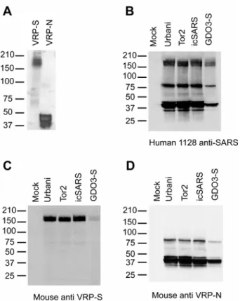
antigenically relevant recombinant proteins. VRP-infected cell lysates were probed with antiserum 1128, derived from a convalescent human SARS patient. Western blot analysis of VRP-S–infected lysates revealed the expected S glycoprotein doublet of approximately 180– 210 kDa, whereas that of VRP-N–infected lysates revealed a major product of less than 50 kDa, the expected sizes for SARS-S and -N, respectively (Fig. 9A). VRP-S was inoculated into BALB/c mice and tested for its ability to induce antigen-specific antibody. Western blots were performed with Vero cell lysates infected with Urbani, SARS-CoV Tor-2, icSARS-CoV (the Urbani recombinant virus), and icGD03-S, a chimeric SARS-CoV expressing the S glycoprotein of the heterologous GDO3 strain. Blots were probed with anti–VRP-S mouse serum, 1128 human convalescent serum, or with anti–VRP-N mouse serum. The Western blots demonstrated that probing with human serum resolved bands corresponding to the major SARS antigens S (a doublet at ~180–210 kDa) and N (triplet at <50 kDa), as well as other unidentified SARS-CoV proteins (Fig. 9B). Serum from mice vaccinated with VRP-S only identified SARS-S (Fig. 9C), whereas serum from mice vaccinated with VRP-N recognized SARS-N in addition to another SARS-CoV protein that is probably a dimer of N (Fig. 9D).
VRP Vaccine Efficacy against icSARS-CoV Replication in the Mouse Model. As a general
measure of vaccine efficacy (Table 1, experiment 1), six 4-wk-old BALB/c mice were vaccinated with either 105 IU of VRP-S or VRP-HA, boosted 4 wk later with an equal amount of VRP, and then i.n. challenged with 105 pfu of icSARS-CoV 8 wk post-boost.
replication of icSARS-CoV following challenge. No virus was detected by plaque assay (250 pfu/g limit of detection) in the lungs of VRP-S–vaccinated animals at 2 d post-infection, whereas the VRP-HA–vaccinated mouse lung had a mean titer of 6.7 ± 0.5 log10 pfu/g (Fig.
10A). Vaccination with VRP-S demonstrated significant protection at the time of peak lung titer relative to VRP-HA–vaccinated control animals (p = 0.007 Fisher exact test).
A second vaccine experiment was completed to evaluate long-term VRP protection. Five-week-old BALB/c mice were vaccinated with 105 IU of VRP-HA, VRP-S, VRP-N, or a combination of VRP-S and VRP-N (VRP-S+N), and boosted 5 wk later. Fifty-four weeks post-boost, mice were i.n. challenged with 105 pfu of icSARS-CoV and lungs removed 4 d post-infection (summarized in Table 1, experiment 2). Although day 2 post-challenge demonstrates peak viral titers, day 4 was chosen to harvest lungs because it is the time at which the highest level of pathology is evident in senescent mice (158). Titers in the lungs (Fig. 10B) of animals vaccinated with VRP-S or the combination of VRP-S+N were below the limit of detection (250 pfu/g). In contrast, the lung titers of VRP-HA–vaccinated animals were 5.8 ± 0.6 log10 pfu/g, comparable to the VRP-N–vaccinated animal titers of 5.3 ± 0.6
log10 pfu/g (p = 0.2). These plaque assay results were confirmed by SARS-CoV–specific in
situ hybridization on lung tissues from the infected mice (Fig. 10C). Radiolabeled
combination of VRP-S+N provided complete long-term protection against challenge with the homologous vaccine strain of SARS-CoV at 4 d post-infection (p < 0.001 Fisher exact test
for both VRP-S- and VRP-S+N–vaccinated groups relative to VRP-HA).
Protection against Heterologous Challenge. To perform cross-protection efficacy studies, it
was necessary to construct a heterologous SARS-CoV. Selection of a likely candidate strain was made after Bayesian analysis of the SARS-CoV S glycoprotein, which demonstrated three main phylogenetic branches. Two of the branches include viruses isolated from animals, such as the palm civet and raccoon dog, and low pathogenic viruses sporadically isolated from humans, such as GDO3 and GZ0401. Viruses representing the 2003 early, middle, and late phases of the epidemic strains form the third branch in the SARS-S
phylogenetic tree (Fig. 11A). We resurrected the S glycoprotein of GDO3, a virus reported from a sporadic SARS case on December 22, 2003. Although GDO3 was not successfully isolated, its S glycoprotein was sequenced and reported. Compared to epidemic strains, GDO3 likely represented an independent introduction, was reported to be less pathogenic, and its S glycoprotein sequence is among the most divergent of all human strains (195). The GDO3-S glycoprotein contains 17 amino acid changes relative to Urbani-S (Fig. 11B), many of which map within neutralizing epitopes between amino acids 130–150 and 318–510, part of the receptor binding domain (RBD) (31, 34, 50, 71, 82, 101, 190, 200, 214). Importantly, polyclonal antibody directed against the late-phase Urbani strain was less effective at
recombinant virus (230). Sequence analysis of plaque-purified icGDO3-S recombinant virus confirmed the presence of the GDO3-S glycoprotein and two additional changes in the S gene relative to Urbani-S (F7L and D613G), which likely arose as tissue-culture adaptations. The chimeric icGDO3-S, which only differs from Urbani SARS-CoV in its S glycoprotein, and wild-type icSARS-CoV recombinant viruses replicated in Vero cells to comparable titers that approached 107 PFU/ml within 24 h (unpublished data) and their proteins were both detected in Western blots with human antiserum from convalescent patients (Fig. 9B). Given the reduced amount of N present in the lysate of icGDO3-S–infected cells, the reduced intensity of the GDO3-S band probed with either anti–VRP-S mouse sera or the convalescent human serum is most likely due to the presence of lower GDO3-S protein rather than a marked difference in antibody specificity between GDO3-S and Urbani-S. icGD03-S replicated efficiently in HAE cells, although its maximum titer was approximately 1 log lower than that of icSARS or Urbani (Fig. 11C). To compare the growth of icSARS and icGDO3-S in animals, 6-wk-old BALB/c mice were i.n. infected with 105 pfu of either icSARS or icGDO3-S. At 2 d post-infection, icSARS-CoV mean lung titer was 6.8 ± 0.5 log10 pfu/g, whereas icGDO3-S titers were lower at 6.3 ± 0.2 log10 pfu/g (p = 0.04). The
mean lung titer of icSARS-CoV–infected mice on day 4 was 4.5 ± 0.5 log10 pfu/g compared
to icGDO3-S titers of 3.7 ± 0.3 (p = 0.04). By the seventh day, virus replication in the lungs
of three of five mice infected with icSARS-CoV and four of five mice infected with
icGDO3-S fell below the limit of detection (50 pfu/g). Average icSARS-CoV and icGDO3-S titers were similar on day 7 with 1.7 ± 0.1 log10 pfu/g and 1.8 ± 0.1 log10 pfu/g (p = 0.9),
To evaluate VRP protection against short-term heterologous challenge, groups of eight animals were primed at 7 wk of age with 106 IU of VRP-S, VRP-N, VRP-S+N, or VRP-HA, boosted 3 wk later, and then challenged 7 wk post-boost with 105 pfu of icGD03-S (summarized in Table 1, experiment 3). Lungs were harvested 2 d after challenge. VRP-S and VRP-S+N protected (p < 0.001 Fisher exact test for both VRP-S and VRP-S+N groups
relative to VRP-HA) against heterologous icGD03-S recombinant virus replication (Fig. 12A). Although high titers of virus were detected in VRP-N– and mock-vaccinated animals with mean titers of 6.3 ± 0.1 and 7.0 ± 0.1 log10 pfu/g, respectively, the VRP-N–vaccinated
animals had a lower mean titer (p < 0.001).
SARS-CoV vaccines should confer protection to elderly subjects who face infection with a new variant of the virus. To model this scenario, we vaccinated 6-mo-old to 1-y-old BALB/c retired breeders with 106 IU of VRP-S, VRP-N, VRP-S+N, or PBS, boosted them 4 wk later, and then challenged them 32 wk post-boost with 105 pfu of icGDO3-S (summarized in Table 1, experiment 4). At 4 d post-infection, mean titers in the lungs of animals
vaccinated with VRP-N and PBS were similar at 4.4 ± 0.5 and 4.7 ± 0.6 log10 pfu/g (p = 0.2),
respectively (Fig. 12B). VRP-S vaccination provided partial protection when compared to the PBS control group (p = 0.026 Fisher exact test), with the lungs of three of eight animals
positive for viral replication at a mean titer of 2.9 ± 1.8 log10 pfu/g. All eight of the lungs
harvested from the VRP-S+N–vaccinated animals were positive for viral replication, although a mean titer of 3.5 ± 1.2 log10 pfu/g was comparable to the mean titer for VRP-S–
vaccinated mice (p = 0.4) and reduced relative to the PBS control (p = 0.02). The presence
sections of mice from PBS-, VRP-N–, VRP-S–, and VRP-S+N–vaccinated groups (Fig. 12C). All tested lung sections from PBS mocks (unpublished data) and VRP-N–vaccinated (Fig. 12C, image a) animals exhibited in situ signal (arrows), although the signal did not appear to be as intense as that of the icSARS-CoV–infected animals (Fig. 10C). Lungs of the VRP-S–vaccinated animals (Fig. 12C, image b) had two of five slides exhibiting SARS-CoV–specific signal above background levels, whereas VRP-S+N (Fig. 12C, image c) had three of four slides.
Senescence and VRP-S Immune Responses. Because neutralizing antibody has been reported
to confer protection from SARS-CoV replication within the lungs of mice (9, 188, 223), it was of interest to determine whether the VRP-S vaccine established high neutralizing
antibody levels that persisted until challenge. Plaque reduction neutralization titer (PRNT) of serum samples harvested prior to vaccination showed no neutralization of icSARS-CoV; similar results were noted from serum collected from mice vaccinated with the negative controls, VRP-HA or PBS (unpublished data). The 80% PRNT values (PRNT80), the dilution
of serum at which plaque numbers are reduced by 80% relative to virus treated with control sera, for VRP-S– and VRP-S+N–vaccinated animals at 5 and 53 wk post-boost against both icSARS-CoV and icGDO3-S were compared (Fig. 13A and Table S1). The mean reciprocal dilutions for the PRNT80 of VRP-S and VRP-S+N against icSARS were measured at 796 ±
307 at 5 wk post-boost and 628 ± 363 at 53 wk. Sera from mice vaccinated with the combination of VRP-S+N had mean reciprocal PRNT80 of 1,091 ± 361 and 370 ± 179 at 5
significant waning of the icSARS-neutralizing activity over the 48-wk period in the VRP-S– vaccinated animals (p = 0.3 Wilcoxin matched pairs signed-rank test), VRP-S+N serum was
diminished by about 3-fold (p = 0.03 Wilcoxin matched pairs signed-rank test). All tested
sera remained above the lower limit of detection (1:100) and were sufficient to prevent icSARS replication within the lungs of challenged animals (Fig. 10B). The neutralizing activity of sera from the vaccinated animals was more effective against the vaccine strain than against heterologous icGDO3-S virus for both sera harvests and vaccine combinations. The reciprocal dilutions for the PRNT80 of the VRP-S samples at 5 wk post-boost were
below the limit of detection, whereas two samples of the week 53 bleed were measured above the limit of detection with a mean value of 112 ± 28. The icGDO3-S PRNT80
measurements for VRP-S+N at weeks 5 and 53 post-boost were below the limit of detection with one exception for each time point: one mouse was measured to have a PRNT80 of 124 at
5 wk, for an average titer of 103 ± 8, and another a PRNT80 of 107 at 53 wk post-boost, an
average of 101 ± 3.
Given that the VRP-S vaccine’s ability to provide long-term protection was likely due to the strong SARS-CoV–neutralizing response induced in vaccinated mice, we measured the PRNT80 of the VRP-S immune sera from mice vaccinated when old (Table 1, experiment 4)
to determine if the incomplete protection seen in that study could be linked to a reduced neutralizing antibody response in the senescent animals (Fig. 13B and Table S1). Against the vaccine strain, the reciprocal dilutions of the mean PRNT80 were 170 ± 82 with two of six
measurable sample at 114, for an average of 103 ± 6 and no icSARS-neutralizing ability detected at week 29. Against icGDO3-S, the VRP-S PRNT80 values were below the limit of
detection with the exception of a single VRP-S–vaccinated animal showing a neutralizing titer of 179 at 12 wk post-boost, for an average dilution of 116 ± 35 for the group. Sera harvested from these animals exhibited a marked reduction in neutralizing ability when compared to the response in animals vaccinated when young, even against the vaccine strain (p = 0.008 for VRP-S week 5 versus VPP-S week 12 post-boost; p = 0.006 VRP-S+N week 5
versus VRP-S+N week 12 post-boost). A strong anti–SARS-CoV neutralizing response was not induced by the VRP vaccines when administered to senescent mice.
ELISAs for total IgG specific for SARS-S and influenza-HA were performed to compare the VRP vaccines’ ability to induce antibody to those antigens in mice vaccinated when young or senescent. ELISA for SARS-S was performed on sera collected prechallenge from the VRP-S– and VRP-S+N–vaccinated mice of experiments 2 and 4 (Fig. 14A). Mice vaccinated young with VRP-S (experiment 2) had an average log10 half-maximum ELISA
titer of 2.6 ± 0.6 at 53 wk post-boost, whereas that of the senescent animals was
approximately a log lower at 1.5 ± 0.9 at 29 weeks post-boost (p = 0.007). The difference
between animals of the two age groups was even more striking when anti-S IgG levels were compared in the VRP-S+N mice. The animals vaccinated young with VRP-S+N had an average titer of 2.6 ± 0.3, whereas the average for senescent animals was at the limit of detection of 0.02 log10 half-maximum ELISA titer. To verify that the reduced ability of the