CHARACTERIZATION OF A PHASE VARIABLE REGULATORY SYSTEM AND ITS IMPACTS ON CLOSTRIDIOIDES DIFFICILE PHYSIOLOGY
Elizabeth Mathias Garrett
A dissertation submitted to the faculty at the University of North Carolina at Chapel Hill in partial fulfillment of the requirements for the degree of Doctor of Philosophy in the
Department of Microbiology and Immunology.
Chapel Hill 2020
© 2020
ABSTRACT
Elizabeth Mathias Garrett: Characterization of a phase variable regulatory system and its impacts on Clostridioides difficile physiology (Under the direction of Rita Tamayo)
Clostridioides difficile is a Gram-positive, spore-forming, obligate anaerobic bacterium that causes gastrointestinal disease and is the leading cause of nosocomial infections in the U.S. Despite its significant public health burden, key aspects of
pathogenesis including the regulatory strategies that benefit C. difficile fitness during infection are not well understood. In this work, we identify and characterize a two-component regulatory system with significant impacts on C. difficile physiology as well as elucidate the complex regulatory networks that govern its expression. We find that expression of cmrRST, a highly conserved operon that encodes two response
regulators (CmrR and CmrT) and a histidine kinase (CmrS), is subject to phase
motility. Additionally, we provide evidence that CmrRST has a role in virulence and that phase variation occurs in vivo. In addition to phase variable regulation by the cmr
ACKNOWLEDGMENTS
First, I must thank my advisor, Rita Tamayo. Rita is an amazing mentor, and I benefited greatly from her advice and her guidance throughout my time in her lab. She is the type of advisor that every grad student wishes to have. I have always enjoyed our scientific discussions and I admire her ability to turn everything including negative data into a positive step forward. Her mentorship has been invaluable, and I will always be grateful for her part in my development as a person and as a scientist.
I am also grateful to all my lab mates through the years. I appreciate their discussion, ideas, and feedback. I have been lucky to have such friendly and
collaborative colleagues. Brandon Anjuwon-Foster and Robert McKee served as great examples to look up to as I started in the lab. I appreciate all the undergraduate
students that helped me through the years. I am also grateful to Ognjen Sekulovic for his collaboration, which made so much of this project possible.
Finally, I want to thank my friends and family. I am grateful to my friends in Chapel Hill for commiserating and also celebrating the peaks and valleys of the
TABLE OF CONTENTS
LIST OF FIGURES... ix
LIST OF TABLES ... xi
LIST OF ABBREVIATIONS ... xii
CHAPTER 1. INTRODUCTION ... 1
CLOSTRIDIOIDES DIFFICILE EPIDEMIOLOGY, RISK FACTORS, AND TREATMENT ... 1
C. DIFFICILE TOXINS ... 6
MOTILITY AND SURFACE BEHAVIORS OF C. DIFFICILE ... 9
Flagella ... 9
Type IV pili ... 11
Biofilm formation ... 11
PHENOTYPIC HETEROGENEITY AND PHASE VARIATION ... 13
Slipped strand mispairing ... 15
Methylation ... 16
Site-specific recombination ... 17
Phase variation in C. difficile ... 20
C-DI-GMP SIGNALING ... 24
C-di-GMP signaling in C. difficile ... 30
CHAPTER 2: PHASE VARIATION OF A SIGNAL TRANSDUCTION SYSTEM CONTROLS CLOSTRIDIOIDES DIFFICILE COLONY
MORPHOLOGY, MOTILITY, AND VIRULENCE ... 35
INTRODUCTION ... 35
MATERIALS AND METHODS ... 38
RESULTS ... 48
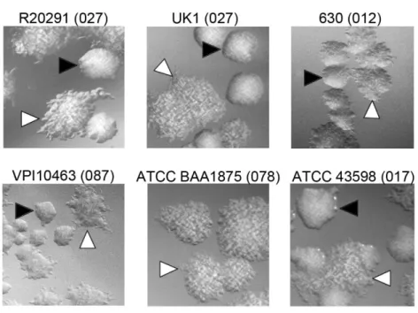
C. difficile strains of diverse ribotypes develop two distinct, phase- variable colony morphotypes ... 48
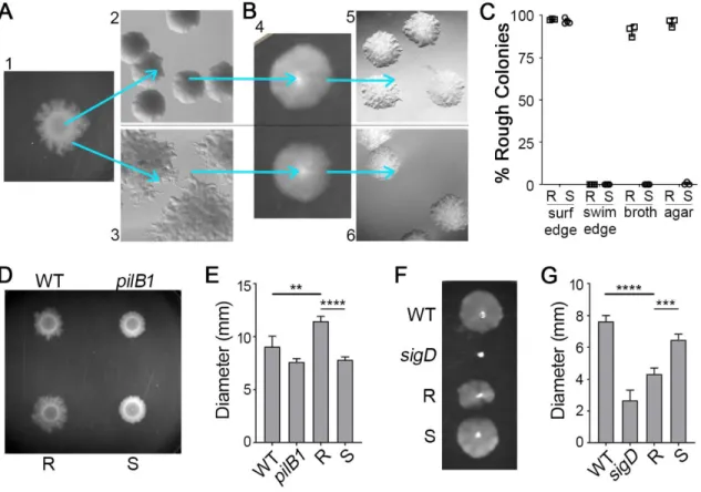
Rough and smooth colony isolates exhibit distinct motility behaviors ... 49
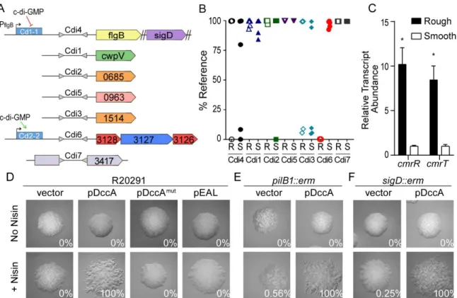
Colony morphology is regulated by a phase variable signal transduction system and c-di-GMP ... 50
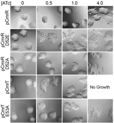
The response regulators CmrR and CmrT regulate colony morphology ... 53
CmrR and CmrT promote bacterial chaining ... 56
Phase variable colony morphology via CmrRST impacts C. difficile virulence ... 58
DISCUSSION ... 61
FIGURES AND TABLES ... 68
CHAPTER 3. MULTIPLE REGULATORY INPUTS CONTROL THE EXPRESSION OF cmrRST ENCODING AN ATYPICAL SIGNAL TRANSDUCTION SYSTEM IN CLOSTRIDIOIDES DIFFICILE ... 93
INTRODUCTION ... 93
MATERIALS AND METHODS ... 97
RESULTS ... 104
Generation and phenotypic characterization of phase locked cmr ON and cmr OFF strains ... 104
C-di-GMP regulates cmrRST expression independently of the invertible element ... 106
CmrR positively autoregulates expression of cmrRST ... 109
cmrRST and its upstream regulatory elements are well conserved in C. difficile ... 110
DISCUSSION ... 111
FIGURES AND TABLES ... 116
CHAPTER 4. TRANSCRIPTOME ANALYSIS OF THE PHASE VARIABLE SIGNAL TRANSDUCTION SYSTEM CmrRST IN CLOSTRIDIOIDES DIFFICILE ... 134
INTRODUCTION ... 134
MATERIALS AND METHODS ... 137
RESULTS ... 141
CmrR and CmrT regulate many genes of diverse functions ... 141
Cross-talk between phase variable genes ... 143
Regulation of swimming motility and colony morphology by CDR20291_1689-1690 ... 144
Contribution of CmrR and CmrT to regulation ... 146
DISCUSSION ... 147
FIGURES AND TABLES ... 151
CHAPTER 5. DISCUSSION ... 165
GENE REGULATION BY CmrR AND CmrT ... 166
SIGNALS FOR THE REGULATION OF cmrRST EXPRESSION ... 167
REGULATION OF MOTILITY, CELL MORPHOLOGY, AND COLONY MORPHOLOGY BY CmrRST ... 172
IMPLICATIONS OF CmrRST PHASE VARIATION FOR PATHOGENESIS ... 175
LIST OF FIGURES
Figure 2.1. Formation of rough and smooth colonies by multiple
C. difficile strains. ... 68
Figure 2.2. Reversible selection of distinct colony morphotypes with opposing motility phenotypes. ... 69
Figure 2.3. Expression of a c-di-GMP regulated phosphorelay system promotes rough colony formation in a TFP- and flagellum-independent manner. ... 71
Figure 2.4. The putative response regulators CmrR and CmrT promote the development of rough colonies. ... 73
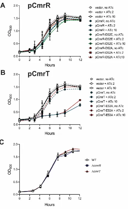
Figure 2.5. Growth of mutant and over-expression strains in vitro. ... 74
Figure 2.6. CmrT is required for rough colony formation. ... 75
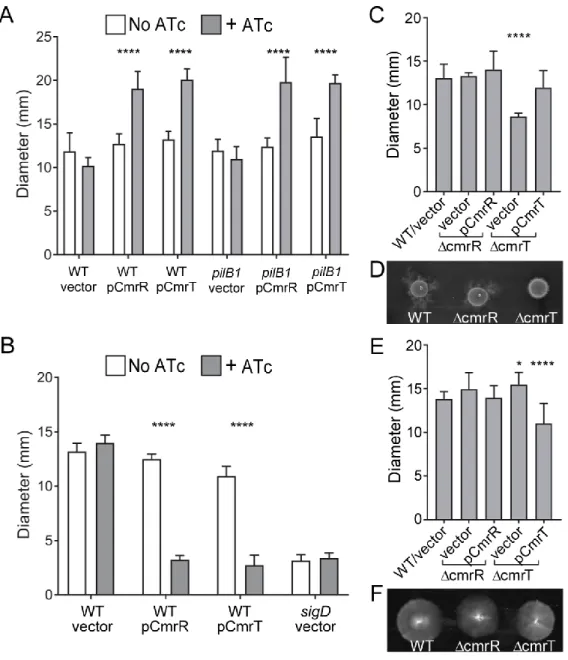
Figure 2.7. CmrR and CmrT inversely regulate surface and swimming motility. ... 76
Figure 2.8. Amino acid substitutions of the phosphorylation sites in CmrR and CmrT alter activity. ... 78
Figure 2.9. CmrR and CmrT do not regulate flagellum or TFP gene expression. ... 80
Figure 2.10. CmrR and CmrT promote bacterial chaining. ... 81
Figure 2.11. The cmrT mutant is defective in cell elongation and chaining. ... 83
Figure 2.12. The cmrR mutant exhibits an increase in biofilm formation. ... 84
Figure 2.13. cmrR and cmrT mutants are not defective in sporulation, germination or toxin production. ... 85
Figure 2.14. Colony morphology and CmrR impact C. difficile virulence. ... 87
Figure 3.1. Generation of genetically phase-locked strains. ... 116
Figure 3.2. Phase-locked cmr ON strain phenocopies the WT rough morphotype. ... 118
Figure 3.3. Regulation of cmrRST by c-di-GMP occurs independent of cmr switch orientation. ... 120
Figure 3.5. The cmr switch is phase locked in recV-deficient strains. ... 123 Figure 3.6. The cmr switch contains a promoter in the ON orientation ... 124 Figure 3.7. Identification of multiple transcriptional start sites upstream
of cmrRST. ... 125 Figure 3.8. CmrR positively regulates cmrRST expression ... 126 Figure 3.9. cmrRST and regulatory element sequences are highly
conserved across C. difficile strains and ribotypes. ... 129 Figure 4.1. Orientation of invertible DNA elements varies between strain
populations. ... 151 Figure 4.2. CmrR and CmrT regulate CDR20291_1689-1690. ... 152 Figure 4.3. CDR20291_1689-1690 regulates colony morphology. ... 153 Figure 4.4. CDR20291_1689-1690 inhibits swimming motility but not
LIST OF TABLES
Table 2.1. Strains and plasmids used in this study. ... 89
Table 2.2 Primers used in this study. ... 91
Table 3.1. Strains and plasmids used in this study. ... 130
Table 3.2. Primers used in this study. ... 133
Table 4.1. Strains and plasmids used in this study. ... 157
Table 4.2. Primers used in this study. ... 158
LIST OF ABBREVIATIONS
5’RACE 5’ rapid amplification of cDNA ends 5’UTR 5’ untranslated region
Amp ampicillin
AP alkaline phosphatase
ATc anhydrotetracycline
BHIS Brain Heart Infusion plus yeast
bp base pair
CA-CDI community acquired Clostridioides difficile infection
cDNA complementary DNA
CDI Clostridioides difficile infection c-di-GMP 3’-5’-Cyclic diguanylic acid CFU colony forming units
Cm chloramphenicol
cmr colony morphology regulator
CO2 Carbon dioxide
cwp cell wall protein
DAR Division of Animal Resources
DGC diguanylate cyclase
EPS exopolysaccharide
FESEM field emission scanning electron microscope
gDNA genomic DNA
H2 molecular hydrogen
IACUC Institutional Animal Care and Use Committee
IFN type 1 interferon
IR Inverted repeat
Kan kanamycin
LIR left inverted repeat
mut mutant
N2 molecular nitrogen
OD optical density
PaLoc pathogenicity locus
PBS phosphate buffered saline PCA principle component analysis PCR polymerase chain reaction
PDE Phosphodiesterase
qPCR quantitative polymerase chain reaction
qRT-PCR quantitative reverse transcription polymerase chain reaction
R rough
RBS ribosomal binding site
REC receiver domain
RM restriction-modification
RPKM reads per kilobase of transcript per million mapped reads
RT ribotype
S smooth
SBP solute binding proteins
SEM scanning electron microscope SIE superinfection exclusion system SSM slipped strand mispairing
T3SS type III secretion system T6SS type IV secretion system
TCCFA taurocholate cycloserine cefoxitin fructose agar
TCS two component system
TFP type IV pili
Tm thiamphenicol
TSS transcriptional start site
TY tryptone yeast
UTI urinary tract infection wHTH winged helix turn helix
CHAPTER 1. INTRODUCTION
CLOSTRIDIOIDES DIFFICILE EPIDEMIOLOGY, RISK FACTORS, AND TREATMENT Clostridioides difficile is a Gram-positive, opportunistic bacterial pathogen. C. difficile infection (CDI) causes mild to severe diarrhea, with a risk of complications including pseudomembranous colitis, toxic megacolon, and sepsis [1-3]. The CDC estimates that there were 233,900 cases of CDI with nearly 13,000 fatalities in 2017 in the U.S [4]. C. difficile causes an estimated 20-30% of antibiotic-associated diarrhea cases [5]. Furthermore, C. difficile is the leading cause of nosocomial infections in the U.S. One study of a hospital over an 11-month period found that 21% of patients acquired C. difficile during their hospital stay, illustrating the extent of this problem [6]. Additionally, community-acquired CDI (CA-CDI) incidence has increased over the last decade, constituting about a third of cases in the U.S [7-9].
C. difficile is a sporulating bacterium and an obligate anaerobe [2, 3]. Because C. difficile dies in environmental oxygen conditions, the transmission of the spore is the primary means of infection through the fecal-oral route [10]. The spore is a metabolically inactive form of the bacterium in which the genetic material is protected by thick
glycoprotein walls. When the spore is ingested by an individual, it passes through gastrointestinal tract until it encounters primary bile acids such as cholate in the
The normal gut microbiota in healthy individuals is very protective against CDI. However, antibiotic use can disturb the gut microbiome, making the individual more susceptible to CDI [11-14]. Much research in recent years has been focused on elucidating the relationship between the gut microbiota and CDI. Antibiotic treatment reduces the number and the diversity of commensal bacteria that appear to prevent CDI [13, 14]. One mechanism by which commensal gut microbes protect against CDI is through the conversion of primary bile acids necessary for spore germination into secondary bile acids [15, 16]. These secondary bile acids do not stimulate C. difficile spore germination and instead inhibit vegetative cell growth. Mice that have been treated with the antibiotic clindamycin produce more of the primary bile acid
taurocholate and less secondary bile acids [11, 15]. Many commensal bacterial families including Lachnospiraceae and Ruminococcaceae are associated with transformation of primary to secondary bile acids [15, 17]. Members of these families have been shown to provide protection from CDI in animal models. Other Clostridia such as Clostridium scindens also can convert primary bile acids into secondary bile acids and are associated with providing resistance to C. difficile colonization [18]. Gut microbes
additionally may prevent CDI by direct bacterial competition. A human intestinal derived strain of Bacillus thuringiensis was found to produce a bacteriocin, named Thuricin CD, that specifically targets spore-forming Gram-positive bacteria including C. difficile [19].
with limited nutrient availability; however, this effect was lost when the medium was supplemented with additional carbon sources including components of mucins [21]. Additionally, Ng et al. demonstrated that antibiotic treatment increased free sialic acid in a murine model and that this nutrient source benefitted C. difficile expansion [20]. These studies suggest that commensal microbes prevent C. difficile colonization by limiting nutrient availability. Not all commensal bacteria are protective against CDI. Bacterioides thetaiotamicron is a common gut microbe that produces an enzyme that cleaves mucin, therefore increasing the availability of sialic acid for C. difficile [20]. B.thetaiotamicron also produces high levels of succinate, which C. difficile can utilize for energy through fermentation [22]. In an animal model, co-infection of C. difficile and B.thetaiotamicron increased C. difficile bacterial burden as compared to C. difficile infection alone [20]. Additionally, in a study surveying the effect of clindamycin treatment on the gut flora of healthy individuals over two years, B.thetaiotamicron was much more highly
represented than non-antibiotic treated controls [23]. Together, these data suggest that the ability of C. difficile to colonize the gut and cause infection is intimately tied to the commensal gut microbiome and that disruption of the gut microbiome increases the risk of CDI.
toxins TcdA and TcdB than non-epidemic isolates and are also able to produce the binary toxin CDT [28]. Additionally, 027 strains are more antibiotic resistant. More recently, the number of cases associated with 027 strains has decreased, and it has been replaced as the most prevalent RT by 012-020 in the U.S., with 002 also becoming more common [29]. In addition to the public health burden of nosocomial cases of CDI, the incidence of CDI has increased over the last decade [7-9]. CA-CDI is more prevalent among females and affects many patient populations previously considered to be low risk, including children and young adults [30]. In Europe, CA-CDI is commonly caused by RT 027 and 014-020, while RT 027 is the most common cause in the U.S [31, 32]. Additionally, new RT are being identified regularly as the cause of both CA-CDI and nosocomial cases. Surveillance in the U.S. has led to 49 unique RT designations from 2010-2014 [29]. An outbreak of severe and recurrent cases of CDI in 2018 was shown to be caused by a new RT, 826 [33]. Therefore, continued surveillance is important to our understanding and control of CDI.
The antibiotics metronidazole, vancomycin, and fidoxomicin are commonly used to treat symptomatic CDI [34]. However, CDI has a high rate of recurrent infection after conventional antibiotic treatment; about 20-30% of patients experience a second
C. DIFFICILE TOXINS
C. difficile produces toxins TcdA and TcdB during colonization of the colon [53]. Both TcdA and TcdB target factors in the host cell that result in disassembly of the cytoskeleton, cell lysis, and a robust immune response [54]. While not all strains produce both, symptomatic CDI is associated with the production of at least one of these toxins [43, 55]. Therefore, their role in disease has been thoroughly studied both in vitro and in vivo.
The genes encoding TcdA and TcdB are within the 19.6 kb pathogenicity locus (PaLoc) of the genome along with tcdE, tcdR, and tcdC [2]. TcdE is suggested to function as a holin for the secretion of toxins TcdA and TcdB [56, 57].TcdR is an RNA polymerase sigma factor that positively regulates the transcription of tcdA and tcdB [58]. TcdR also has additional regulatory targets that promote sporulation [59]. TcdC is an anti-sigma factor that antagonizes TcdR to negatively regulate expression of tcdA and tcdB [60, 61]. RT 027 strains, which are associated with large outbreaks of CDI and hypervirulence, produce high amounts of toxin which has been attributed to an inactivating mutation in tcdC [28]. In vitro data to support this has been inconclusive. Overexpression of tcdC in 027 strain M7404 reduced toxin production as well as
virulence in a mouse model of infection [62]. In contrast, restoration of a functional tcdC allele into 027 strain R20291 was not sufficient to reduce toxin levels or cytotoxicity [63]. Therefore, role of TcdC in infection is unclear.
their activity results in many cytopathic and cytotoxic effects. Rho inactivation causes a redistribution of the cytoskeleton, resulting in cell rounding [64, 65]. Tight junctions are also disrupted, which alters cell-to-cell contact and causes permeabilization of the epithelial cell layer [66, 67]. TcdA and TcdB also induce apoptosis, activate the
inflammasome, and trigger pyroptosis [53, 68]. Together, these cytotoxic effects result in a massive inflammatory response, causing diarrhea and colitis [53, 69]. These toxins are highly potent, and administration of these toxins alone is sufficient to cause disease in an animal model [70].
The B domain is important for the binding of the toxins to host receptors [54]. The B domains of TcdA and TcdB differ in structure, and data suggest that they have
different receptors as well. While many receptors for TcdA and TcdB have been identified, the receptors important for pathogenesis in a human host are still unclear. TcdA has been shown to bind host cell glycans [71, 72]. TcdA also binds gp96, a glycoprotein that is found on human colonocytes [73]. However, loss of gp96 binding only partially protects from TcdA cytotoxicity, suggesting other factors are at play. Chondroitin sulfate proteoglycan 4 and the Wnt receptor FZD have been shown to bind TcdB and promote internalization of the toxin, but their role in pathogenesis is unclear [74, 75].
cleavage, the glucosyltransferase domain of TcdA or TcdB is free to modify target Rho proteins.
There has been much investigation into the individual roles of TcdA and TcdB in pathogenesis. Several studies have presented data suggesting that while both toxins can cause disease, TcdB is more potent and associated with increased virulence in animal models [78-81]. For example, Carter et al. demonstrated using three different mouse models of CDI that a mutation in tcdB, but not tcdA, caused severe attenuation of a C. difficile RT 027 strain [78]. A study in a hamster model of CDI showed similar data that hamsters infected with a strain of C. difficile only producing TcdA had significantly higher survival than those infected with wild-type or a tcdA mutant strain [79]. Characterization of clinical isolates from patients with CDI seem to support these results. Only one disease-causing isolate that does not produce TcdB has been identified [82]. In contrast, virulent strains that do not produce TcdA are far more common [83-85].
Some C. difficile RT including epidemic RT 027 produce a third toxin called binary toxin or CDT [86-88]. CDT consists of two subunits, CDTa and CDTb [54]. CDTb binds an unknown receptor on host cells to mediate uptake while CDTa functions as an actin specific ADP-ribosyltransferase. This toxin prevents actin polymerization, causing severe disruptions in the cell cytoskeleton. Treatment of epithelial cells with CDT
several studies have found that CDT production is associated with increased CDI mortality [91]. However, the role of CDT in pathogenesis is still undefined.
MOTILITY AND SURFACE BEHAVIORS OF C. DIFFICILE Flagella
Flagella serve as important virulence factors in many bacterial pathogens [92]. In other gastrointestinal pathogens including Campylobacter jejuni and Vibrio cholerae, flagella contribute motility, adherence, and colonization of the host [92-94]. Flagellar-mediated swimming motility occurs when the flagellar motor rotates the flagellar filament in a propeller-like manner. C. difficile produces peritrichous flagella that are essential for swimming motility in vitro [95, 96]. Flagellar biosynthetic genes are organized into three major operons called F1, F2, and F3 [97]. F3, also called the early stage flagellar
operon, is the largest of these operons with 29 genes. This operon encodes many of the motor and structural components necessary for flagellar assembly. F1, also called the late stage operon, is not expressed until the proteins encoded by F3 have assembled. F1 encodes proteins including the flagellin, FliC, and the cap protein, FliD. F2 encodes proteins that function in glycosylation of the flagellar filament. The F2 operon is the most divergent of the flagellar operons between C. difficile strains, leading to differences in glycosylation patterns [98-100].
links this to flagellar biosynthesis. SigD has additional regulatory targets outside the flagellar operon and the PaLoc including genes involved in sporulation [101].
Type IV pili
Type IV pili (TFP) are extracellular appendages produced by a wide range of both Gram-positive and Gram-negative bacteria [106, 107]. Smaller and thinner than flagella, TFP similarly can function in motility and adherence. TFP-mediated motility occurs by a grappling hook mechanism in which TFP extend, attach to a surface, and retract, pulling the bacterium along [108]. TFP are a key virulence factor in many bacteria including Clostridium perfringens, where they contribute to a surface motility termed gliding motility, adhesion to host tissues, and biofilm formation [109-112].
Genes encoding components for TFP are considered core genes and are found in all sequenced C. difficile strains [105, 106]. In C. difficile, TFP contribute to surface motility and autoaggregation in vitro [113, 114]. TFP also promote adherence to host cells. McKee et al. showed that TFP-deficient mutants had reduced adherence to
mammalian cell lines at 24 hours post inoculation but not at 1 hour, suggesting that TFP are not required for initial attachment but contribute to long term adherence [115].
Similarly, in a mouse model of CDI, TFP mutants behaved similarly to WT at early stages of infection but were cleared more rapidly, indicating a role for TFP not in initial colonization but in persistence. In a hamster model of infection, C. difficile was shown to express TFP genes, and microscopy showed the attachment of bacteria to the host epithelia by structures resembling TFP [116]. However, the significance of TFP-mediated motility versus adherence during infection is unknown.
Biofilm formation
surrounded by a protective matrix composed of polysaccharides, proteins, and extracellular DNA. For many bacterial pathogens, biofilm formation is a key virulence strategy [117, 118]. Biofilms are highly protective against antimicrobials, environmental stresses, and the host immune response. Additionally, they contribute to bacterial persistence and the recurrence of infection.
C. difficile biofilms were first described in 2012 [119], and the ability of C. difficile to form biofilms in vitro is now well documented [120-123]. Several factors have been shown to contribute to biofilm formation in vitro. Multiple studies have shown using different models of biofilm formation that TFP contribute to biofilm formation in C. difficile [113, 121, 122]. Additionally, genes encoding TFP components are more highly expressed during biofilm growth than planktonic growth [122]. Like that of other
bacteria, the C. difficile biofilm matrix consists of DNA, protein, and polysaccharides [123-125]. Treatment of biofilms with proteinase K or DNAse I prevents biofilm formation and disperses established biofilms, suggesting that these molecules are important structural components of the matrix [123, 124]. Furthermore, flagella and components of the S-layer have been shown to contribute to biofilm formation as well [123, 126, 127].
and colons of hamsters [125]. Further studies have suggested that C. difficile can form polymicrobial biofilms with gut microbiota [130]. While these studies suggest that C. difficile forms biofilms in vivo, their role in infection remains unclear.
Biofilms are linked to recurrent infection in many bacterial pathogens [117, 118]. Therefore, whether C. difficile biofilms contribute to recurrent infection is of great
interest. Semenyuk et al. showed that mature biofilms contain a dense concentration of spores [125]. As spores are highly resistant to antimicrobials, biofilms might serve as a reservoir of spores for reinfection. However, other studies have shown conflicting
results, possibly due to differences in growth conditions and strains tested [123, 129]. C. difficile in biofilms are more resistant than planktonic cells to metronidazole and
vancomycin, antibiotics commonly used to treat CDI [123-125, 131]. This observation suggests that biofilms protect vegetative cells from antibiotics, and these cells could reestablish infection once antibiotic treatment has ceased. Further study on the role of biofilms in recurrent CDI is necessary.
PHENOTYPIC HETEROGENEITY AND PHASE VARIATION
Many bacterial pathogens have been demonstrated to generate phenotypic heterogeneity, which can contribute to virulence as a survival strategy that promotes persistence during infection. One well characterized example of phenotypic
heterogeneity as a virulence strategy is the development of persisters [132, 134]. Many pathogens including Staphylococcus aureus, Mycobacterium tuberculosis, and
Escherichia coli produce a small subpopulation of slow-growing or dormant cells called persisters. Under favorable conditions, this subpopulation may go unnoticed. However, persisters are highly resistant to antibiotic treatment as well as many host immune responses. Thus, they have a fitness advantage compared to vegetative cells during these stressful conditions. Once in more favorable conditions (such as the cessation of antibiotic treatment), persisters can resume normal growth. Persister cells have been shown to perpetuate chronic and recurrent infections and serve as an important example of how phenotypic heterogeneity can aid pathogens in infection.
Phenotypic heterogeneity can be introduced into a population through many mechanisms including phase variation [135-137]. Phase variation is a reversible change, often a recombination event, which happens stochastically at a low rate per bacterial generation. This reversible change results in a change in gene expression and an ON/OFF phenotypic switch. The subpopulation with the switch in the state that confers the greatest fitness advantage, ON or OFF, may become the dominant subpopulation as a result of selective pressure. Phase variable factors are typically surface components including pili, flagella, and capsule. These factors often provide distinct advantages under specific conditions but may be costly due to energy
the bacterial kingdom, including commensal and pathogenic bacteria, and can occur through multiple mechanisms [136, 138, 139].
Slipped strand mispairing
Small, repetitive DNA sequences are highly mutagenic due to the tendency for slippage during DNA replication [140, 141]. One mechanism of phase variation called slipped strand mispairing (SSM) has evolved to utilize these “mistakes” in order to regulate gene expression in an ON/OFF manner. Repetitive sequences can cause a misalignment between daughter and parent strands during replication, resulting in an expansion or contraction of the repeated sequence. If SSM occurs in a promoter region, transcription of the downstream may be affected. Alternately, SSM in a coding region can alter the open reading frame, affecting translation. However, because either
expansion or contraction can occur in these repetitive sequences, the sequence change is reversible. Sequences involved in SSM have been shown to occur with 1 to 7
nucleotide repeats, though tetrameric repeats are most common [136, 142-144]. SSM has been demonstrated in multiple pathogens including Neisseria
Methylation
Reversible changes in the DNA methylation can alter gene expression in a phase variable manner [136, 137]. Methylation of important regulatory elements can affect the binding of transcription factors or other DNA-binding proteins. In contrast to other mechanisms of phase variation, this mechanism does not alter the DNA sequence but rather is epigenetic.
Phase variable methylation has been identified in Salmonella enterica
Typhimurium and E. coli [136]. Several loci in E. coli are phase variably regulated by the activity of Dam methyltransferase including pap, which encodes pili that are a key
virulence factor contributing to pyelonephritis during urinary tract infections (UTI), and agn43, which encodes the outer membrane protein Ag43. Ag43 promotes
autoaggregation as well as biofilm formation, contributing to bacterial virulence, persistence, and recurrent UTI [147-150]. Dam methyltransferase globally recognizes GATC sequences in the E. coli genome and methylates the adenine [151]. When three GATC sequences near the -10 site of the ag43 promoter are methylated, the
transcriptional regulator OxyR is unable to bind [152, 153]. Upon DNA replication, the genome becomes hemimethylated, allowing OxyR to bind and repress expression of ag43. Both Dam and OxyR are constitutively expressed [151]. Therefore, both compete for the ag43 promoter sequence in its hemi- or nonmethylated state, leading to ON and OFF phase cells within the bacterial population. Importantly, the methyltransferase itself is not phase variably regulated; only the methylation state and therefore downstream gene expression is phase variable.
that is responsible for recognition of DNA sequences and the specificity of binding [154]. S. pneumoniae has three genes encoding S subunits. Only one of these genes encodes a functional protein; however, extensive recombination by inversion occurs between these three genes, altering the sequence of the S subunit and therefore the recognized binding sequences of the methyltransferse [155-157]. Altered methylation patterns result in differential production of many factors including capsular exopolysaccharides, causing phase variable opaque and transparent colonies. Studies using animal models of infection indicate that the transparent variant has improved colonization while the opaque is better adapted for later stages of infection [154].
Site-specific recombination
Another mechanism of phase variation involves site-specific DNA recombination [136, 137]. In contrast to homologous recombination, which typically requires more than 50 base pair (bp) of sequence homology, site-specific recombination can occur with cognate sequences of less than 30 bp. Site-specific recombination includes inversion, excision, and insertion [158]. While excision and insertion events have been shown to mediate phase variation, inversion of DNA elements is more common. During inversion, also called conservative site-specific recombination, DNA sequences flanked by
inverted repeats can undergo strand exchange mediated by a recombinase, resulting in “flipping” of the region between the inverted repeats. Crucially, no sequence is lost during this event, and the sequence can repeatedly invert without sequence
through bacteria, and advances in the field continue to elucidate the extent to which they are present [138, 139].
One well-characterized example of phase variation mediated by an invertible element occurs in E. coli [136, 159]. The fimS invertible element consists of a 296 bp sequence flanked by 9 bp inverted repeats. Within the invertible element is a promoter. One orientation of the invertible element, called the ON orientation, contains the
promoter in the proper orientation to promote transcription of the downstream gene, fimA; in the OFF orientation, the promoter is divergently positioned, and fimA is not transcribed. FimA is a major component of type 1 fimbriae. Type 1 fimbriae are a key virulence factor in UTIs. Strains lacking type 1 fimbriaeare highly attenuated in an animal model as well as in human patients with UTI [160]. Additionally, the ability to phase vary the production of type 1 fimbriae is an important virulence strategy. E. coli in which the fimS is locked in the OFF orientation was shown to be significantly attenuated in a mouse model of UTI [161]. Further studies have shown that during infection of the lower urinary tract, fimS is found predominantly in the ON orientation [162, 163]. The invertible element is predominantly OFF in bacteria recovered from the urine,
Site-specific recombination requires the activity of recombinases [158]. Nearly all of these recombinases are either in the tyrosine or serine recombinase families. While these two families of recombinases act through different mechanisms, the end result is the same. Recombinases bind the inverted repeats flanking the invertible DNA
sequence. After the DNA-protein complex has been formed, the recombinase cleaves, rearranges, and rejoins the DNA strands such the intervening sequence has been inverted. In the case of the fim system, the tyrosine recombinases FimB and FimE are encoded directly upstream of fimS [136, 159]. FimB mediates inversion of fimS
bidirectionally (i.e., ON to OFF and OFF to ON) [167]. In contrast, FimE predominantly inverts this sequence from ON to OFF, and only acts in the opposite direction at a very low rate. This selectivity occurs due to binding affinity of FimE to the left inverted repeat with fimS in the OFF orientation, impeding inversion from this orientation [167, 168].
The amount of a recombinase within the cell affects the rate of inversion [138, 169]. Therefore, factors that regulate FimB and FimE production affect the proportion of ON/OFF cells within a population. The DNA binding protein H-NS represses expression of both recombinase genes by blocking the promoter of each [170, 171]. Additionally, the orientation of fimS affects fimE expression. In the OFF orientation, the fimE
transcript is destabilized by the formation of a Rho-dependent terminator at the 3’ end of the coding sequence [172, 173]. Multiple factors called recombination directionality factors have been shown to affect inversion by changing the interaction of the
decreased in Lrp-deficient cells, while fimS is unable to invert without IHF [174-177]. The expression of the recombination directionality factors can be regulated by environmental factors. For example, the expression of lrp is regulated by nutrient availability [178]. While phase variation is canonically stochastic, the dynamics of inversion are subject to a network of interactions and multiple levels of regulation. The well-characterized fimS invertible element serves as an important model for further understanding of phase variation through conservative site-specific recombination.
Phase variation in C. difficile
Recent work by our lab and others has revealed that C. difficile has the potential to generate extensive phenotypic heterogeneity through phase variation. Several invertible DNA elements have been identified in C. difficile. The first invertible element to be identified, Cdi1, was found by Emerson et al. in a 2009 study that investigated the cell wall protein CwpV [179]. This study found that within a clonal population of bacteria only 5-10% of cells expressed cwpV. They identified a putative invertible element
flanked by inverted repeats directly upstream of cwpV and confirmed that this sequence undergoes inversion. Transcriptional analysis revealed that transcription was initiated upstream of the invertible element. The ON orientation permits transcription to continue through the invertible element, allowing cwpV expression. In contrast, in the OFF
orientation, the nascent mRNA forms an intrinsic transcriptional terminator that
The significance of the phase variable regulation of cwpV is unclear. CwpV is a cell wall protein that is antigenically distinct among C. difficile strains [180]. Despite the differences in protein structure, its phase variable regulation seems to be conserved. Overexpression of cwpV results in autoaggregation of cells in vitro, and CwpV also confers resistance to phage predation by allowing adsorption of phage but preventing DNA injection [181]. Based on these results, Sekulovic et al. speculated that CwpV functions as a superinfection exclusion (SIE) system, a mechanism of phage defense [181, 182]. If so, a subpopulation of CwpV-producing cells may provide protection to neighbor cells by binding and disarming phage. However, the significance of this phenomenon to infection remains unknown.
Stabler et al. compared whole genomes of a three C. difficile strains [183]. In addition to multiple mutations, insertions, and excisions, they identified three sequences found in inverted orientations between strains and flanked by inverted repeats. One of these was the previously identified Cdi1 invertible element, regulating cwpV. The other two were named Cdi2 and Cdi3. Both are upstream of genes encoding c-di-GMP phosphodiesterases. They speculated that these sequences are also invertible elements with a role in phase variation but did not characterize them further.
Anjuwon-Foster et al. identified the Cdi4 invertible element, also called the “flg switch”, upstream of the early stage flagellar operon [103]. They verified that the sequence flanked by inverted repeats inverts and regulates expression of the
terminator as observed for the cwpV invertible element. However, this sequence does stop transcription from the promoter found directly upstream of the flg invertible
element, PflgB, suggesting another mechanism of transcriptional termination.
The flg switch regulates the expression of the early stage flagellar operon [103]. As a result, flg ON cells are flagellated while flg OFF cells are aflagellate and do not undergo swimming motility. The early stage flagellar operon encodes SigD, an alternate sigma factor that promotes the expression of the toxin genes tcdA and tcdB. Therefore, the production of toxins is phase variable as well through regulation by SigD. flg ON bacteria produce significantly more toxin than flg OFF bacteria and are more cytotoxic to mammalian cell lines. Together, these results indicate that C. difficile populations consist of motile/toxigenic and non-motile/non-toxigenic subpopulations. Because of the significance of these factors to virulence, the phase variable production of flagella and toxins likely has significant ramifications for infection, though this has not been
experimentally supported.
The initial characterization of flg phase variation was done in RT 027 strain R20291, an epidemic and hypervirulent isolate. RT 012 strain 630, isolated from a patient in 1982, was found to have the invertible element only in the ON orientation [184]. Additionally, Anjuwon-Foster et al. investigated the flg invertible element in RT 012 clinical and environmental isolates and found that it was only in the OFF orientation. These data suggest that in contrast to RT 027 strains, RT 012 strains may only invert at an extremely low rate. The underlying reason is unknown but may be related to
of the dynamics of phase variation in vivo might provide important information about C. difficile pathogenesis.
Sekulovic et al. utilized a new method for detecting invertible elements using deep sequencing of whole bacteria genomes [138]. Analysis of the reads from pair-end sequencing shows specific patterns in regions of inversion. Pairs of reads are typically of opposing orientations and at a certain inner-mate distance. In regions of inversion, these read-pairs are in the same orientation and at a longer inner-mate distance. These atypical reads are typically trimmed or discarded during mapping, but they can predict the location of invertible elements. When accounting for such atypical reads in whole genome sequencing of C. difficile strain R20291, the four previously recognized invertible elements were detected and three additional elements were identified. Sekulovic et al. demonstrated that these predicted invertible elements undergo
inversion and at least one of them regulates downstream genes in a manner consistent with phase variation. Application of this method in C. difficile RT 078 and 017 identified one additional invertible element not found in RT 027 [185]. Additionally, Sekulovic et al. found that some invertible elements are widely conserved with a high degree of
Additional work has been focused on the identification of the recombinases responsible for inversion. Emerson et al. investigated the ability of seven conserved recombinases to invert the Cdi1 invertible element and found that only one, designated RecV, had this ability [179]. RecV was able to invert Cdi1 in a heterologous organism, suggesting that no additional C. difficile-specific factors are required. Deletion of the gene encoding this recombinase locked the invertible element, indicating that RecV is required for inversion [180]. Anjuwon-Foster et al. similarly found that RecV was both sufficient and required for inversion of the flg switch [103]. Further study found that RecV affected the orientation frequency of five of the seven invertible elements in R20291 [138]. Typically, invertible elements have a dedicated recombinase encoded in their vicinity, but the regulation of multiple invertible elements by one recombinase has been described [158, 159]. The Mpi recombinase in Bacteroides fragilis regulates the inversion of at least 13 sequences throughout the genome. Most of these loci are associated with the regulation of polysaccharide production, allowing for a high degree of antigenic variability [186]. Similarly, regulation of multiple invertible elements by RecV may suggest some coordination or common function between the regulated loci or that high phenotypic diversity resulting from one recombinase is beneficial under some conditions. Whether RecV regulates these invertible elements in a hierarchical manner is unknown but may provide important insights into the dynamics of phase variation in C. difficile.
C-DI-GMP SIGNALING
identified in all major bacterial phyla, evidencing the ubiquity of this molecule [187]. C-di-GMP was first discovered in 1987 as a signal stimulating cellulose production in Gluconacetobacter xylinus [188]. Since then, c-di-GMP has been shown to have roles in the regulation of many types of motility, polysaccharide production, biofilm formation, toxin production, attachment and adherence, and many other phenotypes.
Most bacteria exist in a surface-associated state [189, 190]. However, the ability to move is important for nutrient acquisition, the avoidance of detrimental conditions, and other essential functions. C-di-GMP has an important role in many bacteria in the transition between sessile, community-associated and motile, free-living lifestyles. Broadly, c-di-GMP is a negative regulator of factors that promote motility, such as flagella, and is a positive regulator of factors that promote surface attachment, including extracellular matrix components, pili, and other adhesins [189, 191, 192]. C-di-GMP is well-noted as a positive regulator of biofilm formation. C-di-GMP also plays a role in sensing and responding to surface contact. Several studies have shown that
intracellular c-di-GMP levels increase with surface contact in several bacteria [113, 193-195]. Additionally, c-di-GMP regulated factors like flagella and TFP have a role in surface sensing in many bacteria. For example, flagella can mediate transient
c-di-GMP [198]. Attachment to a surface is a key step in colonization, tissue invasion, and biofilm formation.
Numerous bacterial pathogens utilize c-di-GMP signaling pathways to control the production of virulence factors [192, 199]. In P. aeruginosa, c-di-GMP is a negative regulator of the type III secretion system (T3SS), a major virulence determinant that delivers toxic effector proteins to host cells [200, 201]. Furthermore, c-di-GMP positively regulates the type VI secretion system (T6SS). The T6SS contributes to interbacterial killing and is associated with chronic infection [202-204]. C-di-GMP also regulates the production of cholera toxin by Vibrio cholerae, which drives pathogenesis [205]; the production of the Dot/ICM type IV secretion system in Legionella pneumophila, which is required for intracellular growth of the bacteria [206]; and T3SS effector production in S. enterica serovar Typhimurium, which contributes to host cell invasion [207, 208].
Together, these examples convey the important role that c-di-GMP plays in the virulence of many bacterial pathogens.
initiates a type 1 interferon (IFN) response. The host protein DDX41 also binds c-di-GMP and contributes to an IFN response, though this role may be dependent on
STING. Binding of c-di-GMP to DDX41 promotes the interaction of DDX41 with STING, potentially increasing the affinity of STING for c-di-GMP and potentiating the
inflammatory response. While the pro-inflammatory effects of c-di-GMP are well established, it is unclear how c-di-GMP becomes extracellular to the bacterium. It is possible that c-di-GMP exits bacteria through non-specific transporters [212].
Alternatively, it may be released as a consequence of bacterial cell death and lysis. The conditions under which this is relevant for infection are unclear.
C-di-GMP is synthesized from two GTP molecules by diguanylate cyclases (DGCs), which often act as homodimers. DGCs contain a GGDEF domain containing the active site necessary for c-di-GMP synthesis. C-di-GMP is degraded by
phosphodiesterases (PDEs), which contain either EAL or HD-GYP domains for hydrolysis. The activity of these enzymes is often regulated through signal input
domains, such as PAS or REC response regulator domains. Through these domains, c-di-GMP metabolic enzymes can respond to environmental signals, and the opposing activities of these enzymes modulate intracellular c-di-GMP concentrations [213-216]. For example, the DGC WspR in Pseudomonas aeruginosa becomes phosphorylated at its REC domain in response to surface contact [217]. As a result, phosphorylated WspR oligomerizes at the cell poles and synthesizes c-di-GMP [218]. Proteins can also
C-di-GMP receptors include both proteins and RNA riboswitches. Several c-di-GMP binding protein domains have been identified [220, 221]. This includes degenerate GGDEF and EAL domains as previously mentioned, which can regulate the activity of the c-di-GMP receptor. PilZ domains are a well-characterized c-di-GMP binding domain found in many bacteria [222]. PilZ domains canonically contain two conserved motifs: an arginine rich motif (RXXXR) and a (D/N)XSXXG motif within a β-barrel. This domain can be found alone or in conjunction with other domains. Single-domain PilZ proteins are thought to function as adaptor proteins, interacting with and regulating the function of proteins that don’t directly bind c-di-GMP. PilZ domains can also be found alongside a multitude of domains with diverse functions, regulating their function through c-di-GMP. For example, YcgR-like proteins, found in diverse bacteria including E. coli and Bacillus subtilis, contain an N-terminal β-barrel domain and C-terminal PilZ domain [222-225]. In response to c-di-GMP binding, the N-terminal domain interacts with flagellar motor or stator proteins, resulting in the modulation of motility. In several proteins, a binding site for another nucleotide has evolved to bind c-di-GMP, such as FleQ in P. aeruginosa and Clp in E. coli [220].
C-di-GMP can also bind riboswitches, which are secondary structures that form in the 5’ untranslated region (5’UTR) of RNA transcripts [226, 227]. Riboswitches consist of an aptamer domain, which binds a ligand, and an expression platform, which regulates transcription or translation of the mRNA. Ligand binding to the aptamer
conformational change, abolishing the terminator and allowing transcription to continue. Alternatively, the expression platform may permit transcription in the absence of ligand, and binding results in the formation of a transcriptional terminator. Riboswitches can also regulate translation by altering the availability of the ribosomal binding site (RBS) through conformational changes in the RNA secondary structure. Lee et al.
demonstrated that a c-di-GMP riboswitch regulated the spliced products of an allosteric ribozyme [228]. In the presence of c-di-GMP, the ribozyme splices such that the product contains an RBS and start codon, allowing translation. In the absence of c-di-GMP, an alternate secondary structure forms such that the spliced product is lacking an RBS. These examples demonstrate that riboswitches can function in many different ways. As detailed in the next section, riboswitches appear to be the primary receptors of c-di-GMP leading to transcriptional and physiological changes in C. difficile.
effector. For example, a DGC can cause a local increase in c-di-GMP concentrations such that a co-localized effector, but not a distal effector, can respond. For example, the P. fluorescens DGC GcbC directly binds the effector LapD [231, 232]. LapD contains a degenerate EAL domain that binds c-di-GMP, activating the protein and contributing to biofilm formation [233, 234]. Furthermore, this interaction occurs regardless of c-di-GMP production by GcbC, suggesting that c-di-GMP can be quickly produced and sensed within this preformed complex [231, 232]. These studies indicate that c-di-GMP signaling is dynamic with both global and specific regulatory roles.
C-di-GMP signaling in C. difficile
C-di-GMP is a key regulator in C. difficile [235]. Transcriptome analysis of a strain overexpressing a DGC revealed 166 genes that are differentially regulated in response to elevated c-di-GMP levels [236]. C-di-GMP promotes aggregation, biofilm formation, adherence to mammalian cells, and surface motility in C. difficile [113-115, 237]. These phenotypes may in part be due to the regulation of TFP, which contribute to these behaviors as discussed above. However, TFP-deficient mutants still exhibit
increased biofilm formation and adherence, suggesting other c-di-GMP-regulated
factors at play [113, 115]. C-di-GMP regulates multiple known or putative adhesins, and several of these have been demonstrated to increase biofilm formation when
tcdA and tcdB [101, 104]. Therefore, c-di-GMP is an important regulator of known and putative virulence factors of C. difficile.
C. difficile has a large c-di-GMP network including 37 genes encoding predicted DGCs and PDEs, consisting of nearly 1% of its total genome [238, 239]. Based on two separate studies expressing these genes in heterologous bacteria, 28 DGCs and PDEs have been demonstrated to be enzymatically active. Several of these proteins, including some not found to be active, contain both GGDEF and EAL domains, suggesting that they may be c-di-GMP receptors. However, the regulation and function of specific DGCs and PDEs is largely unknown. The exception is PdcA, an EAL-domain PDE that also contains a PAS domain and a degenerate GGDEF domain [120]. Removal of the PAS and GGDEF domains decreased enzyme activity, suggesting that these domains regulate the EAL domain activity. Expression of pdcA is regulated by CodY, a global transcription factor that represses gene expression during nutrient-rich conditions such as exponential phase. Consistent with this, pdcA is expressed during the stationary phase of growth. Additionally, PdcA activity is regulated by GTP, which is more
No c-di-GMP binding protein effectors have been demonstrated in C. difficile, though TFP ATPase PilB1 has a predicted c-di-GMP binding MshEN domain [240]. However, Soutourina et al. identified and confirmed the expression of 16 c-di-GMP riboswitches in strain 630Δerm [241]. Mckee et al. confirmed that 11 of these are
functional and differentially regulate downstream genes in response to altered c-di-GMP levels [236]. Seven riboswitches are class I and negatively regulate gene expression while four are class II and positively regulation expression. Therefore, it seems that riboswitches are the primary mechanism by which c-di-GMP signals in C. difficile.
Several of these riboswitches have been characterized. One riboswitch, Cdi-2-4, regulates the major pilin gene pilA1 and is therefore responsible for the regulation of TFP by c-di-GMP. Cdi-2-4 positively regulates pilA1; binding of c-di-GMP to this
riboswitch abrogates the formation of a transcriptional terminator [113, 114]. The Cdi-1-3 riboswitch is directly upstream of flgB and the early stage flagellar operon. This
riboswitch, when bound to c-di-GMP, negatively regulates expression of flagellar genes as well as toxin genes through SigD. The early stage flagellar operon is also phase variably regulated by an invertible element. Under basal c-di-GMP levels, the Cdi-1-3 riboswitch permits transcription and therefore the invertible element is the determinant of flagellar gene expression. However, when the riboswitch binds c-di-GMP,
SUMMARY
Many aspects of C. difficile biology and pathogenesis remain poorly understood. Several factors contribute to this lack of information. C. difficile has only emerged as a major public health threat in recent decades [1]. Additionally, as suggested by its name, C. difficile can be difficult to culture as an obligate anaerobe, and many techniques utilized with oxygen-tolerant microbes do not function in anaerobic environments [242, 243]. C. difficile is not naturally competent and was considered to be genetically
intractable until recent years; the first plasmids were not conjugated into this organism until 2002 and the first targeted gene disruption was not created until 2006 [244-247]. Advances in the field have allowed new insights into C. difficile physiology, but large gaps in our understanding remain, including many key aspects of virulence and the regulatory strategies that allow adaptation and persistence in infection.
Recent studies have identified that with multiple invertible DNA elements, C. difficile is capable of considerable phenotypic heterogeneity, but the extent and impact of this heterogeneity is unclear [138, 185]. This work began with the goal of
CHAPTER 2: PHASE VARIATION OF A SIGNAL TRANSDUCTION SYSTEM CONTROLS CLOSTRIDIOIDES DIFFICILE COLONY MORPHOLOGY, MOTILITY,
AND VIRULENCE1
INTRODUCTION
Phenotypic heterogeneity within bacterial populations is a widely established phenomenon that allows the population to survive sudden environmental changes [248-250]. Heterogeneity serves as a “bet-hedging” strategy such that a subpopulation can persist and propagate an advantageous phenotype. Phase variation is one mechanism that imparts phenotypic heterogeneity, typically by controlling the ON/OFF production of a surface-exposed factor that directly interfaces with the environment [136, 158]. Many human pathogens, including pathogenic Escherichia coli, Neisseria meningitidis, and Streptococcus pneumoniae, employ phase variation to evade the host immune system and increase host colonization, persistence, and virulence [136, 164, 251-257]. Phase variable phenotypes are heritable yet reversible, allowing the surviving subpopulation to regenerate heterogeneity. In phase variation by conservative site-specific
recombination, a serine or tyrosine recombinase mediates the inversion of a DNA sequence adjacent to the regulated gene(s) [158]. The invertible DNA sequences are
flanked by inverted repeats and contain the regulatory information, such as a promoter, that when properly oriented controls gene expression in cis.
Clostridioides difficile (formerly Clostridium difficile) is a gram-positive, spore-forming, obligate anaerobe and a significant public health burden globally, causing gastrointestinal disease ranging from diarrhea to potentially fatal complications such as pseudomembranous colitis, toxic megacolon, bowel perforation, and sepsis. C. difficile pathogenesis is primarily driven by the toxins TcdA and TcdB, which inactivate host Rho-family GTPases resulting in actin cytoskeleton dysregulation, tight junction
disruption, and host cell death; consequently, TcdA and TcdB compromise the epithelial barrier and elicit diarrheal symptoms and inflammation [78, 258, 259]. Many aspects of C. difficile physiology and pathogenesis remain poorly understood. For example, some Clostridioides species, including C. difficile, are capable of forming two distinct colony morphologies; one is smooth and circular, and the other is rough and filamentous [103, 260-262]. However, the underlying mechanisms and physiological relevance of this dimorphism are unknown. Many bacterial species develop multiple colony morphologies as a result of the regulated production of surface factors, which can impact diverse physiological traits and behaviors [263-268]. In multiple species, the production of surface factors is subject to phase variation, leading to changes in gross colony morphology. In S. pneumoniae and Acinetobacter baumannii, phase variation of
Phase variation of colony morphology is therefore an important adaptive strategy during infection for multiple pathogens.
The biological significance and mechanisms underlying colony morphology development of C. difficile have not been reported. C. difficile encodes multiple factors that are regulated through phase variation. In C. difficile, eight invertible DNA
sequences, or “switches”, have been identified, seven of which are present in the
epidemic strain R20291 [138, 185]. Two have been characterized: one controlling phase variation of the cell wall protein CwpV and the other controlling flagellar phase variation [103, 181, 184, 270]. The phase variable flgB flagellar operon encodes the sigma factor SigD, which coordinates flagellar gene expression and positively regulates the tcdA and tcdB toxin genes [101, 102, 237]. Consequently, the flagellar switch mediates phase variation of flagella and toxin production, highlighting the potential impact of phase variation on C. difficile physiology and virulence.
formation, and colonization of host tissues [104, 113-115, 120, 237]. Additionally, c-di-GMP regulates the expression and cell wall anchoring of surface proteins and adhesins [228, 241, 273]. Therefore, c-di-GMP coordinates the expression of multiple surface factors with implications for pathogenesis.
In this study, we characterized rough and smooth colony isolates of C. difficile R20291 and determined that they exhibit distinct motility behaviors in vitro. Colony morphology and associated motility phenotypes are controlled by both phase variation and c-di-GMP, and colony morphology is independent of TFP and flagella. We identified the c-di-GMP regulated, phase-variable signal transduction system, consisting of a putative histidine kinase and two DNA-binding response regulators, that modulates colony morphology, surface migration, and swimming motility. Finally, we provide evidence using a hamster model of infection for phase variation and a role for the CmrRST system in virulence. The link between phase variation and c-di-GMP signaling to control CmrRST production suggests a mechanism for switching a global expression program during infection, which appear to have critical implications for disease
development.
MATERIALS AND METHODS
Growth and maintenance of bacterial strains
with static growth. Escherichia coli strains were grown in Luria Bertani medium at 37 °C with aeration. Antibiotics were used where indicated at the following concentrations: chloramphenicol (Cm), 10 µg/mL; thiamphenicol (Tm), 10 µg/mL; kanamycin (Kan), 100 µg/mL; ampicillin (Amp), 100 µg/mL. Table S1 lists descriptions of the strains and plasmids used in this study.
Differentiation of rough and smooth colonies
To differentiate rough and smooth colonies from wild-type C. difficile strains, 5 µL of liquid cultures were spotted onto BHIS agar plates and grown for 48-72 hours. For rough and smooth colonies, all growth was collected and plated in dilutions on BHIS agar. For predominantly rough colonies, growth was collected from the filamentous edges [237, 261]. For predominantly smooth colonies, growth was collected from the center of the spot (our stock of C. difficile R20291 consists primarily of bacteria yielding smooth colonies, so the spot center represents the inoculum). For enumeration of rough and smooth colonies, serial dilutions were plated, rough and total colony forming units (CFU) were counted, and data were expressed as percent rough CFU.
Microscopy
Colonies were imaged at 2-8x magnification using a Zeiss Stereo Discovery V8 dissecting microscope with a glass stage and light from the top.
For imaging of motile spots, R20291 WT, ΔcmrR, ΔcmrT, and pilB1-null strains were grown overnight in BHIS and then spotted on BHIS 1.8% agar 1% glucose. After 72 hours of growth, colonies were images using a Keyence BZ-X810 microscope.
For Gram stain imaging, R20291 WT rough or smooth, ΔcmrR, or ΔcmrT colonies were differentiated as previously described and heat fixed on a glass slide. Cells were Gram-stained (BD Kit 212524) and visualized at 40-80x magnification using an Olympus BX60 compound microscope or a Keyence BZ-X810 microscope. For quantification of cell length, at least two images from two biological replicates were analyzed using ImageJ.
Peabody, MA). For quantification of cell length, at least seven images from two biological replicates were analyzed using ImageJ.
Quantitative PCR analysis of invertible element orientations
Quantitative PCR (qPCR) was used to quantify the percent of the population with each of the seven invertible elements in a given orientation as described previously [138]. Rough and smooth colonies were collected from a BHIS plate, and genomic DNA was purified. qPCR was performed with SensiMix SYBR (BioLine). Twenty μL reactions with 100 ng of genomic DNA and 100 nM primers were used. Reactions were run on
Lightcycler 96 system (Roche) with the following three-step cycling conditions: 98°C for 2 min, followed by 40 cycles of 98°C for 30 s, 60°C for 1 min, and 72°C for 30 sec. Quantification was done using the ΔΔCT method, with rpoA as the reference gene and
the indicated reference condition [138]. All primers used in this and other experiments are listed in Table S2.
Construction of ∆cmrR and ∆cmrT in C. difficile R20291
Markerless deletions of cmrR (locus tag CDR20291_3128) and cmrT (locus tag
CDR20291_3126) were done using a previously described codA-based allelic exchange method with minor modifications [63]. Briefly, approximately 1100-1300 bp genomic fragments were PCR-amplified upstream and downstream of cmrR and cmrT with the following primers sets: OS158 and OS266 (cmrR, upstream), OS163 and OS267 (cmrR, downstream); OS268 and OS269 (cmrT, upstream), OS271 and OS283 (cmrT,
primers to allow for accurate fusion of all PCR products into PmeI-linearized pMTL-SC7215 vector using Gibson Assembly Master Mix (New England BioLabs). The resulting plasmids were then conjugated into C. difficile R20291 strain (GenBank accession: CBE06969.1) using the heat-stimulated conjugation method described elsewhere [274]. Mutants were selected as previously described and screened by colony-PCR for the presence of the correct left and right homology-chromosomal junction and the absence of respective cmrR and cmrT coding sequence [63]. All PCR products were Sanger-sequenced to confirm the correct genetic construct and the absence of any secondary mutations.
Surface and swimming motility assays
For surface motility assays, 5 µL of overnight (16-18hr) cultures were spotted onto BHIS-1.8% agar supplemented with 1% glucose [235]. For swimming motility assays, 1 µL of overnight cultures was inoculated into 0.5x BHIS-0.3% agar [103]. For plasmid-bearing strains in both assays, the medium was supplemented with 10 µg/mL Tm. Where indicated, ATc was included at the indicated concentrations to induce
ANOVA to determine statistical significance. The plates were photographed using a Syngene G:Box imager.
Biofilm Assay
Biofilm assays were done as previously described [120]. Overnight cultures of C. difficile were diluted 1:100 in BHIS 1% glucose 50 mM sodium phosphate buffer (pH 7.5) in 24-well polystyrene plates. After 24 hours of growth, supernatants were removed, the biofilms were washed once with PBS and then stained for 30 minutes with 0.1% crystal violet. After 30 minutes, the biofilms were washed again with PBS, and the crystal violet was solubilized with ethanol. Absorbance was read at 570 nm. Four independent
experiments were performed.
RNA isolation and real-time PCR
BHIS agar. After 24 hours growth, colonies were collected and preserved in 1:1 ethanol-acetate.
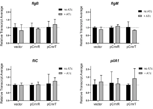
RNA was isolated, treated with DNase I, and reverse transcribed including a no-reverse transcriptase control as previously described[184, 237]. Real-time PCRs were done using 2 ng of cDNA, primers at a final concentration of 500 nM, and SYBR Green Real-Time qPCR reagents (Thermo Fisher) with an annealing temperature of 55°C. Primers used were as follows:R856-R857, flgB; R858-R859, flgM; R1063-R1064, fliC; R930-R931, pilA1; R852-R853, tcdA; R2298-R2299, cmrR; and R2537-R2538, cmrT). The data were analyzed using the ΔΔCt method with rpoC (R850-R851) as the
reference gene [103].
Animal experiments
All animal experimentation was performed under the guidance of veterinarians and trained animal technicians within the Emory University Division of Animal Resources (DAR). Animal experiments were performed with prior approval from the Emory University Institutional Animal Care and Use Committee (IACUC). C. difficile spores were collected from 70:30 sporulation broth after 3 days of growth [275]. The spores were purified using a sucrose gradient and stored in PBS with 1% bovine serum albumin as described previously [115, 276]. Prior to inoculation, the spores were enumerated by plating serial dilutions on BHIS-agar containing 0.1% sodium taurocholate.