THE ROLE OF AUTOTRANSPORTER PROTEINS IN BURKHOLDERIA PSEUDOMALLEI PATHOGENESIS
Cristine Gomes Campos
A dissertation submitted to the faculty of the University of North Carolina at Chapel Hill in partial fulfillment of the requirements for the degree of Doctor of Philosophy in the Department of Microbiology and Immunology of the School of Medicine.
Chapel Hill 2013
ABSTRACT
CRISTINE GOMES CAMPOS: The Role of Autotransporter proteins in Burkholderia pseudomallei Pathogenesis
(Under the direction of Peggy A. Cotter, Ph.D.)
Burkholderia pseudomallei is a tier 1 select agent, and the causative agent of
melioidosis, a disease that ranges from chronic abscesses to fulminant pneumonia and septic shock, which can be rapidly fatal. Autotransporters (ATs) are outer membrane proteins belonging to the Type V Secretion System family, and many have been shown to play crucial roles in pathogenesis in other bacterial species. The genome of B.
pseudomallei strain 1026b encodes two putative classical ATs, and nine putative trimeric
AT proteins.
However, significantly fewer bacteria were recovered from the spleen of Bp340ΔbcaA-infected mice, supporting a role for this AT in dissemination or in survival from the site of infection to the spleen.
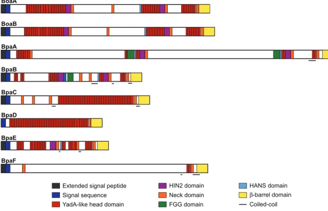
Using a bioinformatics approach, we annotated eight putative domains within each trimeric AT protein, excluding the well-studied BimA, and found short repeated sequences unique to Burkholderia species, as well as an unexpectedly high proportion of ATs with extended signal peptide regions (ESPRs). To characterize the role of trimeric ATs in pathogenesis, we constructed disruption or deletion mutations in each of eight AT-encoding genes and evaluated the resulting strains for adherence, invasion, and plaque formation in A549 cells. Five of the ATs, BoaA, BoaB, BpaA, BpaC, and BpaD, contribute to adherence, and four of the ATs, BpaA, BpaC, BpaE, and BpaF, are
necessary for efficient internalization in A549 cells. Using a BALB/c mouse model of infection, we then determined the contribution of each AT to bacterial burden in lungs, liver, and spleen. At 48 hours post-inoculation, only one strain, Bp340::pDbpaC,
ACKNOWLEGEMENTS
I would like to thank Dr. Peggy A. Cotter, my graduate student advisor, for generously providing me with the opportunity to work under her supervision, and for all she taught me. From sterile technique to infection models, you have provided me with the foundation to all of my microbiology knowledge. I will always be thankful.
I would like to thank present and past member of the Cotter laboratory for all of their help, especially Brandt Burgess, Sabrina Adelaine, Robin Hulbert, Matthew Byrd, and Eliza Mason for the countless hours of scientific and non-scientific conversation that were so instrumental in getting me through graduate school.
I would also like to thank Dr. Herbert Schweizer and Dr. Tung Hoang, as well as their graduate students, for all of their help. Without you this project would not have been possible. My heartfelt thanks also goes to Dr. Todd French and Dr. Erin Folchi for helping me through this journey. Dr. Sharon Taft-Benz, thank you for teaching me so much in the BSL-3, and for all your help. Your kindness and patience will never be forgotten.
TABLE OF CONTENTS
LIST OF TABLES...vii
LIST OF FIGURES...viii
LIST OF ABBREVIATIONS...ix
Chapters I. Introduction...1
Burkholderia classification and Epidemiology...1
Burkholderia pathogenesis...3
Putative Burkholderia virulence regulators...3
Putative Burkholderia virulence factors...5
Burkholderia putative autotransporter proteins...8
References...14
II. Characterization of BcaA, a putative autotransporter protein found in Burkholderia pseudomallei...24
Introduction...24
Materials and Methods...25
Results...30
Discussion...36
Figures...41
References...48
III. Functional characterization of Burkholderia pseudomallei trimeric autotransporters...52
Introduction...52
Materials and Methods...55
Results...58
Discussion...66
Figures...74
References...84
APPENDICES...99 I. Caspase-11 protects against bacteria that escapes the
vacuole...100 II. Discovery of Inhibitors of Burkholderia pseudomallei
methionine aminopeptidase with Antibacterial Activity...129
LIST OF TABLES
LIST OF FIGURES
Figure 2.1: bcaA and bcaB form an operon...42
Figure 2.2: Immunoblot analysis of Bp340::pCCS12HA1...43
Figure 2.3: Plaque analysis of Bp340, Bp340ΔbcaA and Bp340ΔbcaB...44
Figure 2.4: Colony forming units recovered after two-hour adherence and invasion assays with Bp340, Bp340ΔbcaA and Bp340ΔbcaB...45
Figure 2.5: In vivo analysis of Bp340, Bp340ΔbcaA and Bp340ΔbcaB...46
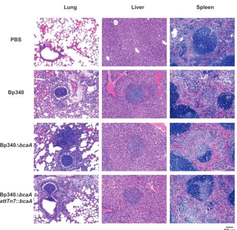
Figure 2.6: Histological analysis of B. pseudomallei-infected tissues...47
Figure 3.1: B. pseudomallei 1026b putative trimeric AT gene loci...75
Figure 3.2: B. pseudomallei 1026b putative trimeric AT protein domains...76
Figure 3.3: ESPRs of putative B. pseudomallei 1026b trimeric AT proteins...77
Figure 3.4: Schematic of deletion and disruption mutations used in trimeric AT study...78
Figure 3.5: Plaque formation by B. pseudomallei 1026b trimeric AT disruption mutants...79
Figure 3.6: Contribution of trimeric AT proteins to adherence and invasion...80
LIST OF ABBREVIATIONS
aa amino acid
AT autotransporter
Bca Burkholderia classical autotransporter Boa Burkholderia oligomeric coiled-coil adhesin Bpa Burkholderia pseudomallei autotransporter CFU colony forming unit
ESPR extended signal peptide region LPS lipopolysaccharides
Mb megabase pair
CHAPTER I Introduction
BURKHOLDERIA CLASSIFICATION AND EPIDEMIOLOGY
Burkholderia pseudomallei is a Gram-negative, rod-shaped, non-spore forming,
motile saprotrophic bacterium with a 7.24 megabase pair (Mb) genome divided into two chromosomes (90). The large 4.07 Mb chromosome (chromosome 1) contains a high proportion of coding sequences related to metabolic functions, such as macromolecule biosynthesis and amino acid (aa) metabolism, and the small 3.17 Mb chromosome (chromosome 2) contains coding sequences for accessory proteins and a vast number of coding sequences predicted to encode proteins of unknown function (37). B.
pseudomallei often forms dry, wrinkly colonies on Ash-down’s agar, but colony
morphology can vary considerably (15). Seven major colony morphotypes have been described, and morphology switching appears to occur in response to different environmental cues, including stress and interactions with epithelial cells or
B. pseudomallei infects a wide-variety of organisms, including human and
non-human mammals (19, 72). It causes Melioidosis, a disease that can range from chronic abscesses to fulminant pneumonia and septic shock and which can be rapidly fatal. First described by Dr. Whitmore, an army pathologist working in Burma, in 1912 (88), it has often been called the “great mimicker”, because it can present a vast array of clinical signs and symptoms. The disease can vary from an acute septic illness to a chronic infection with the persistence of symptoms for over two months. Primary clinical presentation varies from pneumonia to genitourinary infection, skin infections,
bacteremia without focus, septic arthritis, internal-organ abcesses, suppurative parotitis and brain stem encephalitis (89). Infections can be a result of percutaneous inoculation, inhalation, or ingestion. Melioidosis primarily affects people who have been exposed to environments containing B. pseudomallei (90); no reports of transmission between animals and humans have been described so far (9). Several conditions have been
disease without a vaccine (none has been developed to date) nearly impossible (87). Studies suggest that the intracellular nature of B. pseudomallei when infecting eukaryotic cells means that a vaccine will most likely not provide complete immunological
protection, unless T-cell immunity could be engaged (40), which makes the task of developing the vaccine even more difficult. Endemic to southeast Asia, and northern Australia, sporadic cases and clusters have been reported in Brazil (64), the Caribbean, Africa, the Middle East and even in the United States (22).
BURKHOLDERIA PATHOGENESIS
A protein microarray containing over a thousand B. pseudomallei surface exposed proteins was probed with sera from several melioidosis patients, and 170 immunoreactive proteins were identified. Amongst these were autotransporter proteins, lipoproteins, flagellin, and stress response proteins (27). To date only a limited number of virulence regulators and factors have been characterized for B. pseudomallei including quorum sensing and two-component regulatory system regulators, capsular polysaccharides, Type III Secretion System (T3SS), Type VI Secretion System (T6SS), lipopolysaccharides (LPS), flagella, Type IV pilli, and one Type V Secretion System protein named BimA. How these proteins are involved in pathogenesis is unclear and still under investigation.
PUTATIVE BURKHOLDERIA VIRULENCE REGULATORS Quorum sensing
pseudomallei led to decrease virulence in the Syrian hamster and BALB/c models of
infection (81). Quorum sensing is known to regulate the expression of several genes. Metalloproteases, phospholipases, and siderophores are among a few genes that were affected by luxI and luxR mutations in B. pseudomallei (82). The BpeAB-OprB multidrug efflux system found in B. pseudomallei is also involved in quorum sensing, since it is responsible for the extracellular secretion of homoserine-lactones. Deletion of the system caused reduced levels of cell invasion, and cytotoxicity for both epithelial and
macrophage cell lines, and the same were partially restored by addition of homoserine-lactones (14). A second quorum sensing system using
4-hydroxy-3-methyl-2-alkylquinolone signaling molecules has also been described (83), however its function is yet to be determined.
Two-component regulatory system
A complex two-component transcriptional regulatory system (VirAG) is found in B. pseudomallei, but little is known about the role this system has in this bacterium
pathogenesis (11, 78). Two-component regulatory systems allow bacteria to sense and respond to changes in may different environmental conditions. Typically, they consist of a membrane-bound histidine kinase that senses an environmental stimulus, and a
corresponding response regulator that mediates expression of target genes (52). VirAG is encoded immediately upstream of T6SS-1 gene cluster in B. pseudomallei, and it has been shown to transcriptionally activate T6SS-1 genes while inside a macrophage prior to escape from the phagosome (11, 78). It also induces expression and export to the
functional T6SS (11). T6SS-1 is a virulence factor in the Syrian hamster model of melioidosis, however the environmental cues sensed by the VirAG system is still unknown, and whether transcription activation of T6SS-1 genes occurs directly or indirectly via VirAG is still under investigation (11).
PUTATIVE BURKHOLDERIA VIRULENCE FACTORS Capsular polysaccharides
B. pseudomallei has an extracellular capsular polysaccharide,
-3)-2-O-acetyl-6-deoxy-β-D-manno-heptopyranose-(1-, that has been shown to be required for virulence in animal models (61). Although three other morphologically distinct variants have been observed by electron microscopy, little is known about these variants or their role in disease (58). In the presence of serum, capsule expression is increased, and the addition of purified B. pseudomallei capsule to serum bactericidal assays will increase the survival of a serum-sensitive strain (SLR5). Phagocytosis is also increased for capsule-deficient mutants when compared to wild type in the presence of human serum (62). Deposition of the complement C3b is enhanced in capsule mutants, suggesting that the persistence of B. psedomallei in the blood may be due to prevention of opsonization by complement in the
blood (47).
Type III Secretion System
Salmonella SPI-1 T3SS, and highly conserved across Burkholderia species. T3SS can
translocate multiple effectors into the host cell cytosol, where these effectors can subvert cell signaling to the benefit of the bacterium (31). T3SSBsa has been implicated in
invasion, escape from endosomes, intracellular survival, and evasion of autophagy (32) in B. pseudomallei. T3SSBsa is required for virulence in hamster and mouse infection models (75, 86), but the complete repertoire of Bsa effectors remain largely unknown. Recently, the T3SSBsa has been shown to be required for plaque formation and endosome escape, but dispensable for invasion (29).
Type VI Secretion System
Genes encoding a fairly recently discovered secretion system named T6SS have been identified in over one-fourth of all sequenced Gram-negative bacteria. T6SS function appears to be diverse and to be involved in interbacterial competition (67), increasing bacterial fitness in the environment, as well as being involved in pathogenesis (41). B. pseudomallei has six T6SSs. T6SS-1 has been shown to be necessary for
virulence in an acute model of melioidosis and contributes to lethality in hamsters in Burkholderia mallei (a clonal descendent of B. pseudomallei that has undergone genome
decay losing its capability for environmental survival) (66, 67). T6SS-1 has also recently been shown to be responsible for multinucleated giant cell (MNGC) formation in
RAW264 cells (11), and wild-type levels of cell invasion and intracellular survival was observed in the same cell line when T6SS-1 was mutated (68).
Lipopolysaccharides
differ from other LPS, for it exibits weaker murine pyrogenic activity when compared to enterobacterial LPS but stronger mitogenic activity in murine splenocytes (53).
Recognition of LPS by the host is crucial for innate immune response to Gram-negative bacteria through activation of the pattern-recognition receptor Toll-like receptor (TLR) 4 (8). B. pseudomallei infections have been shown to not elicit a TLR4 response, and little is known about how B. pseudomallei LPS plays a role in pathogenesis. B. thailandensis, a non-pathogenic bacterium (sometimes associated with human disease) that has a highly similar genome to B. pseudomallei (44), has a nearly identical LPS to B. pseudomallei. Both bacteria have a similar immunoblot profile against pooled sera from patients with melioidosis, and also similar LPS shedding profile, suggesting that LPS may not be involved in virulence and pathogenesis (1, 2).
Flagella and Type IV pili
B. pseudomallei is flagellated and motile. No difference was seen in the ability of
wild type and aflagellate mutant B. pseudomallei to invade and replicate in human cells in vitro (20). In a diabetic rat and Syrian hamster infection study no difference between
wild type and the aflagellate mutant was seen (24), however another study suggested that the aflagellate mutant was less virulent in a BALB/c intranasal model of infection (20).
Type IV pili have been shown to function as adhesins, and have an important role in virulence in many Gram-negative bacteria. Electron microscopy has shown the
presence of flagella and variable expression of pili on B. pseudomallei (85).
the eight type IV pili associated loci found in B. pseudomallei, which encodes pilA, when mutated was shown to cause decreased adhesion to epithelial cells, and reduced virulence in an intranasal route of infection BALB/c model (26).
BURKHOLDERIA PUTATIVE AUTOTRANSPORTER PROTEINS
Autotransporters (ATs) are outer membrane proteins belonging to the Type V Secretion System family, the largest family of extracellular proteins in Gram-negative bacteria (34). The AT secretion mechanism is remarkably simple, comprising a signal sequence, which targets the protein for secretion across the inner membrane through the Sec-system, a passenger domain (the secreted mature protein), and a C-terminal β-barrel domain that forms the pore in the outer membrane through which the passenger domain passes to the cell surface (33). Secretion of AT proteins have long been believed to be an energy independent, self- sufficient process (35). Many ATs have been shown to be virulence factors playing crucial roles in how bacteria cause disease (6, 10, 43).
Classical and trimeric autotransporter protein secretion
from Bordetella and the adhesin involved in diffuse adherence of E. coli (AIDA-I)) (6, 48).
Trimeric ATs require three proteins to form a functional unit, with the C-terminal 67 to 76 aa of each monomer contributing one-third of the β-barrel (21). Once at the bacterial surface, trimeric AT passenger domains remain intact as a large protein with a membrane bound C-terminus, and an N-terminal domain extending into the extracellular millieu (e.g. the adhesin protein from Yiersinia species YadA, and the adhesin from Haemophilus influenzae Hia) (63, 73). Oligomerization of the passenger domains occurs
via a coiled-coil domain believed to be ~70 to 100 residues from the C-terminal end of the protein (36).
The mechanism of classical and trimeric AT translocation across the outer membrane has been the focus of constant intense debate. According to the initial model, the β-barrel domain is inserted into the outer membrane, and mediates the translocation of the passenger domain covalently linked to it without the aid of any other proteins (54, 57). Recently several autotransporters (e.g. AIDA-I, and the Nisseria meningitides immunoglobin A1 (IgA1)) have been shown to require the Bam complex for biogenesis (55, 84); depletion of BamA abrogates secretion of mature AT passenger domains to the exterior of the cell, and BamD has also been implicated in autotransporter biogenesis (65). How BamA and BamD are involved in inserting the autotransporter β-barrel into the outer membrane is still unknown, but models involving periplasmic chaperones have been proposed (45).
passenger domain secretion, but the hairpin model holds the largest amount of supporting evidence currently. A hairpin structure is formed at the most C-terminal portion of the passenger domain, and maintained until the N-terminus of the passenger domain passes through the β-barrel (42, 49).
Following passenger domain secretion, some classical autotransporters are cleaved and released from their β-domain. Some mechanisms of passenger domain proteolysis have been elucidated. For example, the autotransporter IcsA required for intra- and inter-cellular motility in Shigella flexneri is cleaved by an exogenous protease (69). AIDA-I on the other hand is processed by autoproteolysis by two acidic residues that are contained within the passenger domain (16).
Classical autotransporter passenger domains
Although the β-domains generally show aa sequence conservation, which is consistent with their conserved function, passenger domains can vary widely reflecting their many roles. The IgA1 protease from N. meningitides for example is a serine protease, with a trypsin-like active domain (57) that is responsible for outer membrane release of this classical autotransporter protein. The mature protein is then capable of cleaving human-IgA1, and has a role in human mucosal colonization (43). IgA1 has also been shown to aid in trans-epithelial trafficking in T84 (human colon epithelial cells) monolayers, and to cleave LAMP1 (major integral glycoprotein of lysosomes) contributing to intracellular reproduction of the bacteria (38, 51).
inhibition of bacterial uptake in human tracheal epithelial cells (5), and cytotoxicity in phagocytic cells (28). AIDA-I, an adhesin found in some diffusely adherent E. coli strains (7), like pertactin remains associated with its β-barrel, and electron microscopy has shown it to be evenly distributed around the cell surface. AIDA-I (α-domain), which mediates the specific attachment of bacteria to target cells, in order to be fully functional,
needs to be post-translationally modified by heptose residues at multiple sites (46). Also
found in E. coli is EspP, an autotransporter protein of the enterohaemorrhagic (EHEC) O157:H7 strain. This AT is a protease capable of cleaving pepsin A and human
coagulation factor V, and is believed to contribute to the mucosal haemorrhage observed in patients with haemorrhagic colitis (10).
Trimeric autotransporter passenger domains
The trimeric AT Yersinia adhesin A (YadA) found in some Yersinia species was first described because of its capacity to promote auto-agglutination of Y. enterocolitica
and Y. pseudotuberculosis, as well as adherence to many substrates including epithelial
cells, extracellular matrix, collagen, cellular but not plasma fibronectin, and laminin (36).
The main function of Yad-A is believed to lie in its ability to confer resistance to
bactericidal activity of human serum by binding factor H (13). Non-typeable and
encapsulated Haemophilus influenzae have Hia (H. influenzae adhesin), a trimeric
B. pseudomallei putative autotransporter proteins
B. pseudomallei strain K96243 contains genes predicted to encode nine trimeric
ATs and two classical ATs. These are conserved across other B. pseudomallei genomes and some homologues are found in B. mallei, B. thailandensis, and B. gladioli strains. BPSS0962 (bcaA) and BPSL2237 are putative classical autotransporters predicted to function as a putative serine protease (12) and a lipase/esterase, respectively
(UniProtKB/TrEMBL). BPSL1631, BPSL1705 (boaB), BPSL2063, BPSS0088,
BPSS0796 (boaA), BPSS0908, BPSS1434 (bpaA), BPSS1439 and BPSS1492 (bimA) are all putative trimeric autotransporters.
Two trimeric ATs BoaA and BoaB have been expressed in E. coli and shown to be displayed on the bacterial surface and promote attachment to epithelial cell lines. B. pseudomallei strains with mutations in boaA and boaB exhibited reduced adherence to
epithelial cells, but no growth defect in phagocytic cell lines (4). Although this suggests these genes have a role in virulence, animal studies are required to establish the role of BoaA and BoaB in pathogenesis. The trimeric AT BpaA head domain crystal structure has been resolved (25), but its function remains to be elucidated.
Serum from 21 B. pseudomallei infected patients were used to probe a K96243 expression library, and five autotransporter proteins (BPSL2036, BPSS0908, BoaA, BPSS1439 and BimA) were shown to be potentially immunogenic during human melioidosis (80). A protein microarray probed with pooled melioidosis patients sera identified BPSS0088, BoaA, BpaA and BimA as also having the ability to interact with melioidosis-specific antibodies (27), further suggesting that B. pseudomallei
autotransporters play a role in pathogenesis.
References
1. Anuntagool, N., P. Intachote, V. Wuthiekanun, N. J. White, and S. Sirisinha. 1998. Lipopolysaccharide from nonvirulent Ara+ Burkholderia pseudomallei isolates is immunologically
indistinguishable from lipopolysaccharide from virulent Ara- clinical isolates. Clin Diagn Lab Immunol 5:225-229.
2. Anuntagool, N., T. Panichakul, P. Aramsri, and S. Sirisinha. 2000.
Shedding of lipopolysaccharide and 200-kDa surface antigen during the in vitro growth of virulent Ara- and avirulent Ara+ Burkholderia
pseudomallei. Acta tropica 74:221-228.
3. Attree, O., and I. Attree. 2001. A second type III secretion system in Burkholderia pseudomallei: who is the real culprit? Microbiology 147:3197-3199.
4. Balder, R., S. Lipski, J. J. Lazarus, W. Grose, R. M. Wooten, R. J. Hogan, D. E. Woods, and E. R. Lafontaine. Identification of Burkholderia mallei and Burkholderia pseudomallei adhesins for human respiratory epithelial cells. BMC microbiology 10:250.
5. Bassinet, L., P. Gueirard, B. Maitre, B. Housset, P. Gounon, and N. Guiso. 2000. Role of adhesins and toxins in invasion of human tracheal
epithelial cells by Bordetella pertussis. Infection and immunity 68:1934-1941.
6. Benz, I., and M. A. Schmidt. 1992. AIDA-I, the adhesin involved in diffuse adherence of the diarrhoeagenic Escherichia coli strain 2787
(O126:H27), is synthesized via a precursor molecule. Mol Microbiol 6:1539-1546.
7. Benz, I., and M. A. Schmidt. 1989. Cloning and expression of an adhesin (AIDA-I) involved in diffuse adherence of enteropathogenic Escherichia coli. Infection and immunity 57:1506-1511.
8. Beutler, B., and E. T. Rietschel. 2003. Innate immune sensing and its roots: the story of endotoxin. Nat Rev Immunol 3:169-176.
9. Brett, P. J., and D. E. Woods. 2000. Pathogenesis of and immunity to melioidosis. Acta tropica 74:201-210.
11. Burtnick, M. N., P. J. Brett, S. V. Harding, S. A. Ngugi, W. J. Ribot, N.
Chantratita, A. Scorpio, T. S. Milne, R. E. Dean, D. L. Fritz, S. J. Peacock, J. L. Prior, T. P. Atkins, and D. Deshazer. The cluster 1 type VI secretion system is a major virulence determinant in Burkholderia pseudomallei. Infection and immunity 79:1512-1525.
12. Campos, C. G., L. Borst, and P. A. Cotter. Characterization of BcaA, a putative classical autotransporter protein in Burkholderia
pseudomallei. Infection and immunity.
13. Casutt-Meyer, S., F. Renzi, M. Schmaler, N. J. Jann, M. Amstutz, and G. R. Cornelis. Oligomeric coiled-coil adhesin YadA is a double-edged sword. PLoS One 5:e15159.
14. Chan, Y. Y., and K. L. Chua. 2005. The Burkholderia pseudomallei BpeAB-OprB efflux pump: expression and impact on quorum sensing and virulence. J Bacteriol 187:4707-4719.
15. Chantratita, N., V. Wuthiekanun, K. Boonbumrung, R. Tiyawisutsri, M.
Vesaratchavest, D. Limmathurotsakul, W. Chierakul, S.
Wongratanacheewin, S. Pukritiyakamee, N. J. White, N. P. Day, and S. J. Peacock. 2007. Biological relevance of colony morphology and
phenotypic switching by Burkholderia pseudomallei. J Bacteriol 189:807-817.
16. Charbonneau, M. E., J. Janvore, and M. Mourez. 2009. Autoprocessing of the Escherichia coli AIDA-I autotransporter: a new mechanism
involving acidic residues in the junction region. J Biol Chem 284:17340- 17351.
17. Charles, I., N. Fairweather, D. Pickard, J. Beesley, R. Anderson, G. Dougan, and M. Roberts. 1994. Expression of the Bordetella pertussis P.69 pertactin adhesin in Escherichia coli: fate of the carboxy-terminal domain. Microbiology 140 ( Pt 12):3301-3308.
18. Chen, Y. S., S. C. Chen, C. M. Kao, and Y. L. Chen. 2003. Effects of soil pH, temperature and water content on the growth of Burkholderia
pseudomallei. Folia Microbiol (Praha) 48:253-256.
19. Cheng, A. C., and B. J. Currie. 2005. Melioidosis: epidemiology, pathophysiology, and management. Clinical microbiology reviews 18:383-416.
21. Cotter, S. E., N. K. Surana, and J. W. St Geme, 3rd. 2005. Trimeric autotransporters: a distinct subfamily of autotransporter proteins. Trends Microbiol 13:199-205.
22. Currie, B. J., D. A. Dance, and A. C. Cheng. 2008. The global distribution of Burkholderia pseudomallei and melioidosis: an update. Trans R Soc Trop Med Hyg 102 Suppl 1:S1-4.
23. Dejsirilert, S., E. Kondo, D. Chiewsilp, and K. Kanai. 1991. Growth and survival of Pseudomonas pseudomallei in acidic environments. Jpn J Med Sci Biol 44:63-74.
24. DeShazer, D., P. J. Brett, R. Carlyon, and D. E. Woods. 1997. Mutagenesis of Burkholderia pseudomallei with Tn5-OT182: isolation of motility mutants and molecular characterization of the flagellin structural gene. J Bacteriol 179:2116-2125.
25. Edwards, T. E., I. Phan, J. Abendroth, S. H. Dieterich, A. Masoudi, W. Guo, S. N. Hewitt, A. Kelley, D. Leibly, M. J. Brittnacher, B. L. Staker, S. I. Miller, W. C. Van Voorhis, P. J. Myler, and L. J. Stewart. Structure of a
Burkholderia pseudomallei trimeric autotransporter adhesin head. PLoS One 5.
26. Essex-Lopresti, A. E., J. A. Boddey, R. Thomas, M. P. Smith, M. G. Hartley, T. Atkins, N. F. Brown, C. H. Tsang, I. R. Peak, J. Hill, I. R. Beacham, and R. W. Titball. 2005. A type IV pilin, PilA, Contributes To Adherence of Burkholderia pseudomallei and virulence in vivo. Infection and immunity 73:1260-1264.
27. Felgner, P. L., M. A. Kayala, A. Vigil, C. Burk, R. Nakajima-Sasaki, J. Pablo, D. M. Molina, S. Hirst, J. S. Chew, D. Wang, G. Tan, M. Duffield, R. Yang, J. Neel, N. Chantratita, G. Bancroft, G. Lertmemongkolchai, D. H. Davies, P. Baldi, S. Peacock, and R. W. Titball. 2009. A Burkholderia pseudomallei protein microarray reveals serodiagnostic and cross-reactive antigens. Proceedings of the National Academy of Sciences of the United States of America 106:13499-13504.
28. Fleckenstein, J. M., D. J. Kopecko, R. L. Warren, and E. A. Elsinghorst. 1996. Molecular characterization of the tia invasion locus from
enterotoxigenic Escherichia coli. Infection and immunity 64:2256-2265.
30. Gal, D., M. Mayo, H. Smith-Vaughan, P. Dasari, M. McKinnon, S. P. Jacups, A. I. Urquhart, M. Hassell, and B. J. Currie. 2004. Contamination of hand wash detergent linked to occupationally acquired melioidosis. Am J Trop Med Hyg 71:360-362.
31. Galan, J. E., and H. Wolf-Watz. 2006. Protein delivery into eukaryotic cells by type III secretion machines. Nature 444:567-573.
32. Gong, L., M. Cullinane, P. Treerat, G. Ramm, M. Prescott, B. Adler, J. D. Boyce, and R. J. Devenish. The Burkholderia pseudomallei type III secretion system and BopA are required for evasion of LC3-associated phagocytosis. PLoS One 6:e17852.
33. Henderson, I. R., and J. P. Nataro. 2001. Virulence functions of autotransporter proteins. Infection and immunity 69:1231-1243. 34. Henderson, I. R., F. Navarro-Garcia, M. Desvaux, R. C. Fernandez, and D.
Ala'Aldeen. 2004. Type V protein secretion pathway: the autotransporter story. Microbiol Mol Biol Rev 68:692-744.
35. Henderson, I. R., F. Navarro-Garcia, and J. P. Nataro. 1998. The great escape: structure and function of the autotransporter proteins. Trends Microbiol 6:370-378.
36. Hoiczyk, E., A. Roggenkamp, M. Reichenbecher, A. Lupas, and J.
Heesemann. 2000. Structure and sequence analysis of Yersinia YadA and Moraxella UspAs reveal a novel class of adhesins. EMBO J 19:5989- 5999.
37. Holden, M. T., R. W. Titball, S. J. Peacock, A. M. Cerdeno-Tarraga, T. Atkins, L. C. Crossman, T. Pitt, C. Churcher, K. Mungall, S. D. Bentley, M. Sebaihia, N. R. Thomson, N. Bason, I. R. Beacham, K. Brooks, K. A. Brown, N. F. Brown, G. L. Challis, I. Cherevach, T. Chillingworth, A. Cronin, B. Crossett, P. Davis, D. DeShazer, T. Feltwell, A. Fraser, Z. Hance, H.
Hauser, S. Holroyd, K. Jagels, K. E. Keith, M. Maddison, S. Moule, C. Price, M. A. Quail, E. Rabbinowitsch, K. Rutherford, M. Sanders, M. Simmonds, S. Songsivilai, K. Stevens, S. Tumapa, M. Vesaratchavest, S. Whitehead, C. Yeats, B. G. Barrell, P. C. Oyston, and J. Parkhill. 2004. Genomic plasticity of the causative agent of melioidosis, Burkholderia pseudomallei.
Proceedings of the National Academy of Sciences of the United States of America 101:14240-14245.
38. Hopper, S., B. Vasquez, A. Merz, S. Clary, J. S. Wilbur, and M. So. 2000. Effects of the immunoglobulin A1 protease on Neisseria gonorrhoeae trafficking across polarized T84 epithelial monolayers. Infection and immunity 68:906-911.
invasion of Acanthamoeba astronyxis. Infection and immunity 71:2280- 2282.
40. Inglis, T. J., and J. L. Sagripanti. 2006. Environmental factors that affect the survival and persistence of Burkholderia pseudomallei. Appl Environ Microbiol 72:6865-6875.
41. Jani, A. J., and P. A. Cotter. Type VI secretion: not just for pathogenesis anymore. Cell Host Microbe 8:2-6.
42. Junker, M., R. N. Besingi, and P. L. Clark. 2009. Vectorial transport and folding of an autotransporter virulence protein during outer membrane secretion. Mol Microbiol 71:1323-1332.
43. Kilian, M., J. Reinholdt, H. Lomholt, K. Poulsen, and E. V. Frandsen. 1996. Biological significance of IgA1 proteases in bacterial colonization and pathogenesis: critical evaluation of experimental evidence. APMIS 104:321-338.
44. Kim, H. S., M. A. Schell, Y. Yu, R. L. Ulrich, S. H. Sarria, W. C. Nierman, and D. DeShazer. 2005. Bacterial genome adaptation to niches: divergence of the potential virulence genes in three Burkholderia species of different survival strategies. BMC Genomics 6:174.
45. Knowles, T. J., A. Scott-Tucker, M. Overduin, and I. R. Henderson. 2009. Membrane protein architects: the role of the BAM complex in outer membrane protein assembly. Nat Rev Microbiol 7:206-214.
46. Laarmann, S., and M. A. Schmidt. 2003. The Escherichia coli AIDA autotransporter adhesin recognizes an integral membrane glycoprotein as receptor. Microbiology 149:1871-1882.
47. Larsen, J. C., and N. H. Johnson. 2009. Pathogenesis of Burkholderia pseudomallei and Burkholderia mallei. Mil Med 174:647-651.
48. Leininger, E., M. Roberts, J. G. Kenimer, I. G. Charles, N. Fairweather, P. Novotny, and M. J. Brennan. 1991. Pertactin, an Arg-Gly-Asp-containing Bordetella pertussis surface protein that promotes adherence of
mammalian cells. Proceedings of the National Academy of Sciences of the United States of America 88:345-349.
49. Leo, J. C., I. Grin, and D. Linke. Type V secretion: mechanism(s) of autotransport through the bacterial outer membrane. Philos Trans R Soc Lond B Biol Sci 367:1088-1101.
Risk factors for recurrent melioidosis in northeast Thailand. Clin Infect Dis 43:979-986.
51. Lin, L., P. Ayala, J. Larson, M. Mulks, M. Fukuda, S. R. Carlsson, C. Enns, and M. So. 1997. The Neisseria type 2 IgA1 protease cleaves LAMP1 and promotes survival of bacteria within epithelial cells. Mol Microbiol 24:1083-1094.
52. Mascher, T., J. D. Helmann, and G. Unden. 2006. Stimulus perception in bacterial signal-transducing histidine kinases. Microbiol Mol Biol Rev 70:910-938.
53. Matsuura, M., K. Kawahara, T. Ezaki, and M. Nakano. 1996. Biological activities of lipopolysaccharide of Burkholderia (Pseudomonas) pseudomallei. FEMS Microbiol Lett 137:79-83.
54. Meng, G., N. K. Surana, J. W. St Geme, 3rd, and G. Waksman. 2006. Structure of the outer membrane translocator domain of the
Haemophilus influenzae Hia trimeric autotransporter. EMBO J 25:2297- 2304.
55. Mogensen, J. E., J. H. Kleinschmidt, M. A. Schmidt, and D. E. Otzen. 2005. Misfolding of a bacterial autotransporter. Protein Sci 14:2814-2827. 56. Ngauy, V., Y. Lemeshev, L. Sadkowski, and G. Crawford. 2005. Cutaneous
melioidosis in a man who was taken as a prisoner of war by the Japanese during World War II. J Clin Microbiol 43:970-972.
57. Pohlner, J., R. Halter, K. Beyreuther, and T. F. Meyer. 1987. Gene structure and extracellular secretion of Neisseria gonorrhoeae IgA protease. Nature 325:458-462.
58. Puthucheary, S. D., and S. A. Nathan. 2006. Comparison by electron microscopy of intracellular events and survival of Burkholderia pseudomallei in monocytes from normal subjects and patients with melioidosis. Singapore Med J 47:697-703.
59. Raetz, C. R., and C. Whitfield. 2002. Lipopolysaccharide endotoxins. Annu Rev Biochem 71:635-700.
60. Rainbow, L., C. A. Hart, and C. Winstanley. 2002. Distribution of type III secretion gene clusters in Burkholderia pseudomallei, B. thailandensis and B. mallei. J Med Microbiol 51:374-384.
identification of capsular polysaccharide of Burkholderia pseudomallei as a major virulence determinant. Infection and immunity 69:34-44. 62. Reckseidler-Zenteno, S. L., R. DeVinney, and D. E. Woods. 2005. The
capsular polysaccharide of Burkholderia pseudomallei contributes to survival in serum by reducing complement factor C3b deposition. Infection and immunity 73:1106-1115.
63. Roggenkamp, A., N. Ackermann, C. A. Jacobi, K. Truelzsch, H. Hoffmann, and J. Heesemann. 2003. Molecular analysis of transport and
oligomerization of the Yersinia enterocolitica adhesin YadA. J Bacteriol 185:3735-3744.
64. Rolim, D. B., D. C. Vilar, A. Q. Sousa, I. S. Miralles, D. C. de Oliveira, G. Harnett, L. O'Reilly, K. Howard, I. Sampson, and T. J. Inglis. 2005. Melioidosis, northeastern Brazil. Emerg Infect Dis 11:1458-1460.
65. Rossiter, A. E., D. L. Leyton, K. Tveen-Jensen, D. F. Browning, Y. Sevastsyanovich, T. J. Knowles, K. B. Nichols, A. F. Cunningham, M.
Overduin, M. A. Schembri, and I. R. Henderson. The essential beta-barrel assembly machinery complex components BamD and BamA are
required for autotransporter biogenesis. J Bacteriol 193:4250-4253.
66. Schell, M. A., R. L. Ulrich, W. J. Ribot, E. E. Brueggemann, H. B. Hines, D. Chen, L. Lipscomb, H. S. Kim, J. Mrazek, W. C. Nierman, and D. Deshazer. 2007. Type VI secretion is a major virulence determinant in
Burkholderia mallei. Mol Microbiol 64:1466-1485.
67. Schwarz, S., T. E. West, F. Boyer, W. C. Chiang, M. A. Carl, R. D. Hood, L. Rohmer, T. Tolker-Nielsen, S. J. Skerrett, and J. D. Mougous.
Burkholderia type VI secretion systems have distinct roles in
eukaryotic and bacterial cell interactions. PLoS Pathog 6:e1001068.
68. Shalom, G., J. G. Shaw, and M. S. Thomas. 2007. In vivo expression technology identifies a type VI secretion system locus in Burkholderia pseudomallei that is induced upon invasion of macrophages.
Microbiology 153:2689-2699.
69. Shere, K. D., S. Sallustio, A. Manessis, T. G. D'Aversa, and M. B. Goldberg. 1997. Disruption of IcsP, the major Shigella protease that cleaves IcsA, accelerates actin-based motility. Mol Microbiol 25:451-462.
71. Spahich, N. A., and J. W. St Geme, 3rd. Structure and Function of the Haemophilus influenzae Autotransporters. Front Cell Infect Microbiol 1:5.
72. Sprague, L. D., and H. Neubauer. 2004. Melioidosis in animals: a review on epizootiology, diagnosis and clinical presentation. J Vet Med B Infect Dis Vet Public Health 51:305-320.
73. St Geme, J. W., 3rd, and D. Cutter. 2000. The Haemophilus influenzae Hia adhesin is an autotransporter protein that remains uncleaved at the C terminus and fully cell associated. J Bacteriol 182:6005-6013.
74. Stevens, J. M., R. L. Ulrich, L. A. Taylor, M. W. Wood, D. Deshazer, M. P. Stevens, and E. E. Galyov. 2005. Actin-binding proteins from
Burkholderia mallei and Burkholderia thailandensis can functionally compensate for the actin-based motility defect of a Burkholderia pseudomallei bimA mutant. J Bacteriol 187:7857-7862.
75. Stevens, M. P., A. Haque, T. Atkins, J. Hill, M. W. Wood, A. Easton, M. Nelson, C. Underwood-Fowler, R. W. Titball, G. J. Bancroft, and E. E. Galyov. 2004. Attenuated virulence and protective efficacy of a Burkholderia pseudomallei bsa type III secretion mutant in murine models of melioidosis. Microbiology 150:2669-2676.
76. Stevens, M. P., J. M. Stevens, R. L. Jeng, L. A. Taylor, M. W. Wood, P. Hawes, P. Monaghan, M. D. Welch, and E. E. Galyov. 2005. Identification of a bacterial factor required for actin-based motility of Burkholderia pseudomallei. Mol Microbiol 56:40-53.
77. Stevens, M. P., M. W. Wood, L. A. Taylor, P. Monaghan, P. Hawes, P. W. Jones, T. S. Wallis, and E. E. Galyov. 2002. An Inv/Mxi-Spa-like type III protein secretion system in Burkholderia pseudomallei modulates intracellular behaviour of the pathogen. Mol Microbiol 46:649-659.
78. Sun, G. W., Y. Chen, Y. Liu, G. Y. Tan, C. Ong, P. Tan, and Y. H. Gan. Identification of a regulatory cascade controlling Type III Secretion System 3 gene expression in Burkholderia pseudomallei. Mol Microbiol 76:677-689.
79. Tandhavanant, S., A. Thanwisai, D. Limmathurotsakul, S. Korbsrisate, N.
80. Tiyawisutsri, R., M. T. Holden, S. Tumapa, S. Rengpipat, S. R. Clarke, S. J. Foster, W. C. Nierman, N. P. Day, and S. J. Peacock. 2007. Burkholderia Hep_Hag autotransporter (BuHA) proteins elicit a strong antibody response during experimental glanders but not human melioidosis. BMC microbiology 7:19.
81. Ulrich, R. L., D. Deshazer, E. E. Brueggemann, H. B. Hines, P. C. Oyston, and J. A. Jeddeloh. 2004. Role of quorum sensing in the pathogenicity of Burkholderia pseudomallei. J Med Microbiol 53:1053-1064.
82. Valade, E., F. M. Thibault, Y. P. Gauthier, M. Palencia, M. Y. Popoff, and D. R. Vidal. 2004. The PmlI-PmlR quorum-sensing system in Burkholderia pseudomallei plays a key role in virulence and modulates production of the MprA protease. J Bacteriol 186:2288-2294.
83. Vial, L., F. Lepine, S. Milot, M. C. Groleau, V. Dekimpe, D. E. Woods, and E. Deziel. 2008. Burkholderia pseudomallei, B. thailandensis, and B.
ambifaria produce 4-hydroxy-2-alkylquinoline analogues with a methyl group at the 3 position that is required for quorum-sensing regulation. J Bacteriol 190:5339-5352.
84. Volokhina, E. B., J. Grijpstra, M. Stork, I. Schilders, J. Tommassen, and M. P. Bos. Role of the periplasmic chaperones Skp, SurA, and DegQ in outer membrane protein biogenesis in Neisseria meningitidis. J Bacteriol 193:1612-1621.
85. Vorachit, M., K. Lam, P. Jayanetra, and J. W. Costerton. 1995. Electron microscopy study of the mode of growth of Pseudomonas pseudomallei in vitro and in vivo. J Trop Med Hyg 98:379-391.
86. Warawa, J., and D. E. Woods. 2005. Type III secretion system cluster 3 is required for maximal virulence of Burkholderia pseudomallei in a hamster infection model. FEMS Microbiol Lett 242:101-108.
87. White, N. J. 2003. Melioidosis. Lancet 361:1715-1722.
88. Whitmore A, K. C. 1912. An account of the discovery of a hitherto undescribed infective disease occurring among the population of Rangoon. Indian Med Gaz 92:262-267.
89. Wiersinga, W. J., B. J. Currie, and S. J. Peacock. Melioidosis. N Engl J Med 367:1035-1044.
91. Wuthiekanun, V., M. D. Smith, and N. J. White. 1995. Survival of Burkholderia pseudomallei in the absence of nutrients. Trans R Soc Trop Med Hyg 89:491.
CHAPTER II
Characterization of BcaA, a putative classical autotransporter protein,
in Burkholderia pseudomallei1
Introduction
Burkholderia pseudomallei is a Gram-negative saprotroph that causes
melioidosis, a disease that ranges from chronic abscesses to fulminant pneumonia and septic shock and which can be rapidly fatal. B. pseudomallei has an approximately 7.2-megabase pair (Mb) genome divided into two chromosomes (19), affording this bacterium the metabolic repertoire necessary to adapt to and survive in a variety of different habitats, including soil, the rhizosphere of plants, and a wide variety of human and non-human mammals (36). Melioidosis, first reported by Dr. Whitmore, an army pathologist working in Burma, in 1912 (41), emerged as an infectious disease of serious public health concern in the latter part of the 20th century, manifesting as a rapidly progressing septicemia, with or without pneumonia; a localized soft-tissue infection; or a sub-clinical infection with delayed evidence of clinical infection (21, 42). B.
pseudomallei is endemic to southeast Asia and northern Australia, however sporadic B.
pseudomallei infections have been reported worldwide (4), including but not limited to
the United States, Puerto Rico, El Salvador, and Brazil (10, 21, 34, 43). B. pseudomallei
is classified as an NIH category B priority pathogen and select agent (40), and has recently been reclassified as a CDC Tier 1 select agent due to its virulence in animals, low infectious dose, robust environment stability, and possible delay in diagnosis since the bacterium is not endemic to the United States (http://www.selectagents.gov).
Autotransporters (ATs) are outer membrane proteins belonging to the Type V Secretion System family, the largest family of extracellular proteins in Gram-negative bacteria (18). These proteins have been shown to function as adhesins, degradative enzymes, and cytotoxins, as well as having roles in cell-to-cell spread and serum resistance (17). The AT family is divided into classical and trimeric proteins. The C-terminal 250 to 300 amino acids of classical ATs form a β-barrel that is inserted into the outer membrane where it facilitates the translocation of the N-terminal passenger domain to the cell surface. Trimeric ATs require three proteins to form a functional unit, with the C-terminal 67 to 76 C-terminal amino acids of each monomer contributing one-third of the barrel (7). The β-barrel domains of AT proteins are well conserved, while the passenger domains can vary substantially and have distinct functions (31).
trimeric autotransporter BimA, which contains proline-rich motifs and WH2-like domains that are believed to be responsible for actin polymerization at the pole of the bacterial cell where BimA is localized (38).
The genome of B. pseudomallei strain 1026b contains genes predicted to encode nine trimeric ATs and two classical ATs. The purpose of this study was to determine the role of the predicted classical AT encoded by Bp1026b_II1054 in pathogenesis.
Materials and Methods
Bacterial Strains. All manipulations of B. pseudomallei were conducted in a CDC/USDA-approved animal biosafety level 3 (ABSL3) facility at the University of North Carolina at Chapel Hill. The bacterial strains used in this study are listed in Table 1. B. pseudomallei strains were cultured in Low Salt Lysogeny Broth (LSLB), or on Low Salt Lysogeny Broth Agar (LSLBA) (Sigma-Aldrich, St. Louis, MO) for 24 hours at 37ºC. Escherichia coli strains were cultured on LB or LBA. When appropriate, culture media were supplemented with Kanamycin (Km, 125 µg/ml for B. pseudomallei and 50
Construction of B. pseudomallei bcaA and bcaB mutant strains and plasmids. Deletion of the bcaA and bcaB genes from B. pseudomallei strain Bp340 (a derivative of strain1026b containing a Δ(amrRAB-oprA) mutation. Strain was shown by Dr. Herbert Schweizer to be as virulent as 1026b in the BALB/c acute model of infection) (14, 30) was carried out by allelic exchange using pEXKm5 (28) derivatives. The DNA fragments used to construct pCCX1 and pCCX2 (Table 1) were generated using a two-step, overlap polymerase chain reaction (PCR) method and cloned into pEXKm5. DNA fragments contained approximately 500 bp 5ʹ′ to the gene including the first three codons and 500 bp 3ʹ′ to the gene including the last three codons. pCCX1 and pCCX2 were transformed into E. coli RHO3 cells, and were delivered to Bp340 by conjugation. For Bp340ΔbcaA and
Bp340ΔbcaB complementation strains, the gene and promoter region was cloned into pUC18T-mini-Tn7-Zeo and delivered as previously described (5). All plasmids were verified to be correct and contain no unintended nucleotide changes by DNA sequence analysis.
with Bp340ΔbcaA and Bp340ΔbcaB, respectively, and cointegrants were selected on LSLB-Zeo (5). Integration at the correct location was confirmed by PCR. All DNA regions encompassing about 2 Kb across the deletion junction were PCR amplified and verified to be correct and to contain no unintended nucleotide changes by DNA sequence analysis. DNA sequence inserted in the att Tn7 site in complementation strains were also PCR amplified and verified by DNA sequence analysis.
Total RNA isolation and cDNA synthesis. Total RNA was isolated in Trizol (Invitrogen, Grand Island, NY) from Bp340 grown overnight in LSLB at 37ºC according to the manufacturer’s protocol. For the reverse transcription step, 5 ng of total RNA was transcribed using Super Script III reverse transcriptase (Invitrogen, Grand Island, NY) with oligo(dT) and random primers according to the manufacturer’s instructions. Transcripts were determined by PCR primers described in Table 2.
(diluted 1:5,000) followed by an IR800-conjugated secondary antibody (diluted 1:20,000) (Rockland, Gilbertsville, PA). Antigen-antibody complexes were visualized using the Odyssey infrared imaging system (Li-Cor Biosciences, Lincoln, NE).
Plaque Assay. B. pseudomallei strains were grown overnight in LSLB at 37ºC. Each well of a 6-well plate was seeded with A549 human lung cells such that confluent monolayers contained approximately 1x10⁶ cells per well. Cells were incubated in F12K (Cellgro, Circle Westwood, MA) supplemented with 10% fetal bovine serum (Gibco, Grand Island, NY) at 37ºC with 5% CO2. Bacterial strains were diluted to an OD600 of 0.1 in fresh tissue culture medium, further diluted 1:10, and 25 µl of the diluted culture was
added to each well (MOI of 0.1). Plates were incubated for 2 hours and each well was washed thoroughly with fresh culture medium and overlayed with a mixture containing 1.2% low-melting agarose (Fisher Scientific, Fairlawn, NJ), F12K with 10% FBS, and 0.01% Neutral red (Fischer Scientific, Waltham, MA). Plates were incubated for 24 hours at 37ºC with 5% CO2, and plaques were enumerated in each well. Experiments were performed three times in duplicate, and the results were combined. The combined results were analyzed using a one-way analysis of variance (ANOVA) with Tukey’s post-test at a 95% confidence interval.
Adherence and invasion assay. Bacterial strains and A549 cells were grown as described above. Bacteria were diluted to an OD600 of 0.1 in fresh tissue culture medium, and 250µL of the diluted cultures added to each well (MOI of 100). Plates were
forming units (CFU) in each well. For the invasion assay, cells were incubated an additional hour and a half with Gentamicin (90 µg/ml), washed with fresh culture medium, and were lysed using 1% Triton X-100. Lysates were diluted and plated to determine the total CFU in each well. To calculate the percentage of adherent or invading bacteria, the number of adherent or invading bacteria was divided by the total number of bacteria in the inoculum and multiplied by 100. Experiments were performed three times in duplicate, and the results were combined. The combined results were analyzed using a one-way ANOVA with Tukey’s post-test at a 95% confidence interval.
Animal experiments. All animal experiments were approved by the Animal Studies Committee of the University of North Carolina at Chapel Hill (protocol 10-165). Six- to eight-week-old female BALB/c mice (The Jackson Laboratory, Bar Harbor, ME) were allowed free access to sterilized food and water. Animals were anesthetized prior to infection with Avertin (140mg/Kg) by intraperitoneal injection. For all infections, the desired inoculum of B. pseudomallei was suspended in sterile phosphate buffered saline (PBS). Mice were inoculated intranasally with 500 CFU (LD50 for 1026b has been determined to be around 900 CFU (14)), and at the indicated time points were euthanized by CO2 overdose. Organs were aseptically harvested, homogenized, and the bacterial burden of each organ was determined by plating serial dilutions of the homogenates. Animal experiments were performed twice, with three to four animals per strain per time point and the results combined. Animal experiments were terminated at 48 hours at which time all animals had become moribund.
in 10% neutral buffered formalin for 24 hours. Samples were stored in 70% ethanol until processed into paraffin using routine methods. Paraffin embedded tissues were sectioned 3 to 5 microns thick, captured onto glass slides and stained with hematoxylin and eosin (HE). Sections of liver, spleen and lung were analyzed using light microscopy by a single pathologist (LB) who was blinded to group. Microscopic lesions consisting primarily of necrosis and acute inflammation (neutrophils, macrophages, apoptotic bodies, and fibrin) were quantified as follows: In the lung, the affected bronchi per 20 bronchiolar profiles were counted starting with an affected bronchiole. In the liver and spleen, inflammatory foci were enumerated in 10 consecutive fields at 20x magnification starting on an area of inflammation identified on low magnification.
Results
Bioinformatic analysis of the bcaA and bcaB genes of B. pseudomallei strain 1026b Bp1026b_II1054 is a 3393 base pair (bp) gene predicted to encode a classical AT that we named bcaA for Burkholderia classical autotransporter A. Forty base pairs 3′ to bcaA is Bp11026_II1055 (which we named bcaB), a 687 bp gene predicted to encode a
hypothetical protein. There is a 480 bp gene predicted to encode a hypothetical protein 1522 bp 5′ to bcaA, and 198 bp 3′ to bcaB is a 1593 bp gene oriented in the opposite direction, predicted to encode a periplasmic solute-binding protein (Fig. 1A).
(SMART) predicts BcaA to have a serine protease domain, from aa 64 to 380, belonging to the Peptidase S8 or Subtilase family, and a classical AT β-domain from aa 857 to 1121. BcaB, is a 255 aa protein predicted to contain a prolyl 4-hydroxylase domain from aa 41 to 225 (Fig.1B) (27).
BLAST indicated that homologues of bcaA are present in B. pseudomallei, B. mallei, B. thailandensis, and B. gladioli strains but not in Ralstonia or Cupriavidus
species, close relatives of the Burkholderia genus. All B. pseudomallei and B. mallei strains for which genome sequence is available are predicted to encode proteins with 99% identity with BcaA, 89% identity is found in B. thailandensis and 82% identity in B. oklahomensis. Predicted homologues are not found in bacteria outside of the
Burkholderia genus, suggesting this gene is unique to Burkholderia.
bcaA and bcaB appear to form an operon
RT-PCR was used to determine if bcaA and bcaB are cotranscribed. cDNA was prepared from Bp340 grown overnight at 37°C in LSLB, and used as template for PCR
using the primer pairs (Table 2) shown in Figure 1A. Products of expected sizes were obtained for each primer pair (Fig. 1C), including the pair spanning the intergenic region between bcaA and bcaB, suggesting that these genes are cotranscribed. No product was observed in the mock RT samples demonstrating the lack of DNA contamination in the isolated RNA samples, and consequently the cDNA samples.
BcaA appears to be proteolyzed to smaller polypeptides
To visualize the production of BcaA protein, we constructed Bp340 derivatives in which nucleotides encoding HA epitopes were inserted either immediately following the protein’s predicted signal sequence cleavage site or between codons 58 and 59. Both strains were grown overnight in LSLB at 37°C but no polypeptide was recognized by
Western blotting using an anti-HA antibody. After several unsuccessful attempts to visualize the BcaA protein when the gene was expressed from its native promoter, we constructed a strain in which bcaA was expressed from a strong, constitutively active promoter (the promoter for the rpsL gene from Burkholderia pseudomallei 1026b). First, we inserted the HA epitope-encoding codons immediately following the predicted signal sequence cleavage site, but again were unsuccessful at visualizing BcaA. Finally, we inserted the HA epitope-encoding codons between codons 58 and 59
between 250 and 75 kDa were observed in whole cell lysates of Bp340::pCC12HA1, but not in whole cell lysates of the wild-type strain lacking the HA epitope tag (Figure 2). These data suggest that BcaA is proteolyzed to smaller polypeptides. The smaller polypeptides were not detected in western blots of concentrated supernatant fractions (data not shown), suggesting that they remain associated with the bacterial cell.
bcaA and bcaB are required for efficient plaque formation
B. pseudomallei can spread from cell to cell without exiting the cytoplasm and
can form plaques in a cell monolayer (22). To determine if bcaA or bcaB contribute to plaque formation, A549 respiratory epithelial cell monolayers were infected at an MOI of 0.1 and plaques were counted after 24 hours. Bp340 formed an average of 100
plaques/well, while Bp340ΔbcaA (Fig. 3A) and Bp340ΔbcaB (Fig. 3B) each formed only about 50 to 60 plaques/well. Complementation of both mutations restored plaque-forming ability to wild type levels, indicating that both bcaA and bcaB are required for efficient plaque formation.
bcaA and bcaB are required for efficient invasion of A549 cells
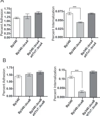
The plaques formed by Bp340ΔbcaA and Bp340ΔbcaB strains appeared to be of similar sizes to those formed by wild type bacteria, suggesting that bcaA and bcaB are involved in the first steps of plaque formation, and therefore required for either adhesion or invasion. To determine if bcaA and bcaB are required for efficient adhesion or
percent adhesion, or were washed, incubated with 90µg/mL of Gentamicin for an
additional hour and a half, then lysed, serially diluted, and plated to determine percent invasion. No difference was observed in adhesion between Bp340 and Bp340ΔbcaA or Bp340 and Bp340ΔbcaB (Fig. 4A). However, Bp340ΔbcaA, and Bp340ΔbcaB
demonstrated significantly decreased invasion compared to Bp340 (Fig. 4B), suggesting that bcaA and bcaB are required for efficient invasion of A549 cells. Complementation of ΔbcaA and ΔbcaB at the Tn7 att site restored invasion to wild type levels. C57BL/6 bone marrow-derived macrophages were also used in similar experiments, but no difference was observed between Bp340, Bp340ΔbcaA, and Bp340ΔbcaB (data not shown). bcaA and bcaB, therefore, are required for invasion of non-phagocytic cells, but do not appear to affect uptake by phagocytic cells.
bcaA is required for efficient dissemination to or survival in the spleen
To determine the contribution of bcaA and bcaB to B. pseudomallei pathogenesis we used an acute intranasal (i.n.) mouse model of infection. Bp340 has been shown to have the same LD50 as 1026b (14) when delivered by the i.n. route, so 6- to 8-week-old female BALB/c mice were inoculated with 500 CFU of B. pseudomallei delivered in a 25
µL volume to the nose. All animals showed signs of respiratory distress by 48 hours post inoculation, becoming moribund and marking the end point of our experiments. The number of CFU in the lungs, liver, and spleen was determined at 48 hours post-inoculation. The number of CFU recovered from the lungs and livers of animals