Mieiwari I. Jumbo et al JMSCR Volume 06 Issue 10 October 2018 Page 101
Immunological Parameters in Breast Cancer Patients Attending University
of Port Harcourt Teaching Hospital, Nigeria
Authors
Mieiwari I. Jumbo
1*, Evelyn N. Eze
3, Zaccheaus A. Jeremiah
21Dept. of Haematology/Blood Transfusion, University of Port Harcourt Teaching Hospital, Choba, Port Harcourt, Nigeria 2
Dept. of Medical Laboratory Science, Niger Delta University, Wilberforce Island, Bayelsa State, Nigeria
3
Dept. of Medical Laboratory Science, Rivers State University, Npkolu, Port Harcourt, Nigeria
*Corresponding Author
Mieiwari I. Jumbo
Email: favourm2002@yahoo.com, phone: +2348038866275
Abstract
Breast cancer is the commonest site specific malignancy affecting mostly women and the most common cause of cancer mortality in women worldwide. Any kind of severe disease or abnormality (especially cancer) has a direct impact on the host’s immune cells, so it is necessary to investigate the changes in some immunological parameters of breast cancer subjects. This study aimed to assess the degree of derangements of some immunological parameters like CD4,CD8,CD4/CD8 ratio and percentages and a biomarker(anti-HER2 protein IgG) and provide mean +SEM values in breast cancer subjects. This was an observational cross-sectional study among histologically diagnosed breast cancer subjects attending surgery outpatients’ clinic and female surgery ward of University of Port Harcourt Teaching Hospital, Port Harcourt. A Questionnaire was designed to collect information, and data was analyzed using (SASR version 9.4).A total of 6.0mls of venous blood was collected from each study participant into EDTA bottles; out of which 3.0mls was used for full blood count, CD4, CD8, absolute counts done on the same day of sample collection. 3.0mls serum of the same blood was used for the analysis of anti-HER2 protein IgG using ELISA method for the determination of the HER2 Receptor status of study participants. A total of 80 subjects;41 histologically diagnosed, consenting breast cancer patients and 39 age- matched (20-70years) staff Medical Laboratory Scientists of the hospital as apparent controls were studied. Demographic characteristics of study participants showed the age group 31-40 had majority of breast cancer 14(17.5%). It was observed that the tribe most affected was the Igbos 33(41.3%). Majority of the study participants were not in their pre-menopausal stage 31(41.9%). Many were not also on therapy 35(89.7%). Blood group distribution of the study participants showed ABO blood group O, 28(34.2%),and Rhesus D positive,36(43.9%).Chi-square did not reveal any association between blood group and breast cancer.(x2 =4-67,p=0.192 and 2-71, p=0-236 respectively. The mean values of the immunological parameters of CD4 and CD8 in the breast cancer subjects were 715.51 ± 51.62 and 386.54 ± 27.50 respectively. These values were significantly lower when compared to 909.37 ± 61.08 and 559.39 ± 43.66 in the control (p=0.018,and p=0.001) respectively. The anti-HER2 protein IgG concentration in the breast cancer subjects was 10.82 ± 1.20 µ/L. This value was found to be significantly lower when compared to 15.69 ± 1.19µ/L in the control subjects (P=0.005). The mean CD4/CD8 % of 33.99 ± 2.65% / 20.94 ± 2.16% in the breast cancer subjects and that of control subjects 35.95 ± 2.16 / 22.46 ± 1.56 % was found not to be statistically significant (P= 0.569). Similarly, the mean CD4/CD8 ratio of 2.11 ± 0.19 in the breast cancer subjects was compared to that of control subjects 1.96 ± 0.15 and was also observed not to be statistically significant (P=0.538). This study revealed that there was evidence of immunological derangement in the breast cancer patients. The anti-HER protein IgG, which measures the status of human epidermal growth factor receptor 2 for breast cancer (one of the breast cancer signaling receptors) was surprisingly significantly elevated in normal controls than the breast cancer patients. Other causes of breast cancer other than HER2 positive status needs to be explored to determine the possible causes of breast cancer in this part of the globe.
Keywords:Immunological, Breast Cancer Patients, University of Port Harcourt Teaching Hospital, Nigeria.
www.jmscr.igmpublication.org Impact Factor (SJIF): 6.379
Index Copernicus Value: 79.54 ISSN (e)-2347-176x ISSN (p) 2455-0450
Mieiwari I. Jumbo et al JMSCR Volume 06 Issue 10 October 2018 Page 102
Introduction
Cancer is a class of diseases characterized by uncontrollable growth of cells. Medically, it is known as a malignant neoplasm and a broad group of diseases involving unregulated cell growth. Breast cancer refers to several types of neoplasm arising from breast tissue. The most common being adenocarcinoma of the cell lining of the terminal duct lobular unit.[1] These cells divide and grow uncontrollably, forming malignant tumors which invades nearby parts of the body. Cancer is regarded as a public health problem worldwide affecting all categories of people. It is a life threatening disease and among the three leading causes of death in developing countries.[2] There are over 200 different types of cancers that affect humans and over 100 different cancers are classified by the type of cells that is initially affected. The following types have been identified: Bladder cancer, Breast cancer, colon and rectal cancer, endometrial cancer, kidney (renal) cancer, leukemia, lung cancer, prostate cancer, thyroid cancer, Non-hodgkin lymphoma, pancreatic cancer.[3][4] identified and listed six common cancers in Nigeria in descending order of frequency as follows: Breast cancer, cervical cancer, prostate cancer, colorectal cancer, liver cancer and anal cancer. By this study [4] identified breast cancer as the most frequently occurring cancer in Nigeria. They occur primarily in the ducts that transport milk to the nipple during breast feeding (lactation) and secondarily in the lobules, (the glands that produce milk)[2]; but can start at any part of the breast. These cells usually form a tumor that can often be seen on an x-ray or felt as a lump. The tumor is malignant; if the cells can grow into (invade) nearby tissues; or even spread (metastasize) to distant parts of the body. Breast cancer occurs majorly in women, but men also suffer from it though of lesser incidence: 3.7% to 8.7% in Nigeria.[4][5]
The presence of a malignant cell triggers immune responses that constitute an important first-line protection against cancer progression. The immune system plays an antagonistic role in the tumoral environment. It detects and destroys abnormal neoplastic cells during a monitoring process called
immunosurveillance.[6] Apart from immunosurveillance, immunoediting (a complex interplay between a tumor and the body’s defenses) results in three different phases namely; elimination, equilibrium, and escape. Most patients are diagnosed during the escape phase at which cancer cells evade immunosurveillance[7]. Any kind of severe disease or abnormality (especially cancer) has a direct impact on blood parameters, so it is necessary to investigate the changes in some haematological and immunological parameters of breast cancer patients. The evaluation of immunological parameters of breast cancer patients is of absolute importance for cancer counseling, diagnosis, cancer progression, treatment, monitoring, therapy response and management. The aim of this study was to assess some immunological parameters in breast cancer patients attending University of Port Harcourt Teaching Hospital.
Materials and Methods Study Design
This study is an observational; cross-sectional design conducted among patients clinically and histologically diagnosed with breast cancer attending the University of Port Harcourt Teaching Hospital. Participants that were recruited for this study were every female who attended the Surgery, Oncology Outpatient clinic/ward with clinical diagnosis of breast cancer disease; who gave informed consent and met all the inclusion and exclusion criteria. Apparently normal healthy staff female medical laboratory scientists and female medical laboratory scientist (interns) age between 20 – 70 years with no history of breast cancer constituted the control population.
Study Area
Mieiwari I. Jumbo et al JMSCR Volume 06 Issue 10 October 2018 Page 103 State, Uyo - Akwa Ibom State and the Atlantic
Ocean.
Port Harcourt is a major industrial centre as it has a large number of multinational firms as well as other industrial concerns, particularly business related to the petroleum industry. It is the chief oil refinery city in Nigeria. The city has an over 500bed prominent health tertiary institution the University of Port Harcourt Teaching Hospital (UPTH), situated on East/ West Road, Port Harcourt. It is a major tertiary care, teaching and research and training facility in Port Harcourt, Rivers State, Nigeria. It is accessed mainly by people from the Southern region, which is made up of neighbouring states, of which major tribes are Igbos, Ikwerres, Ijaws, Ogonis, AkwaIbomites and Deltans. The hospital receives referrals mainly from these states. Therefore the majority of the target population came from these ethic groups [8].
Study Population
This study was carried out on a total of 80 participants; between October, 2017 and March, 2018, out of which 41 were patients between (20 – 70 years) clinically diagnosed with early breast cancer, advanced breast cancer, metastatic breast cancer It also included those undergoing chemotherapy and/or radiotherapy attending UPTH Surgery clinic, and the female surgery ward(FSW)in the Department of Surgery and Accident and Emergency Department and 39 apparently medically healthy females aged between 20 – 70 years; staffs and interns of the medical laboratory services without breast malignancies/cancer, were selected as control. A structured questionnaire was issued to the participants to extract medical information that was useful for the study.
Sample Size
The sample size was determined using[9]formula. Prevalence of breast cancer incidence in Nigeria was taken to be 52.0 per 100,000 cancer cases as at 2012, reported by[10]. Seven six (76) was determined by calculation as sample size.
Ethical Approval and Informed Consent
In line with Helsinki Declaration, approval for this study was obtained for the participation of human subjects in medical research, from the Research Ethics Committee of the University of Port Harcourt Teaching Hospital (UPTH), Port Harcourt. The participants were informed on the aim, objectives, benefits and procedure of the study. They were assured of confidentiality. Also a written consent was obtained from each participant before enrolling them into the study. The investigations were carried out at no cost or risk to the participant. The data were de-identified to protect client confidentiality.
Collection of Blood Sample
Six milliliters (6mls) of blood was drawn from each participant using a standard venepuncture technique as described by Dacie and Lewis (Practical Haematology, 2010), with sterile disposable needle into Plain bottles and EDTA bottles respectively and mixed. The blood in EDTA bottles was mixed for the estimation of complete blood count (CBC), CD4 and CD8 T absolute counts, ABO / Rh blood grouping.
Blood in plain bottle was allowed to clot to generate serum, needed for the analysis of immunoglobulin G (IgG) using human anti-HER2 protein, rapid screening test for antibodies to HIV1and 11, HCV and HBsAg. The separated serum was taken to the laboratory, centrifuged at 2500 rpm for two minutes to harvest clear serum. Labeled specifically for each participant and stored in the refrigerator at 2 – 80C.
Laboratory Analysis
Measurement of CD4+T-lymphocytes (USING SysmexR CYFLOW COUNTER)
Mieiwari I. Jumbo et al JMSCR Volume 06 Issue 10 October 2018 Page 104
Measurement of CD8+T-lymphocytes (USING SYSmexR CYFLOW COUNTER)
Enumeration of CD8+ T-Cells was determined by flowcytometric technique as described by Sysmex Cyflow, Germany operation manual, published in 2009 using CyflowR counter (Sysmex, Germany); equipment of choice for the enumeration ofCD8+ T-Cells of the 80 participants. The CyflowR counter is a fully equipped compact desktop flow Cytometer; dedicated for routine CD4+ and CD4%, CD8+ and CD8% counting. The method of choice was flow Cytometry.
CD8-PE fluorescence was analyzed on a Sysmex Flow Cytometer with an excited light source of 488 nm or 532 nm (blue or green solid state laser). CD8+ T- cells was counted by transfering the test tube with 840µl of the ready prepared blood sample. The dilution factor is 42. This dilution factor is already set internally. Counting results was displayed automatically as CD8+ T-cells per µl whole blood.
Quantitative Investigation of Immunoglobulin G in Breast Cancer
This was done using the human anti-HER2 Protein IgG ELISA Kit: The enzyme-linked immunosorbent Assay (ELISA) technique as described by Alpha Diagnostics International, USA, 2017 was used for the assay of the Human Anti-HER2 Protein (IgG) which is based upon capture of active IgG antibodies to HER2 antigen coated on the plate.
HIV Screening and Confirmation
HIV-1 and II screening was carried out using determine HIV-1/2 (Abbott Laboratories, Illinois, USA), Uni-gold HIV 1and11 rapid test kit,Statpak HIV test kit (Chembio Diagnostic Systems USA) according to the Nigeria national algorithm for HIV screening, which involves the use of three test kits- two for parallel testing and one for a tie breaker. The Manufacturer’s Standard Operating Procedures (SOP) was strictly followed. HIV-Seropositivity was defined as a reactive result on two of the test kits. Subjects not reactive were considered HIV-Seronegative.
HCV Rapid Test
The HCV Rapid Test (Serum/Plasma) is a rapid chromatographic immunoassay for the qualitative detection of antibody to Hepatitis C Virus in serum or plasma. HCV was determined using the Reagents from LabACONR (BiotesT, China).
HBsAg Rapid Test
The HBsAg Rapid Test Strip is a rapid chromatographic immunoassay for the qualitative detection of Hepatitis B Surface Antigen in serum or plasma using Reagents: (from LabACONR- BiotesT, China)
Data Analysis
Questionnaire was given to all participants to gather information (Appendix iii). Data extracted was analyzed using SAS, version 12 of 2013 (USA) for descriptive statistics of mean, standard error of mean (SEM) and ANOVA for mean separation .Box plot analysis was done for graphical presentation and comparison. The relationship existing among the parameters was done using Correlation matrix. Statistical level of significance was set at p≤ 0.05.
Results
Mieiwari I. Jumbo et al JMSCR Volume 06 Issue 10 October 2018 Page 105
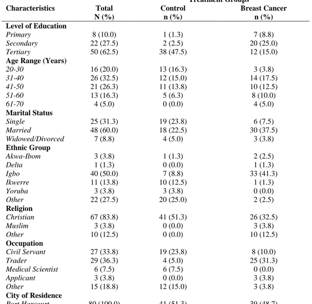
Table 1.1: Demographic Characteristics of Study Participants
Characteristics Total N (%) Treatment Groups Control n (%) Breast Cancer n (%) Level of Education
Primary Secondary Tertiary 8 (10.0) 22 (27.5) 50 (62.5) 1 (1.3) 2 (2.5) 38 (47.5) 7 (8.8) 20 (25.0) 12 (15.0)
Age Range (Years) 20-30 31-40 41-50 51-60 61-70 16 (20.0) 26 (32.5) 21 (26.3) 13 (16.3) 4 (5.0) 13 (16.3) 12 (15.0) 11 (13.8) 5 (6.3) 0 (0.0) 3 (3.8) 14 (17.5) 10 (12.5) 8 (10.0) 4 (5.0) Marital Status Single Married Widowed/Divorced 25 (31.3) 48 (60.0) 7 (8.8) 19 (23.8) 18 (22.5) 4 (5.0) 6 (7.5) 30 (37.5) 3 (3.8) Ethnic Group Akwa-Ibom Delta Igbo Ikwerre Yoruba Other 3 (3.8) 1 (1.3) 40 (50.0) 11 (13.8) 3 (3.8) 22 (27.5) 1 (1.3) 0 (0.0) 7 (8.8) 10 (12.5) 3 (3.8) 20 (25.0) 2 (2.5) 1 (1.3) 33 (41.3) 1 (1.3) 0 (0.0) 2 (2.5) Religion Christian Muslim Other 67 (83.8) 3 (3.8) 10 (12.5) 41 (51.3) 0 (0.0) 0 (0.0) 26 (32.5) 3 (3.8) 10 (12.5) Occupation Civil Servant Trader Medical Scientist Applicant Other 27 (33.8) 29 (36.3) 6 (7.5) 3 (3.8) 15 (18.8) 19 (23.8) 4 (5.0) 6 (7.5) 0 (0.0) 12 (15.0) 8 (10.0) 25 (31.3) 0 (0.0) 3 (3.8) 3 (3.8)
City of Residence
Port Harcourt 80 (100.0) 41 (51.3) 39 (48.7)
Note: Percentages may not add up to 100 due to rounding up; Frequency for each variable may vary due to nonresponses or missing values.
The reproductive characteristics of the study participants revealed that all except two were not pregnant (48.1%) and neither of the subjects were lactating. Majority of them had one child each 30 (39.5%) while the rest had two children each 9 (11.8%). Their menopausal history revealed that majority were not in their pre-menopausal stage 31
(41.9%), only 8(10.8%) were in their premenopausal stage, majority of the subjects were not post-menopausal women 26(34.7%). Only 13 (17.3%) were in their post-menopausal ages. 24 (30.4%) of the women were still seeing their menstrual period. (Table 1.2)
Table 1.2: Reproductive Characteristics of Study Participants
Characteristics Total N (%) Treatment Groups Control n (%) Breast Cancer n (%) Number of Children
1 2 44 (57.9) 32 (42.1) 14 (18.4) 23 (30.3) 30 (39.5) 9 (11.8)
Mieiwari I. Jumbo et al JMSCR Volume 06 Issue 10 October 2018 Page 106
Note: Percentages may not add up to 100 due to rounding up; Frequency for each variable may vary due to non-responses or missing values.
Table 1.3 shows the participants medical history, breast cancer diagnosis and therapy. All the breast cancer patients 39(48.8%) had no previous history of cancer in their families.
Majority of them had been diagnosed 1-3 years ago 21 (53.9%), only 4 (10.3%) of them were on therapy. Majority of them were not on therapy 35(89.7%) as at the time of this study. Majority of
those on therapy 11(84.6%) had been on it for few months. Only 2 (15.4%) of the subjects had received treatment for 1-2 years and the treatment mostly orthodox in nature 11(91.7%). No other diseases neither any cancer type were found co-existing with the condition studied 39 (48.7%). None of them smokes.
Table 1.3: Study Participants Medical History, Breast Cancer Diagnosis and Therapy
Characteristics@ Total
N (%) Treatment Groups Control N (%) Breast Cancer N (%) Any History of Cancer
No Yes 78 (97.5) 2 (2.5) 39 (48.8) 2 (2.5) 39 (48.8) 0 (0.0)
Years Since Diagnosis Few Months 1-3 years 4-6 years 7+ years 14 (35.9) 21 (53.9 3 (7.7) 1 (2.6) 0 (0.0) 0 (0.0) 0 (0.0) 0 (0.0) 14 (35.9) 21 (53.9 3 (7.7) 1 (2.6) On Therapy No Yes 35 (89.7) 4 (10.3) 0 (0.0) 0 (0.0) 35 (89.7) 4 (10.3)
Type of Therapy Orthodox Unorthodox 11 (91.7) 1 (8.3) 0 (0.0) 0 (0.0) 11 (91.7) 1 (8.3)
How Long on Therapy Few Months 1-2 years 11 (84.6) 2 (15.4) 0 (0.0) 0 (0.0) 11 (84.6) 2 (15.4)
Have any Other Disease No Yes 80 (100) 0 (0.0) 41 (51.3) 0 (0.0) 39 (48.7) 0 (0.0)
Any Other Cancer Type No Yes 80 (100) 0 (0.0) 41 (51.3) 0 (0.0) 39 (48.7) 0 (0.0) Cancer Metastasis No Yes 39 (98) 2 (2.0) 0 (0.0) 0 (0.0) 39 (98) 2 (2.0)
Are you Lactating No Yes 66 (97.1) 2 (2.9) 31 (45.6) 2 (2.9) 35 (51.5) 0 (0.0) Pre-menopausal Stage No Yes 53 (71.6) 21 (28.4) 22 (29.7) 13 (17.6) 31 (41.9) 8 (10.8) Post-menopausal Stage No Yes 59 (78.7) 16 (21.3) 33 (44.0) 3 (4.0) 26 (34.7) 13. (17.3
Still Seeing Menstrual Period No Yes 22 (27.9) 57 (72.2) 7 (8.9) 33 (41.8 15 (18.9) 24 (30.4)
Mieiwari I. Jumbo et al JMSCR Volume 06 Issue 10 October 2018 Page 107 Do You Smoke
No Yes
58 (72.5) 22 (27.5)
41 (51.3) 0 (0.0)
17 (21.3) 22 (27.5)
On Chemo No
Yes
33 (84.6) 6 (15.4)
0 (0.0) 0 (0.0)
33 (84.6) 6 (15.4)
Note: Percentages may not add up to 100 due to rounding up; Frequency for each variable may vary due to non responses or missing values.
@Characteristics associated with breast cancer are not applicable to the control group.
Table 1.4 shows the blood groups and Rhesus factor of the study participants.
Majority of them were of blood group O 28(34.2%) and Rhesus D positive 36(43.9%). Chi-Square (X2)
test did not reveal any association between blood groups and breast cancer. (X2 = 4.67; p = 0.192 and 2.71, p = 0.236 respectively).
Table 1.4: Blood Groups and Rhesus (Rh) Factor of Study Participants by Treatment Groups
Note: Percentages may not add up to 100 due to rounding up; Frequency for each variable may vary due to non-responses or missing values.
CI: Confidence Interval
Table 1.5 shows the mean ± SEM of immunological parameters of the study participants. The mean values of absolute CD4+ count for breast cancer subjects was (715.51± 51.62), while that of controls subjects was (909.37 ± 61.08). A p-value (p = 0.018) was observed. A similar trend was noticed in the absolute CD8+ count where the mean values of 386.54 ± 27.50 was significantly lower than
559.39 ± 43.66 among controls (p = 0.001). No statistically significant differences occurred in the CD4+ and CD8+ percentages and ratios. The serum concentration of anti-HER2 in the breast cancer subjects, 10.82 ± 1,20 was significantly reduced when compared with 15.69 ± 1.19 mg/ in the control subjects (p = 0.005).
Table 4.5: Mean ± SEM of Hematological Parameters by Treatment Groups
Parameter
Treatment Group
Test Statistics
Breast Cancer (n=41) Control (n=41)
Mean ± SEM Mean ± SEM p-value Sig. Status
Absolute CD4 Count 715.51±51.62 909.37±61.08 0.018 *
Absolute CD8 Count 386.54±27.50 559.39±43.66 0.001 ***
CD4 % 33.99±2.65 35.95±2.16 0.568 Ns
CD8 % 20.94±2.16 22.46±1.56 0.569 Ns
CD4:CD8 Ratio 2.11±0.19 1.96±0.15 0.538 Ns
Anti-HER 2 Conc. 10.82±1.20 15.69±1.19 0.005 **
Within parameter, mean ± SEM are significantly different at p<0.05. Significance Level: *=p<0.05; **=p<0.01; ***=p<0.001; ****=P<0.0001. SEM: Standard Error of Mean, NS: Not significant, Significant status: *
Characteristics Total
N (%)
Treatment Groups Test Statistics
Control N (%)
Breast Cancer N (%)
X2(df) P-Value Odds Ratio (95% CI)
Blood Group A
B AB O
24 (29.3) 6 (7.3) 3 (3.7) 49 (59.8)
16 (19.5) 2 (2.4) 2 (2.4) 21 (25.6)
8 (9.8) 4 (4.9) 1 (1.2)
28 (34.2) 4.67 (3) 0.192 ----
Rhesus (Rh) Factor Negative
Positive
7 (8.5) 75 (91.5)
2 (2.4) 39 (47.6)
5 (6.1)
Mieiwari I. Jumbo et al JMSCR Volume 06 Issue 10 October 2018 Page 108
Figure 1.1 shows the box plot graphs for selected parameters, anti-HER2
Discussion
This study was designed to assess immunological changes associated with breast cancer patient. In this part of the world and also to ascertain the possible role played by human epidermal growth receptors in the spread of breast cancer in this region. The major findings in this study are {a} High prevalence of breast cancer in women 40 years and below. {b} Mild anaemia as a complication of breast cancer in women.{c}Significant mild elevation in total WBC. {d} Immunological derangement in parameters of CD4 and CD8. {e} Uncommon elevation of anti-HER2 concentration in non-breast cancer patients.
In this study 17.5% of the breast cancer patients which constituted the majority were in the age range of 31 – 40 years. These within the age of 41 – 50% constituted 10% of the breast cancer patients. This observation is at variance with the report of American Cancer Society in 2011 which reported that breast cancer mostly occurs in older women of 45 years and above. However, the result from this study corroborated with the findings of [11] who reported similar findings with this study.
Significant in the current study is the immunological derangements in parameters of
Mieiwari I. Jumbo et al JMSCR Volume 06 Issue 10 October 2018 Page 109 All normal breasts contain the HER2 gene which is
a transmembrane protein. HER2 (also Her-2/neu or ErbB2) stands for Human Epidermal Growth Factor Receptor 2. If the HER2 gene mutates, it causes an uncontrolled increase in HER2 protein. The job of HER2 is to control a protein on the surface of cells that helps them grow. Healthy cells have two copies of the HER2 gene. When the HER2 genes become too many in a cell, it leads to too much HER2 protein being made. Having too many copies of HER2 genes is called Amplification of HER2. Having too many copies of HER2 protein is called over expression of HER2 [13].
In ~30% of breast cancers, HER2 is over expressed and it is associated with the aggressive form of the disease and poor clinical prognosis [14]. The level reaches 45% in patients with breast cancer metastasis.
The present study found that serum HER2 (ECD) level (>15.2 ngl ml) had worse prognosis. There is still no general consensus HER2 (ECD) cut-off for clinical use, commonly used value of 15.2ng/ml to define elevated or normal HER2 (ECD) was used. In this present study, elevated HER2 (ECD) levels were observed in (9/41) which represents 22% and 36% in non-breast patients. Interestingly, elevated Serum HER2 (ECD) levels were detected in 15 apparently healthy controls. [15] reported similar findings that 95% of serum values of HER-2/neu in normal women were found to be <13.37 ng/ml; 5% had values at 17ng/ml and >17ng/ml. Possible explanations for these may be, differences in the control group .May also be that benign breast disease affects HER2 levels normal elevation of Serum HER2 (ECD) levels as reported in healthy controls. Differences in their menopausal status may also contribute. The clinical implication of this unexpected serum ECD elevation could not be further investigated.
This present study, showed serum HER2 levels significantly higher in healthy individuals. This may be as a result of other implicating malignancies since HER-2/neugene is normally expressed on the epithelial cells of numerous organs, including lungs,
bladder, pancreases, breast and prostate and are found to be over expressed in cancer cells [15].
Conclusion
The anti-HER2 protein IgG which measures the HER-2 status for breast cancer (one of the breast cancer signaling receptors) was surprisingly significantly elevated in normal controls than in the breast cancer patients. There is need for routine immunological monitoring of breast cancer patients. Other causes of breast cancer other than HER-2 receptor positive needs to be explored to determine the possible causes of breast cancer in this part of the globe. Based on the findings and limitations of this study, more research is advocated to determine the prevalent hormone receptor positive breast cancer, for example oestrogen positive or progesterone positive breast cancer in this region. A down regulation of absolute CD4 and CD8 counts as an immunological implications in breast cancer patients was also observed.
References
1. Kamangaret, F., Dores, G. M. & Anderson, W. F. (2008). Patterns of cancers incidence, mortality and prevalence across five continents: Defining priorities to reduce cancer disparities in different geographic regions of the world. Journal of Clinical
Oncology, 24, 2137 – 2150.
2. Shrivastava,S., Singh, N., Akshay, K.N., Sanjay, S.C., Shrivastava, R. & Kumar, S. (2017). Comparative study of haemato-logical parameters along with effect of chemotherapy and radiotherapy in different stages of breast cancer. International
Journal of Research in Medical Sciences, 5,
2320-6071.
3. National Cancer Institute, (2015) Cancer
facts and figures.
https://doi.org/10.1016/j.ejca.2012.18.028 4. Abdulkareem, F.(2009). Epidemiology and
incidence of common cancers in Nigeria.
Cancer Registry and Epidemiology, 8(2),
Mieiwari I. Jumbo et al JMSCR Volume 06 Issue 10 October 2018 Page 110 5. Huang, Y. (2015). CD4+ and CD8+ T-cells
have opposing, roles in breast cancer progression and outcome. Oncology Target, 6(19), 17462 – 17478.
6. Isabelle, G., Itatem, A., Azim, J.R. & Ignatiadis, M. (2015). Immunology and breast cancer: toward a new way of understanding breast cancer and developing novel therapeutic strategies. Journal of Clinical Advances in Hematology and
Oncology, 13(6), 23 - 29.
7. Mittal, D., Gubbin, M.M., Schreiber,R,D&, Smyth, M.J.(2014). New insights into cancer immunoediting and its three component phases-elimination, equilibrium, and escape.
Current Opinion of Immunology, 27, 16-25.
8. Robinson-Bassey, G. & Festus, E. (2016). Knowledge and Attitude of Undergraduate Female Students towards breast cancer screening in the University of Port Harcourt, Rivers State. Journal of Nursing and Health. 3(5, 44-45.
9. Araoye, M.O. (2004). Sample size determination. Research methodology with statistics for health and social sciences. 1st Ed. Ilorin: Nathadex Publishers.
10.Jedy-Agba, E., Curado, M.P., Ogunbiyi, O., Oga, E. &Fabowale, T. (2012). Cancer incidence in Nigeria: a report from population- based cancer registries. Cancer
Epidemiology,.36(5), 271-278.
11.Khan, S. (2017). Study on some haematological parameters as biomarkers for breast cancer. Sindh University Research
Journal (Science Series), 49 (1) 23-28.
12.Onyema, O.O., Decoster, L., Njemini, R., Forti, L.N., Bautmans, I., De-Waele, M., and Mets, T. (2015). Chemotherapy – induced changes and Immunosenescence of CD8+ T-cells in patients with Breast Cancer. Anti-Cancer Research International Journal of
Cancer Research and Treatment, 35(3),
1481-1489.
13.Asgeirsson, K.S., Agrawal, A., Allen, C., Hitch, A., Ellis, I.O., Chapman Caroline, Cheung, K.L. & Robertson, J.F.R. (2007). Serum epidermal growth factor receptor and HER2 expression in primary and metastatic breast cancer patients.
14.English, D. P., Rogue, D. M., Santin, A. D. (2013). HER2 expression beyond breast cancer: Therapeutic implications for gynaelogic malignancies. Molecular
Diagnostic Therapeutics, 17(2), 85 – 99.
15.Mitri, Z., Constantine, T. &O’Regan, R. (2012) “The HER2 Receptor in Breast Cancer: Pathophysiology Clinical use and New Advances in Therapy”. Chemotherapy